ALMA MATER STUDIORUM – UNIVERSITÀ DI BOLOGNA
DOTTORATO DI RICERCA IN BIOINGEGNERIA
Ciclo XXV
Settore concorsuale di afferenza: 09/G2
Settore scientifico disciplinare: ING-INF/06
Computational Modeling of
Cardiac Excitation-Contraction Coupling
in Physiological and Pathological Conditions
Presented by
Stefano Morotti
Ph.D. Coordinator
Prof. Angelo Cappello, Ph.D.
Supervisor
Prof. Mauro Ursino, Ph.D.
Reviewers
Prof. Gianni Gnudi
Prof. Massimiliano Zaniboni, Ph.D.
Co-Supervisor
Stefano Severi, Ph.D.
ACKNOWLEDGEMENTS
I owe my gratitude to all those people who have made this thesis possible and because of whom this experience has been one that I will cherish forever.
First, I would like to acknowledge my advisor, Dr. Stefano Severi, who introduced me to the world of scientific research. Thank you for your guidance, friendship, understanding, and support during these years.
I want to express my sincere gratitude also to the reviewers of this thesis and to all the Ph.D. committee members for their helpful suggestions through all stages of my doctoral studies.
My special thanks go to Dr. Donald Bers for welcoming me (many times) in Davis and guiding my research to meet his high standards.
I am most grateful to Dr. Ele Grandi, my unofficial mentor, for the time and energy spent in supporting me as well as her commitment to my projects. Your help and friendship have been invaluable for me.
Finally, I would like to thank all the other precious collaborators I have been honored to work with for their fundamental contributions to the achievement of the results which this thesis will present.
KEYWORDS
Computational Biology Cardiac Electrophysiology Excitation-Contraction Coupling Ventricular Myocytes ArrhythmogenesisABSTRACT
The cardiomyocyte is a complex biological system where many mechanisms interact non-linearly to regulate the coupling between electrical excitation and mechanical contraction. For this reason, the development of mathematical models is fundamental in the field of cardiac electrophysiology, where the use of computational tools has become complementary to the classical experimentation. My doctoral research has been focusing on the development of such models for investigating the regulation of ventricular excitation-contraction coupling at the single cell level. In particular, the following researches are presented in this thesis:
1) Study of the unexpected deleterious effect of a Na channel blocker on a long QT syndrome type 3 patient. Experimental results were used to tune a Na current model that recapitulates the effect of the mutation and the treatment, in order to investigate how these influence the human action potential. Our research suggested that the analysis of the clinical phenotype is not sufficient for recommending drugs to patients carrying mutations with undefined electrophysiological properties.
2) Development of a model of L-type Ca channel inactivation in rabbit myocytes to faithfully reproduce the relative roles of voltage- and Ca-dependent inactivation. The model was applied to the analysis of Ca current inactivation kinetics during normal and abnormal repolarization, and predicts arrhythmogenic activity when inhibiting Ca-dependent inactivation, which is the predominant mechanism in physiological conditions.
3) Analysis of the arrhythmogenic consequences of the crosstalk between β-adrenergic and Ca-calmodulin dependent protein kinase signaling pathways. The descriptions of the two regulatory mechanisms, both enhanced in heart failure, were integrated into a novel murine action potential model to investigate how they concur to the development of cardiac arrhythmias.
These studies show how mathematical modeling is suitable to provide new insights into the mechanisms underlying cardiac excitation-contraction coupling and arrhythmogenesis.
CONTENTS
ACKNOWLEDGEMENTS ... I
KEYWORDS ... III
ABSTRACT ... V
CONTENTS ... VII
INTRODUCTION ... 1
Historical development of experimental electrophysiology ... 1
ECC in ventricular myocyte... 4
Modeling the mechanisms underlying ECC at the single cell level ... 7
Thesis outline ... 9
CHAPTER 1 – Trafficking Defects and Gating Abnormalities of a Novel
SCN5A Mutation Question Gene-Specific Therapy in Long QT Syndrome Type
3 ... 15
INTRODUCTION ... 16 METHODS ... 18 In vitro ... 18 In silico ... 23 RESULTS ... 25Clinical history of the carrier of F1473S mutation in the SCN5A gene ... 25
Response of F1473S-NaV1.5 channel to exposure to Na channel blockers ... 28
Mexiletine exposure and trafficking ... 28
In silico evaluation of F1473S-NaV1.5 and mexiletine effects on AP ... 30
DISCUSSION ... 30
Heterogeneous biophysical properties of SCN5A mutations in LQT3 ... 31
Mexiletine and the unexplained mechanisms for rescuing trafficking of NaV1.5 mutants ... 32
The need to revisit recommendations for the use of Na channel blockers in LQT3 .... 32
Study limitations ... 33
CHAPTER 2 – Theoretical Study of L-type Ca Current Inactivation Kinetics
during Action Potential Repolarization and Early Afterdepolarizations ... 37
INTRODUCTION ... 38
METHODS ... 41
INS measurements ... 41
Modeling Ba-dependent inactivation ... 42
ICa and SR Ca release models development ... 42
RESULTS ... 46
Experimental analysis of INS and IBa inactivation kinetics ... 46
Quantitative assessment of Ba-dependent inactivation ... 49
ICa model identification ... 52
AP model validation ... 54
Analysis of inactivation mechanisms and sources of CDI ... 55
Effects of reduced VDI ... 59
Effects of reduced CDI ... 59
Effects of IKr and IKs block... 61
ICa inactivation kinetics during EADs ... 62
DISCUSSION ... 62
Inactivation of INS and IBa through the LTCC ... 62
Inactivation of ICa during normal and abnormal AP repolarization ... 66
CHAPTER 3 – Arrhythmogenic Consequences of the Crosstalk between
β-Adrenergic and CaMKII Signaling in a Murine Model of Heart Failure ... 75
INTRODUCTION ... 76 METHODS ... 79 RESULTS ... 81 Cell geometry ... 81 K currents ... 81 Ca and Na handling ... 84
CaMKII and PKA effects ... 84
Model validation in WT and CaMKII-OE conditions ... 85
Consequences of βAR stimulation ... 88
DISCUSSION ... 89
Conclusion ... 94
CONCLUSION ... 101
INTRODUCTION
The term "excitation-contraction coupling" describes the fundamental electrophysiological process that links the electrical stimulation and the mechanical contraction in a muscle cell. Given the impact of heart diseases in the developed societies, a deep investigation of the mechanisms underlying cardiac excitation-contraction coupling and its derangements is extremely important. In the past 60 years, the research in cardiac electrophysiology has been benefiting from the use of mathematical models, now commonly adopted to support and drive the classical experimental work. Here, I will summarize the foundations of computational cardiac electrophysiology and provide with an outline of the subsequent chapters of this thesis.
Historical development of experimental electrophysiology
Electrophysiology is the branch of physiology that deals with the electrical properties of excitable cells and tissues (such as neurons and muscles). The history of this field dates back to the 17th century, when Jan Swammerdam first induced the contraction in a frog leg muscle by stimulating a nerve (Cobb, 2002). Swammerdam came close to understanding the nature of signal propagation between nerve and muscle, but at that time, the theory of electricity was still at its dawn. In fact, only about 100 years later, Benjamin Franklin, believed by many to be the "father of electricity", performed his famous experiment of flying a kite during a thunderstorm. At the end of 18th century, Luigi Galvani proposed his theory of electrical excitation of biological tissues (Galvani, 1791). He observed the relationship between stimulus density and muscle contraction, described the muscle refractoriness, and demonstrated the propagation of the stimulus, or action potential (AP). Galvani first hypothesized the existence of ion channels, described as water-filled channels penetrating into the fibers to allow excitability. Although the innovative concept of "animal electricity" proposed by Galvani has been initially countered by Alessandro Volta (a physicist known for the invention of the electric battery), it rapidly spread throughout Europe, also because of new findings by Giovanni Aldini, Galvani's nephew, which reinforced the theory of electrical excitation of biological tissue (Aldini, 1804) by studying the stimulation of body parts of freshly executed criminals. Furthermore, his experiments made a strong impact on the public opinion (Verkhratsky et al., 2006), inspiring even the famous Mary Shelley's "Frankenstein" (1817).Table 1. Perspectives of historical development of electrophysiology. From (Verkhratsky et al., 2006;
Luderitz, 2009; Priori, 2010).
Year
Event
1660s Swammerdam pioneered the frog nerve muscle preparation. 1790s Galvani developed the theory of electrical excitation of biological tissues, speculating about the existence of ion channels . 1800s Aldini applied electrical currents to mammalian brains, reinforcing Galvani's theory.
1828 Nobili instrumentally recorded animal electricity using an electromagnetic galvanometer 1840s Matteucci demonstrated AP-induced muscle contraction, and measured the resting current between the intact and cut surface of the muscle potential. 1848-1884 refined the methods to record muscle currents from frogs and humans. Du Bois-Reymond correctly interpreted Matteucci's experiments and
1850s Von Helmotz determined the AP propagation speed along the nerve cell. 1868 Bernstein made the first true recordings of resting and action potentials. 1887 Waller first studied the electrical phenomena of the heart and recorded the first human ECG. 1895 Langendorff pioneered the isolated perfused mammalian heart. 1896-1902 excitability by Bernstein and Overton and it has been suggested that the Nernst electrolytic theory has been adopted to explain the membrane
AP results from the exchange of Na and K cations. 1902 Einthoven developed the first practical ECG.
1910s Morgan demonstrated that genes are carried on chromosomes and are the mechanical basis of heredity. 1925 Gorter and Grendel found the bilayer structure of the cellular membranes. 1930s Pioneering experiments on Chara and Nitella (algae in the family Characeae) by Harris, Osterhout, and Hill.
1936 Young introduced the squid axon into physiological practice. 1939 Cole and Curtis directly measured the giant squid axon AP by performing impedance measurements. 1943 Hämmerling discovered the role of the nucleus as the repository of genetic information in eukaryotes. 1944 Avery, McLeod and McCarty identified the molecule responsible for transformation as DNA. 1949 Cole and Marmont developed the voltage-clamp technique. 1952 Hodgkin and Huxley formulated the ionic theory of membrane excitation, and developed a squid axon AP model. 1953 Watson and Crick determined the DNA double helix structure. 1960 Noble developed the first cardiac AP model.
1960s Nirenberg, Khorana and Holley discovered the genetic code. 1963 Müller and Rudin measured ion currents in artificial lipid membranes. 1976 Neher and Sakmann developed the patch-clamp technique. 1977 Sanger developed the “chain termination” method for sequencing DNA. McAllister, Noble, and Tsien developed the first ventricular AP model. 1980 Discovery of giga-seal between micropipette tip and surface cell membrane. 1983 Mullis developed the polymerase chain reaction.
1990s Development of species-specific ventricular AP models, and introduction of Markov models. 1995 The Keating group identified mutations responsible for long QT syndrome.
During the following century many scientists gave important contributions to the progress of experimental electrophysiology (Table 1), both by developing new instruments and by providing novel interpretations of observed phenomena. Thus, by the mid-1930s, the fundamental features of AP were already known, the structure of the cell membrane was identified and the existence of ion channels suggested. The introduction of the giant squid axon into physiological practice allowed the first direct measures of APs by Cole and Curtis (Cole & Curtis, 1939), obtained by performing impedance measurements (first using extracellular, and then intracellular electrodes). About 10 years later, the voltage-clamp technique has been developed by Cole (Cole, 1949) and Marmont (Marmont, 1949), and applied by Hodgkin and Huxley to formulate the ionic theory of membrane excitation (Hodgkin & Huxley, 1952; Hodgkin et al., 1952), which demonstrated that membrane excitability is determined by passive ion fluxes, according to their electro-chemical gradients. The importance of their findings has also been recognized by the Nobel committee in 1963, when the Hodgkin and Huxley were awarded with the Nobel Prize in Physiology or Medicine.
Moreover, the Hodgkin and Huxley theory implied the existence of ion channel currents, whose instrumental recordings required to overcome several technical barriers, like the need to access mammalian cells, to control both the extra- and intracellular environments, and, above all, to deal with electrical signals characterized by a very small amplitude and a strong noise (Verkhratsky et al., 2006). The improvement in measurement techniques culminated with the development of the patch-clamp technique (Neher & Sakmann, 1976; Sigworth & Neher, 1980), which allowed the systematic measurement of ion currents and entirely new types of electrophysiological experiments. Patch-clamp technique became not only the workhorse of modern electrophysiology (Petersen et al., 2005), but also one of the most important techniques for the study of signal-transduction mechanisms. Thus, Neher and Sakmann, the developers of this technique, received the Nobel Prize in Physiology or Medicine in 1991.
The 20th century saw also the parallel development of modern genetics. Although genes were known to exist on chromosomes, only in the 1940s the role of DNA as molecule responsible for inheritance has been discovered. In 1953 Watson and Crick (then awarded with the Nobel Prize in Physiology or Medicine in 1962) determined the double helix structure of DNA (Watson & Crick, 1953), suggesting the fundamental mechanisms of DNA semi-conservative duplication (based on the separation of the two strands and the following reconstruction of two DNA filaments). How DNA influences the behavior of cells became clear only in the 1960s, when the studies of Nirenberg, Khorana and Holley (Nobel Prize in Physiology or Medicine in 1968) contributed to discover the role of RNA, messenger created from the DNA and responsible for protein synthesis (Rammler & Khorana, 1962; Nirenberg & Leder, 1964; Holley et al., 1965). With the understanding of the genetic code, the introduction of the Sanger method for sequencing DNA (Sanger & Coulson, 1975) and the development of the polymerase chain reaction for isolating and amplifying a specific section of a DNA (Mullis & Faloona, 1987), scientific research accelerated and, in late 1980s, the search for disease-causing mutations started. Like several other fields, electrophysiology has been strongly impacted by the innovation brought by these techniques (Priori, 2010). The search for the genetic bases of inherited arrhythmogenic diseases was initiated by the Keating group, that first identified genomic mutations able to disrupt structure and function of cardiac ion channels and to cause life threatening arrhythmias (Curran et al., 1995). Thus, the concept
of "cardiac channelopathies" emerged, leading to the redefinition of the paradigms for management of patients with inherited arrhythmogenic diseases.
Hodgkin and Huxley laid the foundation for the use of integrative models in biology, that have been progressively developed (and widely used) in the second half of 20th century. The close interplay between model development and experimentation, together with the improvement in laboratory instrumentations and the introduction of novel techniques (such as patch-clamp and genetic manipulation), led to the discovery of a great number of cellular structures and regulatory mechanisms, thus to a deeper comprehension of the behavior of the cardiomyocyte both in health and in disease (Grandi & Bers, in press).
ECC in ventricular myocyte
Cardiac excitation-contraction coupling (ECC) is the physiological process that links the electrical excitation of the membrane potential (Em) to the consequent mechanical contraction
of the myocyte. ECC is mediated by Ca (Fig. 1, inset), an universal intracellular second messenger that is released in the cytosol at every beat to directly activate the myofilaments (Bers, 2001).
Under physiological conditions and in absence of excitation, ventricular cells maintain a relatively stable Em of about -85 mV (resting potential). When an excitatory stimulus
depolarizes the membrane beyond the threshold potential (about -70 mV), an AP is produced (Fig. 1, inset). The AP is manifest as a propagating wave of transient depolarization (ten Eick
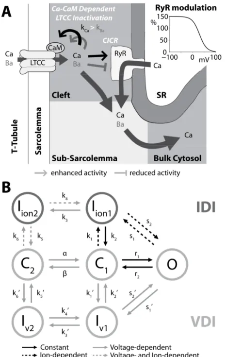
Figure 1. Schematic representation of cardiac ECC. Main panel summarizes Ca transport mechanisms in
ventricular myocytes; inset shows the time course of AP, CaT and contraction measured in a rabbit ventricular myocyte at 37°C. From (Bers, 2002).
et al., 1992), and arises as a result of the dynamic behavior of a diverse population of membrane ion channels. Thus, the AP is characterized by different morphologies across the regions of the heart (Fig. 2) and in different species. Ventricular AP in human (depicted in Fig. 3A) is similar to that in rabbit (Fig. 1, inset), and exhibits a steep upstroke (phase 0), followed by a brief steep repolarization (phase 1) and a sustained slowly decaying plateau phase (2). The plateau phase is followed by a steeper repolarization (phase 3) that restores the resting potential (phase 4) (Grandi & Bers, in press).
Depolarization to the threshold potential activates Na channels and results in a large Na current (INa, Fig. 3B), responsible for the AP upstroke, which quickly inactivates. Upon
membrane depolarization, L-type Ca current (ICa, Fig. 3B) activates as well, but inactivates
slowly and incompletely, allowing for the inward ICa to balance the outward K currents and
maintain the plateau phase of the AP. About the 90% of the L-type Ca channels (LTCCs) are localized in the transverse-tubules (t-tubules), deep invaginations of the sarcolemma (Fig. 1), and this particular localization facilitates the Ca entered via LTCC to trigger the Ca release from the sarcoplasmic reticulum (SR). This phenomenon is called Ca-induced Ca release (CICR) and is mediated by the ryanodine receptors (RyRs), that are sensitive to Ca in the dyadic space (the space between t-tubules and SR). During diastole, most of the intracellular Ca is stored in the SR. Once CICR occurs, Ca is free to diffuse throughout the sarcomere, where it binds to the Ca binding protein troponin in the myofilaments and initiates cell contraction, then the SR actively resequesters Ca via the sarco/endoplasmatic reticulum Ca pump (SERCA).
Membrane repolarization is mainly due to various types of K currents (Fig. 3C): the transient outward currents (Ito and IClCa, carried by K and Cl respectively) determine the notch
that follows the upstroke, the components of the delayed rectifier K current (IKr and IKs)
Figure 2. AP waveforms in different regions of the heart. AP waveforms are displaced in time to reflect
the temporal sequence of propagation through the heart (SA, sino-atrial node; AV, atrio-ventricular node; RV, right ventricle; LV, left ventricle). From (Nerbonne, 2000).
contribute to complete the repolarization, and the inward rectifier K current (IK1) maintains
the resting potential. Ca entered the cell is extruded by Na-Ca exchanger (NCX), that yields a net inward charge movement (INaCa, Fig. 3D) when 1 Ca cation (divalent) is exchanged for 3
Na cations (monovalent); also the plasma membrane Ca pump contributes to Ca extrusion and regulation of cytosolic intracellular [Ca] ([Ca]i). The Na-K pump (NKA) generates an
outward current (INaK, Fig. 3D) by extruding 3 Na ions and importing 2 K cations
(monovalent) on each cycle (Grandi & Bers, in press).
While Em influences [Ca]i, changes in [Ca]i and Ca transporters in turn alter Em. For
example, the intracellular Ca transient (CaT) feeds back on the LTCC, mediating ICa
inactivation, and therefore influences AP morphology. In pathological conditions, these mechanisms can also lead to triggered activity (Bers, in press), especially early and delayed
Figure 3. Schematic representation of ionic currents and CaT underlying the ventricular AP.
Simulated (Grandi et al., 2010) human ventricular AP and CaT (panel A), Na and Ca currents (B, inset shows the different activation and decay times), K currents (C), and NKA and NCX currents. Modified from (Grandi & Bers, in press).
afterdepolarizations (EADs and DADs). EADs and DADs are abnormal depolarizations of the cell membrane (respectively occurring during AP phases 2 or 3, and during phase 4) associated to the development of cardiac arrhythmias. DADs usually occur in a condition of Ca overload, where an increased Ca leak from the SR triggers Ca extrusion via NCX, which depolarizes the membrane (Priori & Corr, 1990). The same mechanism has also been associated to phase 3 EADs (Luo & Rudy, 1994b), that are characterized by low take-off potential. Phase 2 EADs are typically observed when the APD is prolonged (e.g., IKr block or
long QT syndromes), and are commonly associated to ICa reactivation during the prolonged
plateau (January & Riddle, 1989; Luo & Rudy, 1994b; Zeng & Rudy, 1995).
Cardiac electrophysiology, contraction, metabolism, and gene regulation are also subject to modulation by a number of subcellular signaling pathways, which involve cascades of signaling molecules resulting in post-translational modifications (e.g., phosphorylation) of target proteins (Grandi & Bers, in press). One of the most widely studied signaling pathways is that induced by β-adrenergic (βAR) receptor stimulation, whose macroscopic effect is an enhancement of cardiac function. At the single cell level, βAR regulation of ECC is mediated by the phosphorylation of a wide spectrum of target proteins (Bers, 2002), including LTCC, RyR, and phospholamban (PLB, which controls SERCA activity), which leads to an enhancement of CaT (and thus contraction). Recently, Ca-calmodulin dependent protein kinase (CaMKII) has emerged as important in ECC regulation (Maier & Bers, 2007), and its overexpression has been associated to pathological phenotypes. Furthermore, CaMKII and βAR signaling pathways are closely interrelated, since they share some phosphorylation targets and, above all, βAR stimulation can directly or indirectly (via enhanced intracellular Ca) activate CaMKII.
Modeling the mechanisms underlying ECC at the single cell level
The cardiomyocyte is a complex biological system where many regulatory mechanisms interact with dynamically changing ionic concentrations and varying Em. These non-linearinteractions make the single cell an interactive system where a high degree of synthesis and integration occurs (Rudy & Silva, 2006). Thus, analysis and synthesis of such complex systems can benefit from a mathematical approach.
The first computational AP model was formulated by Hodgkin and Huxley for the giant squid axon in 1952 (Hodgkin & Huxley, 1952; Hodgkin et al., 1952), and has been used as the basis to develop many modern neuronal and cardiac AP models. In Hodgkin and Huxley (H&H) model the inward movement of Na is responsible for the strong positive deflection of the Em observed in intracellular recordings, upon depolarization, while outward
flow of K causes repolarization to the resting state. In this formulation the gating mechanisms regulate membrane permeability, whereby distinct entities (i.e. gates) controls the flux of both Na and K ions (Grandi & Bers, in press).
In the early 1960s Noble developed the first models of a cardiac myocyte AP (Noble, 1960, 1962), adapting the H&H formulation to the Purkinje cell. Since ICa had not yet been
discovered at that time, the plateau was supported by inward Na flux. LTCC model was introduced only in the 1975 (McAllister et al., 1975), and the idea that a balance between K and Ca currents maintains the plateau was reinforced in the first computational ventricular
myocyte model (Beeler & Reuter, 1977). Aspects of intracellular Ca handling were first introduced in the Purkinje cell in 1985 (DiFrancesco & Noble, 1985). In 1994 Luo and Rudy published a ventricular AP model (Luo & Rudy, 1994b, a) that included the intracellular SR compartment, time varying intracellular ion concentrations, and ion pumps and exchangers. The Luo-Rudy model (Luo & Rudy, 1994a) became one of the landmarks in the development of computational ventricular electrophysiology. Based on data recorded mostly in guinea pig, it was formulated using the traditional H&H scheme.
More recently, a large body of knowledge has been accumulated on the relationships between ion channel structure and function, the kinetic properties of single ion channels, and the modification of ion channel structure and function by genetic defects (Nickerson & Hunter, 2010). Thus, a major challenge has been the integration of new findings into the AP model in order to mechanistically relate molecular level findings to whole cell function, and it has become clear that models with explicit representation of single ion channel states are required. For this reason, Markov models have been adopted (Clancy & Rudy, 1999, 2002) to replace the H&H formulation (where the gating parameters do not represent specific kinetic states of ion channels). Markov models are based on the assumption that transitions between channel states depend on the present conformation of the channel, but not on previous behavior. As the molecular interactions of channels are often state-dependent, Markov model transitions typically represent specific channel movements that have been characterized experimentally (Rudy & Silva, 2006).
While most of the early AP models generically integrated experimental data from mammalian hearts (mostly guinea pig), many electrophysiological studies had shown species differences in AP waveforms and ionic currents. Thus, based on data obtained from isolated cells from one particular species, ventricular AP models have been developed for mouse (Bondarenko et al., 2004; Li et al., 2010), rat (Pandit et al., 2001), rabbit (Puglisi & Bers, 2001; Shannon et al., 2004; Mahajan et al., 2008), and dog (Winslow et al., 1999; Greenstein et al., 2000; Hund & Rudy, 2004). The first human ventricular cell model was developed by Priebe and Beuckelmann (Priebe & Beuckelmann, 1998), adapting the major ionic currents of the Luo-Rudy model to the data available for human ventricular cells. More recently developed human models (Iyer et al., 2004; ten Tusscher et al., 2004; ten Tusscher & Panfilov, 2006; Grandi et al., 2010; O'Hara et al., 2011) included new measured data in human, providing a more accurate description of electrophysiological properties and Ca and Na handling in the human ventricular myocyte.
The Ca handling system was represented in the Luo-Rudy model by a phenomenological description (mimicking CICR) unable to capture the biophysical details involved (Grandi & Bers, in press). In 1998 Jafri et al. incorporated in a model of the guinea pig ventricular myocyte mechanistic Markov models for both LTCCs and RyRs and a restricted subspace (a single compartment representing the total volume of all dyads) into which all Ca fluxes through these channels are directed (Jafri et al., 1998). This "common pool" model, whereby sarcolemmal Ca influx enters the same Ca pool into which SR Ca is released and by which is regulated, cannot reproduce both high gain and graded release (Stern, 1992), that arises from local stochastic interactions between LTCCs and RyRs in thousands of Ca release units. In 2002 Greenstein and Winslow (Greenstein & Winslow, 2002) developed a comprehensive model of the ventricular myocyte based on the theory of local control of SR Ca release. Another cytosolic Ca compartment (sub-sarcolemma) has been
then introduced (Shannon et al., 2004), based on experiments suggesting that NCX senses elevated Ca levels (compared to the bulk [Ca]), contributing to the improvement of Ca handling description.
Thesis outline
Nowadays computational models play an important role in electrophysiology research and their use is complementary to that of classical experimentation. The general purpose of a model is to translate a set of hypotheses into predictions of observable events, and these predictions can be compared to observations, and the underlying hypotheses can be rejected or corroborated (Potse, 2012). In the past 50 years, many mathematical models of cardiac AP have been developed for different mammalian species and different heart regions, and successfully applied to test whether and how the known mechanisms contribute for all the experimentally observed phenomena, to predict the effects on cell function induced by a mutation (Clancy & Rudy, 1999), or to understand under which circumstances a cell would contribute to arrhythmogenesis (Kurata et al., 2005). By coupling AP models together, it is possible to create tissue models in which study AP propagation and arrhythmia (Comtois et al., 2008). Furthermore, it is possible to study the effect of modified cardiac activation on cardiac pump function, by integrating the electro-mechanical coupling, mechanical contraction, mechano-electric feedback (Trayanova & Rice, 2011).
The present thesis will focus on the regulation of AP and Ca handling, which comprehension is essential to understand cardiomyocyte physiology and arrhythmogenesis (Bers, in press). I will describe three projects, in which computational models have been developed to investigate different mechanisms underlying ventricular ECC in different mammalian species, both in physiological and pathological conditions.
The next chapter will describe the use of computational modeling in a clinical context. The clinicians treated a patient presenting the phenotype of long QT syndrome type 3 (LQT3, an inherited arrhythmogenic disease) with a drug commonly used as a LQT3 gene-specific therapy. Unexpectedly, administration of the drug worsened the patient conditions, thus a deeper investigation of the effects of the specific mutation was needed. Our collaborators from the group of Dr. Silvia Priori (at the Salvatore Maugeri Foundation, Pavia, Italy, and Leon H. Charney Division of Cardiology, New York University School of Medicine, USA) performed the characterization in vitro, while we investigated in silico the effects of both mutation and drug administration on the corresponding gene/protein (encoding the cardiac Na channel) and on the human ventricular AP.
The second project focused on the study of the biophysical properties of ICa, which is
one of the most important players in cardiomyocytes electrophysiology. The inactivation of this current is regulated by both Ca- and voltage-dependent inactivation mechanisms (CDI and VDI). Since the inhibition of both mechanisms has been associated to perturbed cardiomyocyte function, understanding of how CDI and VDI contribute to total inactivation is critical. Different strategies have been adopted in both experimental and theoretical studies to differentiate between the two mechanisms, and their accuracy is debatable. In collaboration with researchers from the group of Dr. Donald M. Bers (at the Department of Pharmacology, University of California in Davis, USA), we analyzed those strategies to select the most
accurate one. Based on this, we identified and validated an improved model of LTCC inactivation in rabbit ventricular myocyte, which was applied to study the relative roles of CDI and VDI during normal and abnormal repolarization.
The third chapter will present a study carried out in the Bers Lab in collaboration with researchers from the group of Dr. Andrew D. McCulloch (at the Department of Bioengineering, University of California in San Diego, USA), with the aim of investigating the crosstalk between CaMKII and βAR signaling pathways in the context of heart failure (HF). It has been shown that both CaMKII expression and βAR tone are upregulated in HF, thus it is important to understand how their interaction could contribute to ECC regulation and arrhythmogenesis. To this aim, we developed a model of murine ventricular ECC, where descriptions of Ca, βAR and CaMKII signaling pathways are integrated. The use of transgenic mouse models has proven to be particularly appropriate in the context of HF, given the wealth of experimental data that can be measured in mice, but cannot be obtained as easily in humans. Our work established the basis for the development of a systematic framework that, accounting for electrophysiological differences among species, is necessary to extrapolate findings in the genetically engineered mouse to the most relevant clinical setting.
References
Aldini G. (1804). Essai théorique et expérimental sur le galvanisme, vol. 2. Fournier et Fils, Paris.
Beeler GW & Reuter H. (1977). Reconstruction of the action potential of ventricular myocardial fibres. The Journal of physiology 268, 177-210.
Bers DM. (2001). Excitation-contraction coupling and cardiac contractile force. Kluwer Academic Publishers, Dordrecht ; Boston. Bers DM. (2002). Cardiac excitation-contraction
coupling. Nature 415, 198-205.
Bers DM. (in press). Excitation-Contraction Coupling. In Cardiac Electrophysiology: From Cell to Bedside, 6th edn, ed. Zipes DP & Jalife J. Expert Consult.
Bondarenko VE, Szigeti GP, Bett GC, Kim SJ & Rasmusson RL. (2004). Computer model of action potential of mouse ventricular myocytes. American journal of physiology Heart and circulatory physiology 287, H1378-1403.
Clancy CE & Rudy Y. (1999). Linking a genetic defect to its cellular phenotype in a cardiac arrhythmia. Nature 400, 566-569.
Clancy CE & Rudy Y. (2002). Na(+) channel mutation that causes both Brugada and long-QT syndrome phenotypes: a
simulation study of mechanism. Circulation 105, 1208-1213.
Cobb M. (2002). Timeline: exorcizing the animal spirits: Jan Swammerdam on nerve function. Nature reviews Neuroscience 3, 395-400.
Cole KS. (1949). Dynamic electrical characteristics of the squid axon membrane. Arch Sci Physiol 3, 253-258.
Cole KS & Curtis HJ. (1939). Electric Impedance of the Squid Giant Axon during Activity. The Journal of general physiology 22, 649-670.
Comtois P, Sakabe M, Vigmond EJ, Munoz M, Texier A, Shiroshita-Takeshita A & Nattel S. (2008). Mechanisms of atrial fibrillation termination by rapidly unbinding Na+ channel blockers: insights from mathematical models and experimental correlates. American journal of physiology Heart and circulatory physiology 295, H1489-1504.
Curran ME, Splawski I, Timothy KW, Vincent GM, Green ED & Keating MT. (1995). A molecular basis for cardiac arrhythmia: HERG mutations cause long QT syndrome. Cell 80, 795-803.
DiFrancesco D & Noble D. (1985). A model of cardiac electrical activity incorporating
ionic pumps and concentration changes. Philosophical transactions of the Royal Society of London Series B, Biological sciences 307, 353-398.
Galvani L. (1791). De viribus electricitatis in motu musculari commentarius. Bon Sci Art Inst Acad Comm 7, 363–418.
Grandi E & Bers DM. (in press). Models of the Ventricular Action Potential in Health and Disease. In Cardiac Electrophysiology: From Cell to Bedside, 6th edn, ed. Zipes DP & Jalife J. Expert Consult.
Grandi E, Pasqualini FS & Bers DM. (2010). A novel computational model of the human ventricular action potential and Ca transient. Journal of molecular and cellular cardiology 48, 112-121.
Greenstein JL & Winslow RL. (2002). An integrative model of the cardiac ventricular myocyte incorporating local control of Ca2+ release. Biophysical journal 83, 2918-2945.
Greenstein JL, Wu R, Po S, Tomaselli GF & Winslow RL. (2000). Role of the calcium-independent transient outward current I(to1) in shaping action potential morphology and duration. Circulation research 87, 1026-1033.
Hodgkin AL & Huxley AF. (1952). A quantitative description of membrane current and its application to conduction and excitation in nerve. The Journal of physiology 117, 500-544.
Hodgkin AL, Huxley AF & Katz B. (1952). Measurement of current-voltage relations in the membrane of the giant axon of Loligo. The Journal of physiology 116, 424-448.
Holley RW, Everett GA, Madison JT & Zamir A. (1965). Nucleotide Sequences in the Yeast Alanine Transfer Ribonucleic Acid. The Journal of biological chemistry 240, 2122-2128.
Hund TJ & Rudy Y. (2004). Rate dependence and regulation of action potential and calcium transient in a canine cardiac ventricular cell model. Circulation 110, 3168-3174. Iyer V, Mazhari R & Winslow RL. (2004). A
computational model of the human left-ventricular epicardial myocyte. Biophys J 87, 1507-1525.
Jafri MS, Rice JJ & Winslow RL. (1998). Cardiac Ca2+ dynamics: the roles of ryanodine receptor adaptation and sarcoplasmic reticulum load. Biophysical journal 74, 1149-1168.
January CT & Riddle JM. (1989). Early afterdepolarizations: mechanism of induction and block. A role for L-type Ca2+ current. Circulation research 64, 977-990.
Kurata Y, Hisatome I, Matsuda H & Shibamoto T. (2005). Dynamical mechanisms of pacemaker generation in IK1-downregulated human ventricular myocytes: insights from bifurcation analyses of a mathematical model. Biophysical journal 89, 2865-2887.
Li L, Niederer SA, Idigo W, Zhang YH, Swietach P, Casadei B & Smith NP. (2010). A mathematical model of the murine ventricular myocyte: a data-driven biophysically based approach applied to mice overexpressing the canine NCX isoform. American journal of physiology Heart and circulatory physiology 299, H1045-1063.
Luderitz B. (2009). Historical perspectives of cardiac electrophysiology. Hellenic journal of cardiology : HJC = Hellenike kardiologike epitheorese 50, 3-16.
Luo CH & Rudy Y. (1994a). A dynamic model of the cardiac ventricular action potential. I. Simulations of ionic currents and concentration changes. Circulation research 74, 1071-1096.
Luo CH & Rudy Y. (1994b). A dynamic model of the cardiac ventricular action potential. II. Afterdepolarizations, triggered activity, and potentiation. Circulation research 74, 1097-1113.
Mahajan A, Shiferaw Y, Sato D, Baher A, Olcese R, Xie LH, Yang MJ, Chen PS, Restrepo JG, Karma A, Garfinkel A, Qu Z & Weiss JN. (2008). A rabbit ventricular action potential model replicating cardiac dynamics at rapid heart rates. Biophysical journal 94, 392-410.
Maier LS & Bers DM. (2007). Role of Ca2+/calmodulin-dependent protein kinase (CaMK) in excitation-contraction coupling in the heart. Cardiovascular research 73, 631-640.
Marmont G. (1949). Studies on the axon membrane; a new method. Journal of cellular physiology 34, 351-382.
McAllister RE, Noble D & Tsien RW. (1975). Reconstruction of the electrical activity of cardiac Purkinje fibres. The Journal of physiology 251, 1-59.
Mullis KB & Faloona FA. (1987). Specific synthesis of DNA in vitro via a polymerase-catalyzed chain reaction. Methods in enzymology 155, 335-350. Neher E & Sakmann B. (1976). Single-channel
currents recorded from membrane of denervated frog muscle fibres. Nature 260, 799-802.
Nerbonne JM. (2000). Molecular basis of functional voltage-gated K+ channel diversity in the mammalian myocardium. The Journal of physiology 525 Pt 2, 285-298.
Nickerson DP & Hunter PJ. (2010). Cardiac Cellular Electrophysiology Modeling. In Cardiac Electrophysiology Methods and Models, ed. Sigg DC, Iaizzo PA, Xiao YF & He B. Springer, Inc.
Nirenberg M & Leder P. (1964). Rna Codewords and Protein Synthesis. The Effect of Trinucleotides Upon the Binding of Srna to Ribosomes. Science 145, 1399-1407. Noble D. (1960). Cardiac action and pacemaker
potentials based on the Hodgkin-Huxley equations. Nature 188, 495-497.
Noble D. (1962). A modification of the Hodgkin--Huxley equations applicable to Purkinje fibre action and pace-maker potentials. The Journal of physiology 160, 317-352. O'Hara T, Virag L, Varro A & Rudy Y. (2011).
Simulation of the undiseased human cardiac ventricular action potential: model formulation and experimental validation. PLoS computational biology 7, e1002061. Pandit SV, Clark RB, Giles WR & Demir SS.
(2001). A mathematical model of action potential heterogeneity in adult rat left ventricular myocytes. Biophysical journal 81, 3029-3051.
Petersen OH, Michalak M & Verkhratsky A. (2005). Calcium signalling: past, present and future. Cell calcium 38, 161-169. Potse M. (2012). Mathematical modeling and
simulation of ventricular activation
sequences: implications for cardiac resynchronization therapy. Journal of cardiovascular translational research 5, 146-158.
Priebe L & Beuckelmann DJ. (1998). Simulation study of cellular electric properties in heart failure. Circulation research 82, 1206-1223.
Priori SG. (2010). The fifteen years of discoveries that shaped molecular electrophysiology: time for appraisal. Circulation research 107, 451-456.
Priori SG & Corr PB. (1990). Mechanisms underlying early and delayed afterdepolarizations induced by catecholamines. The American journal of physiology 258, H1796-1805.
Puglisi JL & Bers DM. (2001). LabHEART: an interactive computer model of rabbit ventricular myocyte ion channels and Ca transport. American journal of physiology Cell physiology 281, C2049-2060.
Rammler DH & Khorana HG. (1962). A new approach to the specific synthesis of the C3'-C5' inter-ribonucleotide linkage. Biochemical and biophysical research communications 7, 147-150.
Rudy Y & Silva JR. (2006). Computational biology in the study of cardiac ion channels and cell electrophysiology. Quarterly reviews of biophysics 39, 57-116.
Sanger F & Coulson AR. (1975). A rapid method for determining sequences in DNA by primed synthesis with DNA polymerase. Journal of molecular biology 94, 441-448. Shannon TR, Wang F, Puglisi J, Weber C & Bers
DM. (2004). A mathematical treatment of integrated Ca dynamics within the ventricular myocyte. Biophysical journal 87, 3351-3371.
Sigworth FJ & Neher E. (1980). Single Na+ channel currents observed in cultured rat muscle cells. Nature 287, 447-449.
Stern MD. (1992). Theory of excitation-contraction coupling in cardiac muscle. Biophysical journal 63, 497-517.
ten Eick RE, Whalley DW & Rasmussen HH. (1992). Connections: heart disease, cellular electrophysiology, and ion channels. FASEB journal : official publication of the Federation of American
Societies for Experimental Biology 6, 2568-2580.
ten Tusscher KH, Noble D, Noble PJ & Panfilov AV. (2004). A model for human ventricular tissue. American journal of physiology Heart and circulatory physiology 286, H1573-1589.
ten Tusscher KH & Panfilov AV. (2006). Alternans and spiral breakup in a human ventricular tissue model. American journal of physiology Heart and circulatory physiology 291, H1088-1100.
Trayanova NA & Rice JJ. (2011). Cardiac electromechanical models: from cell to organ. Frontiers in physiology 2, 43. Verkhratsky A, Krishtal OA & Petersen OH.
(2006). From Galvani to patch clamp: the development of electrophysiology.
Pflugers Archiv : European journal of physiology 453, 233-247.
Watson JD & Crick FH. (1953). Molecular structure of nucleic acids; a structure for deoxyribose nucleic acid. Nature 171, 737-738.
Winslow RL, Rice J, Jafri S, Marban E & O'Rourke B. (1999). Mechanisms of altered excitation-contraction coupling in canine tachycardia-induced heart failure, II: model studies. Circulation research 84, 571-586.
Zeng J & Rudy Y. (1995). Early afterdepolarizations in cardiac myocytes: mechanism and rate dependence. Biophysical journal 68, 949-964.
CHAPTER 1
Trafficking Defects and Gating Abnormalities of a
Novel SCN5A Mutation Question Gene-Specific
Therapy in Long QT Syndrome Type 3
Short title: Paradoxical Effect of Mexiletine on LQT3.
___________________________________________________________________________ The content of this chapter has been published in:
Ruan Y, Denegri M, Liu N, Bachetti T, Seregni M, Morotti S, Severi S, Napolitano C & Priori SG. (2010). Trafficking defects and gating abnormalities of a novel SCN5A mutation question gene-specific therapy in long QT syndrome type 3. Circulation Research 106, 1374-1383.
Abstract
Na channel blockers are used as gene-specific treatments in long QT syndrome type 3 (LQT3), which is caused by mutations in the Na channel gene (SCN5A). The response to treatment is influenced by biophysical properties of mutations and we sought to investigate the unexpected deleterious effect of mexiletine in a mutation combining gain-of-function and trafficking abnormalities. A LQT3 child experienced paradoxical QT prolongation and worsening of arrhythmias after mexiletine treatment. The SCN5A mutation F1473S expressed in HEK293 cells presented a right-ward shift of steady-state inactivation, enlarged window current, and huge sustained Na current (INa). Unexpectedly, it also reduced INa peak by 80%.
Immunostaining showed that mutant NaV1.5 is retained in the cytoplasm. Incubation with 10
µmol/L mexiletine rescued the trafficking defect of F1473S, causing a significant increase in peak current, whereas sustained current was unchanged. Using a INa Markov model and a
model of human ventricular action potential (AP), we showed that simulated exposure of F1473S to mexiletine paradoxically increased AP duration, mimicking QT prolongation seen in the index patient on mexiletine treatment. We provided evidence that Na channel blockers, which are largely used to shorten QT intervals in carriers of SCN5A mutations, may facilitate trafficking of mutant proteins, thus exacerbating QT prolongation. These data suggest that caution should be used when recommending this class of drugs to carriers of mutations with undefined electrophysiological properties, and in vitro and in silico analysis may help to choose the proper treatment.
INTRODUCTION
Long QT syndrome (LQT) is an inherited arrhythmogenic disease characterized by QT interval prolongation and susceptibility to ventricular tachyarrhythmias. LQT type 3 (LQT3) is a variant of LQT characterized by high lethality (Priori et al., 2003), marked prolongation of repolarization, poor response to β-blockers (Priori et al., 2004), and cardiac events occurring preferentially at rest. LQT3 is caused by mutations in the SCN5A gene that encode for the α subunit of the channel that conducts the inward Na current (INa) responsible for fast
depolarization and critical for maintenance of intracardiac conduction (Clancy & Kass, 2005; George, 2005). Following the identification of the first SCN5A mutation published in 1995 (Wang et al., 1995), more than 80 SCN5A mutations have been identified in LQT3 patients.
SCN5A mutations associated with LQT3 increase INa by augmenting either the
sustained current (Isus) or the window current, thus prolonging cardiac repolarization (Ruan et
al., 2009). Based on this evidence, the use of Na channel blockers to treat LQT3 patients and reduce QT interval has been proposed. Early in vitro studies (Priori et al., 1996) demonstrated that mexiletine is effective in shortening action potential duration (APD) in cardiac myocytes exposed to anthopleurin, a compound that mimics LQT3 cellular phenotype. Furthermore early clinical studies on the use of mexiletine or flecainide were successful in shortening repolarization (Schwartz et al., 1995; Windle et al., 2001) and paved the way to the clinical use of Na channel blockers in LQT3 patients as a gene-specific therapy.
As the experience with the use of these drugs accumulated, the early enthusiasm was dimmed, and it became clear that flecainide might not be always appropriate for LQT3 patients. Indeed, the response of selected mutations could induce an electrocardiographic
pattern of coved type ST segment elevation in right precordial leads typical of Brugada syndrome (Priori et al., 2000; Makita et al., 2008). Shortly thereafter, it also became clear that Na channel blockers do not invariably shorten the QT interval, suggesting that SCN5A mutations act through other mechanisms, questioning the "gain-of-function" paradigm that all LQT3 mutations lead to a common channel dysfunction.
This view is supported by the fact that "some" SCN5A mutations cause "overlapping syndromes" in which QT prolongation is associated with "loss-of-function" clinical phenotypes (Brugada syndrome or conduction defects) (Bezzina et al., 1999; Grant et al., 2002; Antzelevitch et al., 2005). It has been recently demonstrated that biophysical diversity of SCN5A mutations is a determinant of response to mexiletine in the clinical setting (Ruan et al., 2007).
Here, we bring the characterization of the diversity of SCN5A mutations to the next level by showing that the interplay between protein trafficking and biophysical abnormalities may detrimentally affect the response to Na channel blockers and represent a hazard rather than a cure.
Figure 1. Mexiletine failed to shorten QTc and to prevent malignant ventricular arrhythmia. Panel A,
DNA sequence encoding a portion of DIII~DIV is shown for a normal control and for the index patient. The affected codon is enclosed by a rectangle indicating the heterozygous F1473S missense mutation. B, ECG recorded at day 1 (upper trace) and day 10 (lower trace) of mexiletine treatment. Further QTc prolongation is evident and T wave is almost fused with the subsequent stimulation spike. C, ECG tracing recorded at day 10 on mexiletine showing failures of pacemaker capture with extremely prolonged cardiac repolarization.
METHODS
In vitro characterization has been performed by our collaborators from Priori group, while we investigated in silico the consequences of mutation and mexiletine administration on the human ventricular action potential (AP).
In vitro
Molecular Screening
Genetic analysis was performed by screening of the open reading frame of the SCN5A, KCNH2, KCNQ1, KCNE1, and KCNE2 genes as previously reported (Napolitano et al., 2005).
Site-directed Mutagenesis and Transfection in HEK293 Cells
The SCN5A mutations were engineered into wild-type (WT) cDNA cloned in pcDNA3.1 (Invitrogen, Carlsbad, CA, USA) and confirmed by sequence analysis of the entire cDNA of
Figure 2. Gating properties of F1473S. Panel A, The voltage dependence of SSI and SSA of F1473S,
measured with standard pulse protocols shown in inset. B, Recovery from inactivation was assessed by the double-pulse protocol shown in inset and fitted using a biexponential function. C, Representative current traces evoked by 150 ms pulses to -10mV for cells expressing WT or F1473S channels. Currents have been normalized to facilitate comparison of WT and F1473S current traces. Besides reduced peak current, F1473S impair channel inactivation and induce a huge Isus. D, The amplitude of Isus in F1473S expressed in percentage of peak current is more than 20-fold larger than in WT; and it is also significantly larger than in P1332L, S941N, R1626P and M1652R, the LQT3-causing SCN5A mutations previously studied. N=7 to 21 cells for each group. * P < 0.001 compared with WT, P1332L, S941N, R1626P and M1652R. E, WT and F1473S currents in response to a ramp test.
SCN5A. HEK293 cells were transfected with equal amount of Na channel α and β1 subunit by
lipofectamine as previously described (Ruan et al., 2007). Electrophysiology
Membrane currents were measured using whole-cell patch-clamp procedures, with Axopatch 200B amplifiers (Axon Instruments, Foster City, CA, USA). Internal pipette solution contained (mmol/L) aspartic acid 50, CsCl 60, Na2-ATP 5, EGTA 11, HEPES 10, CaCl2 1,
and MgCl2 1, with pH 7.4 adjusted with CsOH. External solutions consisted of (mmol/L)
NaCl 130, CaCl2 2, CsCl 5, MgCl2 1.2, HEPES 10, and glucose 5, with pH 7.4 adjusted with
CsOH. In experiments designed to measure the voltage dependence of activation, external Na was reduced to 30 mmol/L with N-methyl-glucamine used as a Na substitute. Recordings were made at room temperature. Holding potentials were -100 mV. Isus was measured 150 ms
Figure 3. F1473S induced a large Isus and reduced peak current in SCN5A channel. Panel A,
Representative whole-cell current traces in HEK293 cell expressing WT (upper) and F1473S (middle) channels. Lower: Current-voltage (I-V) relationship of peak inward current. B, Cells expressing either WT (left) or F1473S (right) SCN5A were immunostained with anti-SCN5A primary antibody (Alomone ASC-005, green). The nuclei were highlighted by the DAPI staining (blue) (scale-bar 10 nm). WT channels were distributed mainly in the cytoplasm and on the plasma membrane; F1473S mutant channels show many condensed spots within the cytoplasm, suggesting a trafficking impairment (white arrows). C, Cytoplasmic distribution of F1473S mutant channel in HEK-SCN5A stable cell lines. Fixed cells were co-stained with the anti-SCN5A polyclonal antibody (green, a and d) and either with anti-Calnexin mAb for the ER (red, b) or anti-Golgin 97 mAb for the Golgi Apparatus (red, e). Protein localization was revealed by indirect immunofluorescence using DyLight 488-conjugated anti-rabbit IgG donkey antibodies and DyLight 594-conjugated anti mouse IgG donkey antibodies. The nuclei were stained with DAPI (blue) (scale-bar 10 nm).F1473S showed many condensed spots (white arrows) proximal to and partially co-localizing with the ER but not with the Golgi apparatus revealing that F1473S mutant channels are either kept in the ER or in the cytoplasm.
after depolarization to -10 mV and determined by subtracting background currents measured in the presence of tetrodotoxin (30 μmol/L, Sigma) from tetrodotoxin-free records. This subtraction procedure was also used for voltage ramp experiments. Steady-state inactivation (SSI) was determined after application of conditioning pulses (500 ms) applied to a series of voltages with an interpulse interval of 5 seconds for control and 30 seconds for mexiletine. Steady-state activation (SSA) was estimated by measuring peak current during a variable test potential. Current at each membrane potential was divided by the electrochemical driving force for Na ions and normalized to the maximum Na conductance. Data for the voltage dependence of activation and inactivation were fitted with the Boltzmann equation. Recovery from inactivation was measured in paired pulse experiments. Details of pulse protocol are given schematically in the figures. Data for the time course of recovery were fitted with biexponential function. Tonic block was measured at 0.033 Hz after steady-state was
Figure 4. Rescue effect and block effect of mexiletine. Panel A, Tonic block for WT, R1626P (mexiletine
sensitive), S941N (mexiletine insensitive), and F1473S, determined by the first test pulse after application of mexiletine. B, F1473S depolarizing shift SSI produced an enlarged window current (black solid line) compared with WT (block dash line); effect of 10 µmol/L mexiletine on window current was marked as grey solid line. C, Peak current density of three conditions: F1473S control (control, 20.54±2.22 pA/pF at -5mV, n=33), F1473S incubated with 10 µmol/L mexiletine for 48 hours with current recording without the drug (w/out tonic block, 29.17±2.87 pA/pF at -5mV, n=44, P<0.05 vs. control), and F1473S incubated with 10 µmol/L mexiletine for 48 hours and current recording in the presence of mexiletine (mex w/ tonic block, 27.17±2.3 pA/pF at -5mV, n=36, P<0.05 vs. control). D, Isus density of three conditions: F1473S control, F1473S incubated with 10 µmol/L mexiletine for 48 hours with current recording without (and with) the drug.
achieved in the presence of mexiletine. In drug rescue experiments, freshly prepared 10 μmol/L mexiletine was added to the culture media.
Immunofluorescence
For immunofluorescence (IF) experiments HEK293 cells were seeded on 0.1% gelatincoated coverslips and allowed to attach for 24 h, where indicated 10 μmol/L mexiletine was added for 48 hours. Following PBS washed cells were fixed with 4% PFA for 10 min and permeabilised with 0.2% Triton for 10 min. Blocking of unspecific sites were achieved by incubation with 10% BSA for 1 hour at room temperature. Primary antibodies used were: polyclonal NaV1.5 (Alomone, ASC-005, 1:100), monoclonal Calnexin (Alexis, clone AF18,
1:200), monoclonal Golgin 97 (Molecular Probes, CDF4, 1:50). Secondary antibodies used were: DyLight 488-conjugated donkey anti-rabbit IgG and Dy-Light-conjugated 594-conjugated donkey anti-mouse IgG (Jackson Laboratories, 1:100). The coverslips were then mounted using a DAPI-containing mounting medium (Vectashield, Vector Laboratories, Burlingame, CA, USA). Confocal microscopy was performed with a Leica TCS-SP2 digital scanning confocal microscope equipped with a HCX PL APO 40x/numerical aperture=1.25 oil immersion objective. The 488-nm Argon laser line has been used for excitation of DyLight 488-conjugated donkey anti-rabbit IgG (detected at 500-530 nm) and the 594 nm He/Ne laser
Figure 5. Rescued membrane expression of F1473S SCN5A channel with 10 µmol/L mexiletine. Panel
A, mexiletine incubation increased the cytoplasmic distribution of F1473S and released the intracellular condensed spots of F1473S (scale-bar 20 nm). B, Confocal images of F1473S distribution with phase contrast in control condition (upper) and with 10 µmol/L mexiletine incubation (lower) (scale-bar 10 nm). C, Plasma membrane fraction of F1473S SCN5A was increased by mexiletine incubation. PM indicates plasma membrane, Cyt, cytosol, CL, total cell lysate; Con, control, no mexiletine incubation, Mex, 10 µmol/L mexiletine incubation for 48 hours. β-Actin was used as the normalizing control.
line for excitation of Dy-Light-conjugated 594-conjugated donkey anti-mouse IgG (detected at 580-630 nm). The pinhole diameter was kept at Airy 1. Images were exported to Adobe Photoshop (Adobe Systems, Mountain View, CA, USA).
Immunoblotting for Quantification of Plasma Membrane Protein Expression
First our collaborators confirmed that the INa observed in stable cell lines expressing
F1473S-NaV1.5 was identical to the current observed in cells with transient expression of the mutant α
and β1 subunit and they were able to show that the same large Isus, reduced peak current,
trafficking defect and gating properties were also present in the transiently transfected cells expressing F1473S-NaV1.5 (data not shown). HEK293 cells which stably expressed F1473S
SCN5A were lysed and total proteins extracted; plasma membrane and cytosolic fractions were isolated using a plasma membrane protein extraction kit according to manufacturer's instructions (Biovision). Total proteins (15 micrograms/sample, quantified by the BCA assay) were resolved by SDS-gel electrophoresis on Novex 4-12% BisTris gradient gels using MES-SDS buffer (Invitrogen), and blotted on nitrocellulose membranes using a submarine system (Invitrogen). A monoclonal anti-human NaV1.5 antibody (1:500, Alomone, Asc-005) was
used to probe for the Na channel while a polyclonal anti β-Actin antibody (1:5000, Affinity Bioreagents) was used to detect a reference protein. Secondary antibodies were conjugated with HRP (1:3000, Promega). Mouse heart was used as positive control. Specific signals were developed using the Supersignal West Pico Chemiluminescent substrate (Pierce) and detected using X-ray films (Kodak).
Statistics
Pclamp9.2 (Axon Instruments, Foster City, CA, USA) and Excel (Microsoft, Seattle, WA, USA) were used for data acquisition and analysis. Data are presented as mean values ± S.E. Statistical comparisons were made using an unpaired 2-tailed t-test or ANOVA with the Tukey post hoc test to evaluate the significance of differences between means. P<0.05 was considered statistically significant.
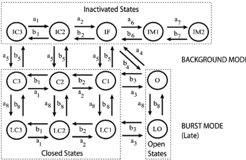
Figure 6. Markov model of the cardiac Na channel. The channel model contains background
(upper nine states) and burst (lower four states) gating modes. The burst mode reflects a population of channels that transiently fail to inactivate. From (Grandi et al., 2007).
In silico
The Na channel Markov model was based on the structure originally proposed by Clancy and colleagues (Clancy & Rudy, 1999; Clancy et al., 2002), depicted in Fig. 6. The whole-cell INa
is given by: ) ( m Na O Na Na G P E E I ,
where the variable PO represents the sum of the probabilities to be in the open states (O and
LO), Em is the membrane potential, ENa is the Na Nerst potential and GNa is the maximum
conductance. The expressions of the transition rates are reported in Table 1. The transition rate parameters were identified by a manual fitting procedure to simulate the experimental data of whole-cell cardiac INa from HEK cells expressing WT or F1473S SCN5A. The
following voltage-clamp protocols were simulated for parameter identification: SSA and SSI, recovery from inactivation, positive voltage ramp and long-depolarization voltage step to assess Isus. The model results for each protocol were compared with experimental data. The
parameter values proposed by in a previous study (Grandi et al., 2007) were chosen where possible as initial guesses in the identification procedure of the transition rate parameters that allowed the best fitting of our WT data. We modified a2 and a4 to reproduce the experimental
data on SSA; a5 was tuned to correctly simulate the channel availability curve; a6, b6 and a7, b7
were adjusted to represent the observed characteristics of recovery from inactivation, a5, b6, b7
were modified to reproduce the response to voltage ramp in the mutant channels and the ratio a8/b8 was tuned to reproduce the experimental data accounting for Isus. The identified WT set
was subsequently used as initial guess to identify the F1473S channel parameters, and two parameters of the F1473S set were tuned to reproduce the effect of mexiletine treatment: P2a5
was modified to introduce the SSI shift and P1a8 to reduce Isus. Table 2 reports the three sets of
model parameters.
We integrated the INa model into a model of AP in human epicardial ventricular
myocyte (ten Tusscher & Panfilov, 2006) as modified by Severi et al. to correctly reproduce
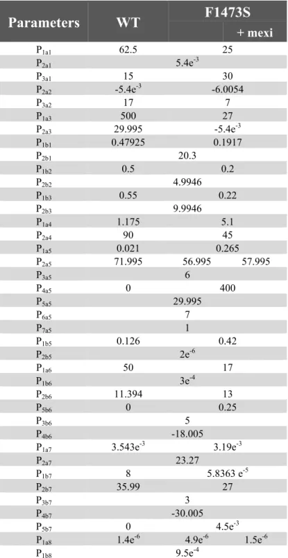
Table 1. Na channel model transition rate expressions.
Transition rate expressions
a1 = P1a1 /(1+exp((-Em+P2a1)/P3a1))a2 = P1a1 /(1+exp((-Em+P2a2)/P3a2))
a3 = P1a3 /(1+exp((-Em+P2a3)/P3a1))
b1 = P1b1 exp((-Em+P2a1)/P2b1)
b2 = P1b2 exp((-Em+P2b2)/P2b1)
b3 = P1b3 exp((-Em+P2b3)/P2b1)
a4 = P1a4 exp((Em-P2a1)/P2a4)
a5 = (P1a5 /(1+exp((Em+P2a5)/P3a5)))(P7a5+P4a5 /(1+exp((-Em+P5a5)/P6a5)))
a6 = a4/P1a6
b4 = (a3 a4 a5)/(b3 b5)
b5 = P1b5 +P2b5 (Em-P2a1)
b6 = P1b6 exp((-Em+P2a1)/P2b6)+P5b6 /(1+exp((-Em+P4b6)/P3b6)))
a7 = P1a7 exp((Em-P2a1)/P2a7)
b7 = P1b7 exp((-Em+P2a1)/P2b7)+P5b7 /(1+exp((-Em+P4b7)/P3b7)))
a8 = P1a8
the experimental APD inverse dependence on extracellular [Ca] (Severi et al., 2009). The kinetic rates were normalized to 37°C using a Q10 of 2.1 (Maltsev & Undrovinas, 2006).
Extracellular [Ca] was set to the physiological value of 1.15 mmol/L. Since mutant patients reveal stronger Isus and thereby higher intracellular [Na], the initial value of this concentration
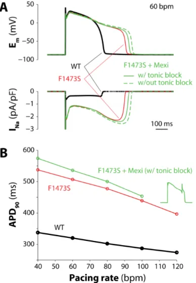
was set at 15 mmol/L in order to reach quickly the steady-state condition. Pacing was simulated by a current pulse train (pulse 1 ms duration, 52 pA/pF amplitude) at different frequencies (from 30 to 120 bpm, i.e. from 0.5 to 2 Hz). The stimulation was maintained at
Table 2. Na channel model parameters for WT and F1473S.
Parameters
WT
F1473S
+ mexi P1a1 62.5 25 P2a1 5.4e-3 P3a1 15 30 P2a2 -5.4e-3 -6.0054 P3a2 17 7 P1a3 500 27 P2a3 29.995 -5.4e-3 P1b1 0.47925 0.1917 P2b1 20.3 P1b2 0.5 0.2 P2b2 4.9946 P1b3 0.55 0.22 P2b3 9.9946 P1a4 1.175 5.1 P2a4 90 45 P1a5 0.021 0.265 P2a5 71.995 56.995 57.995 P3a5 6 P4a5 0 400 P5a5 29.995 P6a5 7 P7a5 1 P1b5 0.126 0.42 P2b5 2e-6 P1a6 50 17 P1b6 3e-4 P2b6 11.394 13 P5b6 0 0.25 P3b6 5 P4b6 -18.005P1a7 3.543e-3 3.19e-3
P2a7 23.27 P1b7 8 5.8363 e-5 P2b7 35.99 27 P3b7 3 P4b7 -30.005 P5b7 0 4.5e-3
P1a8 1.4e-6 4.9e-6 1.5e-6
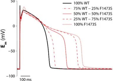
least for 200 s. To analyze the effects of mutations on cardiac AP in the heterozygous condition of F1473S patients, we replicated conditions with different percentage of mutant and WT Na channels.
Matlab and Simulink (Mathworks, Natick, MA, USA) were used for all the numerical computations. A variable order solver based on the numerical differentiation formulas (NDFs) was used to numerically solve the model equations (ode15s).
RESULTS
Clinical history of the carrier of F1473S mutation in the SCN5A gene
The mutation here characterized was identified in a patient born in 2003; no ECG was recorded at birth, and the baby was discharged as a healthy infant. At 12 months of age, he experienced 5 syncopal spells within a few days, was hospitalized and long QT syndrome was diagnosed. Propranolol (4 mg/kg per day) was initiated, and a blood sample was sent to the Priori group in Maugeri Foundation for genetic screening. They sequenced the opening reading frame of KCNQ1, KCNH2, SCN5A, KCNE1, and KCNE2 and identified a heterozygous single-nucleotide transition (4418T>C) in the SCN5A gene, leading to a single amino acid replacement at position 1473 (F1473S, Fig. 1A). This mutation was not present in either the parents of the index case or in 300 control DNA samples.
The patient remained asymptomatic for 2 years on β-blockers; however, in 2006, syncope recurred and torsade de pointes was documented; a ventricular-inhibited pacemaker was implanted, and the dose of β-blockers was increased. One year later, the patient experienced another syncopal episode, accompanied by several runs of torsade de pointes. Oral mexiletine was started in an attempt to shorten QT interval and to control arrhythmias; sinus rhythm was regained and QT duration appeared stable at day 1 of administration (Fig. 1B, upper trace); unfortunately, the effect of mexiletine (60 mg tid) decayed as the treatment continued. After 10 days, corrected QT interval (QTc) persistently exceeded 650 ms (Fig. 1B, lower trace); furthermore, progressive worsening of intraventricular conduction with QRS widening, and occasional pacemaker capture failures (Fig. 1C) were observed. At day 11, the patient developed incessant, intractable tachyarrhythmias and died. The clinicians who treated the patient were confounded by the worsening clinical status during mexiletine treatment. The inspection of the available ECG showed QTc shortening just at the beginning of therapy followed by prolongation (Fig. 1B). The Priori group and us attempted to understand this uncommon and paradoxical response to mexiletine by functional characterization of the mutant.
Functional in vitro characterization of F1473S mutation
As shown in Fig. 2A, F1473S depolarized the SSI curve by 18.9 mV and SSA curve by 4.4 mV. The SSI pattern of mutant channels was consistent with previous observations (Ruan et al., 2007) showing that mexiletine-insensitive mutations are those causing a depolarizing shift of SSI and it also produced a greater overlap of activation and inactivation relations. As a consequence, the window current was markedly enhanced. Recovery from inactivation is shown in Fig. 2B.
Besides enlarged window current, F1473S showed a striking increase of Isus. At -10
mV, F1473S channels continued to show 4.46±0.74 pA/pF (5.39±0.2% of the peak current) at 150 ms as compared to 0.64±0.22 pA/pF (0.25±0.05%) in WT (P<0.0001). Overall, F1473S showed a much higher Isus than other mutations previously characterized (Ruan et al., 2007)
(Fig. 2C).
Our collaborators further characterized the gain-of-function behavior by ramp voltage protocol (from -100 mV to +50 mV). F1473S conducted larger peak currents than WT (F1473S -6.38±0.71 pA/pF, n=9 vs. WT -2.28±0.14 pA/pF, n=8; P<0.0001), and the voltage of the peak was shifted by about +20 mV (F1473S -17.1±3.5 mV, n=9 vs. WT -37.8±3.1 mV, n=8; P<0.0001). WT channels conduct current over a narrow range of voltages near -37 mV, whereas F1473S mutant channels conduct current over a broad range of voltages and increase the inward INa (Fig. 2D). The Priori group concluded that the remarkable increase of both the
window current and the Isus account for QT prolongation and the malignant clinical
phenotype.
On the other hand, F1473S peak current density was unexpectedly reduced to 20% of
Figure 7. Experimentally determined and simulated effects of F1473S mutation on Na channel properties. Panel A, SSI and SSA, the inset highlights in silico results in window current region. B,
recovery from inactivation. Dots represent data from in vitro experiments, solid lines represent simulated results. C, experimental (black) and simulated (white) ratio between sustained and peak current for WT and F1473S channels. D, experimental (symbols) and simulated (solid lines) I-V relations for WT and F1473S channels.