Copyright © 2004, American Society for Microbiology. All Rights Reserved.
Biochemical Characterization of the THIN-B Metallo-
-Lactamase
of Janthinobacterium lividum
Jean-Denis Docquier,
1† Teresa Lopizzo,
1Sabrina Liberatori,
2Manuela Prenna,
3Maria Cristina Thaller,
4Jean-Marie Fre
`re,
5and Gian Maria Rossolini
1*
Dipartimento di Biologia Molecolare, Laboratorio di Fisiologia e Biotecnologia dei Microrganismi,
1and Laboratorio di
Proteomica Funzionale,
2Universita
` di Siena, Siena, Dipartimento di Biologia Molecolare, Cellulare e Animale,
Universita
` di Camerino, Camerino,
3and Dipartimento di Biologia, Universita
` di Roma “Tor Vergata,”
Rome,
4Italy, and Laboratoire d’Enzymologie and Centre d’Inge´nierie des Prote´ines,
Institut de Chimie, Universite´ de Lie`ge, Lie`ge, Belgium
5Received 21 January 2004/Returned for modification 8 May 2004/Accepted 4 August 2004
The THIN-B metallo-
-lactamase, a subclass B3 enzyme produced by the environmental species
Janthino-bacterium lividum, was overproduced in Escherichia coli by means of a T7-based expression system. The enzyme
was purified (>95%) by two ion-exchange chromatography steps and subjected to biochemical analysis. The
native THIN-B enzyme is a monomeric protein of 31 kDa. It exhibits the highest catalytic efficiencies with
carbapenem substrates and cephalosporins, except for cephaloridine, which acts as a poor inactivator.
Indi-vidual rate constants for inactivation by chelators were measured, suggesting that inactivation occurred by a
mechanism involving formation of a ternary complex.
Metallo-
-lactamases (MBLs) are the focus of increasing
investigation both as resistance determinants and as model
enzymes. As resistance determinants their relevance depends
on their functional features (broad substrate specificity,
ef-ficient carbapenemase activity, resistance to the so-called
“mechanism-based”
-lactamase inactivators) and on the
re-cent emergence of MBLs encoded by genes associated with
mobile DNA among major bacterial pathogens (19, 21, 23, 24).
The interest in MBLs as model enzymes arises from the as yet
superficial understanding of their catalytic mechanism and
structure-function relationships, which could be essential to
the development of new
-lactams and enzyme inhibitors. On
the other hand, the MBL fold is conserved within a large
protein superfamily that includes a growing number of proteins
which do not hydrolyze
-lactams (2, 6, 8).
MBLs belong to molecular class B (1) and constitute a
fam-ily of very diverse enzymes. Based on structural relatedness,
they can be grouped into three different subclasses: B1, B2, and
B3 (14, 23). Subclass B3, originally represented by the L1
enzyme from Stenotrophomonas maltophilia (4, 26), has
re-cently expanded to include several enzymes from primarily
environmental bacteria (FEZ from Legionella gormanii [5],
GOB from Chryseobacterium meningosepticum [3], THIN-B
from Janthinobacterium lividum [25], and CAU from
Caulo-bacter crescentus [9]), some of which can occasionally behave as
opportunistic pathogens. The MBLs of subclass B3 are highly
divergent from those of subclass B1 at the sequence level (14)
and, although they retain a three-dimensional fold that is
sim-ilar overall, exhibit an organization of the metal-binding sites
that differs significantly from that of enzymes of subclass B1
(15, 28).
The THIN-B enzyme from J. lividum was identified
follow-ing an environmental screenfollow-ing of MBL-producfollow-ing bacteria
(25) and currently is the only known MBL from a member of
the
-Proteobacteria class. Compared to the other enzymes of
subclass B3, THIN-B is quite divergent and exhibits some
unique structural features, including a larger size and a higher
number of cysteine residues (25). However, the biochemical
properties of this enzyme have not been investigated.
In this paper we describe a system for overproduction of the
THIN-B enzyme in Escherichia coli, the protocol for
purifica-tion of the recombinant protein, and the biochemical and
ki-netic characterization of THIN-B.
MATERIALS AND METHODS
Bacterial strains.E. coli XL-1 Blue (Stratagene, Inc., La Jolla, Calif.) was used as a host for recombinant plasmids. E. coli strains BL21(DE3) (Stratagene), BL21-SI (Invitrogen, Carlsbad, Calif.), and MCT236(DE3) {E. coli CGSC6159 [lacIqrrnB
T14⌬lacZWJ16hsdR514⌬araBA-DAH33⌬rhaBADLD78⌬pyrC (cIts857 ind1 Sam7 nin5 lacUV5-T7 gene 1)]} (constructed in our laboratory) were used as hosts for T7 promoter-based expression plasmids for blaTHIN-Boverexpression
experiments.
Media and culture conditions.Bacteria were always grown aerobically at 37°C unless otherwise specified. Luria-Bertani medium (27) was routinely used for the propagation of E. coli strains. SB medium (20 g of yeast extract/liter, 35 g of tryptone/liter, and 5 g of NaCl/liter; buffered with 50 mM sodium phosphate buffer [pH 7.0]) was used in overexpression experiments with BL21(DE3) and MCT236(DE3) strains. LBON (5 g of yeast extract/liter and 10 g of tryptone/ liter) was used in overexpression experiments with BL21-SI.
Recombinant DNA methodologies.The open reading frame encoding THIN-B was amplified by PCR using primers THIN-B-EXP/f (5⬘-CAT ATG ACA CTA TTG GCG AAG TTG ATG CTG), which added an NdeI linker (boldfaced) to the 5⬘ end, and THIN-B-EXP/r (5⬘-GGA TCC TAG TGC GCG TGC TGG G), which added a BamHI linker (boldfaced) to the 3⬘ end. PCR was performed in a volume of 50l, with 3.5 U of the Expand High Fidelity PCR system (Roche Biochemicals, Mannheim, Germany) in the buffer provided by the manufacturer, 200M deoxynucleoside triphosphates, 50 pmol of each primer, and 10 ng of plasmid pBCIRO-K (25) as the template for the blaTHIN-Bgene. The following
cycling conditions were used: initial denaturation at 94°C for 3 min; denaturation
* Corresponding author. Mailing address: Dipartimento di Biologia
Molecolare, Universita
` di Siena, Policlinico Le Scotte, 53100 Siena,
Italy. Phone: 39 0577 233327. Fax: 39 0577 233325. E-mail: rossolini
@unisi.it.
† Present address: Laboratoire d’Enzymologie et Centre d’Inge
´-nierie des Prote
´ines, Institut de Chimie, Universite
´ de Lie
`ge, B-4000
Lie
`ge, Belgium.
at 94°C for 60 s, annealing at 58°C for 60 s, and extension at 72°C for 90 s, repeated for 30 cycles; and a final extension step at 72°C for 10 min. The amplified DNA was cloned into the SmaI site of plasmid pUC-18 by using the Sure Cloning kit (Amersham Biosciences, Uppsala, Sweden), yielding the re-combinant plasmid pUC-CIRO. Finally, the 0.95-kb NdeI-BamHI fragment of pUC-CIRO, containing the blaTHIN-B gene, was subcloned into the pET-9a
expression vector to produce the recombinant plasmid pET-THIN-B. The cloned blaTHIN-Bgene was sequenced to rule out the presence of any PCR-generated
mutations.
Expression experiments.THIN-B production was tested by using three dif-ferent expression systems, obtained by transformation of E. coli BL21(DE3), BL21-SI, or MCT236(DE3) with pET-THIN-B. With each system MBL produc-tion was monitored, over a 24 h time course, in both supernatants and cell extracts of cultures growing in SB medium containing 50g of kanamycin/ml at either 25 or 37°C. Individual cultures were incubated until the A600reached 0.8
and then were split into two identical flasks, to one of which was added 1 mM isopropyl--D-thiogalactopyranoside (IPTG) [for the BL21(DE3) and MCT236 (DE3) hosts] or 0.3 M NaCl (for the BL21-SI host). Aliquots (1 ml) were sampled at different times and centrifuged (12,000⫻ g, 5 min, 4°C), and the culture supernatant was stored at 4°C. The bacterial pellet was resuspended in 1 ml of 10 mM HEPES-NaOH (pH 7.5) supplemented with 50M ZnSO4and
was disrupted by sonication (5 cycles, for 20 s each cycle, at 45 W) using a B. Braun (Melsungen, Germany) Labsonic L sonicator. The supernatant obtained after centrifugation at 10,000⫻ g for 15 min, to remove cell debris, represented the cell extract. MBL activities in supernatants and cell extracts were determined spectrophotometrically at 30°C by using 150M imipenem as the substrate (wavelength, 300 nm;⌬ε,⫺9,000 M⫺1䡠 cm⫺1) in 10 mM HEPES-NaOH buffer (pH 7.5) (20). The reaction volume was 500l.
Purification of the THIN-B enzyme.The THIN-B MBL was purified from a culture of E. coli MCT236(DE3)(pET-THIN-B) grown in 0.5 liter of SB medium at 25°C. The culture was induced with 1 mM IPTG when the A600was equal to
0.8. Cells were collected, 24 h after induction, by centrifugation (10,000⫻ g, 15 min, 4°C), resuspended in 20 ml of 20 mM ethanolamine-NaOH buffer (pH 9.8) containing 1 mM MgCl2and 50M ZnSO4, and disrupted by sonication as
described above. Cellular debris was removed by centrifugation (12,000⫻ g, 60 min, 4°C). The cleared supernatant was desalted by using a HiPrep 26/10 desalt-ing column (Amersham Biosciences) and loaded (flow rate, 2 ml/min) onto an HR column (1.6 by 5 cm) packed with 10 ml of Source 15Q (Amersham Bio-sciences), equilibrated with 20 mM ethanolamine-NaOH buffer (pH 9.8) (buffer A). Proteins were eluted (flow rate, 4 ml/min) by using a linear gradient of NaCl (0 to 0.2 M, in 40 ml) in buffer A. Fractions exhibiting imipenemase activity were pooled and concentrated by ultrafiltration using a Centriprep YM10 filter unit (Millipore, Bedford, Mass.), and the buffer was changed to 50 mM sodium acetate buffer (pH 5.0) (buffer B) by using a PD-10 disposable column (Amer-sham Biosciences). The sample was then loaded (flow rate, 1 ml/min) onto a Resource S column (6.4 by 30 mm; Amersham Biosciences) preequilibrated with buffer B and was eluted by using a linear NaCl gradient in buffer B (0 to 1 M, in 20 ml). Fractions exhibiting the highest imipenemase activity were pooled, and the buffer was changed to 50 mM HEPES-NaOH (pH 7.5) containing 50M ZnSO4(HBZ buffer), as above. The purified enzyme was stored in HBZ buffer
at⫺80°C until use. During purification, imipenemase activity was assayed as described for expression experiments (see preceding section).
Protein analysis techniques.Sodium dodecyl sulfate-polyacrylamide gel elec-trophoresis (SDS-PAGE) was performed by the method of Laemmli (18) by using a 12% acrylamide concentration in the resolving gel. After electrophoresis, protein bands were stained with Coomassie brilliant blue. Analytical isoelectric focusing (IEF) of the purified protein and zymographic detection of-lactamase activity were carried out as described previously (21). The molecular mass of native THIN-B was estimated by size exclusion chromatography using a Super-dex 75 HR 10/30 column (Amersham Biosciences) as described previously (9, 10). The column was calibrated by using a mixture of bovine serum albumin (0.2 mg), ovalbumin (0.2 mg), chymotrypsinogen A (0.2 mg), and RNase A (0.5 mg). The protein concentration in solution was determined by using a commercial kit (Bio-Rad [Richmond, Calif.] protein assay) with bovine serum albumin as the standard. The BBL numbering scheme (14) is used throughout this paper.
Mass spectrometry.The mass of the purified THIN-B protein was measured by using an Ettan MALDI-TOF Pro mass spectrometer (Amersham Bio-sciences). The enzyme preparation had previously been desalted by using a ZipTip C4 (Millipore Corporation). Protein identification by mass spectrometry was achieved by peptide mass fingerprinting (PMF) (17). Briefly, the purified THIN-B protein was digested in 50 mM NH4HCO3buffer (pH 7.0) by using
trypsin (Promega, Madison, Wisc.) at a final protease/protein ratio of 1:50; the reaction took place at 37°C for 1 h. The mass spectra of the peptides in the
mixture were acquired by using an Ettan MALDI-TOF Pro mass spectrometer (Amersham Biosciences), and the experimental data were compared to theoret-ical PMFs calculated for all the sequences in DNA and protein sequence data-bases by using the database search engine MASCOT, available online (http: //www.matrixscience.com).
Determination of kinetic parameters.The steady-state kinetic parameters for -lactam hydrolysis were measured in HBZ buffer at 30°C as described previ-ously (9, 10). Competition experiments with aztreonam were carried out by using 200M imipenem as the reporter substrate and aztreonam concentrations up to 1 mM. Inactivation rates (ki) with divalent metal chelators were determined as described previously (10) in 50 mM HEPES-NaOH buffer (pH 7.5) (HB buffer) at 30°C by using 1 mM meropenem as the reporter substrate. Data were analyzed as described previously (16) according to the following scheme:
E䡠 Zn ⫹ C L|; K E 䡠 Zn 䡠 C L|; k⫹2 k⫺2 E⫹ Zn 䡠 C
where E䡠 Zn is the metalloenzyme, E is the inactive apoenzyme, C is the metal chelator, Zn䡠 C is the chelator complex, E 䡠 Zn 䡠 C is the ternary metal-enzyme-chelator complex, K is the dissociation constant for the ternary complex, k⫹2is the rate constant for dissociation of the ternary complex, and k⫺2is the
second-order rate constant for formation of the ternary complex. The individual constants K, k⫹2, and k⫺2⬘ (the pseudo-first-order rate constant for formation of
the ternary complex from the apoenzyme and the metal-chelator complex) were calculated by use of a nonlinear regression by fitting the equation ki ⫽ k⫺2⬘
⫹ kK⫹2⬘ ⫹ 关C兴⫹ 关C兴to the experimental data, where k⫺2⬘ is k⫺2䡠 [Zn 䡠 C] and K⬘ is KKm
S⫹ 关S兴
KmS
, where KmSis the Kmfor the reporter substrate (16). The enzyme concentration in kinetic assays ranged between 3.5 and 95 nM.
Determination of free sulfhydryl groups in the protein.The free sulfhydryl groups were titrated in unfolded purified protein samples (after addition of 6 M guanidinium chloride), in either unreduced or reduced forms, by using Ellman’s assay, as described previously (7). The reduced form was obtained after incuba-tion of the protein in the presence of 10 mM dithiothreitol for 1 h at 20°C and subsequent removal of the excess reducer by desalting the sample by use of a PD-10 column (Amersham Biosciences). The final enzyme concentration in the assay was 0.1 mg/ml.
RESULTS AND DISCUSSION
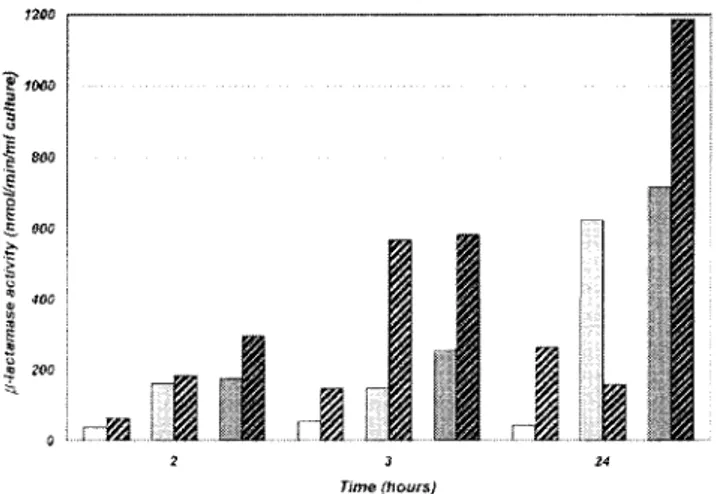
THIN-B production in E. coli.
The bla
THIN-Bgene was
cloned in the T7-based expression vector pET-9a, and enzyme
production was tested by using three different E. coli hosts
producing T7 RNA polymerase. Pilot expression experiments,
in which cultures were grown at 37°C, yielded poor
-lacta-mase activity both in culture supernatants and in cell extracts
(
specific imipenemase activity was always lower than 100 nmol/
min/ml) (data not shown). Lowering the incubation
tempera-ture to 25°C significantly increased the
-lactamase yield
(ap-proximately 3- to 14-fold, depending on the host).
Compara-tive analysis of
-lactamase production in the three different
expression systems showed that addition of the inducer
gener-ally improved the yield of recombinant protein and that the
highest activity was observed in the cell extracts after 24 h (Fig.
1).
-Lactamase activity was also measured in the culture
su-pernatants at different times, but no trace of enzyme was found
in the corresponding samples, except with the BL-21-SI-based
system, for which significant cell lysis was observed when the
inducer (NaCl at 0.3 M) was added to the culture (data not
shown). The highest yield was obtained with MCT236(DE3)
(pET-THIN-B) after induction with 1 mM IPTG, and this
system was adopted for larger-scale THIN-B production and
purification.
THIN-B purification.
THIN-B was purified from a cell
ex-tract of E. coli MCT236(DE3)(pET-THIN-B), obtained from a
0.5-liter culture induced with IPTG and grown at 25°C for 24 h,
by means of two ion-exchange chromatography steps, an anion
exchange at pH 9.8 followed by a cation exchange at pH 5.0.
No detectable changes in enzyme activity were registered
within 2 h of exposure at such pH values. A summary of the
purification process is shown in Table 1. Higher activity was
observed after desalting of the crude extract, presumably due
to removal of low-molecular-weight inhibitors whose nature
was not specifically investigated. The yield of purified protein
was approximately 30 mg per liter of culture. The purity of the
protein preparation was estimated to be
⬎95%, according to
SDS-PAGE analysis (data not shown). Recovery was relatively
low (30%), mainly because, after the cation-exchange step,
only the purest fractions were retained.
Matrix-assisted laser desorption ionization—time-of-flight
(MALDI-TOF) mass spectrometry yielded an average mass of
30,915
⫾ 45 Da for the purified THIN-B protein. The mass
spectrum of the enzyme tryptic digest, obtained by using a
MALDI-TOF spectrometer, yielded nine peaks, all at m/z
ra-tios in agreement with those predicted by computing analysis,
and covering 43% of the protein sequence (Table 2). This
confirmed the authenticity of the enzyme preparation.
Structural features of THIN-B.
Analytical IEF of the
puri-fied protein preparation revealed a pI of 6.3
⫾ 0.3 (data not
shown), in good agreement with the calculated value for the
mature protein (pI 6.22).
Size exclusion chromatography yielded a molecular mass of
32
⫾ 2 kDa, indicating that, under the experimental conditions
adopted, the native enzyme is monomeric, unlike L1 but like
most other MBLs (3, 4, 9, 22). The absence of oligomerization
in THIN-B, despite its longer N-terminal portion, is consistent
with the absence of the residues that are known to be involved
in oligomerization of L1 (28). In particular, the methionine
residue at position 175, which is responsible for hydrophobic
intersubunit interactions involved in formation of the L1
dimer, is replaced in THIN-B by a glycine whose side chain
lacks any possibility of interacting with the hydrophobic pocket
of another subunit. In addition, of the three hydrophobic
res-idues (Leu154, Pro198, Tyr236) that form the L1 hydrophobic
pocket where Met175 is accommodated, only Pro198 is
con-served in THIN-B (Fig. 2).
The THIN-B enzyme contains six cysteine residues,
remark-ably more than are found in the mature GOB-1 (one cysteine
residue), L1 or CAU-1 (two cysteine residues), and FEZ-1
(three cysteine residues) proteins (Fig. 2). Determination of
the free sulfhydryl groups yielded free cysteine/enzyme ratios
of 2.1
⫾ 0.3 and 5.6 ⫾ 0.4 for the unreduced and reduced
proteins, respectively. This suggested that, in THIN-B, four
cysteines might be involved in the formation of two disulfide
bridges. One of these is likely formed by the Cys256 and
Cys290 residues, which in L1 and FEZ-1 are known to form a
disulfide bridge that links the last strand of the
-sheet in the
C-terminal domain with the terminal
␣-helix (15, 28), and
which are conserved in THIN-B. The other disulfide bridge
would most likely involve residues 208 and 213, which are
located at the extremities of the
11 and 12 strands,
respec-tively (15). Alternarespec-tively, the disulfide bridge could involve the
two cysteine residues at positions 32 and 35, which are located
close to the N terminus of the protein (Fig. 2).
Another notable feature of THIN-B, compared to other
enzymes of subclass B3, is its larger protein size (Fig. 2).
THIN-B is 29 and 33 amino acids longer, respectively, than L1
and FEZ-1, the two subclass B3 enzymes whose
three-dimen-sional structures have been solved. Sequence comparison and
FIG. 1. Time-dependent production of the THIN-B enzyme in cell
lysates with different expression systems. E. coli
BL21(DE3)(pET-THIN-B), white; E. coli BL21-SI(pET-BL21(DE3)(pET-THIN-B), light grey; E. coli
MCT236(DE3)(pET-THIN-B), dark grey. Hatched bars represent
sam-ples from cultures to which an inducer (1 mM IPTG, or 0.3 M NaCl for
BL21-SI) was added.
TABLE 1. Summary of a typical purification procedure of the
THIN-B enzyme from E. coli MCT236(DE3)(pET-THIN-B)
Product of purification step Vol (ml) Total amt of protein (mg) Total activitya (U) Sp act (U/mg of protein) Recov-eryc (%) Purifi-cation (fold)
Cell extract
20
680
330
0.49
Sephadex G-25 eluate 36 580 880b 1.51 100 3.12 Source 15Q eluate 27 38 390 10.3 44 21 Resource S eluate 8.8 14.6 300 20.5 34 41aOne unit of activity was defined as the amount of enzyme able to hydrolyze 1mol of substrate in 1 min under the conditions described in Materials and Methods.
bThe higher activity was presumably due to buffer change and/or the elimi-nation of inhibitors potentially present in the crude extract.
cCalculated on the basis of the total activity obtained after buffer exchange (Sephadex G-25 eluate).
TABLE 2. MALDI-TOF mass spectrum data obtained for the
THIN-B protein preparation after digestion with trypsin
a Observed m/z value Predicted mass (Da) Mass difference (Da) Peptide sequence 1,381.708 1,381.7334 0.025 G310 VALPAAAPAAQHAH 1,382.730 1,382.7096 ⫺0.019 A292YAATADAMLTKRb 1,506.764 1,506.7434 ⫺0.02 G236 DGPDISASFAASIAK 1,530.716 1,530.6754 ⫺0.04 S277 GEHNPFIDANACR 1,666.870 1,666.8771 0.007 E308RGVALPAAAPAAQHAHb 1,673.740 1,673.7509 0.011 T21 PAPKPDTPVDCDSCK 1,678.930 1,678.8911 ⫺0.038 D152 DPQFQAKPVVHVAK 2,263.138 2,263.2002 0.062 V252 AALPCDIILSVHPDSTGVLDK 2,515.120 2,515.1333 0.013 C213 LDVVYADSLNPYSSGDFTYTGK aThe observed m/z values are compared to the predicted monoisotopic masses of the different trypsin-generated peptides, whose sequences are shown and which covered 43% of the protein sequence.
b
consideration of structural data indicate that most of those
extra amino acids are likely to be part of a longer N-terminal
coil and of a longer C-terminal
-helix, with no important
modifications of internal elements (Fig. 2).
Kinetic properties of the THIN-B enzyme.
Determination of
the kinetic parameters of THIN-B with several substrates
rep-resentative of different
-lactam families, including penicillins,
narrow- to expanded-spectrum cephems, carbapenems, and
aztreonam, revealed that all the compounds tested, except
aztreonam, were consistently hydrolyzed by the enzyme,
al-though with a remarkable variability of k
cat/K
mratios (from
8
⫻ 10
3to 5
⫻ 10
6M
⫺1䡠 s
⫺1) (Table 3). The highest
cata-lytic efficiencies (k
cat/K
mratios,
⬎10
6
M
⫺1䡠 s
⫺1) were
ob-served with carbapenems, cefuroxime, and cefotaxime, while
cefepime appeared to be a poor substrate. Comparison of K
mvalues indicated a higher apparent affinity for carbapenems
and cephalosporins (except cefepime) than for penicillins. The
highest turnover rate was observed with ampicillin (k
cat, 480
s
⫺1), a substrate for which the enzyme also exhibited the
high-est K
m(1.3 mM), resulting in a relatively low catalytic efficiency
compared to those of expanded- and broad-spectrum
cepha-losporins and carbapenems (Table 3). Aztreonam was not
hy-drolyzed and did not interact with the enzyme (Table 3), as has
also been observed with other MBLs (3, 9–13, 20, 22, 23). With
cephaloridine (at a substrate concentration of 100
M), the
hydrolysis kinetics presented a notable though slow
inactiva-tion pattern until an apparent steady state was reached (rate,
650
mol/min/mg of enzyme) (Fig. 3). Although the
inactiva-tion mechanism has not been further investigated, the most
reliable hypothesis possibly explaining a similar behavior could
be the existence of two enzyme-substrate complex forms with
significantly different turnover rates, leading to accumulation
of the enzyme-substrate complex with minor activity. It would
be interesting to further investigate this point by mass
spec-trometry experiments.
THIN-B exhibits a broad substrate profile, similar to that of
other MBLs of subclass B3 (3, 9, 11, 12, 22). Comparison of the
kinetic parameters of these enzymes reveals both common
FIG. 2. Amino acid sequence alignment of the THIN-B MBL in comparison with the other subclass B3 enzymes (L1 from S. maltophilia
IID1275 [29], FEZ-1 from L. gormanii ATCC 33297 [5], GOB-1 from Chryseobacterium meningosepticum PINT [3], and CAU-1 from Caulobacter
crescentus DSM4727 [9]). Residues that are identical in all sequences are boldfaced. The structural elements of L1 are indicated above the
sequences (
␣ and 3
10, helices;
, strands). Cysteine residues are shaded.
TABLE 3. Kinetic parameters of the purified THIN-B metallo-
-lactamase and comparison with catalytic efficiencies
of other subclass B3 enzymes
aSubstrate kcat(s⫺1) Km(M)
kcat/Km(M⫺1䡠 s⫺1)
THIN-B L1 GOB-1 FEZ-1 CAU-1
Ampicillin
480
1,300
3.7
⫻ 10
54.4
⫻ 10
6—
b1.1
⫻ 10
45
⫻ 10
5Piperacillin
100
500
2
⫻ 10
57
⫻ 10
61.66
⫻ 10
61.2
⫻ 10
45.7
⫻ 10
5Cefuroxime
140
50
2.8
⫻ 10
62.7
⫻ 10
69.8
⫻ 10
56.6
⫻ 10
61.4
⫻ 10
4Cefotaxime
80
40
2
⫻ 10
62.6
⫻ 10
68.5
⫻ 10
52.4
⫻ 10
6—
Ceftazidime
20
140
1.4
⫻ 10
5—
7.6
⫻ 10
54
⫻ 10
32.0
⫻ 10
3Cefepime
⬎2.3
⬎300
7.9
⫻ 10
31.9
⫻ 10
42
⫻ 10
56
⫻ 10
3—
Imipenem
120
80
1.5
⫻ 10
67.5
⫻ 10
56.6
⫻ 10
52
⫻ 10
52
⫻ 10
5Meropenem
200
40
5
⫻ 10
64.5
⫻ 10
65.34
⫻ 10
65
⫻ 10
52.6
⫻ 10
5Aztreonam
NH
c⬎1,000
—
—
—
—
—
aData are means from three independent measurements; standard deviations were below 10%. Data for other enzymes are from the work of Felici and colleagues (11, 12) (L1), Bellais et al. (3) (GOB-1), Mercuri et al. (22) (FEZ-1), and Docquier et al. (9) (CAU-1).
b—, data not available. cNH, no hydrolysis detected.
features and functional differences. The carbapenemase
activ-ity tends to be quite efficient in all cases, with an overall
preference for meropenem, while cefepime tends to behave as
a relatively poor substrate in most cases. On the other hand,
large differences in catalytic efficiency can be observed with
penicillins and some cephems (Table 3). The slow inactivation
of THIN-B by cephaloridine is certainly an uncommon feature.
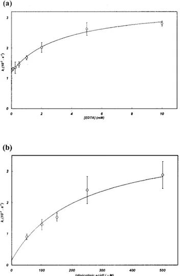
Inactivation of THIN-B by metal chelators.
The THIN-B
enzyme was efficiently inactivated by EDTA,
o-phenanthro-line, and dipicolinic acid, allowing the determination of
pseu-do-first-order inactivation rates (k
i).
With EDTA and dipicolinic acid, the inactivation rates
var-ied with the inactivator concentration, following a hyperbolic
dependence, and individual inactivation constants (K, k
⫹2,
k
⫺2⬘) could be calculated (Fig. 4; Table 4). This behavior
suggests that the inactivator does not act by simply scavenging
the free metal from the buffer but that a ternary
enzyme-metal-inactivator complex is formed during inactivation. No similar
behavior has been observed for other enzymes of subclass B3
(9, 22), and THIN-B represents the first example of a subclass
B3 enzyme for which formation of the ternary complex could
be hypothesized.
With o-phenanthroline, the measured inactivation rate [k
i,
(1.22
⫾ 0.06) ⫻ 10
⫺2s
⫺1] was independent of the inactivator
concentration (in the range of 50 to 500
M) and
correspond-ed to the k
⫹2constant, indicating that formation of the ternary
complex was rather efficient (K,
⬍50 M) (Table 4).
The inactivation efficiencies of chelators were generally high,
with values similar to those observed for subclass B1 enzymes
(10, 13, 20). Interestingly, with THIN-B, EDTA was a better
inactivator than dipicolinic acid or o-phenanthroline (Table 4),
at variance from what has generally been observed for MBLs
(9, 10, 13, 16, 20, 22).
Concluding remarks.
Subclass B3 of MBLs, which until
recently included only the L1 enzyme from S. maltophilia,
currently includes several members of notable structural and
functional diversity. Although produced by a nonpathogenic
bacterium, the THIN-B enzyme exhibits some peculiar
struc-tural and functional features and could be an interesting model
for further structural studies. Moreover, the distribution of
MBLs among an increasing number of bacterial species
typi-cally associated with environmental niches that might act as a
reservoir for efficient resistance determinants might offer an
important clue for MBL evolution.
ACKNOWLEDGMENTS
This work was supported by the European Research Network on
metallo-
-lactamases (contract HPRN-CT-2002-00264) and by a grant
from the Italian Ministero dell’Istruzione, dell’Universita
` e della
Ricerca (PRIN 2003). J.-D.D. is funded by the “Fonds National de la
FIG. 3. Time course for cephaloridine hydrolysis in the presence of
13 nM THIN-B. A steady state was reached after approximately 10
min. Cephaloridine was normally hydrolyzed when 8 nM VIM-2 (10)
was added to the reaction mixture, as indicated by the arrow.
FIG. 4. Dependence of the pseudo-first-order inactivation rate (k
i)
of THIN-B on the concentration of the metal chelator EDTA (a) or
dipicolinic acid (b). The line in each graph represents the best fit
resulting from the nonlinear regression used to calculate individual
inactivation parameters (K, k
⫹2, and k
⫺2⬘).
TABLE 4. Inactivation parameters for the THIN-B
metallo-
-lactamase with various chelating agents
a Chelating agent k⫺2⬘ (s⫺1) k⫹2(s⫺1) (M)K (Mk⫺1⫹2䡠 s/K⫺1)EDTA
1.2
⫻ 10
⫺22.2
⫻ 10
⫺23,140
7.0
⫻ 10
3o-Phenanthroline
⬍10
⫺21.2
⫻ 10
⫺2⬍50
⬎2.4 ⫻ 10
2Dipicolinic acid
1.5
⫻ 10
⫺33.9
⫻ 10
⫺2215
1.8
⫻ 10
2Recherche Scientifique” of Belgium as a “Collaborateur Scientifique
F.N.R.S.”
REFERENCES
1. Ambler, R. P. 1980. The structure of-lactamases. Philos. Trans. R. Soc. Lond. B 289:321–331.
2. Aravind, L. 1999. An evolutionary classification of the metallo--lactamase fold proteins. In Silico Biol. 1:69–91.
3. Bellais, S., D. Aubert, T. Naas, and P. Nordmann. 2000. Molecular and biochemical heterogeneity of class B carbapenem-hydrolyzing-lactamases in Chryseobacterium meningosepticum. Antimicrob. Agents Chemother. 44: 1878–1886.
4. Bicknell, R., E. L. Emanuel, J. Gagnon, and S. G. Waley. 1985. The produc-tion and molecular properties of the zinc-lactamase of Pseudomonas mal-tophilia IID 1275. Biochem. J. 229:791–797.
5. Boschi, L., P. S. Mercuri, M. L. Riccio, G. Amicosante, M. Galleni, J. M.
Fre`re, and G. M. Rossolini.2000. The Legionella (Fluoribacter) gormanii metallo--lactamase: a new member of the highly divergent lineage of mo-lecular-subclass B3-lactamases. Antimicrob. Agents Chemother. 44:1538– 1543.
6. Callebaut, I., D. Moshous, J. P. Mornon, and J. P. de Villartay. 2002. Metallo--lactamase fold within nucleic acids processing enzymes: the -CASP family. Nucleic Acids Res. 30:3592–3601.
7. Creighton, T. E. 1989. Disulfide bonds between cysteine residues, p. 156–168. In T. E. Creighton (ed.), Protein structure, a practical approach. IRL Press, Oxford, United Kingdom.
8. Daiyasu, H., K. Osaka, Y. Ishino, and H. Toh. 2001. Expansion of the zinc metallo-hydrolase family of the-lactamase fold. FEBS Lett. 503:1–6. 9. Docquier, J. D., F. Pantanella, F. Giuliani, M. C. Thaller, G. Amicosante, M.
Galleni, J. M. Fre`re, K. Bush, and G. M. Rossolini.2002. CAU-1, a subclass B3 metallo--lactamase of low substrate affinity encoded by an ortholog present in the Caulobacter crescentus chromosome. Antimicrob. Agents Che-mother. 46:1823–1830.
10. Docquier, J. D., J. Lamotte-Brasseur, M. Galleni, G. Amicosante, J. M.
Fre`re, and G. M. Rossolini.2003. On functional and structural heterogeneity of VIM-type metallo--lactamases. J. Antimicrob. Chemother. 51:257–266. 11. Felici, A., and G. Amicosante. 1995. Kinetic analysis of extension of substrate specificity with Xanthomonas maltophilia, Aeromonas hydrophila, and Bacil-lus cereus metallo--lactamases. Antimicrob. Agents Chemother. 39:192– 199.
12. Felici, A., G. Amicosante, A. Oratore, R. Strom, P. Ledent, B. Joris, L.
Fanuel, and J. M. Fre`re.1993. An overview of the kinetic parameters of class B-lactamases. Biochem. J. 291:151–155.
13. Franceschini, N., B. Caravelli, J. D. Docquier, M. Galleni, J. M. Fre`re, G.
Amicosante, and G. M. Rossolini.2000. Purification and biochemical char-acterization of the VIM-1 metallo--lactamase. Antimicrob. Agents Che-mother. 44:3003–3007.
14. Galleni, M., J. Lamotte-Brasseur, G. M. Rossolini, J. Spencer, O. Dideberg,
and J. M. Fre`re.2001. Standard numbering scheme for class B-lactamases. Antimicrob. Agents Chemother. 45:660–663.
15. Garcia-Saez, I., P. S. Mercuri, C. Papamicael, R. Kahn, J. M. Fre`re, M.
Galleni, G. M. Rossolini, and O. Dideberg.2003. Three-dimensional struc-ture of FEZ-1, a monomeric subclass B3 metallo--lactamase from
Fluori-bacter gormanii, in native form and in complex withD-captopril. J. Mol. Biol.
325:651–660.
16. Hernandez-Valladeres, M., A. Felici, G. Weber, H. W. Adolph, M.
Zeppeza-uer, G. M. Rossolini, G. Amicosante, J. M. Fre`re, and M. Galleni.1997. Zn(II) dependence of the Aeromonas hydrophila AE036 metallo--lactamase activity and stability. Biochemistry 36:11534–11541.
17. James, P., M. Quadroni, E. Carafoli, and G. Gonnet. 1994. Protein identi-fication in DNA databases by peptide mass fingerprinting. Protein Sci.
3:1347–1350.
18. Laemmli, U. K. 1970. Cleavage of structural proteins during the assembly of the head of bacteriophage T4. Nature 227:680–685.
19. Laraki, N., M. Galleni, I. Thamm, M. L. Riccio, G. Amicosante, J. M. Fre`re,
and G. M. Rossolini.1999. Structure of In31, a blaIMP-containing Pseudo-monas aeruginosa integron phyletically related to In5, which carries an un-usual array of gene cassettes. Antimicrob. Agents Chemother. 43:890–901. 20. Laraki, N., N. Franceschini, G. M. Rossolini, P. Santucci, C. Meunier, E. de
Pauw, G. Amicosante, J. M. Fre`re, and M. Galleni.1999. Biochemical char-acterization of the Pseudomonas aeruginosa 101/1477 metallo--lactamase IMP-1 produced by Escherichia coli. Antimicrob. Agents Chemother. 43: 902–906.
21. Lauretti, L., M. L. Riccio, A. Mazzariol, G. Cornaglia, G. Amicosante, R.
Fontana, and G. M. Rossolini.1999. Cloning and characterization of blaVIM,
a new integron-borne metallo--lactamase gene from a Pseudomonas aerugi-nosa clinical isolate. Antimicrob. Agents Chemother. 43:1584–1590. 22. Mercuri, P. S., F. Bouillenne, L. Boschi, J. Lamotte-Brasseur, G.
Amico-sante, B. Devreese, J. Van Beeumen, J. M. Fre`re, G. M. Rossolini, and M. Galleni.2001. Biochemical characterization of the FEZ-1 metallo- -lacta-mase of Legionella gormanii ATCC 33297Tproduced in Escherichia coli.
Antimicrob. Agents Chemother. 45:1254–1262.
23. Nordmann, P., and L. Poirel. 2002. Emerging carbapenemases in Gram-negative aerobes. Clin. Microbiol. Infect. 8:321–331.
24. Osano, E., Y. Arakawa, R. Wacharotayankun, M. Ohta, T. Horii, H. Ito, F.
Yoshimura, and N. Kato.1994. Molecular characterization of an enterobac-terial metallo-lactamase found in a clinical isolate of Serratia marcescens that shows imipenem resistance. Antimicrob. Agents Chemother. 38:71–78. 25. Rossolini, G. M., M. A. Condemi, F. Pantanella, J. D. Docquier, G.
Amico-sante, and M. C. Thaller.2001. Metallo--lactamase producers in environ-mental microbiota: new molecular class B enzyme in Janthinobacterium livi-dum. Antimicrob. Agents Chemother. 45:837–844.
26. Saino, Y., F. Kobayashi, M. Inoue, and S. Mitsuhashi. 1982. Purification and properties of inducible penicillin-lactamase isolated from Pseudomonas maltophilia. Antimicrob. Agents Chemother. 22:564–570.
27. Sambrook, J., and D. W. Russell. 2001. Molecular cloning: a laboratory manual, 3rd ed. Cold Spring Harbor Laboratory Press, Cold Spring Harbor, N.Y.
28. Ullah, J. H., T. R. Walsh, I. A. Taylor, D. C. Emery, C. S. Verma, S. J.
Gamblin, and J. Spencer.1998. The crystal structure of the L1
metallo--lactamase from Stenotrophomonas maltophilia at 1.7 A˚ resolution. J. Mol. Biol. 284:125–136.
29. Walsh, T. R., L. Hall, S. J. Assinder, W. W. Nichols, S. J. Cartwright, A. P.
MacGowan, and P. M. Bennett.1994. Sequence analysis of the L1 metallo--lactamase from Xanthomonas maltophilia. Biochim. Biophys. Acta 1218: 199–201.