U
"TOR VERGATA"
FACOLTA' DI MEDICINA
DOTTORATO DI RICERCA IN
BIOCHIMICA E BIOLOGIA MOLECOLARE
“NATURAL INHIBITORS
A PROPOSED
Dott. Daniele Amadio
ocente Guida: prof. Mauro Maccarrone
vanni Floris;
DIPARTIMENTO DI MEDICINA SPERIMENTALE E SCIENZE BIOCHIMICHE VIA MONTPELLIER, 1 – 00133 ROMA
NIVERSITÀ DEGLI STUDI DI ROMA
2008-XXI CICLO
OF FAAH:
MECHANISM FOR THE MODULATION OF THEIR
ACTIVITY”
D
Esaminatori: proff. Giovanni Antonini; Gio
Alessandro Finazzi Agrò
-Agrò
Coordinatore: prof. Alessandro Finazzi
1.1 Overview of the endocannabinoid system 10
thway
OLASE
2.1 Subcellular localization 21
.1 Effect of 15-(S)-HAEA, 15-(S)-MeOAEA and 15-(S)-AcOAEA
P 1 rece ors ION PERSPECTIVES 57 PROCEDURES .1 Materials 58 ABSTRACT 4 RIASSUNTO 5 INTRODUCTION 6
CHAPTER 1. THE ENDOCANNABINOID SYSTEM 10
1.2 Biosynthesis of endocannabinoid s 11
1.3 Degradation of endocannabinoids 15
1.4 Molecular targets and signalling pa s 20
CHAPTER 2. FATTY ACID AMIDE HYDR 21
2.2 Regulation of gene expression 22
2.3 Structural proprieties 24
2.4 Catalytic reaction 29
2.5 Synthetic inhibitors 32
2.6 Natural inhibitors 36
CHAPTER 3. OBJECTIVE OF THE RESEARCH 38 CHAPTER 4. RESULTS 40
4
on AEA metabolism 40
4.2 Effect of 15-(S)-HAEA, 15-(S)-MeOAEA and 15-(S)-AcOAEA
on 2-AG metabolism 43
4.3 Effect of 15-(S)-HAEA, 15-(S)-MeOAEA and 15-(S)-AcOAEA
on CB1R, CB2 and TR V pt 45
4.4 Molecular modelling of FAAH and molecular dynamics
simulation 47
CHAPTER 5. DISCUSS 52 CHAPTER 6. CONCLUSION AND FUTURE
CHAPTER 7. EXPERIMENTAL 58
7.2 Synthesis of 15-(S)-MeOAEA and 15-(S)-AcOAEA 59
7.3 Assays of AEA metabolism 63
7.4 Assays of 2-AG metabolism 64
7.5 Binding to CB1, CB2 and TRPV1 receptors 65
7.6 Molecular modelling of FAAH 66
7.7 Molecular dynamics simulations 67
7.8 Statistical analysis 68
REFERENCES 69
RINGRAZIAMENTI 84
SHORT CURRICULUM VITAE 85
ABSTRACT
Endocannabinoids are novel endogenous lipid mediators with neuro- and immuno-modulatory functions. They are called endocannabinoids because
they bind the same receptors activated by Δ9-tetrahydrocannabinol (THC),
the active principle of hashish and marijuana. Several enzymes for the biosynthesis and degradation of the endocannabinoids have been discovered and characterized, forming the so called “endocannabinoid system”.
Anandamide (AEA), one of the best studied endocannabinoids, is inactivated by fatty acid amide hydrolase (FAAH). However, in some cells like platelets and polymorphonuclear leukocytes an oxidative pathway has also been described. The main products generated by lipoxygenase (LOX) are hydroperoxyanandamides (HpAEAs), which are quickly reduced to hydroxyanandamides (HAEAs). They have been tested on the enzymes of the endocannabinoid system, suggesting they may act as endogenous FAAH inhibitors.
Of all the LOX-generated oxygenated derivates, 15-HAEA appears to be the most interesting, due to its specific ability to inhibit FAAH but not anandamide membrane transporter (AMT) nor N-acyl-phosphatidylethanolamine-specific phospholipase D (NAPE–PLD), neither to bind the cannabinoid receptors (CBRs).
Methylation and acetylation are biochemical reactions common in the cellular environment and we investigated whether methylated and acetylated derivatives of HAEAs might affect the inhibitory properties of these natural compounds. Moreover, we investigated the effects of these compounds on the other proteins of the endocannabinoid system. We show that methylated and acetylated derivatives of HAEAs are remarkable modulators of molecular recognition of HAEAs by the elements of the endocannabinoid system.
The advantage of these natural substances, unlike the most powerful synthetic compounds, is that HAEAs are reversible rather than irreversible inhibitors of FAAH, and they seem to be suitable tools for flexible modulation of the endocannabinoid system in vivo.
RIASSUNTO
Gli endocannabinoidi sono una classe di composti lipidici con funzioni neuro- ed immuno-modulatorie identificati di recente. Sono chiamati “endocannabinoidi” poiché sono in grado di legare ed attivare gli stessi
recettori attivati dal Δ9-tetraidrocannabinolo (THC), il principio attivo
dell’hashish e della marijuana. Nel corso degli ultimi anni sono stati individuati e caratterizzati diversi enzimi responsabili della biosintesi e della degradazione degli endocannabinoidi e ci si riferisce complessivamente a questi con il termine di “sistema endocannabinoide”.
L’anandamide (AEA), uno degli endocannabinoidi più studiati, viene principalmente inattivata dalla idrolasi delle ammidi degli acidi grassi (fatty
acid amide hydrolase, FAAH). Tuttavia, in alcuni tipi di cellule come
piastrine e leucociti, un metabolismo ossidativo addizionale è stato descritto per questa classe di composti. L’azione della lipossigenasi (LOX) sulla anandamide genera idroperossianandamidi (HpAEA), le quali sono velocemente ridotte a idrossianandamidi (HAEA). Queste ultime sono state testate sugli enzimi del sistema endocannabinoide e risultano possedere una maggiore affinità per la FAAH rispetto all’anandamide stessa, suggerendo una loro azione come inibitori endogeni.
Tra tutti i derivati ossigenati generati dalla LOX, la 15-HAEA risulta essere il più interessante per la sua capacità di inibire selettivamente la FAAH ma non il suo trasportatore (anandamide membrane transporter, AMT), né l’enzima responsabile della produzione di AEA (NAPE-PLD), nè i recettori cannabici (CBRs).
La metilazione e l’acetilazione sono reazioni biochimiche comuni nell’ambiente cellulare ed in questo lavoro abbiamo studiato l’effetto di tali derivati metilati ed acetilati delle HAEA, ipotizzando un’eventuale via di inattivazione di questi composti naturali. Inoltre, abbiamo caratterizzato gli effetti di questi composti sulle altre proteine del sistema endocannabinoide coinvolte sia nel metabolismo dell’AEA che del 2-arachidonilglicerolo (2-AG), dimostrando che questi derivati possono agire come modulatori del riconoscimento molecolare delle HAEA da parte degli elementi del sistema endocannabinoide.
Diversamente dai potenti inibitori sintetici della FAAH finora sviluppati, il vantaggio di questi composti naturali è che tutte le HAEA hanno un effetto reversibile, rappresentando dei candidati ideali per una modulazione flessibile del sistema endocannabinoide in vivo.
INTRODUCTION
The recreational value of Cannabis sativa preparations is known to most people, largely as a result of the explosion in its use in the late 1960s; indeed, marijuana is still one of the most widespread illicit drugs of abuse in the world (Adams and Martin, 1996). The plant contains about 60
cannobinoid compounds (Ross and Elsohly, 1996), among which Δ9
-tetrahydrocannabinol (Δ9-THC) (Figure 1) is the primary psychoactive
component and is thought to mediate most of the physiological effects
associated with marijuana smoking (Dewey, 1986). Δ9-THC was used in
folklore medicine long before the discovery of its mechanism of action. The stringent structural characteristics that cannabinoid compounds must possess in order to exert their psychotropic effects, and the key observation that cannabinoids inhibit adenylate cyclase, supported the presence of a specific, high-affinity binding site for these lipidic substances (Howlett and
Fleming, 1984). Shortly afterwards, the first membrane receptor for Δ9-THC
was identified in rat brain (Devane et al., 1988). Its distribution was consistent with the pharmacological properties of psycotropic cannabinoids, and therefore it was designated type 1 cannabinoid receptor (CB1R) (Devane et al., 1988). A peripheral cannabinoid-binding receptor was identified a few years later in spleen and immune cells, and was called type 2 cannabinoid receptor (CB2R) (Munro et al., 1993).
Since then, a number of endogenous agonists of CB receptors were characterized, i.e. amides, esters and ethers of long chain polyunsaturated fatty acids collectively termed ‘endocannabinoids’. Remarkably, these
compounds are structurally different from Δ9-THC or other plant
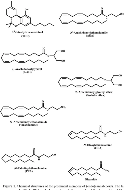
cannabinoids (De Petrocellis et al., 2004). In fact two arachidonate derivatives, the amide N-arachidonoylethanolamine (anandamide, AEA) (Devane et al., 1992) and the ester 2-arachidonoylglycerol (2-AG) (Sugiura et al., 1995), both shown in Figure 1, are the most biologically active endocannabinoids described to date (De Petrocellis et al., 2004). Also the ether 2-arachidonoylglyceryl-ether (noladin ether) (Figure 1) has been shown to act as an endocannabinoid (Hanus et al., 2001), but its actual physiological relevance remains a matter of debate (Oka et al., 2003). Furthermore an ‘inverted anandamide’, O-arachidonoylethanolamine (virodhamine) (Figure 1), has been shown to behave as a partial agonist or as a full agonist at CB1 or CB2 receptors, respectively (Porter et al., 2002).
Instead, the amides N-oleoylethanolamine (OEA), N-palmitoylethanolamine (PEA) and oleamide (Figure 1) are better considered ‘endocannabinoid-like’ compounds, because they do not activate directly CB receptors, but rather prolong the activity of true endocannabinoids within the cell by an ‘entourage effect’ (De Petrocellis et al., 2004).
AEA and 2-AG are present in the central nervous system (CNS) and also in peripheral tissues (Sugiura et al., 2002), but exhibit important differences in their quantitative distribution; 2-AG is more abundant than AEA in the brain and behaves as a full agonist for CB1R and CB2R, while AEA acts as partial agonist for CB1R and as a weak partial agonist for CB2R (Sugiura et al., 2000). AEA levels may vary by 4 to 6-fold in different regions of the rat brain, with the highest levels in the striatum and brainstem and the lowest levels in the cerebellum and cortex (Bisogno et al., 1999a; Yang et al., 1999). AEA was found in regions of both rat and human brains that contain high densities of CB1R (e.g., hippocampus, cerebellum, and striatum) and also in a region that is sparse in CB1R like the thalamus (Felder et al., 1996). It is clear from these data that for AEA the relative regional abundance in the brain does not correlate with the distribution of CB1R. AEA levels in the brain are equivalent to those of other neurotransmitters such as dopamine and serotonin, but at least 10-fold lower than the levels reported for GABA and glutamate. AEA has also been found in peripheral tissues such as human and rat spleen, which expresses high levels of CB2R. Small amounts of AEA were also detected in human serum, plasma, and cerebrospinal fluid (Felder et al., 1996).
The concentration of 2-AG can be up to ~200-fold higher than that of AEA in the brain (Bisogno et al., 1999a). Yet, there are reports showing much lower 2-AG:AEA ratios in rat striatum (~10), substantia nigra (~3), and globus pallidus (~4) (Gubellini et al., 2002) These differences may arise from different methodologies, for instance killing the animals by decapitation without immediate freezing instead of soaking in liquid nitrogen can increase 2-AG levels by ~15-fold (Sugiura et al., 2002). Discrepancies may also be a consequence of the high sensitivity of endocannabinoids to environmental factors like animal diets, caging and bedding systems, viral load, water quality, and pathogen infections. A recent example of the dramatic effect of these factors on endocannabinoid levels has been recently reported (Guo et al., 2005). In this context, it seems noteworthy that a recent study has shown that the extracellular concentrations of AEA and 2-AG are both in the nanomolar range (Caillè et al., 2007), suggesting that these two compounds have a similar availability for their CBR-mediated biological
actions. On the other hand, the spatial distribution of the two endocannabinoids is similar in different regions of the brain. In fact, the highest concentrations of 2-AG were found in the brainstem, medulla, limbic forebrain, striatum, and hippocampus, and the lowest in the cortex, diencephalons, mesencephalon, hypothalamus, and cerebellum (Sugiura et al., 2002). Therefore, much alike AEA, no correlation was found between 2-AG concentrations and CB1R distribution. 2-2-AG was also detected in the peripheral nervous system, i.e. in the sciatic nerve, lumbar spinal cord, and lumbar dorsal root ganglion cells (Sugiura et al., 2002).
In just one decade, endocannabinoids have been shown to play manifold roles, both in the CNS and in the periphery. In particular, it is now widely accepted that the biological activity of AEA and 2-AG is largely dependent on a ‘metabolic control’, that modulates the effects of these substances by modulating their in vivo concentration (or endogenous tone) (Cravatt and Lichtman, 2002).
O OH N H O OH O O OH OH O O NH2 O OH OH N H O OH N H O OH NH2 O Δ9-tetrahydrocannabinol (THC) Ν−Αrachidonoylethanolamide (AEA) 2−Αrachidonoylglycerol (2-AG) 2−Αrachidonoylglyceryl ether (Noladin ether) Ο−Αrachidonoylethanolamide (Virodhamine) Ν−Oleoylethanolamine (OEA) Ν−Palmitoylethanolamine (PEA) Oleamide
Figure 1. Chemical structures of the prominent members of (endo)cannabinoids. The last
three compounds (OEA, PEA and oleamide) are better considered “endocannabinoid-like” compounds, because they do not activate cannabinoid receptors.
CHAPTER 1. THE ENDOCANNABINOID SYSTEM
1.1 Overview of the endocannabinoid system
Investigations of the pathways involved in the metabolism of endocannabinoids have grown exponentially in recent years following the discovery of cannabinoid receptors. As other lipid mediators, AEA and 2-AG are released from cells ‘on demand’ by stimulus-dependent cleavage of membrane phospolipid precursors (Di Marzo et al., 1994).
AEA biosynthesis has been shown to occur through several pathways mediated by N-acylphosphatidylethanolamide-phospholipase D (NAPE-PLD), a secretory PLA2 and PLC. 2-AG is generated through the action of selective enzymes such as phosphatidic acid phsophohydrolase, diacylglycerol lipase (DAGL), phosphoinositide-specific PLC (PI-PLC) and lyso-PLC. A putative membrane transporter, catalyzing a facilitated diffusion process, is involved in the cellular uptake or release of endocannabinoids. AEA is metabolized by fatty acid amidohydrolase (FAAH) and 2-AG is metabolized by monoacylglycerol lipase (MAGL), and to a lesser extent by FAAH.
Taken together AEA and 2-AG, their congeners and metabolic enzymes, their purported transporters and molecular targets form the ‘endocannabinoid system (ECS)’.
1.2 Biosynthesis of endocannabinoids
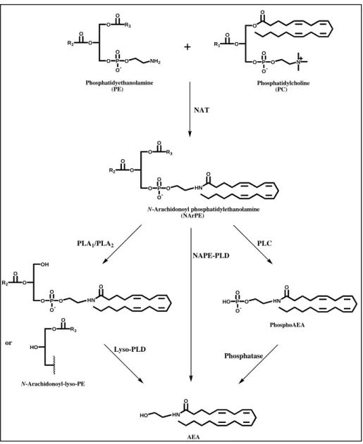
The main route for AEA biosynthesis occurs by two enzymatic steps involving the sequential action of a calcium dependent N-acyltransferase (NAT) and of a NAPE-specific phospholipase D (NAPE-PLD) (Okamoto et al., 2004) (Figure 2). In the first step, NAT catalyzes direct transfer of arachidonic acid from the sn-1 position of phosphatidylcholine (PC), generating N-arachidonoylphosphatidylethanolamine (NArPE), the AEA precursor. This biosynthetic pathway is in agreement with the observation that AEA levels are generally lower than those of the other NAEs in most of the tissue analyzed so far, because the arachidonic acid levels in position 1 of phospholipids are very low.
In the last step, NArPE is hydrolyzed by NAPE-PLD which releases AEA and phosphatidic acid (PA). This enzyme has been cloned and purified from rat heart and classified as a member of the zinc metallo-hydrolase family of the β-lactamase fold (Okamoto et al., 2004). NAPE-PLD does not recognize phosphatidylcholine and phoshatidylethanolamine as substrates, and it is widely distributed in mouse organs, with highest concentrations in brain, kidney and testis (Okamoto et al., 2004). The same group who characterized
NAPE-PLD also suggested that several PLA1/A2 isozymes can generate
N-arachidonoyl-lysoPE (NAr-lysoPE) from NArPE, and that a lysoPLD may
release AEA from NAr-lysoPE. Therefore, the sequential action of PLA1/A2
and lysoPLD may represent an alternative biosynthetic pathway for NAEs, including AEA (Sun et al., 2004) (Figure 2, left side).
Recently, it has been shown that in RAW264-7 macrophages, the lipopolysaccharide-induced anadamide production appears to depend mainly on a pathway whereby NAPE is hydrolysed to yield a phosphor-AEA, which is then dephosphorylated (Liu et al., 2006) (Figure 2, right side).
Furthermore, a non exclusive role of NAPE–PLD in the conversion of NAPE to AEA is clearly indicated by the unchanged brain levels of AEA in NAPE–PLD knockout mice (Leung et al., 2006). In fact, an independent pathway may occur through a double-deacylation of NAPE to generate lyso-NAPE and then glycerophospho-NAE, that is rapidly cleaved to release the corresponding NAE. This novel route is driven by the sequential action of a fluorophosphonate-sensitive serine hydrolase and a metal-dependent phosphodiesterase (Simon and Cravatt, 2006).
The levels of 2-AG in tissues and cells are usually much higher than those of AEA, and in principle they are sufficient to activate both cannabinoid receptor subtypes (Sugiura et al., 1995). At any rate, the 2-AG found in cells
and tissues is probably not uniquely used to stimulate cannabinoid receptors, as 2-AG is at the crossroads of several metabolic pathway. It is likely that a particular pool of 2-AG is produced via a special pathway only for the purpose of functioning as endocannabinoid. In line with this hypothesis, the extracellular levels of 2-AG are close to those of AEA, and are in the nanomolar range (Caillè et al., 2007).
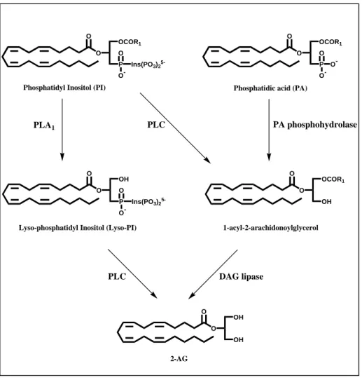
A biosynthetic pathway for 2-AG provides for quick hydrolysis of inositol phospholipids by a specific PLC, generating 1-acyl-2-arachidonoylglycerol (DAG) (Di Marzo, 2008) (Figure 3). DAG is then converted to 2-AG by a
sn-1-DAG lipase (Bisogno et al., 2003) (Figure 3). Another pathway for
2-AG formation involves the hydrolysis of phosphatidylinositol (PI) by PLA1 into lysoPI, followed by hydrolysis by phospholipase C (PLC) to produce 2-AG (Sugiura and Waku, 2000) (Figure 3). Furthermore, 2-2-AG has been shown to be produced also by PLC-independent pathways (Bisogno et al., 1999b).
Very recently, two sn-1-specific DAG lipases (α and β) responsible for the synthesis of 2-AG have been cloned by comparing human genome with Penicillium DAGL sequence. Both DAGL α and β are associated with the
cell membrane and are stimulated by high concentrations of Ca2+ and,
remarkably, by physiological concentrations of glutathione (Bisogno et al., 2003).
O O O O P O O -O N O R1 O O O P O O -O NH2 O R2 O R3 O O O P O O -O O R2 O R3 HN O OH O O P O O -O O R2 HN O O HO O R3 HO HN O HO P O O -O HN O or Phosphatidyethanolamine (PE) Phosphatidylcholine (PC) N-Arachidonoyl phosphatidylethanolamine (NArPE) PhosphoAEA AEA N-Arachidonoyl-lyso-PE NAT NAPE-PLD PLA1/PLA2 Lyso-PLD PLC Phosphatase
O O OCOR1 P O O -Ins(PO3)2 5-O O OCOR1 P O O -O -O O OH P O O -Ins(PO3)2 5-O O OCOR1 OH O O OH OH
Phosphatidyl Inositol (PI) Phosphatidic acid (PA)
Lyso-phosphatidyl Inositol (Lyso-PI)
2-AG
1-acyl-2-arachidonoylglycerol
PLC PA phosphohydrolase
PLC DAG lipase
PLA1
1.3 Degradation of endocannabinoids
The endocannabinoid actions are relatively short-lasteing, due to the presence of effective mechanisms for their cellular removal and subsequent degradation. Because they are lipophilic compounds, endocannabinoids can diffuse through the cell membrane. However, in order to be rapid, selective and subject to regulation, the diffusion process needs to be facilitated by a carrier, or to be driven by a mechanism capable of rapidly reducing the intracellular concentration of endocannabinoids, or both.
Indeed, AEA appears to be taken up by several cells via a facilitated transport mechanism, possibly mediated by a purported anandamide membrane transporter (AMT) (Glaser et al., 2005). In fact, cellular uptake of AEA is saturable, temperature-dependent and sensitive to synthetic inhibitors, as expected for a protein-mediated process (Maccarrone et al., 1998). However, some authors have reported evidence against the existence of AMT, suggesting that the enzyme mostly responsible for AEA hydrolysis, fatty acid amide hydrolase (FAAH) (Figure 4, right side), may be the sole responsible of AEA cellular uptake, by reducing its intracellular concentration (Glaser et al., 2003).
On the other hand, several data are in agreement with a facilitated transport of AEA independent of FAAH. In fact, different cells that do not express FAAH are still able to rapidly take up AEA (Day et al., 2001); compounds that inhibit AEA cellular uptake without affecting FAAH activity have been synthesized (Ortar et al., 2003); saturable AEA accumulation can be still observed in synaptosomes and cells prepared from FAAH-null mice (Ligresti et al., 2004; Fegley et al., 2004). Overall, from the available data it is possible to conclude that FAAH activity can contribute to facilitated AEA transport, yet it is not necessary; other mechanisms different from intracellular hydrolysis may also enhance the rate of endocannabinoid uptake. In line with this, a new model for AEA transport has been proposed, that might engage a caveolae/lipid rafts-related endocytic process (McFarland et al., 2004). Furthermore, the involvement of lipid rafts in the control of AEA uptake in neuronal cells (Bari et al., 2005) and in human immune cells (Bari et al., 2006) has also been documented.
On the other hand, it has been suggested that the 2-AG membrane transporter may be the same used by anandamide, i.e. AMT (Beltramo and Piomelli, 2000; Bisogno et al., 2001).
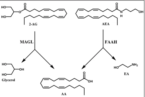
Once inside the cell, endocannabinoids are degraded through mechanisms depending on their chemical nature. FAAH has been identified as the main
responsible for AEA hydrolysis to arachidonic acid and ethanolamine (Cravatt and Lichtman, 2002) (Figure 4, right side). FAAH also catalyzes the hydrolysis of long chain primary fatty acid amide and glycerol esters; its structural and catalytic proprieties have been fully investigated (Fezza et al., 2008). Evidence has accumulated that points to FAAH as a key regulator of fatty acid amides (FAAs) in vivo concentration (Cravatt and Lichtman, 2003).
Although FAAH can catalyze also the hydrolysis of 2-AG (Di Marzo and Deutsch, 1998), the levels of the latter substance, unlike those of AEA, are not increased in FAAH ‘knockout’ mice (Lichtman et al., 2002). This observation is in agreement with the existence of other enzymes catalyzing 2-AG degradation (Di Marzo et al., 1999; Goparaju et al., 1999) (Figure 4, left side). In fact, monoacylglycerol lipase (MAGL) is a cytosolic enzyme that cleaves efficiently 2-AG (Ben-Shabat et al., 1998; Di Marzo and Deutsch, 1998). In rat brain, MAGL is more abundant in regions where also CB1 receptors are highly expressed (hippocampus, cortex, anterior thalamus and cerebellum). Furthermore, immunohistochemical studies in the hippocampus suggested a presynaptic localization of MAGL, supporting its role in the degradation of 2-AG as retrograde messenger. Interestingly, recent studies have confirmed a sort of ‘complementary localization’ of MAGL and FAAH in the brain, pre-synaptic and post-synaptic respectively, suggesting different roles for AEA and 2-AG in endocannabinoid signaling within the CNS (Gulyas et al., 2004). Incidentally, the data on MAGL localization supplement previous observations showing that the diacylglycerol lipases (DAGLs) responsible for 2-AG production are instead post-synaptic in the adult brain (Dinh et al., 2002; Bisogno et al., 2003). The hydrolysis of AEA by FAAH is the most common inactivation pathway. However, anandamide (and other endocannabinoids) can also be susceptible to oxidative metabolism catalyzed by lipoxygenase (LOX), cyclooxygenase-2 (COX-cyclooxygenase-2) and cytocrome P450 oxidase (Kozak and Marnett, cyclooxygenase-200cyclooxygenase-2) (Figure 5). In some cells like platelets and polymorphonuclear leukocytes an oxidative pathway has been described (Edgemond et al., 1998a), which leads to the generation of analogous of prostanoids (by COX-2, but not COX-1) and leukotrienes (by LOX). The COX-2 action on AEA and 2-AG leads to two new families of compounds named “prostamides” (prostaglandin ethanolamide) and prostaglandin glycerol esters (Ross et al., 2002), through the action of cycloxygenase-2 and, subsequentely, by the action of several prostaglandin synthases yet implicated in the arachidonic acid oxidative
metabolism. Prostamides do not affect FAAH or AMT, neither bind the CBRs (Fowler, 2007) and its molecular target is still unknown.
Lipoxygenase metabolism of AEA in mouse brain (Hampson et al., 1995) and more recently in blood cells (Veldhius et al., 2003) has also been demonstrated. Regarding the lipoxygenase products of AEA, they can be formed through the action of 5-, 12- and 15-LOX (Van der Stelt et al., 2002) (Figure 5). The hydroxyl derivates of AEA generated by LOX do not present significant affinity for cannabinoid receptors, but inhibit FAAH in the low µM range (Van der Stelt et al., 2002).
Few investigations of cytocrome P450-mediated endocannabinoid metabolism have been reported. These studies reported the production of more than 20 polar lipids, most of which appear to be mono-oxygenated AEA-derivatives (Bornheim et al., 1995).
In contrast to AEA and 2-AG, which have been detected in biological fluids of humans, the presence of hydroxy-derivatives of endocannabinoids and other related oxygenated metabolites has not yet been reported (in part due to the their very low concentration and to their very short life-time). However, there are no reasons to preclude their existence in vivo in humans, and it has been proposed they might act as fine modulators in the flexible regulation of the elements of the endocannabinoid system (Maccarrone, 2004).
OH O N H O OH HO NH2 OH HO HO AEA 2-AG O O HO HO
Figure 4. Main catabolic routes for AEA and 2-AG.
AA
EA Glycerol
N H O OH Ν−Αrachidonoylethanolamide (AEA) Epoxy-, mono- and
di-oxygenated derivatives CytP450s O OH N H OH N H O OH HO O N H OH OH
Leukotrienes ethanol amide H N O OH O O PGH2-EA OH Other prostamides COX-2 5-(S)-HAEA 15-(S)-HAEA 12-(S)-HAEA LOX 15-LOX 12-LOX 5-LOX
1.4Molecular targets and signalling pathways
Endocannabinoids act primarily at cannabinoid receptors. These are seven trans-membrane spanning receptors that include type-1 cannabinoid receptors (CB1R), which are present mainly in the CNS but are also expressed in peripheral tissues and cells like lymphocytes (Borner et al., 2007), and type-2 cannabinoid receptors (CB2R), expressed predominantly by astrocytes, spleen and immune cells (Lunn et al., 2006), but also present in the brainstem (Van Sickle et al., 2005). CB1R and CB2R belong to the rhodopsin family of G protein-coupled receptors (GPCRs), particularly those of the Gi/o family (Howlett et al., 2002). The binding of endocannabinoids to these receptors induces several biological actions, such as the inhibition of adenylate cyclase (AC), the regulation of ionic currents (inhibition of voltage-gated L, N and P/Q-type Ca2+ channels, activation of K+ channels), the activation of focal adhesion kinase, of mitogen-activated protein kinase (MAPK), and of cytosolic phospholipase A2, and the activation (CB1R) or the inhibition (CB2R) of nitric oxide synthetase (NOS). Additionally, a recent report has shown an unprecedented coupling of CB1R to Gq/11 proteins, suggesting further diversity of CB1R signaling pathways (Lauckner et al., 2005).
Furthermore, there is some evidence that endocannabinoids induce a biological activity via other CB receptors, like a purported CB3 (GPR55) receptor (Baker et al., 2006; McPartland et al., 2006), via non-CB1/non-CB2 receptors, or via non-cannabinoid receptors. In the latter group, transient receptor potential vanilloid 1 (TRPV1) has emerged as an important target of AEA, but remarkably not of 2-AG. TRPV1 is a six trans-membrane spanning protein with intracellular N- and C-terminals, and a pore-loop between the fifth and sixth transmembrane helices (Jung et al., 1999). This ligand-gated and non-selective cationic channel is activated by molecules derived from plants, such as the pungent component of ‘hot’ red peppers capsaicin, by noxious stimuli like heat and protons (Jordt and Julius, 2002), and by peptides contained in spider toxins (Siemens et al., 2006). Also AEA is considered a true ‘endovanilloid’ (Van der Stelt et al., 2004), that behaves as an authentic (though weak) ligand of TRPV1.
CHAPTER 2. FATTY ACID AMIDE HYDROLASE
2.1 Subcellular localization
Fatty acid amide hydrolase (N-arachidonoylethanolamine amidohydrolase, EC 3.5.1.4; FAAH) has been found mainly in microsomal and mitochondrial fractions of rat brain and liver (Deutsch et al., 1993; Desarnaud et al., 1995), and of porcine brain (Ueda et al., 1995).
Recent studies performed with confocal microscopy, showed that FAAH is localized intracellularly as a vesicular-like staining, that has no association with the plasma membranes and is partially co-localized with the endoplasmic reticulum. These morphological data were corroborated by biochemical assays of FAAH activity in subcellular fractions, showing that AEA hydrolysis was primarily confined to the endomembrane compartment (Oddi et al., 2005). Moreover, by means of reconstituted vesicles derived from purified membrane fractions, it was demonstrated that transport activity is retained by plasma membrane vesicles devoid of FAAH, thereby indicating that AEA hydrolase activity is not necessary for AEA membrane transport.
Overall, by means of confocal microscopy, subcellular fractionation, and biochemical analysis it can be demonstrated, at least in some cell types, that transport and hydrolysis of AEA are uncoupled also in cells with a normal genetic background for FAAH. Therefore, it can be concluded that the transport and the hydrolysis steps are two spatially and functionally independent events of the AEA inactivation pathway.
2.2 Regulation of gene expression
The genomic organization of mouse and human FAAH genes was reported in 1998 (Wan et al., 1998). In humans, the FAAH gene is localized to chromosome 1p, while in mice it is on chromosome 4 (Wan et al., 1998). The genomic configuration of the human and mouse FAAH exons is highly conserved, with 15 exons ranging in size from 40-207 bp. Each splice donor and acceptor sites are conserved and, with the exception of two introns (2 and 7), even the intron size of human and mouse FAAH gene is conserved (Wan et al., 1998). With the mouse genomic organization completed, another DNA region amenable to study was the FAAH promoter, and in fact a number of investigators turned their attention to understanding how FAAH gene expression is regulated.
A number of studies showed that in the periphery, estrogen and progesterone would in part regulate FAAH gene expression (Maccarrone et al., 2000; Maccarrone et al., 2001). In 2001, a mouse FAAH promoter analysis using neuronal and muscle cell lines suggested that a tissue-specific expression of
FAAH is accomplished via elements within 700 bp of the FAAH initiation
codon ATG (Puffenbarger et al., 2001). In 2002, further mouse FAAH promoter analysis was published and while these first two studies did not mark identical sites for the start site of mouse FAAH mRNA (+1 of transcription), neither group identified a TATA box which might explain the variation of transcription initiation sites in brain and liver (Puffenbarger et al., 2001; Waleh et al., 2002). Human FAAH promoter studies have been performed in human T lymphocytes, where leptin and progesterone activation of FAAH transcription was shown to occur via STAT3 (signal transduction and activator of transcription-3) and Ikaros transcription factors, respectively (Maccarrone et al., 2003a; Maccarrone et al., 2003b). Neither the human nor mouse FAAH promoters seem to have an active TATA box, while both contain several SP1 binding sites (Maccarrone et al., 2003b). Later on, the human FAAH promoter was examined in lymphoma U937
versus neuroblastoma CHP100 cells (Maccarrone et al, 2004). Interestingly,
it was found that, while leptin and progesterone strongly enhanced FAAH promoter activity in lymphoma cells, neither leptin nor progesterone (alone or in combination) significantly changed FAAH expression in neuroblastoma cells, suggesting significant differences in the response of FAAH promoter sequences along the neuroimmune axis (Maccarrone et al, 2004). Further work will be needed to understand the tissue-specific regulation of FAAH gene, and to determine which transcription factors drive FAAH expression
within the CNS. Nowadays, with FAAH promoter sequences outlined and its genomic organization completed, another avenue of research could be to ‘knockout’ or silence FAAH gene expression, by using homologous recombination to target the FAAH gene. It seems noteworthy that tissue extracts from FAAH(−/−) mice displayed 50 to 100-fold lower hydrolysis rates towards AEA and other FAAs, indicating that FAAH is indeed the primary enzyme responsible for the hydrolytic degradation of these lipids in vivo. Consistent with this premise, the pharmacological administration of AEA produced greatly exaggerated behavioral effects in FAAH(−/−) mice, compared to wild-type littermates, including hypomotility, analgesia, hypothermia, and catalepsy. All the effects of AEA in FAAH(−/−) mice were blocked by a CB1R antagonist, indicating that this substance acts as a selective CB1R ligand in these animal models (Lichtman et al., 2002).
2.3 Structural properties
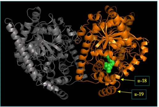
FAAH has been crystallized in complex with an irreversible active site-directed inhibitor, the methoxy arachidonyl fluoro-phosphonate (MAFP), and its three-dimensional structure has been analyzed at a 2.8 Å resolution (Bracey et al., 2002). To obtain the crystalline structure, a catalytically active mutant was generated (ΔTM-FAAH), where the first 29 amino acids were deleted (Patricelli et al., 1998). ΔTM-FAAH is soluble and homogeneous in detergent-containing buffers, opening the avenue to the in vitro mechanistic and structural studies, and is still able to bind membranes. The X-ray structure of this mutant confirmed that FAAH is an integral membrane enzyme with a globular shape: the enzyme crystallized as a homodimer, indicating that it is at least a dimer in solution (Mckinney end Cravatt, 2005) (Figure 6).
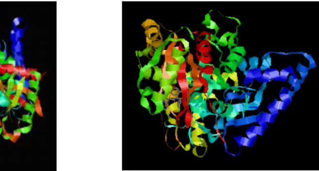
More than 100 members of the AS family of enzymes have been reported in the literature, but only for malonamidase (MAE2) (Shin et al., 2002) and C-terminal peptide amidase (PAM) (Labahn et al., 2002), two soluble bacterial enzymes, structural data are available (Figure 7). All three resolved structures of AS enzymes (FAAH, MAE2, and PAM) revealed a common core, but compared to other AS enzymes, which are mostly soluble proteins, FAAH displays two distinguished features: its integration into membranes and its strong preference for hydrophobic substrates.
Furthermore, three well-defined domains have been identified in FAAH: i) a transmembrane domain at the N-terminus which directs protein oligomerization, ii) a serine- and glycine-rich domain, and iii) a proline-rich domain.
FAAH has several elements of secondary structure: the twisted β-sheet consisting of 11 mixed strands (accounting for ~17% of the whole protein structure) is surrounded by 28 α-helices of various lengths (accounting for ~53% of the whole protein structure) (Figure 6). Recently, the stability of ΔTM-FAAH has been studied as a function of chemical (guanidinium hydrochloride) or physical (high hydrostatic pressure) denaturation (Mei et al., 2007). The unfolding transition of the enzyme was observed to be complex and required a fitting procedure based on a three-state process with a monomeric intermediate. The first transition was characterized by dimer dissociation, with a free energy change of ~11 kcal/mol that accounted for ~80% of the total stabilization energy. This process was also paralleled by a large change in the solvent-accessible surface area, because of the hydration occurring both at the dimeric interface and within the monomers. As a
consequence, the isolated subunits were found to be much less stable (ΔG ~3 kcal/mol). The addition of MAFP enhanced the stability of the dimer by ~2 kcal/mol, toward denaturant- and pressure-induced unfolding. FAAH inhibition by MAFP also reduced the ability of the protein to bind to the membranes. Taken together, these findings suggest that local conformational changes at the level of the active site might induce a tighter interaction between the subunits of FAAH, thus affecting the enzymatic activity and the interaction with membranes (Mei et al., 2007).
ΔTM-FAAH appears to bind membrane lipids via helices α-18 and α-19 (amino acid 410-438), which form a helix-turn-helix motif (Figure 8). This motif interrupts the AS fold and is comprised mainly of hydrophobic residues (with few basic amino acid) that are likely to constitute a membrane binding surface of FAAH. In addition, a predicted N-terminal transmembrane (TM) domain (amino acids 9-29) forms a membrane binding helix that strengthens the interactions of the α-18 and α-19 helices with membranes (McKinney and Cravatt, 2005). Remarkably, sequence comparisons revealed that this domain is not present in other AS enzymes (Cravatt et al., 1996).
The two monomers of FAAH have a parallel alignment, that allows both subunits to function concomitantly by recruiting substrates from the same membrane (Figure 8). The parallel orientation is required to have the α-18 and α-19 membrane cap on the same face of the dimer, thus enhancing membrane binding (McKinney and Cravatt, 2005). Noteworthy, the intimate relationship between the membrane binding surface and the active site of FAAH resembles the membrane-binding domains of two other integral membrane enzymes, like squalene cyclase (Wendt et al., 1997) and prostaglandin H2 synthase (Picott et al., 1994). Also these enzymes act on lipid-soluble substrates and have hydrophobic caps surrounding the entrance of the corresponding active sites. These three enzymes share no sequence or fold homology, indicating that they have evolved independently similar strategies for membrane integration (Bracey et al., 2004). However, all three enzymes are dimeric proteins, with the active site capped by a hydrophobic domain, that is surrounded by basic amino acids in order to interact with negatively charged phospholipids (McKinney and Cravatt, 2005).
It has been suggested that FAAH may have different structural alterations, allowing direct access from the cytosolic and the membrane side to its active site. In fact, X-ray analysis revealed several unusual features of the enzyme: the resolved crystal structure confirms that FAAH has different key regions, including a remarkable collection of channels that form a ‘cytosolic port’
and a ‘membrane port’ to facilitate substrate recognition, binding, hydrolysis and product release (thus improving the catalytic turnover). These ports might grant the simultaneous access to both membrane and cytosolic compartments of the cell, useful for substrate entry and/or product exit during the catalytic reaction (Cravatt et al., 2003).
A potential substrate entryway (which presents anphipathic residues possibly to accommodate polar substrate head groups towards the FAAH active site) has been identified next to α-18 and α-19 helices, and it may indicate direct connection between the FAAH active site and the hydrophobic membrane bilayer. The mode for membrane binding of FAAH may facilitate movement of the FAA substrates directly from the bilayer to the active site, with no need for transport of these lipids through the aqueous cytosol. In this model, the substrate would first enter via the membrane to the active site; following hydrolysis, the released fatty acid (hydrophobic) and amine (hydrophilic) products would then exit through the membrane-access and cytosolic-access channels, respectively. Moreover, the cytoplasmic port may serve the additional function of providing a way for a water molecule required for deacylation of the FAA-FAAH acyl-enzyme intermediate, which has been already characterized by LC-MS (Patricelli and Cravatt., 1999).
α-19
α-18
Figure 6. Schematic representation of rat ΔTM-FAAH complexed with the irreversible
inhibitor MAFP in green (PDB file 1MT5). It should be remarked the parallel alignment of the two monomers, with the lower α-helices (α-18 and α-19) of each subunit constituting the FAAH-membrane binding cap.
Figure 7. Structures of MAE2 (left) (PDB file 1OCL) and PAM (right) (PDB file 1M21).
Relative to FAAH, where monomers orientation is parallel, the two monomers of MAE2 display an anti-parallel alignment.
Helix 19 Helix 19
Figure 8. Left side: common representation of dimeric FAAH. Right side: 90 degree
2.4 Catalytic reaction
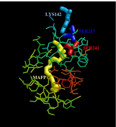
FAAH presents unique biochemical properties due to an unusual serine-serine-lysine (Ser241-Ser217-Lys142) catalytic triad (Figure 9). In fact, differently from the substrate selectivity displayed by most serine hydrolases, which react with esters at rates several orders of magnitude faster than amides, FAAH reacts with esters and amides at equivalent rates. It has been demonstrated that this unusual property depends on a single lysine residue (Lys142), since its mutation to alanine greatly reduces the amidase activity of FAAH, without affecting the esterase activity (Patricelli and Cravatt, 1999). Many investigations have been focused to clarify the catalytic mechanism of FAAH. A number of mutagenesis, kinetic and chemical labeling studies have revealed that the FAAH nucleophile is Ser241 (Patricelli et al., 1999). Mutagenesis studies also invoked the participation of additional residues in the catalytic mechanism of FAAH: in particular, a serine residue (Ser217) mutated to alanine produced a mutant FAAH with a significant reduction of hydrolytic activity (~2000 fold), reduction that was much less severe than that observed with mutants lacking either the serine nucleophile (Ser241) or the lysine base/acid (Lys142). Remarkably, the unusual catalytic core of FAAH is highly conserved among the AS family members. Lys142 appears to play a critical role as both base and acid in the hydrolytic cycle. In fact, several lines of experiments show that Lys142 plays a role as a base that activates the Ser241 nucleophile in FAAH, whereas other kinetic data seem to support a role for Lys142 as an acid that participates in the protonation of the substrate leaving group (Patricelli and Cravatt, 1999). The relative importance of acid-catalyzed leaving group protonation for amide hydrolysis compared to ester hydrolysis has been emphasized previously in semi-empirical studies (Frest, 1971): consistent with these predictions, a tight coupling of base-catalyzed nucleophile activation and acid-catalyzed leaving group protonation might explain the ability of FAAH to normalize the acylation/hydrolysis rates of an amide or an ester substrate (McKinney and Cravatt, 2005). This hypothetical mechanism assumes that Lys142 would be deprotonated in the absence of bound substrate, leading to a constitutively activated nucleophile (Ser241). The structural arrangement analysis of catalytic residues indicates that in FAAH the impact of Lys142 on Ser241 nucleophile strands and the leaving group protonation likely occurs indirectly, via the bridging Ser217 of the triad; the latter may act as a ‘proton shuttle’. In this model, FAAH would force protonation of the substrate leaving group early in the transition state
of acylation, concomitantly with the nucleophile attack on the substrate carbonyl group (McKinney and Cravatt, 2005).
It should be pointed out that the comparable hydrolysis rate for amide and ester bonds has a biological meaning: FAAH must bind and hydrolyze its FAA substrates against a background of a large excess of structurally related esters such as monoacylglycerols (Mechoulam et al., 1995). To reach this goal, the active site of FAAH has specifically evolved and adapted to hydrolysis of FAA substrates in a cellular environment with high concentration of fatty acid esters. Therefore, the unique biochemical proprieties of FAAH permit to this enzyme to act as a lipid amidase in vivo.
LYS142
SER217
SER241
MAFP
Figure 9. Wireframe visualization within 8 Å of the active site of FAAH. The catalytic
2.5 Synthetic inhibitors
Growing evidence demonstrates that FAAH is the critical regulator of the endogenous levels of AEA, suggesting that it may serve as an attractive therapeutic target for the treatment of human disorders (Maccarrone, 2006). Unfortunately, AEA is rapidly inactivated by FAAH, which prevents its therapeutic exploitation. Yet, inhibitors of FAAH that block degradation of AEA and related endocannabinoids might be useful to tackle pathologies in which endocannabinoid levels are reduced.
The first non-specific inhibitor reported for FAAH was the serine protease inhibitor phenylmethylsulfonyl fluoride (PMSF) (Deutsch and Chin, 1993) (Figure 10). Since this compound was not selective for FAAH, there was a growing interest to design more potent and selective inhibitors. New compounds were obtained from the derivatization of various fatty acids with functional groups, previously reported to react and form covalent adducts with catalitycally active serine and cysteine residues. This method allowed the discovery of novel FAAH inhibitors, like diazomethylarachidonoyl ketone (Edgemond et al., 1998b), stearylsulfonyl fluoride (Deutsch et al., 1997a), methyldodecyl fluorophosphonate (Martin et al., 2000), arachidonylsulfonyl fluoride (Segall et al., 2003), and the most potent methyl arachidonyl fluorophosphonate (MAFP) (Figure 10) (Deutsch et al., 1997b). All these compounds are potent irreversible inhibitors of FAAH, however, they also have remarkable affinity for the CB1 receptor.
More recently, a series of irreversible aryl-carbamates inhibitors were described (Mor et al., 2004). URB597 (cyclohexyl carbamic acid 3'-carbamoyl-biphenyl-3-yl ester) (Figure 10), the most potent member of this
family, inhibited FAAH activity with an IC50 value of 4.6 nM in rat brain
extracts (Mor et al., 2004), and of 0.5 nM in intact neurons (Piomelli et al., 2006), without affecting other serine hydrolases. In addition, introduction of small polar groups in meta-position of the distal phenyl ring, and in para-position of the proximal phenyl ring, were found to improve inhibition (Mor et al., 2004; Tarzia et al., 2006). These carbamates inhibit FAAH activity through irreversible interaction based on nucleophilic attack of Ser241 in the active site. Biochemical evidence (Alexander and Cravatt, 2005) showed that these inhibitors covalently modify the active site by adopting an orientation opposite of that originally predicted from modeling (Mor et al., 2004). Indeed, the O-biaryl substituents would reside in the cytoplasmic-access channel (rather than in the acyl-chain-binding channel), where they would be susceptible to enzyme-catalyzed protonation to enhance their function as
leaving groups. Based on these results, a series of carbamates were designed, in which the N-cyclohexyl unit was replaced with various N-alkyl groups mimicking the acyl chains of anandamide. These compounds, of which JP-104 (undec-10-ynyl-carbamic acid 3'-carbamoyl-biphenyl-3-yl ester) is a prototype member (Figure 10), generally exhibited enhanced potency (Alexander and Cravatt, 2005). More recently, Ahn and coworkers have described a new series of ‘PF’ urea-based inhibitors with piperidine/piperazine groups (see PF-750 (N-phenyl-4-(quinolin-3-ylmethyl)piperidine-1-carboxamide) in Figure 10). These compounds have been shown to covalently inactivate FAAH via carbamylation of the serine nucleophile in the active site, and did not show any detectable activity against other serine hydrolases in mammalian proteomes (Ahn et al., 2007). In general, an irreversible mechanism of inhibition might reduce the versatility of a drug for in vivo applications. Thus, a major challenge for the ongoing pharmaceutical research is the development of potent and selective, but reversible, inhibitors of FAAH.
Based on α-ketoheterocycle protease inhibitors (Edwards et al., 1995), potent reversible competitive inhibitors were developed, combining an unsaturated acyl chain and an α-keto-N4-oxazolopyridine, with incorporation of a second weakly basic N-atom. This class of compounds showed potency in the subnanomolar range, with Ki values falling below 200 pM (Boger et al., 2000). The inhibition potency was strongly dependent on the hydrophobicity of the flexible acyl chain, and on the degree of α-substitution (Boger et al., 2001). For example, the compound OL-135 (1-oxo-1[5-(2-pyridyl)-2-yl]-7-phenylheptane) (Figure 10) displayed an exceptional combination of high potency (Ki = 4.7 nM towards rat-recombinant FAAH) and high selectivity
in vivo (Lichtman et al., 2004; Boger et al., 2005).
Several inhibitors have been tested in vivo and their ability to inactivate FAAH was shown to elicit pain and anxiety (Kathuria et al., 2003), without the side effects (hypomotility, hypothermia and catalepsy) that usually
accompany activation of CB1 receptors by exogenous cannabinoids like Δ9
-THC (Fowler, 2003; Cravatt and Lichtman, 2003). To maintain this lack of ‘cannabinoid side effects’, FAAH inhibitors must be devoid of affinity for the cannabinoid receptors. The most studied of the FAAH inhibitors, URB597, does not exhibit affinity for cannabinoid receptors. Thus, at doses that inhibit FAAH and substantially raise brain levels of AEA, but not of 2-AG, this compound did not induce common side effects of typical CB1 agonists, suggesting that they it might be exploited as an innovative anti-anxiety therapeutic (Gaetani et al., 2003).
Also OL-135 (see above) allowed a profound increase of anandamide levels in the brain and spinal cord, and displayed CB1-dependent antinociceptive effects in the hot-plate, tail-immersion, and formalin tests (Lichtman et al., 2004).
S F O O P O O F O NH2 O HN O O NH2 O HN O N N N H O O O N N PMSF MAFP OL-135 PF-750 JP104 URB597
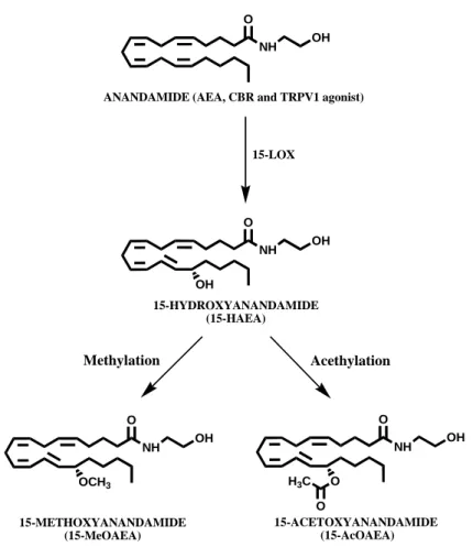
2.6 Natural inhibitors
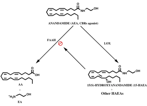
Besides the design of synthetic molecules, several papers reported the presence of specific enzymatic reactions able to produce also in the cells compounds able to act as reversible FAAH inhibitors (Maccarrone and Finazzi-Agrò, 2004). In particular, it has been found that oxidative metabolites of AEA generated by various lipoxygenases, i.e. the hydroxyanandamides (HAEAs; see Figure 11), are powerful inhibitors of FAAH (Van der Stelt et al., 2002).
The main products of LOX action on AEA are hydroperoxyanandamides (HpAEAs), which are quickly reduced in the cellular environment to hydroxyanandamides (HAEAs) (Figure 11). LOXs are enzymes that catalyze the stereospecific conversion of 1Z,4Z-pentadiene system into 1-hydro(pero)xy-2E,4Z-pentadiene system at different positions of the acyl chain (Yamamoto, 1992).
Of interest is the fact that all HAEAs are reversible competitive inhibitors of FAAH (Van der Stelt et al., 2002). In addition, the fact that various lipoxygenases (i.e., 5-, 12-, and 15-LOXs) generate different HAEAs with different inhibition profiles towards the proteins of the ECS, suggests that cells with different LOXs might contribute different selectivity to networks regulating endocannabinoids actions. These compounds may be the ‘physiological’ inhibitors of FAAH, of potential utility in the control of emotional states and of those disorders whose onset or symptoms are associated with defective production or excessive degradation of AEA and congeners. More in general, it should be pointed out that HAEAs represent one of the newest paradigms of the ability of cells to make their own tools to regulate key targets like FAAH, and they seem to do it more simply than is done in the laboratory.
NH O OH NH O OH OH ANANDAMIDE (AEA, CBRs agonist)
15(S)-HYDROXYANANDAMIDE (15-HAEA) OH O AA +H 3N OH EA + FAAH LOX + Other HAEAs
CHAPTER 3. OBJECTIVE OF THE RESEARCH
Here, we extended the characterization of one of the most interesting HAEAs towards the main elements of the endocannabinoid system (i.e., CB1R, CB2R, AMT, FAAH, NAPE-PLD), and also towards the enzymes implicated in 2-AG metabolism (i.e., DAGL, 2-AGMT, MAGL). We focused our attention on 15-HAEA, that is the (S) stereoisomer of the main product generated by 15-LOX from AEA: 15-(S)-hydroxy-eicosa-5Z,8Z,11Z,13E-tetraenoyl-N-(2-hydroxyethyl)amine. 15-HAEA shows the lowest inhibition constant for FAAH, with respect to other hydroxyanandamides (Van der Stelt et al., 2002).
Moreover, we tested the effect of two putative “natural” compounds, 15-(S)-metoxy-eicosa-5Z,8Z,11Z,13E-tetraenoyl-N-(2-hydroxyethyl)amine and 15-(S)-acetoxy-eicosa-5Z,8Z,11Z,13E-tetraenoyl-N-(2-hydroxyethyl)amine, referred from now as 15-MeOAEA and 15-AcOAEA, respectively (Figure 12). In fact, methylation and acetylation are biochemical reactions very common in the cellular environment (Cimato et al., 2002). For instance, these reactions are involved in the dynamic changes at the onset of post-translational modifications like phosphorylation, acetylation and ubiquitination.
Interestingly, Huang and collaborators have found that the methylation of the “endovanilloid” NADA (N-arachidonoyl-dopamine) by catechol-O-methyl-transferase leads to a methyl derivate (3-O-methyl-NADA) that is significantly less potent than NADA at the vanilloid receptor TRPV1, suggesting that methylation might represent a mechanism for the partial inactivation of NADA in nervous tissues where methyl-transferase activity is abundant (Huang et al., 2002). Furthermore, the role of methylation in the development and function of the nervous system is well-known (Mattson et al., 2003), and notably also endocannabinoids have been implicated in these processes (Berrendero et al, 1999). More recently, it has been reported that methylation and acetylation of the phenolic hydroxyls contained in the cannabinoids structure were detrimental for the antibacterial activity of these compounds (Appendino et al., 2008). Therefore, the rationale of our study was that methylation and acetylation of the hydroxyl group of HAEAs might be biologically relevant for the modulation of these hydroxides as natural inhibitors of FAAH. In order to ascertain the specificity of these substances, we investigated also their interaction with the other elements of the endocannabinoid system.
NH O OH NH O OH OH NH O OH OCH3 NH O OH ANANDAMIDE (AEA, CBR and TRPV1 agonist)
15-HYDROXYANANDAMIDE (15-HAEA) 15-METHOXYANANDAMIDE (15-MeOAEA) O H3C O 15-ACETOXYANANDAMIDE (15-AcOAEA) 15-LOX Acethylation Methylation
Figure 12. 15-LOX activity generates from anandamide 15-(S)-hydroxyanandamide
(15-HAEA), from which the methoxy- and acetoxy-derivatives may be formed.
CHAPTER 4. RESULTS
4.1 Effect of 15-(S)-HAEA, 15-(S)-MeOAEA and 15-(S)-AcOAEA on AEA metabolism
We investigated the effect of 15-HAEA, 15-MeOAEA and 15-AcOAEA on NAPE-PLD (Figure 13). The last two compounds were synthesized for the first time in our laboratory according to the procedures described in Materials and Methods. We have found increased NAPE-PLD activity in presence of 15-HAEA (142% at 1 µM, p<0.05) respect to control (set to 100%) but not for MeOAEA (86% respect to control) nor for 15-AcOAEA (100% respect to control) (Table 1). Interestingly, the difference respect 15-HAEA for both compounds results significant (p<0.05).
We also investigated the inhibitory effect of 15-HAEA, 15-MeOAEA and
15-AcOAEA on the transport of 1 μM [3H]-AEA inside the cells (Figure 13
and Table 1). Like 1 μM 15-HAEA, which reduced AEA transport about 28% respect to control (p<0.05), 15-MeOAEA reduced AEA transport about
22% (p<0.05). Instead, 15-AcOAEA did not affect [3H]-AEA uptake (96%
respect to control) and the difference is statistical significant respect 15-HAEA (p<0.05).
The activity of FAAH in the presence of HAEA, MeOAEA and 15-AcOAEA (Figure 13 and Table 1) were assayed by incubating these compounds with mouse brain homogenate as previously described. FAAH activity was found to be drastically decreased when pre-incubated with 1 μM 15-HAEA (28% respect to control set to 100%, p<0.01), as well when pre-incubated with 15-MeOAEA (33% respect to control, p<0.01). Different inhibition values were found for 15-AcOAEA, obtaining FAAH activity about 86% respect to control and resulting in a very significant difference respect to 15-HAEA (p<0.01).
0 50 100 150 200
NAPE-PLD AMT FAAH
# * # * * # ** ** ## ** CTRL 1μM 15-HAEA 1μM 15-MeOAEA 1μM 15-AcOAEA % of c o nt rol
Figure 13. Effects of 15-(S)-HAEA, 15-(S)-MeOAEA and 15-(S)-AcOAEA on AEA
metabolism. The anandamide production by NAPE-PLD in presence of HAEA, 15-MeOAEA and 15-AcOAEA was performed by measuring the production of [3H]-AEA from
[3H]-NArPE as reported in Materials and Methods. The effect of 15-HAEA and its synthetic
derivates 15-MeOAEA and 15-AcOAEA on AMT activity was assayed incubating the synaptosomes obtained from mouse brain with 1µM [3H]-AEA. The effect of HAEA,
15-MeOAEA and 15-AcOAEA on [3H]-AEA hydrolysis by murine brain FAAH was
investigated by using 10 μM [3H]-AEA. * p<0.05 vs control; ** p<0.01 vs control; # p<0.05 vs
Table I. Effect of 1 μM 15(S)-HAEA, 1 μM 15(S)-MeOAEA and 1 μM 15(S)-AcOAEA on
the proteins of the Endocannabinoid System.
Compound Control 15-(S)-HAEA 15-(S)-MeOAEA 15-(S)-AcOAEA NAPE-PLD activity (pmol/min/mg) 14± 1 (100%) 19 ± 2 * (142%) 12 ± 1 # (86%) 14 ± 1 # (100%) AMT activity (pmol/min/mg) 0.80 ± 0.10 (100%) 0.57 ± 0.04 * (72%) 0.62 ± 0.04 * (77%) 0.77 ± 0.05 # (96%) FAAH activity (pmol/min/mg) 210 ± 15 (100%) 59 ± 4** (28%) 69 ± 3 ** (33%) 181 ± 11 ## (86%) DAGL activity (pmol/min/mg) 398 ± 14 (100%) 199 ± 12 ** (50%) 386 ± 15 ## (97) 339 ± 10 ## (85%) 2-AGMT activity (pmol/min/mg) 2.0 ± 0.2 (100%) 3.6 ± 0.3 * (179%) 2.2 ± 0.2 # (109%) 1.9 ± 0.2 # (97%) MAGL activity (pmol/min/mg) 511 ± 41 (100%) 470 ± 38 (92%) 562 ± 56 (110%) 536 ± 38 (105%) CB1R binding (fmol/mg) 112 ± 12 (100%) 32 ± 3 ** (29%) 83 ± 6 ## (74%) 86 ± 5 ## (77%) CB2R binding (fmol/mg) 27 ± 3 (100%) 27 ± 2 (100%) 21 ± 2 # (76%) 23 ± 2 (85%) TRPV1 binding (fmol/mg) 220 ± 25 (100%) 165 ± 14 * (72%) 221 ± 20 # (100%) 213 ± 19 (97%) *p < 0.05 vs control, ** p < 0.01 vs control. # p < 0.05 vs 15-HAEA, ## p < 0.01 vs 15-HAEA.
4.2 Effect of 15-(S)-HAEA, 15-(S)-MeOAEA and 15-(S)-AcOAEA on 2-AG metabolism
First, we studied the action of the compounds on the main enzyme responsible for 2-AG biosynthesis (Figure 14 and Table 1). We have found that 15-HAEA (at 1 μM) exerts a remarkable inhibitory effect on DAG lipase (50% of activity respect to control set to 100%, p<0.01). When we tested 15-MeOAEA at the same concentration, we did not observe any alteration on DAGL activity (97% respect to control), and similar results were obtained for the acetylated derivative (85% respect to control). However, both showed a very significant difference respect to 15-HAEA (p< 0.01).
Then, we investigated the inhibitory effect of 15-HAEA, 15-MeOAEA and
15-AcOAEA on the transport of 1 μM [3H]-2-AG inside the cells.
Interestingly, 1 μM 15-HAEA affect 2-AG transport augmenting 2-AG uptake about 178% respect to control set to 100% (p<0.05) (Figure 14 and Table 1). To test 15-MeOAEA and 15-AcOAEA, the assays were performed incubating the labelled substrate with these compounds at the same concentration that 15-HAEA (1 μM). In particular, 2-AGMT activity in presence of 15-MeOAEA was only slightly increased (109% respect to control), while 2-AGMT activity in presence of 15-AcOAEA was unaffected (97% respect to control). It is worth to notice the difference between 15-HAEA and the methylated and acetylated analogous, which is statistical significant for both the derivatives (p<0.05).
Finally, we extended our investigation on the effect of the compounds to the main enzyme responsible for 2-AG degradation. We tested the effect of 15-HAEA, 15-MeOAEA and 15-AcOAEA on MAGL activity (Figure 14 and
Table 1), by using 2-[3H]-OG as radiolabelled substrate as previously
described in Materials and Methods. At 1 μM, 15-HAEA did not exhibit any significant inhibitory effect on MAGL activity (92% respect to control set to 100%). Analogously, even for 15-MeOAEA and 15-AcOAEA there were not significant alterations (at 1 μM) on MAGL activity (110% and 105% respect to control, respectively).
0 50 100 150 200
DAGL 2-AGMT MAGL
** ## ## * # # CTRL 1μM 15-HAEA 1μM 15-MeAEA 1μM 15-AcOAEA % of c o nt r o l
Figure 14. Effects of 15-(S)-HAEA, 15-(S)-MeOAEA and 15-(S)-AcOAEA on 2-AG
metabolism. The effect of 15-HAEA, 15-MeOAEA and 15-AcOAEA on DAG lipase was investigated in presence of 500 μM [14C]-DAG. The uptake of [3H]-2-AG in presence of
15-HAEA, 15-MeOAEA and 15-AcOAEA was performed incubating the synaptosomes obtained from mouse brain with 1 µM [3H]-2-AG. The effect of HAEA, MeOAEA and
15-AcOAEA on MAG lipase was tested by measuring the production of [3H]-glycerol released
from 10 μM [3H]-2-OG. The results are expressed respect to control (absence of compounds)
set to 100% (see also table 1). * p<0.05 vs control; ** p<0.01 vs control; # p<0.05 vs
4.3 Effect of 15-(S)-HAEA, 15-(S)-MeOAEA and 15-(S)-AcOAEA on CB1R, CB2R and TRPV1
To test the ability of the synthesized compounds to bind to CB1R and CB2R, we performed radioligand competition binding studies and analyzed
the displacement of [3H]-CP55940 from membranes prepared by murine
brain and spleen, respectively, as described in Materials and Methods. As reported in Figure 15 and in Table I, we observed that 1 µM 15-HAEA
strongly reduce [3H]-CP55940 binding to brain membranes (CB1R) (29%
respect to control set to 100%, p<0.01) but not to spleen membranes (CB2R) (101% respect to control). Differently, at the same concentration used for 15-HAEA (1 μM), 15-MeOAEA and 15-AcOAEA present similar slight affinity for CB1R (74% and 77%, respectively) and for CB2R (76% and 85%, respectively) respect to control (set to 100%). Even if these data are not significant respect to control, they are very significant respect to 15-HAEA for CB1R (both p< 0.01). Regard to CB2R, the difference obtained respect to 15-HAEA results significant only when compared to 15-MeOAEA (p< 0.05).
In order to put in a better perspective the effects of HAEA and 15-MeOAEA on the proteins of the endocannabinoid system, we analyzed the
displacement of [3H]-resinferatoxin by these compounds from membranes
prepared by murine brain (Figure 15 and Table 1). While 1 μM 15-HAEA has moderate affinity for TRPV1 receptors (28% of displacement, p<0.05), the same concentrations (1 μM) of 15-MeOAEA and 15-AcOAEA used in
our assays were totally ineffective on the binding of [3H]-resinferatoxin to
mouse brain homogenate (100% and 97% respect to control, respectively). However, the difference between 15-HAEA and 15-MeOAEA has found to be statistical significant (p<0.05).
0 50 100 150 CB 1R ## CB 2R TRP V1 * ** ## # # CTRL 1μ M 15-HAEA 1μ M 15-MeOAEA 1μ M 15-AcOAEA o l o n tr f c % o
Figure 15. Binding affinity of 15-HAEA, 15-MeOAEA and 15-AcOAEA for cannabinoid
and vanilloid receptors. We investigated the effect of 15-HAEA and its synthetic derivates on the binding of 400 pM [3H]-CP55.940 on mouse brain (CB1R) and mouse spleen (CB2R).
The effect of 15-HAEA, 15-MeOAEA and 15-AcOAEA on TRPV1 receptor was obtained from mouse brain homogenate monitoring the effect of the compounds on the binding of 500 pM [3H]-RTX. Data are reported respect to control set to 100%. * p<0.05 vs control; **
4.4 Molecular modelling of FAAH and molecular dynamics simulation
We focused on the internal channel of FAAH, finding possible access paths from the phospholipidic membrane and the cytosolic space. We highlighted how AEA (and probably other endocannabinods) might enter directly from the plasma membrane to be processed (Figure 16, left side). Once processed, the polar amines substituents liberated from the fatty acid amide substrates can be released on the cytosolic port through the internal channel that we have evidenced (Figure 16, right side).
The structural and dynamic features of AEA and its analogues in the binding pocket of FAAH were investigated using molecular dynamics (MD) simulations. The MD trajectories were clusterized to highlight the bound-conformations of AEA and its analogues. The left side of figure 17 shows the representative conformers of AEA in the cavity of the enzyme, while right side of figure 17 is relative to 15-HAEA. The structures of AEA within the enzyme present a close similarity, while the conformations of 15-HAEA are more distinct, especially in the C16-C20 tail, when the hydroxyl group is well oriented in the cavity. A cluster analysis of the MD trajectories was performed to highlight the differences in ligand flexibility in the FAAH pocket (Table II). The clustering was performed using the root mean square deviation (RMSD) computed on the heavy atoms of the ligands; a threshold RMSD value of 0.5Å between cluster representative conformers was used. 15-HAEA, compared to AEA, shows 5 more bound conformers (as obtained from the 1.5 ns MD simulations) and a slightly induced fit (as estimated from the comparison of the bound conformations with the nearest free conformers obtained by the full energy minimization of the ligands).
Figure 18 reports the hydrogen-bonding interactions observed from the MD simulations of 15-HAEA (left side) and 15-MeOAEA (right side). From these calculations result that 15-HAEA complex is favoured by three possible H-bonds, namely with Leu372, Glu373, and Ser376, while 15-MeOAEA gives H-bond interaction with only one amino acid, Tyr335. These simulations have evidenced the possible different interactions of 15-HAEA and its methylated analogous with the binding site environment of FAAH. The presence of a methyl group affects the stability of the complex protein–ligand but 15-MeOAEA still can accommodate in the binding site. For 15-AcOAEA, which neither is able to generate hydrogen bond, it is more difficult to accommodate into the binding site, due to the presence of a more voluminous group.
(A)
(B)
Figure 16. Static view of FAAH from bilayer and cytosolic sides. (A): view of FAAH
from the membrane layer. Only one of the two subunits is filled (accessible surface area represented in orange) and the blue arrow indicates the active site where the substrate (green) can be processed, evidencing the possible entrance channel. (B): view of FAAH from the cytosolic space. Only one subunit is represented. The upper black holes on the subunit are the contact points with the other subunit. The blue arrow remarks the cytosolic port where, once hydrolyzed, the polar head of anandamide (ethanolamine) may be released.