DIPARTIMENTO SCIENZE BIOMOLECOLARI (DISB)
Corso di Dottorato di Ricerca in: Scienze della Vita, Salute e Biotecnologie
Curriculum: Biologia della Cellula e degli Organismi
CICLO XXX
Investigation of the effects of Campylobacter jejuni
virulence factors in human cells:
different pathways involved
Settore Scientifico Disciplinare (SSD): BIO-16
Relatore:
Prof. Stefano Papa
Dottorando:
Dott.ssa Gianna Di Sario
Co-relatore:
Dott.ssa Barbara Canonico
1
Table of contents
Abbreviations 5
Abstract 8
Introduction 9
1. Biology of Campylobacter jejuni 9
1.1 Epidemiology of C. jejuni 10
1.2 Transmission of C. jejuni 11
1.3 Pathogenesis of C. jejuni 12
1.4 Post-infectious sequelae of C. jejuni 13
1.4.1 The Guillain-Barré syndrome 13
1.5 Molecular basis of C. jejuni pathogenesis 17
1.5.1 Lipooligosaccharides 17
1.5.2 Capsule 18
1.5.3 Flagella 18
1.5.4 The cytolethal distending toxin 19
1.5.5 Glycosylation system 20
1.6 The outer membrane vesicles (OMVs) 21
1.7 C. jejuni-host cell interactions 23
2. Mechanisms of cell death 31
2.1 Apoptosis 31
2.1.1 Bcl-2 family proteins 33
2.1.2 Extrinsic and intrinsic pathway 34
2.1.3 pRb, p53 and CD59 35
2.2 Necrosis 37
2.3 Autophagy 37
Aims and objectives 39
Materials and Methods 40
3. Human tumour myeloid cells (U937) and normal monocytes 40
3.1 Ethic statement 40
2
3.3 Growth conditions 41
3.4 Cell lines and monocytes isolation 41
3.5 C. jejuni cell lysate preparation 42
3.6 Detection of cytotoxin activity in C. jejuni lysates 42
3.7 Pre-treatment of U937 cells and monocytes with C. jejuni lysates 42
3.8 Pre-treatment of U937 cells with C. jejuni OMVs 43
3.9 Pre-treatment of U937 cells with Rapamycin 43
3.10 Nanoparticle Tracking Analysis (NTA) 43
3.11 Morphological feature evaluation 43
3.12 Flow cytometry and confocal microscopy stainings 44
3.13 Trypan blue viability test 48
3.14 Confocal microscopy analyses 48
3.15 Statistical analyses 48
4. Human intestinal cell model and molecular work 49
4.1 Bacterial strains, plasmids and primers 49
4.2 Growth conditions and storage 51
4.3 Preparation of a specific OD600 C. jejuni suspension 52
4.4 DNA Purification 53
4.5 Purified DNA Quantitation 54
4.6 Polymerase Chain Reaction Technique 54
4.7 Tris-Acetate-EDTA agarose ethidium bromide gel electrophoresis 55
4.8 Purification of PCR products 55
4.9 Ligation and transformation stages 56
4.10 PCR from boiled/lysed bacteria colonies 57
4.11 Overnight culture preparation 58
4.12 Isolation of plasmid DNA: QIAprep Miniprep production 58
4.13 Restriction endonuclease digestion 58
4.14 Inverse PCR mutagenesis 59
4.15 DNA sequencing 60
4.16 Transformation of C. jejuni by Electroporation 61
3
4.18 Isolation of C. jejuni OMVs 64
4.19 Quantitation of total proteins by bicinchoninic acid assay (BCA) 65 4.20 Pre-treatment of T84 intestinal Eukaryotic cells with C. jejuni whole cells and
OMVs 66
4.21 CHOP and BIP proteins detection by SDS-PAGE and Western blotting 67
4.22 Total RNA isolation 67
4.23 cDNA synthesis 69
4.24 Reverse Transcription PCR (RT-PCR) 70
Results 72
5. Monocytes from donor’s human blood 72
Discussion 85
6. U937 cell model: U937 cells preincubated with C. jejuni lysates 88
Discussion 109
7. U937 cell model: U937 cells preincubated with C. jejuni OMVs 112
Discussion 126
8. Human intestinal cell model 127
Discussion 131
9. Construction of C. jejuni 11168H GFP mutant strain 132
Discussion 136
Conclusions 137
Appendices 143
Appendix 1-Primer design 143
Appendix 2- Different orientations of the KmR cassette 145
5
Abbreviations
AAG, autoagglutination
AIDP, acute inflammatory demyelinating polyneuropathy AMAN, acute motor axonal neuropathy
AMSAN, acute motor sensory axonal neuropathy AnxV, Annexin V
ATF6, activating transcription factor-6 BCA, bicinchoninic acid assay
Bcl-2, B-cell lymphoma-2 protein BSA, albumin standards
CCV, Campylobacter-containing vacuole CDT , cytolethal distending toxin
CHOP, C/EBP homologous protein CL, Cardiolipin
CPSs, Capsular polysaccharides CTRL, untreated control cells
D-MEM, Dulbecco’s modified eagle medium DR, death receptor
eIF2α,eukaryotic transcriptional initiation factor ER, endoplasmic reticulum
ERAD , endoplasmic reticulum-associated degradation FADD, Fas-associated death domain protein
FasL, Fas ligand FBS, fetal bovine serum FCS, foetal calf serum FlaA, flagellin A FlaB, flagellin B,
6 FSC, forward light scatter
FSC, forward scatter
GBS, Guillain-Barré syndrome IBD, inflammatory bowel diseases IECs, intestinal epithelial cells IPCRM, Inverse PCR mutagenesis IRE-1, inositol-requiring protein-1 ISPCR, insert specific primers PCR Km, Kanamycin
LAMP-1, lysosomal-associated membrane protein-1 LB, Luria-Bertani
LBP, LPS binding protein LE, late endosome
LOS, lipooligosaccharides LPS, lipopolysaccharide LTDR, LysoTracker Deep Red LTG, LysoTracker Green m TORC1, mTOR complex 1 m TORC2, mTOR complex 2 MAC, membrane attack complex MDC, Monodansylcadaverine MFI, mean fluorescence intensity
MOMP, mitochondrial outer membrane permeabilization mTOR, mammalian target of rapamycin
MUT, mutant
MVB, multivescicular bodies NAO, Nonyl Acridine Orange NR, Nile Red
7 NTA, nanoparticle tracking analysis
OMVs, outer membrane vesicles
PBMCs, peripheral blood mononuclear cells PERK, protein kinase RNA-like ER kinase PI construct, Plasmid + Insert
PI, Propidium Iodide
PI3K, phosphoinositide 3-kinase
PIK construct, Plasmid Insert Kanamycin cassette pRb, retinoblastoma protein
RM, Rapamycin
ROS, reactive oxygen species
RPMI, Roswell Park Memorial Institute RT, at room temperature
SSC, side light scatter TLR4, Toll-like receptor 4
TMRE, Tetramethylrhodamine ethyl ester perchlorate TNFR1, TNF receptor 1
TNF-α, tumour necrosis factor alpha
TRAIL, TNF-related apoptosis inducing ligand UPR, unfolded protein response
VAIN, variable anaerobe chamber VPPCR, vector primer PCR WR, working reagent WT, wild type
8
Abstract
The gram-negative bacterium Campylobacter jejuni represents a major agent causing food-borne gastroenteritis in humans worldwide. C. jejuni infection is also an important pre-condition for Guillain-Barrè syndrome. The cytotoxic effects of Campylobacter have been ascribed to the actions of several different toxins. The best characterized C. jejunir toxin is the cytolethal distending toxin (CDT), released by bacteria via outer membrane vesicles (OMVs). Many bacterial pathogens, including C. jejuni, utilize OMVs to transport virulence factors, such as the CDT (cytolethal distending toxin), into host cells. To date, different authors described the peculiar relationship among C. jejuni virulence factors and human intestinal and myeloid cells. It is known that resident macrophages contribute to the maintenance of tissue by acting as the first line of defense against pathogens and by initiating wound repair. These monocyte-derived cells establish (together with dendritic cells) the role of “guardian of the gut”. The aim of this work was to investigate the effects of C. jejuni virulence factors in human cells focusing the attention on cellular pathways that this bacterium activates or deactivates during host cell infection. Among the several virulence factors, the CDT was the most investigated. To do that, effects induced by three different C. jejuni wild type strains were compared with the effects induced by the C. jejuni cdtA mutant strain, in three different cell lines: monocytes isolated from donor’s human blood, tumour myeloid cells (U937) and human intestinal epithelial cells (T84). C. jejuni lysates, OMVs and whole cells were separately used for the infections. Carried out cytometric, microscopy and molecular analyses revealed that C. jejuni induce DNA damage, apoptosis, mitochondrial and lysosomal destabilization as well as intracellular lipid content alterations, ICAM-1 upregulation and activation of autophagic, secretory and endocytic pathways in a CDT dependent manner. Nevertheless, although they are important for C. jejuni infection, CDT seems not to have a role in ER stress and UPR activation, CD14 and CD59 upregulation. Involvement of all these pathways enhances C. jejuni invasion, persistence and survival, allowing the possible onset of post infectious sequelae such as the Guillain-Barré syndrome.
9
Introduction
1. Biology of Campylobacter jejuni
The Campylobacter genus belongs to the family Campylobacteraceae, the order Campylobacterales, the class Epsilonproteobacteria, and the phylum Proteobacteria. Since its first description, the genus has grown to include several important human and animal pathogens that are primarily classified through phylogenetic means. The genus Campylobacter consists of 26 species, 2 provisional species, and 9 subspecies (as of December 2014).
Although the infections were attributed to Vibrio fetus (now known to be Campylobacter fetus), the first recognized case of Campylobacter infection (termed campylobacteriosis) were reported in the early 20th century in farm animals, and during the next three decades, it was believed to be a rare, opportunistic, invasive pathogen that occurred principally in debilitated hosts. Only in the early 1980s the importance of these infections was recognized as a cause of human gastrointestinal illness. By the mid-to-late 1980s, Campylobacter species have been identified as one of the most common bacterial causes of diarrhoea worldwide.
C. jejuni like all Campylobacter species, is a microaerophilic, non-fermentative Gram-negative organism. Being structurally similar to other Gram-Gram-negative bacilli, C. jejuni cells are slender, spirally ‘curved rods’, approximately 0.2-0.8 wide and 0.5-5µm long. It is motile with a characteristic corkscrew like motion via a single polar unsheathed flagellum at one or both ends of the cell (Vandamme et al, 2015).
For an efficient cultivation in vitro, the microaerophilic nature of C. jejuni requires an atmosphere with reduced oxygen and elevated carbon dioxide concentrations: gas mixtures of 5% oxygen, 10% carbon dioxide and 85% nitrogen provide optimal cultivation conditions for most C. jejuni isolates (Bolton et al, 1983).
The C. jejuni genome is small (1.6-2.0 megabases) and can establish long-term associations with their hosts, sometimes with pathogenic consequences (Young et al, 2007).
The complete genome sequence of C. jejuni was characterized and hypervariable regions that might be important in the survival of this organism were found (Parkhill et al, 2000). Most of the hypervariable sequences that have been found are in regions that encode proteins that are involved in the biosynthesis or modification of surface-accessible carbohydrate structures, such as the capsule, lipooligosaccharide (LOS) and flagellum; these structures have a key role
10
in C. jejuni biology, in particular for host-bacterium interactions (Young et al, 2007). C. jejuni is naturally competent, meaning that it can take up DNA from the environment. This leads to recombination between strains, which allows the generation of even more genetic diversity. The horizontal transfer of both plasmid and chromosomal DNA occurs both in vitro and in vivo during chick colonization; this indicates that natural transformation may have a significant role in genome plasticity (Wilson et al, 2003) (Avrain et al, 2004).
1.1 Epidemiology of C. jejuni
To date, campylobacteriosis is one of the most widespread infectious diseases of the last century. Recent reports suggested that in the past decade the incidence and prevalence of campylobacteriosis increased globally in both developed and developing countries. In North America, Europe, and Australia was recorded a remarkable increase, and data from parts of Africa, Asia, and the Middle East indicated that campylobacteriosis was endemic in these areas, especially in children (Fig. 1.1) (Kaakoush et al, 2015). In developed countries, where waterborne infection is less likely, animals are the primary source of human infection and disease. By contrast, Campylobacter is hyperendemic in the developing world, because of poor sanitation and close human contact with animals. In these countries Campylobacter species are usually limited to children, with illness/infection ratios decreasing with age, suggesting that exposure in early life might lead to the development of protective immunity (Rao et al, 2001).
Several factors have been hypothesized to influence the prevalence of Campylobacter infections; for example, in the last years it has been hypothesized that it might be influenced by population-level immunity (Havelaar et al, 2009).
11
Fig. 1.1 Incidence and prevalence of Campylobacteriosis worldwide (American Society of Microbiology, 2015).
1.2 Transmission of C. jejuni
C. jejuni is commonly considered to be a commensal organism of chickens and other avian species. Several environmental reservoirs can lead to human infection by C. jejuni. It colonizes the chicken gastrointestinal tract in high numbers, primarily in the mucosal layer, and it is passed between chicks within a flock through the faecal-oral route. C. jejuni can infect humans directly through consumption of contaminated food, particularly poultry products, unpasteurized milk, and water (Fig. 1.2) (Friedman et al, 2004) (Kapperud et al, 1992) (Neimann et al, 2003). In humans, C. jejuni can invade the intestinal epithelial layer, resulting in inflammation and diarrhoea.
12
Fig. 1.2 Consumption of undercooked or contaminated food and contact with animals can lead to transmission of Campylobacter to humans. Depending on species, strains involved and the ingestion dose, gastrointestinal and/or extra-gastrointestinal manifestations can occur (Kaakoush et al, 2015).
1.3 Pathogenesis of C. jejuni
C. jejuni is successful in competing with the human intestinal microbiota; an infectious dose of few hundred bacteria is sufficient to overcome the colonization resistance of humans leading to campylobacteriosis (Hofreuter, 2014). More than 80% of the registered campylobacteriosis are caused by C. jejuni, for this reason the term ‘campylobacteriosis’ is frequently associated to C. jejuni infection. Campylobacteriosis typically causes gastroenteritis/enterocolitis, and clinical picture varies significantly in duration, severity and associated symptoms (Ketley, 1997). Following an incubation of ~24-72 hours, symptoms like cramping abdominal pain, fever, vomiting and headaches can develop (Blaser, 1997). Upon onset of abdominal pain, diarrhoea develops quickly. Depending on virulence of the infecting strain and host immune status, it varies from a mild, non-inflammatory, watery presentation to severe and bloody.
13
Although gastroenteritis is the major clinical condition resulting from C. jejuni infection, other serious conditions within the gastrointestinal tract have been associated with C. jejuni, including inflammatory bowel diseases (IBD), post infectious functional gastrointestinal disorders, such as irritable bowel syndrome and functional dyspepsia, and celiac disease (Kaakoush et al, 2015).
1.4 Post-infectious sequelae of C. jejuni
In addition to gastrointestinal infections, C. jejuni can trigger extra-intestinal manifestations, as either a local isolated infection (which is a systemic manifestation after an episode of enteritis) or a postinfectious immune disorder. Overall, the incidence of extra-intestinal manifestations associated with C. jejuni infection is low compared with gastroenteritis. Post-infectious complications can be severe, and potentially life-threatening. These manifestations include Guillain-Barré syndrome (GBS), Miller Fisher Syndrome, Bell’s palsy (unilateral facial paralysis) and reactive arthritis. To date, the post-infectious disease most extensively studied is GBS.
1.4.1 The Guillain-Barré syndrome
The Guillain-Barre’ syndrome (GBS) is an acute post-infectious ascending paralysis that can affect peripheral and cranial nerves (particularly VII, facial nerve) and manifests as rapidly evolving weakness and sensory disturbance in arms, legs and, in some cases, facial, bulbar and respiratory muscles (Goodfellow & Willison, 2016). This disorder affects children and adults of all ages and both sexes, although men are more frequently affected than women. In most cases (60-70%), the neuropathy is preceded by a bacterial or viral illness. Usually, an initial respiratory tract infection or gastroenteritis is followed by an acute phase in which paralysis of muscles develops and reaches a plateau. After this phase, a gradual resolution of the paralysis follows, that lasts from weeks to months.
Current strategies for GBS therapy include plasma exchange and intravenous immunoglobulin (IVIg), that have been shown to improve outcomes for patients with GBS (Hughes et al, 2014) (van Doorn et al, 2010).
C. jejuni, cytomegalovirus, Epstein-Barr virus, and Mycoplasma pneumonia infections are recognized as triggering agents of about two-third of GBS cases (Sinha et al, 2004). In 1982 Rhodes and Tattersfield reported for the first time the link between C. jejuni infection and GBS development (Rhodes & Tattersfield, 1982). Several subsequent studies documented the
14
prevalence of C. jejuni infection or seropositivity in patients with GBS, confirming the finding to be widespread and reproducible (Rees et al, 1995). At present, C. jejuni is recognized as the most identifiable pathogen associated with development of GBS because 25-40% of GBS patients suffer from C. jejuni infection 1-3 weeks prior to the illness (Mishu & Blaser, 1993); nevertheless, the risk of developing GBS after C. jejuni infection exposition is quite low, only 1 in 1000 patients develops GBS (Magira et al, 2003). The annual incidence of GBS is approximately between 1.2 and 2.3 cases per 100,000 persons (van Doorn et al, 2008).
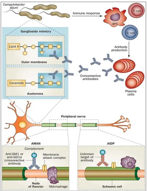
Depending on the electrophysiological properties, GBS is classified in different subtypes: acute inflammatory demyelinating polyneuropathy (AIDP), acute motor axonal neuropathy (AMAN), and a severe form of AMAN that is termed acute motor sensory axonal neuropathy (AMSAN), which is much less common (Griffin et al, 1996). AMAN is an axonal subtype (30 to 65% of patients) particularly widespread in Asia and Central and South America (Kuwabara & Yuki, 2013) (Bae et al, 2014), while AIDP is more prevalent in Europe and North America. Especially for the AMAN type, the pathogenesis of the disease is believed to involve molecular mimicry between sialylated lipooligosaccharide structures, that are contained on the cell envelope, and ganglioside epitopes of C. jejuni and neural gangliosides. This mechanism generates a cross-reactive immune response, resulting in a cascade of immune-mediated inflammatory responses, degeneration of the peripheral nerve and interruption of neurotransmission (Fig. 1.3). In serum samples from numerous patients affected by AMAN, subsequently to C. jejuni infection, high titres of antibodies against the following gangliosides were contained: GM1, GM1b, GD1a or GalNAc-GD1a (Ogawara et al, 2000). Basically, the LPS (lipopolysaccharide) from C. jejuni contains a terminal tetrasaccharide identical to that of GM1 (Yuki et al, 1993). The concept of ‘molecular mimicry’ is gathered from these observations, which imply the sharing of homologues epitopes between bacterial LPS and ganglioside surface components of peripheral nerves, particularly on the axolemma. It was supposed that, because of these molecular analogies, antibodies that are produced to attack bacteria could also attack neuronal axons, leading to GBS.
The immune response is generated by a specific immune recognition which involves T-lymphocytes, monocytes, and various cytokines responsible for causing the demyelination (Fig. 1.4). Recent studies also established the relevant role of macrophages in immune-mediated nerve damage, which are essential in both the effector phase of the disease and initiating the repair phase (Hartung & Toyka, 1990) (Baichwal et al, 1988). Previous studies
15
also demonstrated that human monocytes can phagocytose Campylobacter species in vitro and that C. jejuni, ingested by human macrophages, convert to a coccal form (Kiehlbauch et al, 1985).
Moreover, in the last 10 years, cell sorting investigations (Van Rhijn et al, 2002) revealed that Campylobacter DNA was present in CD14+ and CD33+ populations, indicating that myelomonocytic cells are Campylobacter DNA-carrying cells. A possible explanation was that although ingested bacteria could be killed by monocytes, bacterial DNA is resistant to degradation and persists within the cells, without causing clinical symptoms, in a viable but non culturable form (Hickey et al, 2005).
Fig. 1.3: Molecular mimicry and antiganglioside antibodies are at the base of the immunopathogenesis of GBS. (van den Berg et al, 2014).
16
Fig. 1.4 Monocyte functions. Monocytes and T-cells are critical to the host response to acute bacterial infection but monocytes are primarily viewed as amplifying the inflammatory signal. Although monocytes are specialized in defense against pathogens, C. jejuni elaborated CDT dependent and independent mechanisms to survive in human monocytic cells. (Image created by Gianna Di Sario)
17 1.5 Molecular basis of C. jejuni pathogenesis
To date bacterial factors implicated in host cell invasion and disease pathogenesis are: lipooligosaccharides, capsule, flagellar apparatus, cytolethal distending toxin (CDT) and post-translational glycosylation system (O-linked and N-linked glycosylation).
1.5.1 Lipooligosaccharides
In Gram-negative bacteria a main component of most outer membranes is the lipopolysaccharide (LPS). It has crucial roles in protection of bacteria from harsh environments and toxic compounds (Dong et al, 2014). LOS is analogous to the lipopolysaccharide (LPS) found in other Gram-negative families; LPS is made up of an O-polysaccharide chain, core oligosaccharide and a lipid A component in the outer membrane. LOS, lacking an O-polysaccharide repeating structure, is a low molecular weight form of bacterial LPS (Gilbert et al, 2008). They have a role in adhesion, invasion and colonisation of host and intestinal niches, protection from complement-mediated killing and surviving in different non-intestinal environments (Guerry et al, 2002) (Karlyshev et al, 2005). LOS are highly variable structures and C. jejuni uses this variability as an expedient for avoiding host defences and adapting to different microenvironments. LOS are also capable of mimicking human antigens (Guerry & Szymanski, 2008). In particular, molecular mimicry of C. jejuni LOS with gangliosides in nervous tissue induces the generation of cross-reactive anti-ganglioside antibodies resulting in GBS and Miller Fisher syndrome (see 1.4.1) (Ang et al, 2004). Indeed, it was demonstrated that specific types of the C. jejuni LOS biosynthesis gene locus are clearly associated with immune-mediated neuropathy and with the presence of ganglioside-mimicking structures in LOS (Godschalk et al, 2004).
It is well known that membrane CD14 molecule is involved in LPS-induced cytokine production in human blood cells (Alexander & Rietschel, 2001) (Landmann et al, 1996). CD14 is a 55-kDa glycoprotein, which is mainly expressed on mature monocytes, macrophages and activated granulocytes. It is a myeloid membrane glycoprotein which serves as a receptor for complexes of lipopolysaccharide (LPS) and LPS binding protein (LBP) (Heinzelmann & Bosshart, 2005). The specific cellular recognition of agonistic LPS/lipid A is initialized by the combined extracellular actions of LBP, the membrane-bound or soluble forms of CD14 and the newly identified Toll-like receptor 4 (TLR4) *MD-2 complex, leading to the rapid activation of an intracellular signaling network. Moreover, it is well known that
18
CD14 molecule is involved in a CD14-dependent phagocytosis that monocytes use to phagocytose Gram-negative bacteria (Neu et al, 2013).
1.5.2 Capsule
Capsular polysaccharides (CPSs) are commonly present on bacterial surface and play an important role in bacterial pathogenesis, survival and persistence (Llobet et al, 2008). Although capsule might be crucial for successful adhesion to epithelial cells, it does not provide protection against host innate defence and the potent antimicrobial action of human β-defensins 2 or 3 (Zilbauer et al, 2005). As elucidated during sequencing of the shotgun library of NCTC11168 (Karlyshev et al, 1999) and in subsequent studies (Gundogdu et al, 2007) (Guerry et al, 2012), capsule components are codified by genes that are split in three different regions: the first and the last regions comprise kps genes, that encode the KpS proteins involved in capsular assembly and transport; whereas the central region is highly variable and is involved in polysaccharide synthesis. Therefore, CPSs are involved in structural variation, mimicry to host antigens, and resistance to phagocytosis and complement-mediated killing.
1.5.3 Flagella
Campylobacter motility is mediated by a single unsheathed flagellum that can be contained at one or both poles. Flagella represent the primary adherence factor that creates contact between eukaryotic cell membrane and specific bacterial-invasion factors (Hu et al, 2008). Flagellar motility is vital to many aspects of C. jejuni biology, including motility, host colonization, virulence in ferret models, secretion and host-cell invasion. This apparatus allows C. jejuni to colonize both human and animal host cells (Wassenaar et al, 1993). A correlation between the presence of intact flagellum and the ability of C. jejuni to adhere and invade host cells was demonstrated in the last 20 years (Grant et al, 1993) (Konkel et al, 2004). Flagella are also involved in: secretion of virulence proteins, autoagglutination (AAG), microcolony formation and avoidance of the innate immune response (Guerry, 2007). Structurally, flagella are composed of a major flagellin, FlaA, and a minor flagellin, FlaB, that are both approximately 59 kDa in size and are highly homologous. flaA and flaB genes are respectively regulated by σ28 (encoded by fliA) and a σ54-dependent promoter (encoded by rpoN). Mutation of flaA results in the production of a truncated flagellar filament composed
19
of FlaB and having a severe reduction in motility. On the contrary, mutants in flaB have no significant changes in motility and produce a flagellar filament that appears structurally normal (Nuijten et al, 1990).
1.5.4 The cytolethal distending toxin
CDT is a heterotrimeric holotoxin produced by a diverse group of Gram-negative pathogenic bacteria, belonging to the subclass of AB2 toxin superfamily. As in the case of other AB
toxins, CDT comprise three subunits, CdtA, CdtB, and CdtC (Ge et al, 2008).
The A subunit of the toxin, CdtB, is an enzyme, with an average molecular size of 29 kDa, that exhibits cation-dependent metalloenzyme activities, in vitro, characteristic of endonucleases (Elwell & Dreyfus, 2000; Lara-Tejero & Galan, 2000), inositol polyphosphate 5-phosphatases (Dlakic, 2001) and sphingomyelinases (Hofmann et al, 2000). The B component consist in two heterogeneous subunits, CdtA (23 kDa) and CdtC (21 kDa). CdtA and CdtC act as carriers to deliver the catalytic subunit, CdtB, into host cells (Smith & Bayles, 2006). CdtB reaches the nucleus by an endoplasmic reticulum-associated degradation (ERAD) or non-ERAD pathway (followed by translocation across the nuclear membrane) where exhibits DNase I-like activity and induces limited DNA damage such as double-strand damage, leading to the activation of DNA repair responses and cell cycle arrest at the G2/M
phase (Canonico et al, 2014; Lara-Tejero & Galan, 2001)
The importance of the CdtB is clearly demonstrated in the case of Salmonella typhi that, lacking the genes for CdtA and CdtC, can translate only the CdtB protein. It was proposed that S. typhi synthesizes and secretes CdtB once it has reached an intracellular compartment of the host cell (Haghjoo & Galan, 2004). To interact with the target cells, the A subunit, that imparts biological activity to the holotoxin, is likely translocated across the plasma membrane of the target cells through a receptor-mediated process. In addition, a second important element is thought to be important for the binding of CDT to a specific area of the host membrane: it is known as lipid raft (see Lipid rafts section).
In the last 15 years different authors (Guerra et al, 2005) (DiRienzo, 2014) (Gargi et al, 2013) suggested a novel model system through which all CDTs are trafficked in a retrograde manner from the cell membrane to Golgi and endoplasmic reticulum to translocate to cytosolic targets and nucleus. More studies will be required to better define the nature of this process for C. jejuni CDT.
20 1.5.5 Glycosylation system
C. jejuni possesses two protein-glycosylation systems: The N- linked glycosylation system, which modifies serine or threonine residues, and the O- linked glycosylation system, which modifies asparagine residues (Young et al, 2007).
N-linked glycosylation machinery of C. jejuni is encoded by a single gene cluster of 16 kb named pgl cluster (protein glycosylation) (Szymanski et al, 1999). The first N-linked oligosaccharyltransferase identified in bacteria was PglB, a key enzyme in the pgl locus. Although N-linked glycosylation system is conserved in all C. jejuni strains studied to date, its role in C. jejuni biology is not clear and has to be better investigated.
It was demonstrated a correlation between N-linked glycosylation system and bacterial virulence. Different C. jejuni glycosylation mutants, that were deficient in their ability to glycosylate certain proteins, showed reduction in adhesion and invasion of human intestinal epithelial cells in vitro and colonization of the chick gastrointestinal tract (Karlyshev et al, 2004) (Kakuda & DiRita, 2006). In according to recent studies (Larsen et al, 2004) this system might be also involved in the evasion of the immune system.
Structure of N-linked glycan was clarified by mass spectrometry and NMR spectrometry analyses that revealed that this glycan is a heptasaccharide with the following structure: GalNAc-a1,4-Gal- NAc-a1,4-(Glcb1,3)GalNAc-a1,4-GalNAc-a1,4-Gal- NAc-a1,3-Bac-b1,N-Asn, where Bac is bacillosamine, 2,4-diacetamido-2,4,6-trideoxyglucose (Knauer & Lehle, 1999)
In contrast to the N-linked system, the genetic locus of the O-linked system is more heterogeneous and genetically diverse (Champion et al, 2005); the putative flagellar glycosylation locus of C. jejuni NCTC 11168 contains ∼50 genes, among which are the genes encoding the flagellin structural proteins FlaA and FlaB. O-linked glycosylation is crucial for successful flagellin assembly and motility, therefore influencing adhesion, invasion and virulence in vivo (Thibault et al, 2001). Defects in O‑linked glycosylation result in: loss of motility, decrease in the adherence to and invasion of host cells, and decreased virulence in ferrets (Guerry et al, 2006). O‑linked glycosylation of flagellin is necessary for the proper assembly of the flagellar filament, which has led to the hypothesis that the O‑glycan might have a role in the interactions of flagellin subunits with one or many elements of the flagellar apparatus.
21 1.6 The outer membrane vesicles (OMVs)
OMVs have emerged as pathogenic nanoparticles that can travel beyond the mucosa to distant locations within the host, with the ultimate goals of causing cellular destruction, promoting bacterial survival and facilitating the development of pathogenesis in the host (Pathirana & Kaparakis-Liaskos, 2016) (Fig. 1.5).
OMVs are blebs generated from the outer membrane of the cell envelope of all Gram-negative species of bacteria studied to date (Amano et al, 2010). They are typically 20-200 nm in diameter, and are released only by viable cells during the course of normal metabolism and in all cell growth phases in all environmental conditions studied (Bonnington & Kuehn, 2014). These nanostructures contain outer membrane proteins, phospholipids, lipooligosaccharides (LOS), and numerous periplasmic proteins of wide molecular mass range (Mashburn-Warren et al, 2008).
Once the OMVs are free from the bacterium, they appear as small membrane vessels including periplasmic constituents and outer membrane components. In the last decade, OMVs are increasingly recognized as key determinants for bacterial virulence (Kulp & Kuehn, 2010) and play a major role in host-pathogen interactions, including the trafficking and eventual release of diverse virulence factors from many pathogenic bacteria (Ellis & Kuehn, 2010). Recently, it was reported that the C. jejuni wild type strain 81-176 produces OMVs that contain biologically active CDT: during pathogenesis, release of OMVs by C. jejuni is a route of this bacterium to deliver all CDT subunits to the surrounding environment, infecting host cells and causing the typical cytolethal distending effects in vivo and in vitro (Lindmark et al, 2009). Several roles were attributed to OMVs: they may act as delivery vehicles for bacterial toxins lacking typical signal sequences (Balsalobre et al, 2006; Kouokam et al, 2006), promote cell-cell communication via transit of signaling molecules (Mashburn & Whiteley, 2005), and can inhibit phagosome-lysosome fusion during macrophage infection (Fernandez-Moreira et al, 2006). OMVs are also potentially rich in antigens that serve as initial targets for innate and adaptive immune recognition (Bergman et al, 2005). OMVs may also have defensive roles during infection, by sequestering antibiotics and antibodies, as well as acting as decoy antigens to divert the attention of the immune system away from the invading bacteria (Ellis & Kuehn, 2010) (Chattopadhyay & Jaganandham, 2015). Moreover, Elmi et al. demonstrated that apart from CDT, OMVs secreted by C. jejuni delivered also other virulence-associated C. jejuni proteins (Elmi et al, 2012).
22
Fig. 1.5 Atomic force micrographs of (A) a C. jejuni strain 81-176 cell (Bar: 1 µm) and of (B) OMVs on the surface of a C. jejuni cell (Bar: 100 nm). (C) Electron micrograph of OMVs isolated from C. jejuni strain 81-176 (arrows indicate OMVs) (Bar: 100 nm) (Lindmark et al, 2009).
It was hypothesized that OMVs produced by Gram-negative bacteria can invade host cells through four different routes. These routes can require clathrin coated pits, formation of caveolae, and using of lipid rafts or direct membrane fusion (Fig.1.6).
Recent research within the field have identified mechanisms whereby OMVs interact with host cells to mediate inflammation and immunity have been studied by several authors (Pathirana & Kaparakis-Liaskos, 2016). OMVs from different pathogens including H. pylori (Ismail et al, 2003) C. jejuni (Elmi et al, 2012) and E. coli (Kaparakis-Liaskos & Ferrero, 2015) have shown to trigger the production of a range of proinflammatory molecules, such as interleukin-8, in human epithelial cells resulting in modulation and control of proliferation (Li et al, 2015b), apoptosis, (Mondal et al, 2016) and immune response.
23
Fig. 1.6 O' Donoghue et al. demonstrated that OMVs produced by Gram-negative species enter host cells by several different pathways. OMVs entry can be impaired by the use of inhibitors against components of these pathways such as chlorpromazine, papain, monensin-ionophore, monodansylcadaverine, dynasore, methyl‐β cyclodextrin, filipin and nystatin; wortmannin, wiskostatin, cytocholasin D (O'Donoghue & Krachler, 2016).
1.7 C. jejuni-host cell interactions
Bacterial pathogens, especially those with an intracellular life cycle, have shown to be able to manipulate the human host cell to ensure their own survival. Cytoskeletal rearrangements, induction of anti-apoptotic pathways and control of the host cell cycle are all used to benefit bacterial infection and growth (Siegl & Rudel, 2015)
C. jejuni infection is a multistep process that includes colonization of the intestinal mucosa and interactions with and invasion of the human intestinal epithelial cells (IECs) (Young et al., 2007). Bacterial factors such as motility, glycosylation and capsule are involved in C. jejuni internalization (Szymanski et al, 2002) (Bacon et al, 2001); mutations in these
24
pathways lead to deficiencies in ability to adhere to and invade host human and animal cells (Hendrixson & DiRita, 2004; Morooka et al, 1985; Watson & Galan, 2008).
C. jejuni entry process appears to utilize the host-cell cytoskeleton as observed in many other bacterial pathogens such as Listeria monocytogenes, Shigella flexneri and Salmonella typhimurium (Cossart & Sansonetti, 2004). Unlike these reported bacteria, a recent study suggested that C. jejuni is internalized into human intestinal epithelial cells through a microtubule-dependent, actin-independent fashion.
Different strategies are adopted by bacteria in order to survive and replicate within host cells. As a matter of fact, different behaviours can be adopted by C. jejuni when invade either phagocytic or non-phagocytic eukaryotic cells. Therefore, C. jejuni internalization proceed to specific routes depending on the intracellular environment.
Robert O. Watson et al. reported that C. jejuni survives within intestinal epithelial cells by deviating from the canonical endocytic pathway; the bacterium showed the ability to avoid its delivery to lysosomes, where invading bacteria are commonly killed, shortly after internalization (Watson & Galan, 2008). In epithelial cells, C. jejuni is able to construct an intracellular niche known as Campylobacter-containing vacuole (CCV). C. jejuni can modify the CCV to suit its metabolic needs and survive. CCVs are distinct from lysosomes and are functionally separated from the canonical endocytic pathway; indeed, they are not accessible to endocytic tracers (Watson & Galan, 2008). It was also hypothesized that C. jejuni internalization is caveolin-1 dependent. A caveolin-1-stabilized lipid membrane may be required for a proper signaling through tyrosine kinases, which are also required for C. jejuni internalization rather than for endocytosis. As a matter of fact, efficient signaling through receptor tyrosine kinases requires lipid rafts or caveolae (Helms & Zurzolo, 2004) (Simons & Toomre, 2000). In addition, CCVs showed to contain LAMP-1 (lysosomal-associated membrane protein-1), an endocytic marker, that marks the end stage, when late endosomes are fused with lysosomes (Eskelinen, 2006). At the final stages of endocytosis, to degrade bacteria endolysosomal vesicles are formed in eukaryotic cells (Hamasaki & Yoshimori, 2010; Huang & Brumell, 2009)
On the contrary, C. jejuni seems not to be able to deviate this system in professional phagocytes like macrophages, which are specialized to engulf and rapidly kill bacteria (Kiehlbauch et al, 1985; Wassenaar et al, 1997), but contradictory papers are present in literature about this issue. Despite of the phagocytic activity of macrophages, it was supposed that C. jejuni was able to infect and utilizes monocytes for its spreading in humans and animals (Hickey et al, 2005). Hickey TE et al. showed that C. jejuni 81-176 was capable of
25
extensive replication within human monocytic cell vacuoles and induced apoptotic death by means cytolethal distending toxin. Michael A. Jones et al. also confirmed that monocytes and macrophages are important cell types with which C. jejuni interacts in mammals (Jones et al, 2003). These might be relevant in perpetuating the inflammatory disease because it has been shown that C. jejuni can persist within peripheral blood monocytes for up 7 days (Kiehlbauch et al, 1985). Moreover, monocytic cells could be important contributors to the induction and maintenance of gut inflammation because express a range of cell receptors such as CD14 and CD11a, which are involved in cell signaling in response to different bacterial pathogen-associated molecular patterns (Galdiero et al, 2001).
In addition, previous studies reported that monocytes play an important role in the control of immune-related diseases like GBS. Once in circulation, they can migrate into the tissues and have the potential to differentiate into macrophages or particular types of dendritic cells. Taken together, these data confirm that C. jejuni can survive intracellularly in both intestinal cells and monocytes, even though more studies are needed to investigate the relationship between C. jejuni and monocytes. Understanding C. jejuni ability to modulate the cellular environment is key in understanding its ability to cause disease.
Lipid rafts
Lipid rafts are self-organized parts of the lipid bilayer of eukaryotic cells, enriched in sphingolipids, cholesterol and proteins, that represent sub compartments that serve as stabilized platforms for specific biological functions in eukaryotic cells. Hence, the integrity of lipid rafts is important for the correct functionality of trafficking, signaling cascades and other cellular processes (Michel & Bakovic, 2007).
Bacteria can cross epithelial barriers via a transcellular route by exploiting lipid rafts. Bacteria endocytosed via this route appear to avoid lysosomal fusion (Zaas et al, 2005), hence deviating the classical endocytic pathway generally used by cells to kill invading microorganisms.
OMVs have been shown to deliver bacterial virulence factors into host cells by fusion with the host cell plasma membrane or via receptor-mediated endocytic pathways (Ellis & Kuehn, 2010) (Kulp & Kuehn, 2010). Different bacterial pathogens, including E. coli (Kesty et al, 2004), H. pylori (Kaparakis et al, 2010) and P. aeruginosa (Bomberger et al, 2009), produce OMVs that are able to bind lipid rafts. Binding of OMVs to lipid rafts has been reported also
26
for C. jejuni, which seems to be able to deliver its virulence factors by OMVs into human IECs (see 1.6).
In blood cells as well, in particular T cells, lipid rafts can be involved in bacterial invasion; depending on the differentiation status of T cells, lipid rafts seclude various protein receptors involved in cell signaling, cytoskeleton reorganization, membrane trafficking, and the entry of infectious organisms into the cells (Thomas et al, 2004).
The Endoplasmic reticulum
The ER is a large membrane-bound organelle that ensures intra- and inter- molecular disulphide bond formation, protein N-linked glycosylation, Ca2+ storage, and lipid biosynthesis. These components are subsequently delivered to their destination compartments, which include the ER itself, the Golgi apparatus, the plasma membrane, the extracellular milieu, or the endocytic and autophagic pathways. ER functions are highly interconnected and perturbation in one directly affects the others (Bettigole & Glimcher, 2015). The ER is composed of a series of continuous membranes organized into subdomains that include the rough-, smooth- and transitional-ER, and the nuclear envelope. The rough ER, which is mainly laminar, is associated with polyribosomes for protein synthesis and Ca2+ signaling. The smooth ER is primarily composed of tubular structures providing the site of lipid biosynthesis, has a main role in Ca2+ signaling, and is referred as the chief point of contact with other organelles (Park & Blackstone, 2010). For its role in secretory pathways and its biosynthetic functions, the ER stands as a nutrient-rich intracellular location that is presumably devoid of bactericidal functions, such as antimicrobial peptides or hydrolytic enzymes, representing a suitable niche for the intracellular survival, persistence and proliferation of intracellular bacterial pathogens.
ER plays also a crucial role in cellular homeostasis by modulating processing and folding of secretory membrane proteins. When protein folding in the ER is compromised or protein folding requirements exceeds, unfolded or misfolded proteins accumulate and induce ER stress. ER stress leads to the activation of the unfolded protein response (UPR), an evolutionary conserved cytoprotective signalling pathway, to restore normal ER functioning (Schroder & Kaufman, 2005) (van Schadewijk et al, 2012).
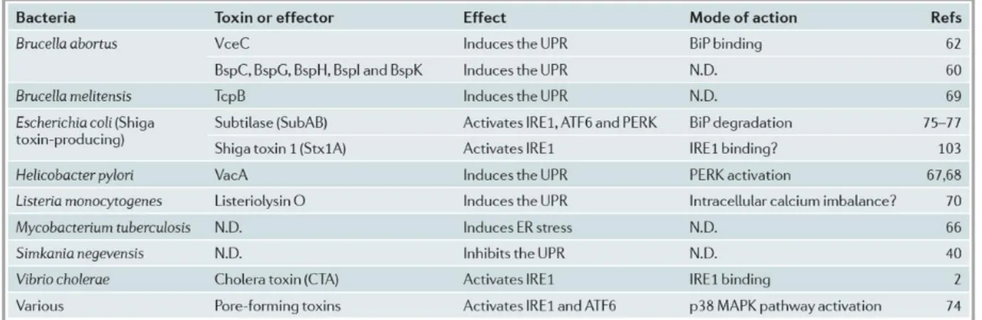
The UPR can be directly activated by certain secreted bacterial virulence factors, such as pore forming toxins (Bischof et al, 2008), subtilase cytotoxin (Wolfson et al, 2008) and cholera toxin (Cho et al, 2013) (Table 1.1). These toxins induce UPR by activating UPR mediators
27
directly. Microorganism-mediated UPR modifications therefore tune the quality and the magnitude of the immune response. On the contrary, bacteria like Legionella pneumophila modulate the cellular environment by suppressing the UPR via multiple mechanisms (Treacy-Abarca & Mukherjee, 2015). L. pneumophila is a Gram negative intracellular bacteria that can survive and replicate within eukaryotic cells; this ability is essential for its virulence.
Table 1.1 At present, it has been found that UPR can be directly activated by many secreted bacterial virulence factors (Celli & Tsolis, 2015).
Bacterial interactions with the UPR are intricate. Recent findings support that UPR is a key component of the crosstalk among ER, intracellular bacteria and their pathogenic activities. Bacteria may adapt to live in the ER resulting in UPR subversion that promotes the proliferation of intracellular bacterial pathogens; meanwhile, UPR may contribute to inflammatory and immune responses against invading bacteria. Modulation of ER functions during infection can promote bacterial infection by providing a replicative niche, but at the same time the resulting disruption of the secretory pathway can provide a pattern of pathogenesis that aids the innate immune system in recognizing intracellular infection and in mounting an appropriate defence. However, considering the more rapid evolution of bacterial pathogens compared to their hosts, it is likely that bacteria have evolved to modulate the UPR to their advantage during infection (Celli & Tsolis, 2015)
UPR modulates genes required for protein folding, protein degradation, glycosylation, lipogenesis, Ca2+ signalling and autophagy in order to correct the offending agent that trigger the ER stress. Biochemically, UPR is mediated by three ER stress receptors: protein kinase RNA-like ER kinase (PERK), inositol-requiring protein-1 (IRE-1) and activating transcription factor-6 (ATF6).
28
During steady state, all these ER stress receptors are maintained in an inactive state through their association with the ER-chaperone protein GRP78 (BiP). Having BiP higher affinity for misfolded proteins than for UPR mediators, under stress conditions, BiP dissociates from IRE-1, PERK and ATF6, leading to their activation and initiation of the UPR (Fig.1.7). Activation of IRE-1, PERK and ATF6 initiates signalling pathways of the UPR to restore homeostasis in the ER, via increasing the production of chaperones to assist in protein folding, arresting translation of proteins not involved in resolving ER stress, and degradation of misfolded proteins via the ER-associated degradation (ERAD) pathway.
ERAD is the process by which misfolded or unassembled proteins are destroyed in eukaryotic cells. This complex system involves recognition of degradation signals, dislocation of proteins across the ER membrane and degradation by the ubiquitin-proteasome system in the cytoplasm.
Dissociation of BiP from PERK leads to autophosphorylation and thereby PERK activation. Activated PERK phosphorylates the α subunit of eukaryotic transcriptional initiation factor (eIF2α), resulting in the inactivation of eIF2α and translational attenuation by interfering with 5’-cap assembly (Harding et al, 1999) (Harding et al, 2000), which prevents further accumulation of unfolded proteins in the ER (Luchetti et al, 2017). General translational inhibition results in an increase in cap-independent translation, facilitating the accumulation of the transcription factor ATF4 (Vattem & Wek, 2004). Afterward, ATF4 transcriptionally upregulates the transcription factor C/EBP homologous protein (CHOP) (Ma et al, 2002), which has been shown to modulate apoptosis in different cellular stress conditions.
The second arm of the UPR is initiated by ATF6. Dissociation of BiP from ATF6 leads to translocation of ATF6 to the Golgi complex where it is cleaved by site-1 and site-2 proteases into an active transcription factor. The resulting cytoplasmic fragment liberated from the membrane enters the nucleus resulting in transcriptional activation of its target genes (Yoshida et al, 2000) (Yoshida et al, 2001), such as BiP, CHOP and X-box binding protein (XBP1). In this manner ATF6 indirectly regulates autophagy and apoptosis via XPB1 and CHOP (Song et al, 2017).
IRE1α is a multifunctional protein that possesses kinase and endonuclease activities. Dissociation of BiP from the luminal domain of IRE1α leads to its homodimerization and autophosphorilation which triggers the excision of 26 base pairs from the XBP1 mRNA, and subsequent mRNA relegation causes a translational reading frame shift yielding the highly active transcription factor known as XBP1s (Calfon et al, 2002). XBP1s forms a heterodimer with pATF6(N) and binds to the enhancer element called the UPR element, resulting in the
29
transcriptional activation of ERAD genes such as HRD1, EDEM and Derlins (Lee et al, 2003), leading to transcription of ER-chaperone proteins. The splicing of XBP1 mRNA is considered to be an ER stress marker. The IRE1α-signalling axis is the most highly conserved branch of the UPR, it is present across species from yeast to humans.
All these three branches work cohesively to correct ER stress but can also lead to apoptosis under unresolvable stress conditions. ATF4, XBP1 and ATF6 direct a transcriptional program that upregulates chaperones, components of the ERAD pathway, and factors involved in autophagy and apoptosis that act to restore cellular homeostasis or, if the ER dysfunction cannot be resolved, initiate programmed cell death (Celli & Tsolis, 2015).
XBP1 and ATF6 support the ER folding capacity by upregulating chaperones, glycosylases, ERAD components, intracellular support machinery and protein disulphide isomerases. Moreover, Xbp1/IRE1α may limit the influx of proteins into the ER by degrading ER-localized mRNA. In a parallel branch, PERK stops protein synthesis to allow time for the ER to correct existing misfolded proteins, while downstream its target genes induce an antioxidant response to oppose ROS production generated by iterative protein folding cycles. Temporal and branch-specific UPR control is crucial for determining adaptation versus survival (Lin et al, 2007): indeed, depending on the stress conditions either prosurvival IRE1 or PERK remain activated. Successful neutralization of the instigating stress results in cell survival, whereas the outcome for failure is cell death.
30
Fig. 1.7 UPR mediators are held inactive by BiP protein, but under stress conditions, BiP dissociates from these three mediators, leading to their activation (Bettigole & Glimcher, 2015).
The UPR can be induced in vitro by chemicals such as Thapsigargin and Tunicamycin. Thapsigargin blocks the ER Ca2+ ATPase pump causing ER Ca2+ store depletion, and Tunicamycin blocks N-linked glycosylation of proteins. These two chemicals, leading to high levels of stressors, rapidly activate all three components of UPR (Rutkowski & Kaufman, 2004)
The effective immunity depends on ER homeostatic processes such as Ca2+ signalling, metabolism of lipids, glycosylation and oxidative protein folding. These same pathways, which are required to support the immune system functioning, are modulated by UPR. For instance, lipid metabolism has a central role in immune cell function. Membrane fluidity regulates lipid rafts formation, which are also crucial in C. jejuni infection, receptor clustering and signalling dynamics. IRE1 and PERK can directly induce biosynthesis of fatty acids, phospholipids and cholesterol, likely to safeguard intracellular membrane homeostasis.
31
Protein glycosylation has also an important role in the immune system by regulating cell-trafficking, surface receptors dynamics and apoptosis.
Autophagy is a key immunological process charged with degrading proteins aggregates, damaged organelles and intracellular pathogens (see 2.2). It facilitates antigen processing and presentation on both MHC-I and MHC-II, capturing and killing of intracellular microbes, efferocytosis, production of type I interferon and cell survival and differentiation. Chemical ER stress can induce autophagy via IRE1 and PERK pathways (Ogata et al, 2006); in vivo other UPR components may be involved.
2. Mechanisms of cell death
While philosophers seek the meaning of life, cell biologists are becoming ever more interested in the meaning of death (Savill & Fadok, 2000). Early and last papers about cell death distinguish three distinct mechanisms of programmed cell death that are named apoptosis, autophagy and necrosis (Fuchs & Steller, 2015).
Apoptotic and autophagic events are going to be described below (see 2.1 and 2.2). Necrosis is characterized by organelle and cellular swelling, rupture of the plasma membrane and release of the intracellular content (Vanden Berghe et al, 2014). The necroptosis is a non-apoptotic mode of cell death that is elicited by TNF receptor 1 (TNFR1) when caspases are inhibited (Degterev et al, 2005); necroptosis has often been regarded as synonymous with regulated necrosis (Galluzzi et al, 2014).
The functional relationship between apoptosis ('self-killing') and autophagy ('self-eating') is intricate in the sense that, under certain circumstances, autophagy constitutes a stress adaptation that avoids cell death (and suppresses apoptosis), whereas in other cellular settings, it constitutes an alternative cell death pathway. Depending on the upstream signals and the different instances, cells activate one or both of these responses; they can combine them or switch between the two responses in a mutually exclusive manner (Maiuri et al, 2007).
2.1 Apoptosis
Previous papers reported that CDT causes cell cycle arrest and cell death in a range of target cells (Canonico et al, 2014; Shenker et al, 2001; Svensson et al, 2001), upon cell cycle arrest DNA repair occurs and cell apoptosis results. Apoptosis has since been recognized and accepted as a distinctive and important mode of “programmed” cell death, which involves the
32
genetically determined elimination of cells. It occurs normally during development and aging and as a homeostatic mechanism to maintain cell populations in tissues. It is also thought that apoptosis is a defense mechanism such as in immune reactions or when cells are damaged by disease or noxious agents (Norbury & Hickson, 2001). Apoptosis marks unwanted cells with 'eat me' signals that are going to be recognized by phagocytes which mediate their engulfment and degradation (Savill & Fadok, 2000).
In the last decade, many apoptotic cascades have been described with the attempt to categorize all signal transduction pathways that lead to cell death, such as intrinsic and extrinsic, mitochondrial and death receptor (DR), p53-dependent and -independent, and caspase-dependent and -independent pathways in association with initiation, commitment, and execution phases (Ashe & Berry, 2003).
Apoptotic event is a highly conserved process, firstly studied on Caenorhabditis elegans (Horvitz, 1999). In biochemical terms, two distinct phases in apoptosis can be recognized, the initiation and execution phase. In accord with the source of the triggers, that can be extracellular or intracellular, intrinsic and extrinsic mechanisms of initiation can be defined. For these reasons, apoptosis can be classified in intrinsic and extrinsic pathways. Intrinsic pathway can be triggered by intracellular signals, for example by DNA damage, and involves specifically mitochondria; whilst the extrinsic pathway can be triggered by extracellular signals, for example, by growth factor withdrawal, steroid hormones, ligation of death receptors. Both these apoptotic cell death systems are mediated by molecular pathways that culminate in the activation of a family of cysteine proteases, known as the caspases, which orchestrate the dismantling and clearance of the dying cell. Activated caspases generate a cascade of proteolytic and nucleolytic events that amplify the initial signal (the execution phase). However, different studies indicate that a cell that has been treated with an apoptotic inducer can also initiate a suicide programme that does not rely on caspase activation (Chipuk & Green, 2005); therefore apoptotic cell death can be distinguished in caspase-dependent and caspase-independent apoptosis.
Light and electron microscopy have identified the various morphological changes that develops during apoptosis. During the early process it is possible to observe cell shrinkage and pyknosis by light microscopy; in this stage, cells are smaller in size, the cytoplasm is dense, the organelles are more tightly packed, and the chromatin is condensate. Then, extensive plasma membrane blebbing occurs followed by karyorrhexis and separation of cell fragments into apoptotic bodies during a process called “budding.” During this process the organelle integrity is still maintained and an intact plasma membrane encloses all the
33
intracellular content. After execution phase, these bodies are subsequently phagocytosed by macrophages, parenchymal cells, or neoplastic cells and degraded within phagolysosomes. Unlike necrosis, this programmed cell death does not cause inflammatory reactions because apoptotic cells are rapidly phagocytosed avoiding the release of the intracellular content and anti-inflammatory cytokines are not produced by engulfing cells (Kurosaka et al, 2003). Phagocytosis is due to the expression of cell surface markers that result in the early phagocytic recognition of apoptotic cells by adjacent cells. This is achieved by the movement of the normal inward-facing phosphatidylserine of the cell’s lipid bilayer to expression on the outer layers of the plasma membrane (Bratton et al, 1997).
Although externalization of phosphatidylserine is a well-known recognition ligand for phagocytes on the surface of the apoptotic cell, recent studies have shown that also other proteins, such as Annexin I and calreticulin, could be as important as phosphatidylserine. Annexin V is a recombinant phosphatidylserine-binding protein that interacts strongly and specifically with phosphatidylserine residues and is largely used in cytometry, for the detection of apoptosis (Arur et al, 2003).
2.1.1 Bcl-2 family proteins
Bcl-2 family proteins are considered to be key regulators of apoptosis. All family members share homology with the archetypal member of the family, B-cell lymphoma-2 protein (Bcl-2), which contains four Bcl-2 homology domains: BH 1-4. Bcl-2 family proteins can be functionally subdivided into anti-apoptotic (Bcl-2, Bcl-XL, Bclw, Mcl-1) and pro-apoptotic members. Two subfamilies of pro-apoptotic Bcl-2 family members have been identified: ‘BH3-only’ family, containing only a catalytic domain BH3 (Bid, Bim, Bik, Bad, Bmf, Hrk, Noxa, and PUMA), and ‘multidomain’ family, also named Bax family, containing BH1, BH2, and BH3 domains (Bax, Bak e Bok). One of the ‘BH3-only’ protein function is activating the multidomain family members Bax and Bak.
Depending on the prevalence of either pro-apoptotic or anti-apoptotic members, the destiny of the cell is decided. It was demonstrated that Bcl-2 proteins are necessary for the completion of apoptotic programs (Wei et al, 2001) (Yin et al, 2002).
Core members of the Bcl-2 family share structural similarity with the pore-forming domains of bacterial toxins, emphasizing the relevance of these proteins to membrane biology (reviewed by (Schendel et al, 1998). Several Bcl-2 family proteins insert into intracellular
34
membranes, particularly membranes of mitochondria and endoplasmic reticulum (ER), operating as guardians of these organelles.
Many Bcl-2-family proteins, both anti-apoptotic and pro-apoptotic, have C-terminal transmembrane domains that insert in the outer membrane of mitochondria. Pro-apoptotic proteins such as Bax and Bak induce mitochondrial outer membrane permeabilization (MOMP), causing the release of caspase-activating proteins and other cell death mediators; Bax or Bak are necessary for MOMP and for controlling the permeability of mitochondrial membranes. Mitochondria induce apoptosis by releasing proteins that participate in caspase activation (for example, cytochrome c) and by neutralizing inhibitors of caspases (Reed, 2002).
About ER, it was found that overexpression of anti-apoptotic Bcl-2 proteins seem to protect cells against cell death induced by ER stress (see The endoplasmic reticulum section), whereas pro-apoptotic Bcl-2-family proteins (such as Bax or Bak) are required (Scorrano et al, 2003). But how Bcl-2-family proteins regulate ER-initiated cell death mechanisms is unclear.
Bcl-2 family proteins are also present in lysosomes where have a critical role of cell life and death mediators. Lysosomes participate to apoptosis and necrosis, and are also critically involved in autophagy. Therefore, upon overexpression of either anti-apoptotic or pro-apoptotic proteins, lysosomes can mediate cell survival or death (Pattingre et al, 2005)
2.1.2 Extrinsic and intrinsic pathway
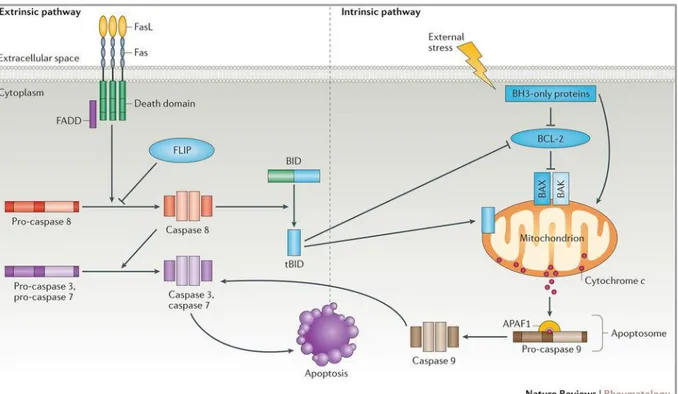
Extrinsic pathway is activated by the transduction of the apoptotic signal after the binding of death receptors, for example Fas, TNF receptor 1 (TNFR1) and TNF-related apoptosis inducing ligand (TRAIL) receptor R1 and R2, to their ligands FasL, TNF and TRAIL. On the contrary, the intrinsic pathway of apoptosis is mediated by mitochondria and is regulated by the Bcl-2 protein family. In a possible scenario of extrinsic pathway, the binding of Fas ligand (FasL) to its receptor Fas leads to the cleavage and activation of pro-caspase 8 to initiator caspase 8. This step can be mediated by the adaptor protein Fas-associated death domain protein (FADD) and can be inhibited by FADD-like apoptosis regulator (also known as FLIP), a catalytically inactive homologue of caspase 8. Subsequently, initiator caspase 8 cleaves and activates caspase 3 and caspase 7 leading to the degradative phase of apoptosis. The intrinsic pathway of apoptosis is regulated by Bcl-2 family proteins (see 2.1.1) ‘BH3-only’ proteins can either sequester anti-apoptotic proteins, or directly activate the
35
‘multidomain’ proteins, such as BAK and BAX. Once the apoptotic signalling is initiated, Bak and Bax induce the release of cytochrome C from the mitochondrion leading to its binding to APAF1 and the formation of a complex with pro-caspase 9, known as apoptosome that is thought to initiate apoptosis (Rodriguez & Lazebnik, 1999). Activation of caspase 9 in the apoptosome in turn induces apoptosis through the activation of caspase 3 and caspase 7. An alternative pathway of Fas-induced cell death involves crosstalk between the extrinsic and the intrinsic apoptotic pathways. This crosstalk is mediated by the cleavage of Bid that leads to the formation of truncated Bid, which interacting with Bak or Bax, is able to modify mitochondrial permeability leading to cytochrome C release from mitochondria. Bak or Bax expression can also be regulated by p53 (Vousden & Lane, 2007) (Marino et al, 2014) (Fig. 1.8).
Fig. 1.8 Extrinsic and intrinsic apoptotic pathways (Cuda et al, 2016).
2.1.3 pRb, p53 and CD59
The tumour suppressors pRb (retinoblastoma protein) and p53 are genes which encode for products that negatively affect the cell cycle progression thus protecting the cell from the accumulation of potentially tumorous mutations. Respectively, to control the cell cycle progression, pRb negatively regulates the transcription factor E2F, whereas p53 inhibits the