UNIVERSITY OF VERONA DEPARTMENT OF BIOTECHNOLOGY
Graduate School of Natural Sciences and Engineering Doctoral Program in Biotechnology
XXX Cycle
Biochemical characterization of ornithine aminotransferase
and cystathionine γ-lyase from Toxoplasma gondii: possible
targets for drug development?
S.S.D. BIO/10
Coordinator: Prof. Massimo Delledonne Tutor: Prof. Paola Dominici Co-Tutor: Dr. Alessandra Astegno
Abstract
Toxoplasmosis is a widespread parasitic disease caused by Toxoplasma gondii, an obligate intracellular protozoa belonging to the phylum Apicomplexa. Toxoplasmosis is a major public health problem, infecting one-third of humans worldwide. Due to the fact that no effective vaccine is currently available and treatment is based on drugs for which resistance is emerging, there is an urgent need to discover novel drug targets that are exploitable for the design of new therapeutics against the pathogen.
A recent proteomic analysis of partially sporulated oocysts of T. gondii showed that oocysts have a greater capability of de novo amino acid biosynthesis, shedding light on several stage-specific proteins whose functional profile is in accord with the oocyst need to resist various environmental stresses [1]. Herein, we focused our attention on two enzymes belonging to these putative oocyst/sporozoite-specific protein group: the ornithine aminotransferase (OAT) and the cystathionine γ-lyase (CGL). OAT is involved in the polyamine metabolism and catalyzes the reversible conversion of L-ornithine (L-orn) into glutamate-5-semialdehyde and glutamate, while CGL catalyzes the cleavage of L-cystathionine (L-cth) to L-cysteine (L-cys),
α-ketobutyrate and ammonia in the reverse transsulfuration pathway. Despite the central
metabolic roles of these enzymes, the functionality of none of them has so far been investigated. Herein, a biochemical characterization of OAT and CGL from T. gondii has been performed, in order to expand the very limited knowledge about the polyamine and cysteine metabolism of the parasite and to explore the possible use of these enzymes as novel drug targets against toxoplasmosis.
Analysis of spectral and kinetic properties of TgOAT revealed that the enzyme is largely similar to OATs from other species regarding its general transamination mechanism and spectral properties of PLP; however, it does not possess a specific ornithine aminotransferase activity, but exhibits both N-acetylornithine (AcOrn) and γ-aminobutyric acid (GABA) transaminase activity, highlighting its possible role both in arginine and GABA metabolism in
vivo. The presence of Val79 in the active site of TgOAT in place of Tyr, as in its human
counterpart, provides the necessary room to accommodate AcOrn and GABA, resembling the active site arrangement of GABA transaminases. Moreover, mutation of Val79 to Tyr resulted in a change of substrate preference between GABA, AcOrn and L-orn, suggesting a key role of Val79 in defining substrate specificity.
The purified TgCGL is a functional enzyme which splits L-cth almost exclusively at the CγS bond to yield L-cys. This finding likely implies that the reverse transsulfuration pathway is operative in the parasite. The enzyme displays only marginal reactivity toward L-cys, which is
also a mixed-type inhibitor of TgCGL activity, therefore indicating a tight regulation of cysteine intracellular levels in the parasite. Structure-guided homology modelling revealed two striking amino acid differences between human and TgCGL active sites (Glu59 and Ser340 in human to Ser77 and Asn360 in toxoplasma). Mutation of these two residues to the corresponding residues in human revealed their importance in modulating both substrate and reaction specificity of the parasitic enzyme.
Altogether our findings could be considered as a first step toward exploring the possible use of TgOAT and TgCGL as an anti-toxoplasmosis drug targets.
Table of Contents
Abstract ... 6
Abbreviations/Acronyms ... 10
CHAPTER 1 ... 12
GENERAL INTRODUCTION ... 12
1. Toxoplasma gondii, the causative agent of toxoplasmosis ... 14
2. Toxoplasmosis. ... 16
3. Pyridoxal 5’-phosphate enzymes ... 18
4. PLP-dependent enzymes as drug targets. ... 20
5. Aims ... 22
CHAPTER 2 ... 23
UNIQUE SUBSTRATE SPECIFICITY OF ORNITHINE AMINOTRANSFERASE FROM TOXOPLASMA GONDII ... 24 1. INTRODUCTION ... 25 1.1 Polyamine metabolism ... 25 1.2 Ornithine aminotransferase ... 26 2. EXPERIMENTAL ... 31 2.1 Materials ... 31 2.2 Protein production ... 31
2.3 Size exclusion chromatography (SEC) ... 32
2.4 Determination of equilibrium dissociation constant of PLP for TgOAT and V79Y variants ... 33
2.5 Steady state analysis ... 33
2.6 Pre-steady state analysis ... 34
2.7 Spectroscopic measurements... 34
2.8 Molecular modelling studies ... 35
2.9 Isothermal titration calorimetry (ITC) ... 35
3. RESULTS ... 37
3.1 Production of recombinant TgOAT ... 37
3.2 Spectral properties of recombinant TgOAT ... 38
3.3 Steady state kinetic studies ... 40
3.4 Absorption changes of TgOAT in the presence of substrates ... 42
3.5 Rapid-scanning stopped-flow kinetics ... 43
3.6 Reaction of TgOAT with β-chloro-L-alanine ... 45
3.7 V79Y variant ... 46
3.9 Regulation of TgOAT activity by reduced TgTrx ... 51
4. DISCUSSION ... 55
CHAPTER 3 ... 58
THE TRANSSULFURATION ENZYME CYSTATHIONINE Γ-LYASE IS FUNCTIONAL IN TOXOPLASMA GONDII ... 59
1. INTRODUCTION ... 60
1.1 Sulfur-containing amino acids metabolism ... 60
1.2 Cystathionine γ-lyase ... 62
2. EXPERIMENTAL PROCEDURES ... 65
2.1 Protein production ... 65
2.2 Size exclusion chromatography (SEC) ... 65
2.3 Apo-proteins preparation ... 66
2.4 Limited proteolysis ... 66
2.5 Differential scanning calorimetry ... 66
2.6 Enzyme activity assays ... 66
2.7 Inhibition assays ... 67
2.8 Spectroscopic measurement ... 68
2.9 Statistical analysis ... 68
2.10 Thin layer chromatography ... 68
2.11 Molecular modelling studies ... 68
3. RESULTS ... 70
3.1 Expression and purification of recombinant TgCGL ... 70
3.2 Properties of recombinant TgCGL ... 70
3.3 Steady State Kinetic Parameters of TgCGL ... 72
3.4 Molecular modelling ... 77 3.5 N360S variant ... 79 3.6 S77E variant ... 82 4. DISCUSSION ... 85 GENERAL CONCLUSIONS ... 87 Appendix ... 88 References ... 90
Abbreviations/Acronyms
AcOAT N-acetylornithine aminotransferase AcOrn N-acetylornithine
ADC Arginine decarboxylase
AdoMet S-adenosylmethionine
AdoMetDC S-Adenosylmethionine decarboxylase
BCA β-chloro-L-alanine CBL Cystathionine β-lyase CBS Cystathionine β-synthase CD Circular dichroism CGL Cystathionine γ-lyase CGS Cystathionine γ-synthase CS Cysteine synthase
dcAdoMet Decarboxylated S-adenosylmethionine DSC Differential scanning calorimetry DTNB 5,5′-dithiobis-(2-nitrobenzoic acid)
DTT DL-Dithiothreitol
EDTA Ethylenediaminetetraacetic acid GABA γ-aminobutyric acid
GABA-AT γ-aminobutyric acid aminotransferase
GOX Glutamate oxidase
IPTG Isopropyl β-D-1-thiogalactopyranoside ITC Isothermal titration calorimetry
L-cth L-cystathionine
L-cys L-cysteine
LDH lactate dehydrogenase
L-hcys L- homocysteine
L-orn L-ornithine
MBP buffer MOPS, bicine, proline buffer
NADH β-Nicotinamide adenine di nucleotide
OAS O-acetylserine
OAT Ornithine aminotransferase ODC Ornithine decarboxylase P5C ∆1-pyrroline-5- carboxylate
PAG DL-proparglyglycine
PAO Polyamine oxidase
PDB Protein Data Bank
PLP Pyridoxal 5’-phosphate PMP Pyridoxamine 5′-phosphate
POSP Putative oocyst/sporozoite-specific protein
SDS-PAGE Sodium dodecyl sulfate polyacrylamide gel electrophoresis SEC Size exclusion chromatography
SHMT Hydroxymethyltransferase SPDS Spermidine synthase
SPMS Spermine synthase
SSAT Spermidine/spermine N-acetyltransferase
Trx Thioredoxin
UV Ultraviolet
UV-Vis Ultraviolet-Visible
wt wild-type
Chapter 1
General introduction
The main topic of this Ph.D. thesis is the biochemical characterization of ornithine aminotransferase (Chapter 2) and cystathionine γ-lyase (Chapter 3) from Toxoplasma gondii. For each enzyme, a brief introduction on the metabolism in which the enzyme is involved and known features of the enzyme is given.
The following sections will give an overview on basic features of Toxoplasma gondii and the general mechanism of action of PLP-dependent enzymes.
1. Toxoplasma gondii, the causative agent of toxoplasmosis
Toxoplasma gondii is an obligate intracellular protozoa that belongs to the phylum
Apicomplexa, precisely to the coccidian subclass. As all the apicomplexa parasites, T. gondii possesses a unique organelle called the apicoplast. The apicoplast has a secondary endosymbiotic origin and is essential for the survival of the parasite. However, the specific functions of this organelle are not fully clarified. Thanks to the genome projects underway for
T. gondii and Plasmodium falciparum, it is known that the apicoplast is involved in the fatty
acid biosynthesis and in the synthesis of isopentenyl diphosphate (IPP), a precursor of isoprenoids. Moreover, subsequent data showed that the apicoplast of these parasites makes iron sulfur complexes and cooperates with the mitochondrion in the synthesis of haem [2].
T. gondii has a complex life cycle consisting of different phases of sexual and asexual
reproduction and uses felidae and warm-blooded vertebrates, i.e., mammals and birds, as final or intermediate hosts, respectively.
There are three infective stages of T. gondii: a rapidly dividing invasive tachyzoite, a slowly dividing bradyzoite contained in tissue cysts, and an environmental stage, the sporozoite, contained in oocysts [3].
Oocysts are produced during the parasite’s sexual cycle that occurs in the intestine of definitive host. Oocysts are excreted through cat feces in the environment, where sporulation takes place and sporozoites become infective. Upon oral uptake of sporulated oocysts by new hosts, sporozoites transform to tachyzoites that actively penetrate all nucleated cells and replicate rapidly by repeated endodyogeny. The tachyzoite form causes tissue destruction and is therefore responsible for clinical manifestations of the disease. The consequent immune-response of the host is accompanied by the formation of tissue cysts in which bradyzoites multiply slowly by endodyogeny. Tissue cysts are the terminal life-cycle stage in the intermediate hosts and are found in the retina, brain, skeletal and heart muscles. Bradyzoites could persist inside cysts for the life of the host or they could be released from cysts, transform back into tachyzoites that reinvade host cells. If ingested by a definitive host the bradyzoites proliferate in epithelial cells of the small intestine. After this asexual multiplication, the sexual phase of the life cycle is restored (Figure 1)[4,5].
Figure 1. The complex life cycle of Toxoplasma gondii [6].
During the developmental transition of these three phases, T. gondii modifies its metabolism, and morphology, to adapt to the environmental changes during its life cycle. Regarding the energy metabolism, significant differences were found between bradyzoites and tachyzoites. Bradyzoites lack a functional TCA (tricarboxylic acid) cycle and respiratory chain. Pyruvate kinase and lactate dehydrogenase (LDH) activities are higher in bradyzoites, suggesting that lactate production is the major metabolic pathway for energy generation during latency. In contrast, tachyzoites use both mitochondrial oxidative phosphorylation and glycolysis to generate ATP [7]. The parasite expresses different stage-specific isoforms of some enzymes in order to adjust glycolysis fluxes to accommodate proliferation or dormancy. T. gondii possesses two isoenzymes of LDH, LDH1 and LDH2, which are respectively tachyzoite- and bradyzoite-specific [7]. Moreover, two stage-specific enolase (ENO) have been described. In
vitro analysis revealed that the tachyzoite-specific ENO2 has a higher specific activity at Vmax and a lower denaturation temperature than those of the bradyzoite-specific ENO1. These enzymatic properties are in agreement with the metabolic and physiological needs of the parasite during differentiation [8].
Due to the difficulty in producing and working with oocysts, the sporozoite is the less biochemically characterized among the infectious stages of T. gondii. Recent proteomic analysis revealed that the metabolic proteins of freshly sporulated sporozoites may be more
similar to tachyzoites than to bradyzoites. Indeed, the ENO2 and LDH1 isoforms, that predominate in tachyzoites, were detected in oocysts [9]. Moreover, T. gondii oocysts were found to possess all the enzymes of both glycolytic and TCA cycle, differing from tachyzoites for the expression of isoenzymes of citrate synthase and phosphoenolpyruvate carboxykinase [1].
It is known that in T. gondii the TCA cycle is not coupled to glycolysis, as the pyruvate dehydrogenase complex is specifically localized to the apicoplast and not to the mitochondrion [10]. Proteomic data suggested that oocysts generate mitochondrial acetyl-CoA, necessary to feed the TCA cycle, through the β-oxidation of fatty acids and the degradation of branched amino acids. Alternatively, intracellular tachyzoites presumably use the enzyme acetyl-CoA synthetase (TGME49_066640), which could produce acetyl-CoA from the acetate scavenged from host cell mitochondria, to fuel the TCA cycle [1].
Other interesting stage-specific differences were found in amylopectin metabolism and amino acid metabolism [1]. T. gondii stores glucose in cytoplasmic granules of amylopectin, found to be more abundant in oocysts and bradyzoites than tachizoytes [11]. Oocysts uniquely express a 4-α-glucanotransferase, resulting in an increase of amylopectin debranching and glucose mobilization. This feature is in agreement with the additional demand of energy typical of the oocyst’s sporulation process.
Regarding amino acid metabolism, oocysts specifically express enzymes with a key role in the synthesis of 6 non essential amino acids, i.e., proline, alanine, threonine, cysteine, lysine and tyrosine, underlying the capability of oocysts to adapt to the nutrient-poor extracellular environment.
2. Toxoplasmosis.
Toxoplasmosis is a zoonotic disease of medical and veterinary importance with worldwide distribution [12]. Seroprevalence varies widely between different countries (from 10 to 80%) and often within a given country. As for animals, seroprevalence in human is affected by many factors, like climatic and anthropogenic factors, including dietary, social or cultural habits, quality of water and sanitation coverage [3, 4].
There are essentially two ways of transmission: the vertical and the horizontal transmission. The vertical or congenital transmission occurs during pregnancy, when the tachyzoites might cross the placenta and infect fetus. Congenital toxoplasmosis may cause abortion, neonatal death, or fetal abnormalities [12]. The horizontal transmission of T. gondii may occurs through the infection by one of the three life-cycle stages of the parasite, i.e. oocysts, tissue
cysts or tachyzoites. Tachyzoites, which are very sensitive to environmental conditions, can be transmitted by transplantation of organs or by consumption of unpasteurized milk. In general, the majority of horizontal transmissions is caused by ingestion of one of the two persistent stages of T. gondii, i.e. tissue cysts in infected meat and oocysts in food or water contaminated with cat faece. In particular, sporulated oocysts are a significant source of infection for intermediate hosts, as they are very resistant to environmental conditions. They are distributed through wind, rain, or harvested feeds and they remain infectious in moist soil or sand more than a year. Sporulated oocysts also are highly impermeable and, therefore, are also very resistant to disinfectants [5].
In immunocompetent individuals, T. gondii infection is asymptomatic in more than 80% of cases. On the other hand, toxoplasmosis is always life threatening in immunocompromised patients. Clinical manifestations include serious encephalitis, mental status changes, seizures, motor deficits, sensory abnormalities, speech abnormalities, hemiparesis and neuropsychiatric findings [3, 12].
Drug treatment is usually not necessary in asymptomatic hosts, except in children younger than 5 years. The most effective treatment for acute cases of toxoplasmosis is the combination of pyrimethamine and sulfadiazine, which have a synergic action and target two different enzymes along the folate synthesis pathway. In congenital toxoplasmosis, spiramycin is used either alone, in order to prevent fetal infection, or combined with sulfadiazine and pyrimethamine when there is substantive evidence of fetal infection. Other current therapies include the use of clindamycin, atovaquone or azithromycin [12].
However, these regimens are inadequate for the treatment of toxoplasmosis for a variety of reasons. First of all, they only control the proliferative tachyzoite stage and are unable to eliminate the cyst stage of the parasite. Then, the combination of these drug result to be toxic and have significant side effects, including hypersensitivity, bone marrow suppression, and teratogenic effects. Moreover, these treatments are no selective for toxoplasmosis therapy as they were used in the treatment of other apicomplexan disease prior to being repurposed [13]. All these motives, in addition to the emerging of parasite drug resistance, suggest that a better understanding of unique T. gondii developmental physiology, metabolism, molecular structure and virulence is required to facilitate the design of novel inhibitors against parasite.
3. Pyridoxal 5’-phosphate enzymes
Pyridoxal 5’-phosphate (PLP) enzymes are characterized by their involvement in different metabolic pathways due to their ability to catalyze a wide repertoire of reactions. Almost all PLP-dependent enzymes are associated with biochemical pathways that involve amino acids, but they also play key roles in the replenishment of one-carbon units, synthesis and degradation of biogenic amines, synthesis of tetrapyrrolic compounds and metabolism of amino-sugars [14].
Despite the variety of catalyzed transformations by the PLP-enzymes, the mechanisms of reaction share several common features. In all PLP-dependent enzymes acting on amino acid substrates, the cofactor is covalently bound to the ε-amino group of an active-site lysine residue through an imine linkage, forming the so-called internal aldimine. Through transimination, the ε-amino group of the lysine residue is exchanged with the α-amino group of the amino acid substrate to form the planar external aldimine. Both types of aldimines react reversibly with primary amines in a transaldimination reaction, with formation of a geminal diamine intermediate, allowing either binding of substrates or release of products (Scheme 1).
Scheme 1. Structures of internal aldimine, the geminal diamine and the external aldimine (adapted from [15]).
The external aldimine is the common central intermediate for all PLP-catalyzed reactions with amino acids. The cleavage of one of the three bonds at Cα gives rise to the quinonoid intermediates; the net negative charge arising from the heterolytic cleavage of sigma bonds is delocalized by the extensive conjugation of the π-electrons of the pyridine ring of the cofactor, that acts as an electron silk [15].
An important factor for the reaction specificity is the orientation of the bonds at Cα of the substrate moiety in the external aldimine adduct. Dunathan [16] pointed out that the bond in the substrate amino acid to be broken by a PLP-dependent enzyme should lie in a plane perpendicular to the plane of the cofactor-imine π system. This would minimize the transition
state energy by allowing maximum σ-π overlap between the breaking bond and the ring-imine
π system. It could also provide the geometry closest to that of the planar quinonoid
intermediate to be formed, thus minimizing molecular motion in the approach to the transition state.
PLP-catalyzed reactions may be classified as reactions proceeding through (Figure 2):
Deprotonation: dissociation of the α-hydrogen from the Schiff base leads to a quinonoid-carbanionic intermediate that can react in several ways: i) racemisation, reprotonation at Cα but without stereospecificity (e.g. alanine racemase); ii) transamination, protonation of C4’ of PLP to form a ketimine intermediate that undergoes hydrolysis to pyridoxamine phosphate (PMP) and an α-oxo acid (e.g. aspartate aminotransferase); or iii) β- (or γ-) elimination and replacement, when a good leaving group is present in the β (or γ) position of the amino acid it can be eliminated (e.g. tryptophanase and tryptophan synthase).
Elimination of CO2: loss of the carboxyl group from external aldimine that most commonly leads to the protonation at the original site of decarboxylation followed by breakup of the Schiff base (e.g. glutamic acid decarboxylase).
Side chain cleavage: Schiff base side chains undergo aldol cleavage. Conversely, a side chain can be added by β condensation. The best known enzyme of this group is serine hydroxymethyltransferase.
Figure 2. A schematic view of the different reaction types catalyzed by PLP-dependent enzymes that act on amino acids [17].
Due to their common mechanistic features, PLP-dependent enzyme often catalyze different chemical reactions, showing ‘catalytic promiscuity’. This feature may have played a
fundamental role in divergent evolution and diversification of catalytic properties. Ancestral enzymes were probably able to catalyze several reactions, but gene duplication and evolutionary pressure may have worked to modify enzymes’ active sites so as to confer narrower substrate and reaction specificity [18].
Initially, PLP-dependent enzymes have been classified into five distinct structural groups [19], which presumably correspond to five independent evolutionary lineages originated very early in the evolution (before the three biological kingdoms diverged) from different protein ancestors [20]. These families have been named from their more representative enzyme. The aspartate aminotransferase family corresponds to fold type I and contains the majority of structurally determined PLP-dependent enzymes, consisting of aminotransferases, decarboxylases as well as of enzymes that catalyze α,β- and α,γ-eliminations. Its members are catalytically active as homodimers, although they may assemble into higher order complexes, and their active site lies at on the dimer interface. The tryptophan synthase β-subunit family corresponds to the fold type II and differ from those of fold Type I in that the active sites are composed entirely of residues from one monomer, with additional regulatory domains. The bacterial alanine racemase family corresponds to the fold type III. These enzymes are obligate dimers, as each monomer contributes residues to both active sites. In enzymes of the D-amino acid aminotransferase family, fold type IV, the cofactor binds with its re side facing the protein rather than the active-site pocket as in the fold-type I family, accounting for the difference in stereochemistry of the products in the D-amino acid reaction. The enzymes of fold type V, e.g. glycogen phosphorylase, are mechanistically distinct in utilizing the cofactor phosphate group for catalysis. Subsequently, two more groups were identified by Percudani and Perracchi, fold type VI and fold type VII that are represented by D-lysine 5,6-aminomutase and lysine 2,3-5,6-aminomutase respectively, and contains enzymes whose structures are unlike all other PLP-dependent enzymes [17].
4. PLP-dependent enzymes as drug targets.
A consequence of their widespread metabolic distribution is that a number of these enzymes are current drug targets. For example, inhibitors of L-DOPA (L-3,4-dihydroxyphenylalanine) decarboxylase are used in the treatment of Parkinson’s disease [21], alanine racemase has been identified as antibacterial drug target [22] and inhibitors of γ-aminobutyric acid aminotransferase (GABA-AT) are used in the treatment of epilepsy [23].
PLP-dependent enzymes are also intimately involved in the metabolic pathways of protozoa [24]. However, so far only serine hydroxymethyltransferase (SHMT) and ornithine
decarboxylase (ODC) have been targeted to develop clinically useful anti-protozoan drugs. Therefore, future studies are required to explore the potential of the other enzymes as anti-protozoan drug targets.
Recent analysis for PLP-dependent enzymes suggests three types of emergent drug targets: (1) enzymes that are present only in pathogens; (2) enzymes common to both humans and pathogens that possess different properties, allowing to discriminate each other; (3) enzymes related to a specialized parasitic life style (distinct host cells or particular life cycle phase). All these groups of enzymes identify targets that may be of interest in the development and design of species-specific therapeutics [24, 25].
Regarding the third group of drug targets, a recent proteomic analysis of partially sporulated oocysts of T. gondii identified a subset of proteins specifically expressed at the oocyst/sporozoite stage of the parasite. Among these putative oocyst/sporozoite-specific proteins (POSPs), four PLP-dependent enzymes were found. In particular, the oocyst resulted to differ from the tachyzoite for the expression of ornithine aminotransferase (OAT, TGME49_069110), an enzyme that takes part in the synthesis of proline and polyamine metabolism. The POPS subset also included three enzymes involved in the biosynthesis of cysteine: cystathionine β-synthase (CBS, TGME49_059180), cystathionine γ-lyase (CGL, TGME49_112930) and cysteine synthase (CS, TGME49_078910). This analysis provided proteomic evidence that the capability of de novo amino acid biosynthesis is modulated between oocysts and tachyzoites, with oocysts showing a greater persistence than tachyzoites. This feature is in accordance with the adaptation of T. gondii oocysts to the nutrient-poor and stressing extracellular environment.
5. Aims
This thesis aims to give a detailed biochemical description of two enzymes from T. gondii, the ornithine aminotransferase (TgOAT) and the cystathionine γ-lyase (TgCGL), in order to expand the very limited knowledge about the polyamine and cysteine metabolism of the parasite and to explore the possible use of these enzymes as novel drug targets against toxoplasmosis.
We overexpressed the proteins in E. coli and we isolated the recombinant enzyme as His-tagged proteins. The kinetic parameters of main and secondary activities were evaluated by steady state and pre-steady state analysis. In particular, pre-steady state analysis has been performed in collaboration with Prof. Mariarita Bertoldi from University of Verona. Moreover, we analyzed oligomeric state, ligand-induced spectral transitions and protein stability through an array of biochemical and biophysical techniques, such as UV-Vis and fluorescence spectroscopy, circular dichroism spectroscopy, size exclusion chromatography, limited proteolysis and differential scanning calorimetry. After a preliminary biochemical characterization, mutagenesis studies were performed to identify the key residues in the active sites that are responsible for reaction and substrate specificity.
Collaboration with Dr. Alessandro Paiardini, researcher at Sapienza University of Rome, was established in order to perform molecular modelling analysis to understand at a molecular level the structure/function relationship, substrate binding and catalytic mechanism of the enzymes.
Unique substrate specificity of ornithine aminotransferase from
Toxoplasma gondii
Abstract
Toxoplasma gondii is a protozoan parasite of medical and veterinary relevance responsible for
toxoplasmosis in humans. As an efficacious vaccine remains a challenge, chemotherapy is still the most effective way to combat the disease. In search for novel druggable targets, herein we performed a thorough characterization of the putative PLP-dependent enzyme TgOAT. We overexpressed the protein in E. coli and analyzed its molecular and kinetic properties by UV-Vis absorbance, fluorescence, and CD spectroscopy, in addition to kinetic studies of both the steady state and pre-steady state. TgOAT is largely similar to OATs from other species regarding its general transamination mechanism and spectral properties of PLP; however, it does not show a specific ornithine aminotransferase activity as its homologs, but exhibits both AcOrn and GABA transaminase activity in vitro, suggesting a role in both arginine and GABA metabolism in vivo. The presence of Val79 in the active site of TgOAT in place of Tyr, as in its human counterpart, provides the necessary room to accommodate AcOrn and GABA, resembling the active site arrangement of GABA transaminases. Moreover, mutation of Val79 to Tyr results in a change of substrate preference between GABA, AcOrn and L-orn, suggesting a key role of Val79 in defining substrate specificity. The findings that TgOAT possesses parasite-specific structural features as well as differs in substrate specificity from its human homolog make it an attractive target for antitoxoplasmosis inhibitor design that can be exploited for chemotherapeutic intervention.
This work was published in the Biochemical Journal (2017) DOI: 10.1042/BCJ20161021
1. INTRODUCTION
1.1 Polyamine metabolism
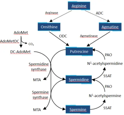
Polyamines are aliphatic hydrocarbon chains with one or more amine groups. Their positive charge at physiological pH enables their interaction with polyanionic molecules such as DNA, RNA, phospholipid head groups in cell membrane or cell wall components. Due to their variety in terms of molecular structure, valence and prevalence, polyamines undertake different roles in the cells such as survival, growth, gene expression, stress response, cell differentiation and parasidic activity [26].
In Eukariota the biosynthesis of polyamines typically starts with the conversion of arginine to ornithine by arginase and the subsequent decarboxylation of ornithine by ODC to form putrescine. Adenosylmethionine decarboxylase (AdoMetDC) produces decarboxylated S-adenosylmethionine (dcAdoMet) which, then gives its aminopropyl group to putrescine for spermidine and spermine synthesis. Spermidine synthase (SPDS) and spermine synthase (SPMS), respectively, catalyse the latter two reactions. An alternative biosynthetic pathway that characterized Bacteria and plants is the arginine decarboxylase (ADC) pathway. ADC converts arginine into agmatine, which is consequently converted into putrescine by agmantinase (Figure 3).
The polyamine metabolism is highly divergent among Protozoa. In Trypanosoma brucei and Leishmania parasites, putrescine and spermidine are synthesized by ODC, AdoMetDC and SPDS. Differently, Trypanosoma cruzi lacks ODC, but does express both AdoMetDC and SPDS, implying that once putrescine is transported into the parasite, it can be converted into spermidine and spermine by the promiscuous activity of SPDS [27]. Moreover, published data have shown that Cryptosporidium parvum has a plant-like pathway that utilizes ADC [28], whereas in P. falciparum ODC and AdoMetDC enzymes were components of a bifunctional protein [29].
Interference with polyamine metabolism has been successfully exploited in the clinical treatment of West African sleeping sickness originated by Trypanosoma brucei gambiense by using α- difluoromethylornithine (DFMO, Eflornithine), a derivative of ornithine that can irreversibly inactivate ODC [30–32]. Furthermore, AdoMetDC, and SPDS have been chemically validated as a drug target in the major protozoan parasites and many specific irreversible inhibitors have been synthetized [27, 33–36]. These results prompted the idea that polyamine metabolism may be an important drug target also in other protozoan parasites responsible for global diseases, including T. gondii.
Recent studies suggest that toxoplasma lacks a forward-directed polyamine biosynthestic pathway. Indeed, it is devoid of the activity of two essential enzymes for polyamine metabolism such as ODC and ADC, and therefore is auxotrophic for polyamines [37, 38]. T.
gondii has previously been shown to have a high affinity putrescine transporter,
demonstrating the ability to scavenge host-derived putrescine [39]. This protozoan synthesizes polyamines by a retroconversion pathway from spermidine to spermine and putrescine via spermidine/spermine N-acetyltransferase (SSAT) and polyamine oxidase (PAO) [38]. Even if polyamine metabolism has not been deeply investigated in T. gondii, it is known that the parasite, as Plasmodium, possesses the polyamine-related PLP-enzyme OAT which is involved in ornithine homeostasis, control of polyamine levels and synthesis, and mitosis.
1.2 Ornithine aminotransferase
OAT (E.C. 2.6.1.13) is a strongly conserved enzyme found in almost all eukaryotic organisms, from protozoans to humans, and from fungi to higher plants.
OAT, like every PLP-dependent transaminase, operates via a “ping-pong” mechanism that requires two half reactions to complete one catalytic cycle of transamination. In the first part of the reaction, L-orn binds to the pyridoxal form of the holo-enzyme (OAT–PLP) with the formation of L-glutamate-γ-semialdehyde, which then cyclizes spontaneously to ∆1
-pyrroline-5- carboxylate (P5C), yielding the pyridoxamine form of the enzyme (OAT–PMP). The second half-transamination proceeds through the reaction of α-ketoglutarate (α-KG) with OAT–PMP to reform OAT–PLP and release L-glutamate (see Scheme 2). P5C is further converted into proline.
Scheme 2. Overview of the two half-transamination reactions catalysed by OAT.
The human OAT is synthesized as a 49 kDa precursor in the cytosol and it is then imported into mitochondria where it reaches the functional conformation upon removal of an N-terminal mitochondrial targeting sequence (residues 1–25) producing a ~45 kDa mature protein [40]. In the PDB databank, six crystal structures of this enzyme, both ligand-free [41] and in complex with different substrate analogues [42, 43], are present. These crystallographic studies showed that the human enzyme assume an hexameric assembly consisting of three homodimers hold together principally by electrostatic interactions. The dimer belongs to the fold type I class of PLP-enzymes and it was suggested that it could represent the functional unit of the enzyme [41]. In the active site, the PLP cofactor is fixed at the interface between subunits and forms an internal aldimine via a Schiff base bond with the catalytic Lys.
Crystal structures of human OAT complexed with inhibitors elucidate the exact mechanism for the specific ω-transaminase reaction of the enzyme: the α-carboxylate of L-orn is bound by R180 with the non-reacting α-amino group held between the OH groups of Y55 and Y85, while R413 interacts with E235 in order to arrange the δ-amino group of L-orn for transimination. In particular, the glutamate residue is important for neutralizing the positive charge of the R413 through a salt bridge during the first half-reaction, when there is an
internal PLP
productive binding of the second half
half-reaction, this hydrogen bond
α-KG to interact with R413 Figure 4. fuoromethylornithine PDB ID code 2OAT. Interestingly, in expressed at th
protein, whose functional behavior is associated to the adaptation of multiple extracellular environmental stresses
treating the parasitic infection.
Despite the presence of eight spatial structures of TgOAT in Protein Data Bank (PDB ID code 4NOG at 1.2 Å; PDB
5EAV at 1.6 Å; PDB ID code 5EQC at 2.2 Å; PDB ID code 5DJ9 at 1.55Å; PDB ID code 5E5I at 1.7 Å; PDB ID code 5E3K at 1.7Å), functional information regarding this enzyme is still lacking.
Herein, in order to evaluate inhibitor design,
from T. gondii
A previous study, describing the human GABA
contributors to the productive binding of
internal PLP-lysine aldimine as the substrate approaches. This closed system allows productive binding of
the second half-reaction. On the other hand reaction, this hydrogen bond
KG to interact with R413
Figure 4. Structure of the a uoromethylornithine
PDB ID code 2OAT.
Interestingly, in T.
expressed at the oocyst/sporozoite stage
whose functional behavior is associated to the adaptation of multiple extracellular environmental stresses
treating the parasitic infection.
Despite the presence of eight spatial structures of TgOAT in Protein Data Bank (PDB ID code 4NOG at 1.2 Å; PDB
5EAV at 1.6 Å; PDB ID code 5EQC at 2.2 Å; PDB ID code 5DJ9 at 1.55Å; PDB ID code 5E5I at 1.7 Å; PDB ID code 5E3K at 1.7Å), functional information regarding this enzyme is still lacking.
in order to evaluate
inhibitor design, significant progress in the spectroscopic characterization of the enzyme OAT
T. gondii ME49
previous study, describing the GABA-AT,
contributors to the productive binding of
lysine aldimine as the substrate approaches. This closed system allows productive binding of L-orn to OAT
reaction. On the other hand reaction, this hydrogen bond
KG to interact with R413 (Figure 4)
Structure of the a uoromethylornithine (5FMOrn)
PDB ID code 2OAT. The figure is carried out
gondii, OAT has been recently identified as a protein specifically
e oocyst/sporozoite stage
whose functional behavior is associated to the adaptation of multiple extracellular environmental stresses
treating the parasitic infection.
Despite the presence of eight spatial structures of TgOAT in Protein Data Bank (PDB ID code 4NOG at 1.2 Å; PDB ID code 4ZLV at 1.8 Å; PDB ID code 4ZWM at 2.3 Å; PDB ID code 5EAV at 1.6 Å; PDB ID code 5EQC at 2.2 Å; PDB ID code 5DJ9 at 1.55Å; PDB ID code 5E5I at 1.7 Å; PDB ID code 5E3K at 1.7Å), functional information regarding this enzyme is
in order to evaluate the TgOAT as a potential target for structure
significant progress in the spectroscopic characterization of the enzyme OAT ME49 and in the determination of its catalytic properties has
previous study, describing the
, demonstrated that Tyr55 and contributors to the productive binding of
lysine aldimine as the substrate approaches. This closed system allows to OAT and
reaction. On the other hand
reaction, this hydrogen bond network is weakened, and the salt bridge opens up to allow (Figure 4) [43]
Structure of the active site of hOAT
(5FMOrn). FMP is the inhibitor adduct formed between PLP and 5FMOrn. The figure is carried out
, OAT has been recently identified as a protein specifically e oocyst/sporozoite stage [1]
whose functional behavior is associated to the adaptation of multiple extracellular environmental stresses
Despite the presence of eight spatial structures of TgOAT in Protein Data Bank (PDB ID code ID code 4ZLV at 1.8 Å; PDB ID code 4ZWM at 2.3 Å; PDB ID code 5EAV at 1.6 Å; PDB ID code 5EQC at 2.2 Å; PDB ID code 5DJ9 at 1.55Å; PDB ID code 5E5I at 1.7 Å; PDB ID code 5E3K at 1.7Å), functional information regarding this enzyme is
the TgOAT as a potential target for structure
significant progress in the spectroscopic characterization of the enzyme OAT and in the determination of its catalytic properties has
previous study, describing the different substrate specifici demonstrated that Tyr55 and
contributors to the productive binding of
lysine aldimine as the substrate approaches. This closed system allows and restrains the binding of
reaction. On the other hand, when there is an external PMP during the second network is weakened, and the salt bridge opens up to allow
[43].
ctive site of hOAT
FMP is the inhibitor adduct formed between PLP and 5FMOrn. The figure is carried out using VMD
, OAT has been recently identified as a protein specifically [1]. Inhibition of a polyamine
whose functional behavior is associated to the adaptation of
multiple extracellular environmental stresses, could constitute an efficacious strategy for
Despite the presence of eight spatial structures of TgOAT in Protein Data Bank (PDB ID code ID code 4ZLV at 1.8 Å; PDB ID code 4ZWM at 2.3 Å; PDB ID code 5EAV at 1.6 Å; PDB ID code 5EQC at 2.2 Å; PDB ID code 5DJ9 at 1.55Å; PDB ID code 5E5I at 1.7 Å; PDB ID code 5E3K at 1.7Å), functional information regarding this enzyme is
the TgOAT as a potential target for structure
significant progress in the spectroscopic characterization of the enzyme OAT and in the determination of its catalytic properties has
different substrate specifici demonstrated that Tyr55 and
contributors to the productive binding of L-orn. In particular, Tyr85
lysine aldimine as the substrate approaches. This closed system allows the binding of
, when there is an external PMP during the second network is weakened, and the salt bridge opens up to allow
ctive site of hOAT in complex with the inhibitor 5 FMP is the inhibitor adduct formed between PLP and 5FMOrn.
using VMD (Visual Molecular Dynamics)
, OAT has been recently identified as a protein specifically Inhibition of a polyamine
whose functional behavior is associated to the adaptation of
could constitute an efficacious strategy for
Despite the presence of eight spatial structures of TgOAT in Protein Data Bank (PDB ID code ID code 4ZLV at 1.8 Å; PDB ID code 4ZWM at 2.3 Å; PDB ID code 5EAV at 1.6 Å; PDB ID code 5EQC at 2.2 Å; PDB ID code 5DJ9 at 1.55Å; PDB ID code 5E5I at 1.7 Å; PDB ID code 5E3K at 1.7Å), functional information regarding this enzyme is
the TgOAT as a potential target for structure
significant progress in the spectroscopic characterization of the enzyme OAT and in the determination of its catalytic properties has
different substrate specifici demonstrated that Tyr55 and Tyr85
. In particular, Tyr85
lysine aldimine as the substrate approaches. This closed system allows the binding of α-KG
, when there is an external PMP during the second network is weakened, and the salt bridge opens up to allow
in complex with the inhibitor 5 FMP is the inhibitor adduct formed between PLP and 5FMOrn.
(Visual Molecular Dynamics)
, OAT has been recently identified as a protein specifically Inhibition of a polyamine-related, stage
whose functional behavior is associated to the adaptation of
could constitute an efficacious strategy for
Despite the presence of eight spatial structures of TgOAT in Protein Data Bank (PDB ID code ID code 4ZLV at 1.8 Å; PDB ID code 4ZWM at 2.3 Å; PDB ID code 5EAV at 1.6 Å; PDB ID code 5EQC at 2.2 Å; PDB ID code 5DJ9 at 1.55Å; PDB ID code 5E5I at 1.7 Å; PDB ID code 5E3K at 1.7Å), functional information regarding this enzyme is
the TgOAT as a potential target for structure
significant progress in the spectroscopic characterization of the enzyme OAT and in the determination of its catalytic properties has
different substrate specificities between human OAT and Tyr85 in human OAT
. In particular, Tyr85 was found
lysine aldimine as the substrate approaches. This closed system allows that are required for , when there is an external PMP during the second network is weakened, and the salt bridge opens up to allow
in complex with the inhibitor 5 FMP is the inhibitor adduct formed between PLP and 5FMOrn.
(Visual Molecular Dynamics).
, OAT has been recently identified as a protein specifically related, stage
whose functional behavior is associated to the adaptation of T. gondii
could constitute an efficacious strategy for
Despite the presence of eight spatial structures of TgOAT in Protein Data Bank (PDB ID code ID code 4ZLV at 1.8 Å; PDB ID code 4ZWM at 2.3 Å; PDB ID code 5EAV at 1.6 Å; PDB ID code 5EQC at 2.2 Å; PDB ID code 5DJ9 at 1.55Å; PDB ID code 5E5I at 1.7 Å; PDB ID code 5E3K at 1.7Å), functional information regarding this enzyme is
the TgOAT as a potential target for structure-based selective significant progress in the spectroscopic characterization of the enzyme OAT
and in the determination of its catalytic properties has been made
ties between human OAT and in human OAT were
was found to be a major lysine aldimine as the substrate approaches. This closed system allows that are required for , when there is an external PMP during the second network is weakened, and the salt bridge opens up to allow
in complex with the inhibitor 5-FMP is the inhibitor adduct formed between PLP and 5FMOrn.
, OAT has been recently identified as a protein specifically related, stage-specific oocysts to could constitute an efficacious strategy for
Despite the presence of eight spatial structures of TgOAT in Protein Data Bank (PDB ID code ID code 4ZLV at 1.8 Å; PDB ID code 4ZWM at 2.3 Å; PDB ID code 5EAV at 1.6 Å; PDB ID code 5EQC at 2.2 Å; PDB ID code 5DJ9 at 1.55Å; PDB ID code 5E5I at 1.7 Å; PDB ID code 5E3K at 1.7Å), functional information regarding this enzyme is
based selective significant progress in the spectroscopic characterization of the enzyme OAT
been made.
ties between human OAT and were major to be a major lysine aldimine as the substrate approaches. This closed system allows that are required for , when there is an external PMP during the second network is weakened, and the salt bridge opens up to allow
, OAT has been recently identified as a protein specifically specific oocysts to could constitute an efficacious strategy for
Despite the presence of eight spatial structures of TgOAT in Protein Data Bank (PDB ID code ID code 4ZLV at 1.8 Å; PDB ID code 4ZWM at 2.3 Å; PDB ID code 5EAV at 1.6 Å; PDB ID code 5EQC at 2.2 Å; PDB ID code 5DJ9 at 1.55Å; PDB ID code 5E5I at 1.7 Å; PDB ID code 5E3K at 1.7Å), functional information regarding this enzyme is
based selective significant progress in the spectroscopic characterization of the enzyme OAT
ties between human OAT and major to be a major
determinant of specificity towards L-orn with respect to GABA [44]. Notably, Tyr85 in human OAT is replaced by isoleucine in GABA-AT.
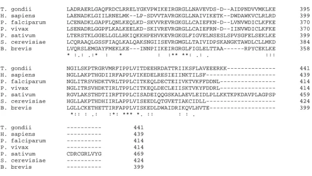
Sequence alignment of OAT from different organisms indicated that both of these tyrosines are highly conserved. However Tyr85 (human OAT numbering) is substituted by Val79 in TgOAT (Figure 5). Thus, a further aim of this study was to determine the contribution of Val79 to the specificity of the TgOAT enzyme by mutating the valine residue to tyrosine.
T. gondii ---MATKSDGSASAAAEGGARKTNIEAYRDGLKLKTEEDFFACDRQYVCQNYA 50 H. sapiens MFSKLAHLQRFAVLSRGVHSSVA----SATSVATKKTVQGPPTSDDIFEREYKYGAHNYH 56 P. falciparum ---MDFVKELKSSQDYMNNELTYGAHNYD 26 P. vivax ---MDFIKELKSSQDYMNNELTYGAHNYD 26 P. sativum ----MAATRQVQCLMRRV---CRGTRTF--AVATQSNASSSSQTIIDKEHQHSAHNYH 49 S. cerevisiae ---MSEATLSSKQTIEWENKYSAHNYH 24 B. brevis ---MSKTNVVIEQTEKFGAHNYH 20 . . : . .:** T. gondii PVPVVISKGKGARVWDINGNEYYDFLAGVSSLSQGHCHPRVIAALCRQAERLTLTLRAFG 110 H. sapiens PLPVALERGKGIYLWDVEGRKYFDFLSSYSAVNQGHCHPKIVNALKSQVDKLTLTSRAFY 116 P. falciparum PIPVVLKRGKGVFVYDIEDRRYYDFLSAYSSVNQGHCHPDILNAMINQAKKLTICSRAFF 86 P. vivax PIPVVLKRGSGVFVYDIEDRRYYDFLSAYSSVNQGHCHPNILNAMINQAKKLTICSRAFF 86 P. sativum PLPIVFAHAKGSSVWDPEGNKYIDFLSGYSAVNQGHCHPKILKALHDQADRLTVSSRAFY 109 S. cerevisiae PLPVVFHKAKGAHVWDPEGKLYLDFLSAYSAVNQGHCHPHIIKALTEQAQTLTLSSRAFH 84 B. brevis PLPIVISKAEGVWVHDPEGNKYLDMLSAYSALNQGHRHPRIIQALKDQADKVTLTSRAFY 80 *:*:.: :..* : * :.. * *:*:. *::.*** ** :: *: *.. :*: *** T. gondii NDVTGPACRFMAEMFGYDRVLLMNTGAEAGESALKIARKWAYEVKEIPPDSAKVILCNNN 170 H. sapiens NNVLGEYEEYITKLFNYHKVLPMNTGVEAGETACKLARKWGYTVKGIQKYKAKIVFAAGN 176 P. falciparum SDSLGVCERYLTNLFGYDKVLMMNTGAEASETAYKLCRKWGYEVKKIPENSAKIIVCNNN 146 P. vivax SDSLGVCERYLTTLFGYDKVLMMNTGAEANETAYKMCRKWGYEVKKIPENEAKIIVCNNN 146 P. sativum NDRFPVFAEYLTALFGYDMVLPMNTGAEGVETALKLARKWGYEKKKIPNDEALIVSCCGC 169 S. cerevisiae NDVYAQFAKFVTEFFGFETVLPMNTGAEAVETALKLARRWGYMKKNIPQDKAIILGAEGN 144 B. brevis NDQLGEFYEKLSAVTGKEMILPMNTGAEAVETALKAVRRWAYDVKKVPENQAEIIVCEGN 140 .: . :: . . . :* ****.*. *:* * *:*.* * : .* :: . . T. gondii YWGRTITACSSSTTFD-CYNNFGPFTPGF---ELIDYDDVGALEEALKD---PNVAA 220 H. sapiens FWGRTLSAISSSTDPT-SYDGFGPFMPGF---DIIPYNDLPALERALQD---PNVAA 226 P. falciparum FSGRTLGCVSASTDKK-CKNNFGPFVPNF---LKVPYDDLEALEKELQD---PNVCA 196 P. vivax FSGRTLGCVSASTDRK-CKNNFGPFVPNF---LKVPYDDLEALEVELQD---PNVCA 196 P. sativum FNGRTLGVISMSCDNE-ATRGFGPLMPGH---LKVDFGDAEAIERIF-KEKGDRVAA 221 S. cerevisiae FHGRTFGAISLSTDYEDSKLHFGPFVPNVASGHSVHKIRYGHAEDFVPILESPEGKNVAA 204 B. brevis FHGRTVTVTSFSSAEE-YRRGFGPFTPGF---KIIPYGDIEALKQAI-T---PNTAA 189 : ***. * * ***: *. : :.. : : ...* T. gondii FFVEPIQGEGGVNVPKPGYLKRAHELCRSKNVLLIVDEIQTGLCRTGRLLAADH--DEVH 278 H. sapiens FMVEPIQGEAGVVVPDPGYLMGVRELCTRHQVLFIADEIQTGLARTGRWLAVDY--ENVR 284 P. falciparum FIVEPVQGEAGVIVPSDSYFPGVASLCKKYNVLFVADEVQTGLGRTGKLLCTHH--YGVK 254 P. vivax FVVEPIQGEAGVILPSDGYFKGVEALCKKYNVLFVADEVQTGLGRTGKLLCTYH--YGVR 254 P. sativum FILEPIQGEAGVVIPPDGYLKAVRDLCSKYNVLMIADEIQTGLARTGKMLACDW--EDVR 279 S. cerevisiae IILEPIQGEAGIVVPPADYFPKVSALCRKHNVLLIVDEIQTGIGRTGELLCYDHYKAEAK 264 B. brevis FMLEPIQGEAGIIIPQEGFLKQAQEVCKANNVLLVSDEIQTGFGRTGKMFASDW--ENVV 247 :.:**:***.*: :* .:: . :* :**:: **:***: ***. :. .
T. gondii PDILLLGKSLSAGVVPISAVMGRADVMDVLKPGTHGSTFGGNPLACAVAVEALTVLKDEK 338 H. sapiens PDIVLLGKALSGGLYPVSAVLCDDDIMLTIKPGEHGSTYGGNPLGCRVAIAALEVLEEEN 344 P. falciparum PDVILLGKALSGGHYPISAILANDDVMLVLKPGEHGSTYGGNPLAAAICVEALKVLINEK 314 P. vivax PDVILLGKALSGGHYPISAILANNDVMLVLKPGEHGSTYGGNPLAAAICVESLNVLINEK 314 P. sativum PDVVILGKALGGGILPVSAVLADKDVMLCIKPGQHGSTFGGNPLASAVAIAALEVIKEER 339 S. cerevisiae PDIVLLGKALSGGVLPVSCVLSSHDIMSCFTPGSHGSTFGGNPLASRVAIAALEVIRDEK 324 B. brevis PDMYIMGKALGGGVFPISAVAADKEILSVFEPGSHGSTFGGNPLGCAVAVAAMDVLADEG 307 **: ::**:*..* *:*.: ::: : ** ****:*****.. :.: :: *: :*
T. gondii LADRAERLGAQFRDCLRRELYGKVPWIKEIRGRGLLNAVEVDS-D--AIDPNDVVMKLKE 395 H. sapiens LAENADKLGIILRNELMK--LP-SDVVTAVRGKGLLNAIVIKETK--DWDAWKVCLRLRD 399 P. falciparum LCENADKLGAPFLQNLKEQLKD-SKVVREVRGKGLLCAIEFKN-D--LVNVWDICLKFKE 370 P. vivax LSENADRLGGPFLKALKEELKD-SKIVREVRGRGLLCAIEFRN-D--IINVWDICLKFKE 370 P. sativum LTERSTKLGGELLGLLHKIQKKHPEHVKEVRGKGLFIGVELNSESLSPVSGFELSEKLKE 399 S. cerevisiae LCQRAAQLGSSFIAQLKALQAKSNGIISEVRGMGLLTAIVIDPSKANGKTAWDLCLLMKD 384 B. brevis LVQRSLEMGAYFMEKLKE---INNPIIKEIRGRGLFIGLELTTAA---RPYCEKLKE 358 * :.: .:* : * : :** **: .: . ::: T. gondii NGILSKPTRGRVMRFIPPLVITDEEHRDATTRIIKSFLAVEEERKK--- 441 H. sapiens NGLLAKPTHGDIIRFAPPLVIKEDELRESIEIINKTILSF--- 439 P. falciparum NGLITRSVHDKTVRLTPPLCITKEQLDECTEIIVKTVKFFDDNL--- 414 P. vivax NGLITRSVHDKTIRLTPPLCITKEQLDECLEIISKTVKYFDDRL--- 414 P. sativum RGVLAKSTHDTIIRFTPPLCISADEIQQGSKALAEVLEIDLPLLKKTKPKDAVPLAGPSP 459 S. cerevisiae HGLLAKPTHDHIIRLAPPLVISEEDLQTGVETIAKCIDLL--- 424 B. brevis LGLLCKETHETTIRFAPPLVISKEDLDWAIDRIKQVLHVTE--- 399 *:: : .: :*: *** *. :: : : . T. gondii --- 441 H. sapiens --- 439 P. falciparum --- 414 P. vivax --- 414 P. sativum CDRCGRLVYG 469 S. cerevisiae --- 424 B. brevis --- 399
Figure 5. Sequence alignment of OAT from different organisms. Black shading indicates the PLP-binding lysine. Gray shading indicates Tyr55 (human OAT numbering) which is conserved in OATs. The target residue for mutational analysis is highlighted in yellow. The OATs used in this alignment (NCBI accession number) are XP_002365604.1, T. gondii ME49; AAA59959.1, H. sapiens; AAA16481.1, P. falciparum; EDL46384.1, P.
vivax; ABZ10818.1, P. sativum; ONH76070.1, S. cerevisiae; WP_012685950.1, B. brevis. All sequence
2. EXPERIMENTAL
2.1 Materials
PLP, L-Ornithine, γ-Aminobutyric acid, α-Ketoglutaric acid, Nα-Acetyl-L-ornithine, β-Chloro-L-alanine, L-Glutamate Oxidase, o-Dianisidine, Peroxidase, L-Lactic Dehydrogenase, IPTG, and NADH were purchased from Sigma.
2.2 Protein production
The complete cDNA of TgOAT (accession number: XM_002365563) in pUC57 vector was obtained from Genscript Corporation with a tag of six His at the C-terminal. The gene was subcloned into the expression vector pET21a, which was used for the heterologous expression of TgOAT in E. coli BL21(DE3) Codon plus cells. After growing cell cultures at 37°C to an absorbance at 600 nm of 0.6, expression was induced with 0.5 mM IPTG at 24°C for 20 h. Cell were then harvested by centrifugation, resuspended in 20 mM sodium phosphate pH 8, 300 mM sodium chloride and 1X protease inhibitor EDTA free and lysed by sonication. After centrifugation for 30 min at 30,000×g, the supernatant was loaded onto an Ni-affinity column equilibrated with 20 mM sodium phosphate at pH 8, 300 mM sodium chloride, and 10 mM imidazole. The imidazole concentration was increased to 500 mM in a linear gradient and soluble TgOAT eluted between 100 and 200 mM imidazole. The fractions containing TgOAT were pooled and, after addition of 100 µM PLP, were concentrated. Elution imidazole and unbound PLP were removed by extensive washing with 20 mM sodium phosphate buffer, pH 8, using Vivaspin concentrators (Sartorius).
The concentration of monomer was calculated with the extinction coefficient ε280 nm = 40255 M-1 cm-1 (http://web.expasy.org/protparam/). PLP content in the holo-enzyme was calculated by addition of 0.1 M NaOH and using ε388 nm = 6600 M-1 cm-1. The yield from a standard purification was approximately 45 mg/L culture.
Generation of the N-terminally truncated TgOAT variant, without the first 16 residues, was performed by PCR (Polymerase chain reaction) amplification using the pET21a-TgOAT construct (forward primer 5’-CATATGGCTAGGAAAACGAACATTGAAGCTTACC-3’; reverse primer 5’-GGATCCTCAGTGGTGGTGGTGGTGGTGTTTTTTG-3’). The resulting DNA fragment was inserted into pET21a at the NdeI/BamHI sites and used to transform the
E. coli strain BL21(DE3) Codon Plus.
Generation of V79Y, C179S, C187S, and the double mutant C179S C187S TgOAT was carried out by site specific mutagenesis on the N-terminally truncated pET21a-TgOAT
construct using the QuikChange® site-directed mutagenesis kit (Agilent Technologies). The sequence of the mutated plasmid was verified by DNA sequence analysis.
Expression and purification of all variants were carried out as described for full length wild-type TgOAT. The yield from a one-litre purification was approximately 50 mg for the N-terminally truncated form and 35 mg for other mutants.
The PLP- and PMP-form of the variants were prepared incubating the enzyme with α-KG and L-orn, respectively, for 20 min at room temperature and washing the excess substrates using Vivaspin concentrators (Sartorius). The complete conversion in the two forms was then monitored by an absorption spectrum.
The coding sequence for thioredoxin from T. gondii (TgTrx, accession number: XM_002370147) was cloned as C-terminal 6xHis-fusion construct in E. coli expression vector pET11 using NdeI and BamHI restriction sites. Protein expression was carried out by growing freshly transformed E. coli BL21(DE3) cells in LB (Luria-Bertani) medium at 37°C to an OD of 0.6 at 600 nm; after induction with 0.5 mM IPTG, cells were grown for 4 hours, harvested by centrifugation, resuspended in extraction buffer (20 mM sodium phosphate pH 8, 300 mM sodium chloride, and 10 mM imidazole and 1X protease inhibitor EDTA free), and lysed by sonication. The cell debris was removed by centrifugation (35,000 ×g for 20min) and the supernatant was loaded onto an Ni-affinity column equilibrated with 20 mM sodium phosphate at pH 8, 300 mM sodium chloride and 10 mM imidazole. The imidazole concentration was increased stepwise, first to 70 mM to remove nonspecifically bounded proteins, and then to 500 mM to elute the enzyme. Monomer concentration was determined from the calculated extinction coefficient ( 280nm = 8543 M−1 cm−1; http://web.expasy.org/protparam/). The yield from a standard purification was approximately 20 mg/L culture.
TgTrx was reduced trough incubation with fresh made DTT solution (1 mM) for 2 h at RT. Excess DTT was removed by extensive washing with 20 mM Hepes pH 8, using Vivaspin concentrators. To prepare oxidized forms, the proteins were reacted with freshly prepared H2O2 (1.1 molar equivalent) for 15 min at room temperature [45].
2.3 Size exclusion chromatography (SEC)
The oligomeric state of TgOAT was investigated by size exclusion chromatography using Superdex 200 HR 10/300 GL column. Protein elution was performed at a flow of 0.5 ml/min in 50 mM sodium phosphate pH 8.5, 150 mM sodium chloride. The calibration curve of the column was obtained following the protocol in [46].
2.4 Determination of equilibrium dissociation constant of PLP for TgOAT and V79Y variants
The apo-proteins were obtained by purification from E. coli without addition of PLP. The apo-form showed no absorbance between 320 - 500 nm. The holo-enzyme was easily reconstituted by the addition of exogenous PLP to the apo-protein.
The KDPLP was obtained by monitoring the change of intrinsic fluorescence of the apo-protein (1µM) in the presence of increasing concentrations of PLP (0.01-10 µM) in 50 mM Bis-Tris propane pH 8, at 25 °C. The KDPLP value was calculated using the following eq:
= + + − + + − 4
2 1
where and are the concentrations of TgOAT dimer and PLP respectively, is the equilibrium dissociation constant, Y is the fluorescence change at the PLP concentration , while is the fluorescence change at saturating PLP concentrations.
2.5 Steady state analysis
Transaminase activity of TgOAT was detected via (i) the GOX-coupled assay and (ii) the ninhydrin assay following the procedures described in [47] and [48], respectively. Briefly, in the GOX-assay a reaction mixture containing L-orn, 5 mM α-KG (or 2 mM for V79Y), 50 µM PLP, 1 µM TgOAT and 50 mM Hepes pH 8 was incubated at 37 °C for 7 min. After addition of 14 mM phosphoric acid followed by incubation at 90°C for 2 minutes to stop the reaction, the mixture was centrifuged.
Then, the mixture was incubated for 90 min at 37 °C in the presence of GOX (0.015 units), 0.75 mM o-dianisidine, and peroxidase (2.25 units). After addition of sulfuric acid (3.36 mM), absorbance was measured at 530 nm. A standard curve was prepared with known concentrations of L-glutamate (10-500 µM) supplemented in the reaction mixture, in the presence of 50 µM PLP, 5 mM α-KG, and 14 mM phosphoric acid (Appendix, Figure A1). The determination of Km and vmax of the substrates was performed varying the concentration of one substrate and keeping the other at a constant concentration value.
In the ninhydrin assay TgOAT was added to an assay reaction mixture containing 50 mM Hepes pH 8, pyruvate or oxaloacetate as substrate, 50 mM L-orn, 0.05 mM PLP, and incubated for 30 min at 37 °C. Then, 0.6 M HCl and 0.26% (wt/vol) ninhydrin were added to stop the reaction and the samples were heated for 5 min at 100 °C. After resuspending the pellet in 1.5 ml 99% ethanol, the absorbance was measured at 510 nm.
The β-lyase activity of TgOAT towards BCA was detected as described previously [49] by monitoring pyruvate formation via NADH-dependent lactate dehydrogenase.
The Michaelis-Menten equation was used in modelling of data and determination of kinetic parameters of TgOAT activity.
For α-KG, data were fit to a non-hyperbolic curve that considers substrate inhibition:
=
1 + ⁄ + ⁄ 2
where is the reaction rate, is the total enzyme concentration, is the Michaelis-Menten constant, is the substrate concentration, is the rate constant, and is inhibition constant.
The inhibition mode of putrescine and spermidine with respect to L-orn was determined from Lineweaver Burk plot. The Ki value was calculated from the secondary plot [50].
The kinetic experiments were carried out at least in triplicate, and reported values represent means ± S.E.M of two or more independent determinations using different batches of protein that were purified separately. Data fitting was carried out with OriginPro8 (OriginLab).
2.6 Pre-steady state analysis
The reaction of TgOAT-PLP (10 µM) or TgOAT-PMP (10 µM) with various concentrations of L-orn (1-100 mM) or α-KG (0.1- 10 mM) were performed in 20 mM sodium phosphate buffer, pH 8 at 12°C in 200 µl. In both reactions, 500 absorbance spectra were collected between 250-550 nm on a J&M Tidas 16256 diode array detector (Molecular Kinetics) using a BioLogic SFM300 instrument, with a dead-time of 3.6 ms at a flow rate of 12 mL/s. The determination of rate constants was performed by following changes at 416 or 331 nm using the non-linear regression equation
= !+ ∆ #$%&' 3
where At is the absorbance at time t, A∞ is the final absorbance, ∆A is the amplitude of the phase, kobs is the measured observed rate constant. Global fitting of the absorbance spectra were performed using SpecFit, and single wavelength increases or decreases were analyzed with Biokine 4.01 (Biologic) software.
2.7 Spectroscopic measurements
Absorption spectra were collected with a Jasco-V560 UV-Vis spectrophotometer (Easton, Maryland, USA), using 20 µM enzyme in a buffer solution containing 50 mM Bis-Tris propane pH 8.
CD spectra were carried out on a Jasco J-710 spectropolarimeter (Easton, Maryland, USA) at 25 °C in 20 mM sodium phosphate pH 8. Protein concentration was ~ 30 µM in a cuvette with a path length of 1 cm. Spectra were recorded as described in [51].
Fluorescence emission spectra were obtained with a Jasco FP8200 spectrofluorometer (Easton, Maryland, USA) at a protein concentration varying from 1 to 10 µM in 50 mM Bis-Tris propane pH 8 at 25 °C upon tryptophans excitation at 280 nm or upon cofactor excitation at 331 or 415 nm. Spectra of blanks were subtracted from each spectrum of samples containing protein.
2.8 Molecular modelling studies
The crystal structure of TgOAT in its internal aldimine form (PDB ID code 4ZLV) was used to generate the V79Y form of the enzyme, using the “mutagenesis” tool of PyMol [52], followed by energy minimization with the BIOPOLYMER package from InsightII (V.2000, MSI, Los Angeles), as already described [53].
The Dundee PRODRG2 Server [54] was employed to build the energy minimized three-dimensional structures of the PLP-GABA, PLP-L-Orn and PLP-AcOrn external aldimines complexes, which were then docked into the active site of wild-type and V79Y form of TgOAT, using the template-based molecular docking approach of Molegro Virtual Docker (MVD) software (®CLCbio). The flexible torsions of external aldimines were automatically distinguished by MVD, and then checked manually in terms of consistency. A search space of 15 Å radius was used for docking, which was centred on the active site. The internal aldimine form of PLP , as present in 4ZLV, was used as the pharmacophoric group for docking based on the template. If an atom of the ligand matched a group definition, a weighted score depending on its distance to the centers of the group was given. A grid-based MolDock score (resolution 0.30 Å) was utilized as a scoring function, while MolDock SE was used for the docking algorithm [55]. A total of 10 runs were used for each ligand. Similar poses (RMSD ≤ 1.0 Å) were clustered, and the one with the best score was considered to be representative. The remaining docking parameters were fixed at default values. Following docking, energy optimization of hydrogen bonds was carried out.
2.9 Isothermal titration calorimetry (ITC)
All ITC experiments were performed on a TA Instrument Nano-ITC (New Castle, Delaware, USA). TgTrx binding to TgOAT was evaluated by titrating 2 µl of 2 mM TgTrx solution into the reaction cell (200 µL) containing 150 µM protein dissolved in 20 mM sodium phosphate
buffer, pH 8 at 25 °C. Prior to each titration Trx and OAT solutions were equilibrated to 25 °C and degassed. The baseline from a buffer blank titration was subtracted from the raw data.
![Figure 1. The complex life cycle of Toxoplasma gondii [6].](https://thumb-eu.123doks.com/thumbv2/123dokorg/8244268.129297/12.892.228.683.108.524/figure-complex-life-cycle-toxoplasma-gondii.webp)
![Figure 2. A schematic view of the different reaction types catalyzed by PLP-dependent enzymes that act on amino acids [17]](https://thumb-eu.123doks.com/thumbv2/123dokorg/8244268.129297/16.892.255.651.701.1025/figure-schematic-different-reaction-types-catalyzed-dependent-enzymes.webp)