Department of Sanità pubblica e biologia cellulare
University of Tor Vergata, Rome, Italy
IMMUNE RECONSTITUTION IN
PAEDIATRIC PATIENTS WITH ACQUIRED
IMMUNODEFICIENCY
Candidate: Simone Pensieroso
ABSTRACT
Background: Immune system plays a crucial role in defending organism from pathogens. However, immunological functions may be severely impaired by a number of disorders including acquired immunodeficiencies, possibly due to infections, chronic diseases, immunosuppressive drugs or surgeon therapies. In order to favour the immune reconstitution of patients with acquired immunodeficiency, various therapeutic treatments are constantly under investigation for both adults and paediatric patients.
Aim of the study: To evaluate immune reconstitution in children with acquired immunodeficiency. Attention was focused on two groups of HIV-1 vertically infected children (subjected the first group to HAART from the third months of life, the second to a simplified treatment) and on a cohort a leukaemia children who underwent Umbilical Cord Blood Transplantation (UCBT).
Materials and Methods: Immune restoration was principally evaluated in terms of lymphoproliferative response and T-cell receptor (TCR) repertoire. Immunological studies were complemented by routine clinical and laboratory analyses.
Results: HIV-infected children showed a significant normalization of immune functions investigated, with a long-term maintaining of good clinical and immunological parameters. Also children who underwent UCBT showed a notable immune restoration.
Conclusions: In HIV-infected children, an early application of antiretroviral treatment from the third month of life favours immune reconstitution and the application of a simplified regimen seems to permit the maintenance of good immunological results obtained during the previous successful HAART. In the context of transplanted children our data underline applicability and advantages of UBC compared to Bone marrow (BM) transplantations.
Key words: Immune reconstitution, Acquired Immunodeficiency, HIV-1, HAART, Umbilical cord blood transplantation, T cell receptor, Spectratyping
IMMUNE RECONSTITUTION IN PAEDIATRIC PATIENTS
WITH ACQUIRED IMMUNODEFICIENCY
Simone Pensieroso
Doctoral thesis from the Department of Sanità pubblica e biologia cellulare, University of Tor Vergata, Rome, Italy
This thesis is based on the following articles and manuscript:
1) Palma P, Romiti ML, Cancrini C, Pensieroso S, Santucci M, Montesano C, Bernardi S, Amicosante M, Di Matteo G, Di Cesare S, Wahren B, Rossi P, Castelli-Gattinara G. Early Highly Active Antiretroviral treatment delayed over 3 months of life is associated with good clinical outcome, long-term viral control and persistent antiviral T-cell response in HIV-1 vertically infected infants. Submitted to Eur. J. of Pediatrics.
2) Palma P, Romiti ML, Cancrini C, Pensieroso S, Montesano C, Cantucci MB, Tozzi A, Bernardi S, Martino AM, Andreoni M, Amicosante M, Freda E, Rossi P & Castelli-Gattinara G. Successful simplification of protease inhibitor-based HAART with a triple nucleosides regimen in HIV-1 vertically infected children. Submitted to Antiviral therapy
3) Pensieroso S, Romiti ML, Palma P, Castelli-Gattinara G, Bernardi S, Freda E, Rossi P, Cancrini C. Switching from protease inhibitor-based HAART to a protease inhibitor-sparing regimen is associated with improved specific HIV-immune responses in HIV-infected children. AIDS. 20, 1893-1896 (2006).
4) Finocchi A, Romiti ML, Di Cesare S, Puliafito P, Pensieroso S, Rana I, Pinto R, Cancrini C, De Rossi G, Caniglia M, Rossi P. Rapid T-cell receptor CD4+ repertoire reconstitution and immune recovery in unrelated umbilical cord blood transplanted pediatric leukemia
ABBREVIATIONS
3TC: Lamivudine
Ab: Antibody
ABC: Abacavir
Ag: Antigen
AIDS: Acquired immunodeficiency syndrome ALL: Acute lymphoblastic leukaemia AML: Acute myeloblastic leukaemia APC: Antigen presenting cell
APC-Cy5 Allophycocyanin
AT-2: Aldhitriol 2
ATG: Antithymocyte globulins
AZT: Zidovudine BCR: B cell receptor BM: Bone marrow BU: Busulfan CCR5: CC-chemokine receptor 5 CD40L: CD40 ligand
CDR: Complementary determining region CLP: Common lymphoid progenitor
CMV: Cytomegalovirus
Cpm: counts per minute
CSA: Cyclosporine A
cTEC: cortical thymic epithelial cell CTL: Cytotoxic T lymphocyte CTX: cyclophosphamide CXCR4: CXC-chemokine receptor 4 CY: Cyclophosphamide d4T Stavudine DC: Dendritic cell
DC-SIGN: DC-specific ICAM3-grabbing non integrin
ddI: Didanosine
DN: Double negative
DP: Double positive
FITC: Fluorescein isothiocyanate
FK406: Tacrolimus
GC: Germinal centre
G-CSF: Granulocyte Colony Stimulating Factor
GM-CSF: Granulocyte/macrophage-colony stimulating factor GVHD: Graft versus host disease
HAART: Highly Active Antiretroviral Therapy HIV: Human immunodeficiency virus
HSCT: Haematopoietic stem cell transplantation
HSV-1: Herpes virus-1
ICAM: Intercellular adhesion molecule IFN-γ: Interferon γ IL-4: Interleukin 4 IL-5: Interleukin 5 IL-6: Interleukin 6 IL-12: Interleukin 12 IVIG: Intravenous Ig LC: Langherans cell LPS: Lypopolisaccaride LPV: Lopinavir MDDC: Monocyte-derived DC
MHC: Major histocompatibility complex MLR: Leucocyte mixed reaction
mTEC: medullar thymic epithelial cell myDC: Myeloid dendritic cell
NK: Natural killer
NNRTI: Non-nucleoside reverse transcriptase inhibitor NRTI: nucleoside reverse transcriptase inhibitor PCR: Polymerase chain reaction
pDCs: Plasmacytoid dendritic cell PDN: Prednisolone
PE R-Phycoerythrin
PHA: Phytohaemaglutinin
PI: Protease Inhibitor
PMA: Phorbol myristate acetate
PWM: Pokeweed mitogen
RAG: Recombination activating gene RT-PCR: Reverse transcriptase PCR
RTV: Ritonavir
SI: Stimulation index
SP: Single positive
TBI: Total body irradiation TCR: T cell receptor
Th: T helper
TLR: Toll like receptor TNF: Tumour necrosis factor TREC: TCR-excision circle
UCBT: Umbilical Cord Blood Transplantation
VP16: Etoposide
CONTENTS
ABSTRACT...3
ABBREVIATIONS ...7
INTRODUCTION ...15
The immune system ...17
B lymphocytes...18
T lymphocytes...21
T-cell receptor (TCR)...24
Antigen presenting cells (APCs)...27
Dendritic cells (DCs) ...27
Major histocompatibility complex (MHC) ...28
Children’ immune system ...30
Immune system disorders and immunodeficiency...31
Pathogenesis of HIV ...33 B-cell compartment...35 T-cell compartment ...36 DCs compartment ...37 Paediatric HIV-infection. ...39 Treatment ...39
Haematological disorders and transplantation: an other case of acquired immunodeficiency...43
Umbilical cord blood transplantation (UCBT) ...45
Immune reconstitution analysis ...49
Cytofluorimetric analysis ...49
TCRBV repertoire analysis - Spectratyping ...49
T-cell receptor excision circles (TRECs) analysis ...52
T-cell proliferation ...53
AIM OF THE STUDY...55
DESIGN ...57
MATERIALS & METHODS ...61
Patients, therapies and clinical investigations...61
Study 1 ...61
Study 2 ...62
Study 3 ...63
TCR Spectratyping...67
Cloning and sequencing of a monoclonal spectratyping profile (Study 1)..70
Analysis of HIV-1-specific CD8+ T-lymphocytes. (Study 1 and 2) ...70
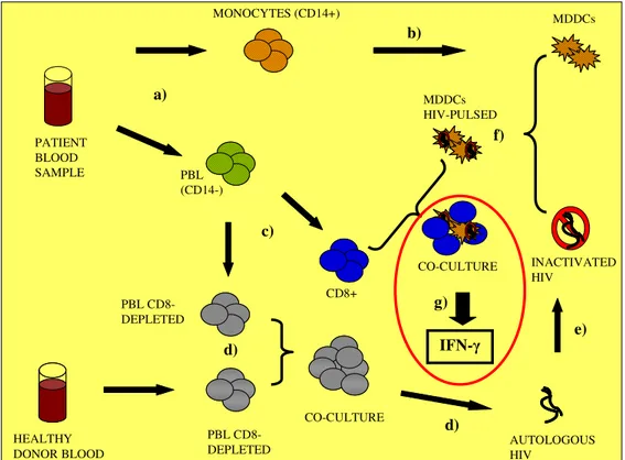
Generation of MDDCs (Study 2) ...71
MLR (Study 2) ...72
MDDCs pulsing and CD8+ IFN-γ production (Study 2)...72
Statistical analysis ...74
RESULTS AND DISCUSSION ...77
Study n° 1 : Early highly active antiretroviral treatment delayed over 3 months of life is associated with good clinical outcome, long-term viral control and persistent antiviral T-cell response in HIV-1 vertically infected infants . (Paper 1) ...77
Clinical parameters ...77
Clinical outcome ...78
Virologic evaluation...78
CD4+ and CD8+ counts ...78
T-cell compartment analyses ...79
T-cell proliferation ...79
TCRBV repertoire...80
CD8+ Lymphocytes cytotoxicity...83
Discussion ...84
Study n° 2: Switching from protease inhibitor-based-HAART to a protease inhibitor-sparing regimen is associated with improved specific HIV-immune responses in HIV-infected children. (Paper 2 & 3)...87
Clinical parameters ...87
Clinical outcome ...89
Virologic evaluation...90
CD4+ and CD8+ counts ...90
T-cell compartment analyses ...91
T-cell proliferation ...91
TCRBV repertoire...93
CD8+ Lymphocytes cytotoxicity...96
Dendritic cells compartment ...98
Phenotype analysis ...98
MLR ...100
DC pulsing and IFN-γ production...101
Discussion ...102
Study n° 3 : Rapid T-cell receptor CD4+ repertoire reconstitution and immune recovery in unrelated umbilical cord blood transplanted paediatric leukaemia patients. (Paper 4) ...107
Clinical parameters ...107
Clinical outcome ...107
HLA typing and chimerism...108
Cell counts...108
T-cell compartment analyses ...111
T-cell proliferation ...111 TCRBV analyses...112 Discussion ...117 CONCLUSIONS...121 AKNOWLEDGEMENTS...122 REFERENCES...123 Paper 1...149 Paper 2...161 Paper 3...175 Paper 4...179
INTRODUCTION
In healthy individuals the organism is protected from pathogens such as bacteria, viruses and parasites through the action of a specialized complex of cells constituting the immune system.
In particular conditions, the immune system could be altered leading to immunological disorders like lymphoproliferative disease, autoimmunity, hypersensitivity, and immunodeficiency.
The latter could be distinguished into two types: primary immunodeficiencies, due to genetic or developmental alterations and secondary immunodeficiencies, induced by viral or bacterial infections, by the use of immunosuppressive drugs, or by chronic disease or surgeon therapy.
Several treatments exist in order to favour the reconstitution of immune system in immune-compromised patients, and new therapeutic approaches are constantly under investigation.
Objective of this study is to evaluate the immune reconstitution in paediatric patients with secondary immunodeficiencies.
Particular attention was focused on immune restoration of:
• Cohorts of human immunodeficiency virus (HIV)-infected children: - Treated with early Protease Inhibitor Highly Active Antiviral
Therapy (PI-HAART) started over three months of life.
- Treated with a simplified regimen (PI-sparing-HAART), after a first line successful PI-HAART.
A number of techniques could be used to study the immune reconstitution: we performed several immunological and molecular analyses, with particular interest in the spectratyping technique, which measure the breadth of the antigenic diversity of the T-cell receptor (TCR) repertoire distribution.
The immune system
The immune system protects the body from infections by pathogenic organism. It is composed by cells, organs and tissues arranged in a dynamic interactive network that can be divided into two major arms: innate and adaptive immunity.
The first kind of protection is non-specific and immediately guaranteed by cells, like as granulocytes, monocytes and macrophages, activated by direct contact with various microbial products via toll-like receptors (TLR). Innate immune functions are also carried out by mast cells and natural killer (NK) beside to antibacterial enzymes and components of the complement system. Innate immunity does not confer long-term protection to the host because there is no creation of immunological memory.
The second kind of protection, the adaptive immunity, is pathogen-specific and requires the recognition of specific “non-self” antigens in the presence of “self”, during the process of antigen presentation. This antigen specificity is possible thanks to B cell receptor (BCR) and T cell receptor (TCR) respectively expressed on surface of B and T lymphocytes, and is necessary for the generation of specific responses, and for the development of an immunological memory able to mount a stronger attack each time that a particular pathogen is re-encountered. Besides T and B lymphocytes, also antigen presenting cells (APCs) play a pivotal role in adaptive immunity, being responsible of antigen presentation: overall these cells guarantee specificity, memory and diversity, which are the three main features of
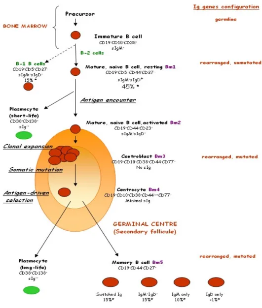
B lymphocytes
B-cells mediate humoral responses via secretion of their BCR in the form of Antibodies (Abs), glycoproteins belonging to gammaglobulin class, able to bind pathogens, facilitating their destruction and neutralization in a specific way. B lymphocytes may also act as antigen presenting cells and as regulators of immunological responses, secreting cytokines and interacting with T cells. B-cells are generated in the bone marrow (BM) from the Common Lymphoid Progenitor (CLP) (Ye 2007). In the bone marrow (BM) rearrangements of genes encoding for the BCR occur and transitional naïve B-cells still functionally immature are formed. (Chung J. 2003). At this point, naïve B-cells migrate in periphery and, following Antigen (Ag) encounter, they can immediately differentiate into short-life low-affinity immunoglobulin (Ig)-secreting cells (Martin 2001), that are responsible of the T-cell independent Ag responses, or can be transported by the bloodstream to secondary lymphoid organs, principally spleen and lymph nodes. Here they mature within germinal centres (GC), during the so called germinal centre reaction, to constitute long-life plasma-cells responsible of the T-cell dependent Ag responses (de Vinuesa 2000, Wolniak 2004). A prerequisite to the formation of a GC is the co-stimulatory interaction of antigen-specific follicular B and T-helper cells (Liu 1991) through their receptors CD40/CD40L: as a consequence of this interaction, GC precursor B-cells emerge and proliferate within the stromal environment created by a special network of dendritic cells (DCs) called follicular dendritic cells (FDCs) (Tew 1990, Kosco 1992), leading to the formation of GC. In a GC two zones could be distinguished: a dark zone and a light zone. In the dark zone B-cells undergo proliferation while their variable genes are subjected to diversification by the somatic hypermutation machinery
(Weigert 1970, Mac Lennan 1994). After somatic hypermutation, B-cells are directed to the light zone, where undergo a selection process based on the affinity of their BCR toward the antigens presented in the form of immune complexes by FDCs in the presence of helper T cells (Nayak 1999). The so formed high-affinity B-cells are positively selected, undergo isotype switching, and may become Abs secreting cells (plasma-cells) or memory B-cells that subsequently exit the GC through the surrounding mantle zone (Mac Lennan 1994). Memory B-cells could be identified by the expression of switched Ig and by the CD27 surface marker (Klein 1998, Agematsu 2000). Interestingly a new memory B-cell population CD27+IgM+, which can be generated in the absence of GC has been recently identified (Weller 2001, 2004). These IgM+ memory B-cells cannot be detected in splenectomised and asplenic individuals (Carsetti 2006), so that they could be related to B1a B-cells population found in mice that require the spleen for their survival and produce natural Abs (Wardemann 2002). Such B-cell population in human, called B-1 B-cells to distinguish from the classical B-2 B-cells, could be responsible for natural Abs production and for T-independent response (Kruetzmann 2003), constituting a bridge between innate and adaptive immunity.
Figure I. B-cell development. In BM rearrangement of genes encoding for
BCR occur. Atypical B-1 B-cells are IgM+ and are immediately ready to produce natural Abs in a T-cell-independent manner, while classical 2 B-cells mature in periphery and, following Ag encounter, become short-life plasma-cells, or migrate in GC, where, in a T-cell-dependent manner, undergo clonal expansion, somatic hypermutation, positive selection and isotype switching. Finally they leave secondary lymphoid organ becoming long-life plasma-cells or constituting the memory pool.
T lymphocytes
T-cells mediate cellular responses through several action mechanisms depending on T-cell subtype involved. Indeed two subsets exist: cytotoxic and helper T-cells. A third subset is constituted by T-cells (expressing transcription factor Foxp3 and CD25 marker on their surface) with regulatory functions. Cytotoxic T lymphocytes (CTLs), identified from surface expression marker CD8+, kill infected cells via secretion of cytokines (like as TNF-α and IFN-α), granules (like as perforin and granzymes) or by the interaction of Fas ligand (FasL) with Fas expressed on target-cells (Berke 1994, Shresta 1998).
T helper (Th) lymphocytes, identified from surface expression marker CD4+, could be distinguished in two further subset: CD4+ Th1 lymphocytes act as cell stimulators via secretion of cytokines like as Interferon-γ (IFN-γ) and Iterleukin-12 (IL-12), while CD4+ Th2 lymphocytes favour antibody production from B cells secreting Interleukin-4 (IL-4), Interleukin-5 (IL-5) and Interleukin-6 (IL-6).
T-cell activation occurs following the antigen presentation process: antigens are presented in form of peptides bound to the Major Histocompatibility Complex (MHC), constituted by molecules expressed on the surface of several autologous cells, mainly DCs.
Although the recent evidences of extrathymic T-cell development pathways in secondary lymphoid organs (Blais 2006), the thymus is the organ that supports the differentiation and selection of T cells (Bevan 1977).
After the CLP entrance into the thymus, thymocyte development follow with the expression of recombination activating gene RAG 1 and RAG 2, which
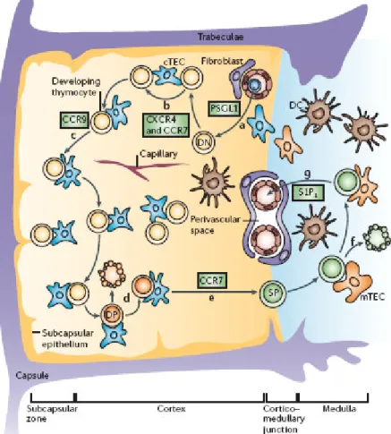
Figure II. T-cell development in the thymus. a) T-lymphoid progenitor
migrates into the thymus. b-c) DN thymocytes, after migration (regulated by chemokine signals) towards thymus cortex, become DP and d) undergo positive and negative selection. e) Positively selected thymocytes differentiate in CD4+ or CD8+ SP and f) in the medulla are further selected to delete tissue-specific-antigen-reactive T-cells and to generate regulatory T-cells. g) SP T-cells express shingosine-1-phosphate receptor 1 (S1P1) and are directed
back trough circulation that contain high concentration of sphingosine-1-phosphate. CXCR4: CXC-chemokine receptor 4; CCR7: CC-chemokine receptor 7; CCR9: CC-chemokine receptor 9; PSGL1: platelet-selectin glycoprotein ligand. (Adapted from Nature Rev. Takahama 2006).
During this phase thymocyte are characterised by the surface expression of CD25, whereas are CD44- and CD4 and CD8 double negative (DN) (Pearse 1989, Shinkai 1992).
Only the cells that succeed in in-frame rearrangement of the gene encoding the TCR β-chain are selected for further differentiation, which consists in the rearrangement of genes encoding the TCR α-chain and thereby in the expression of functional TCRαβ antigen receptors (von Boehmer 1997). During this phase thymocytes become CD4+CD8+ double positive (DP) (Takahama 2006) and undergo positive selection (Kisielow 1988, Jameson 1995), which consists in the interaction between their TCR with peptide-MHC complexes expressed by stromal cells, such as cTECs and DCs in the cortex (Bousso 2002). Only tymocyte with a low-avidity interaction, receive survival signals and become single positive thymocytes (SP) for CD4 or CD8, while those without any interaction or with an high-avidity interaction die for apoptosis (negative selection) to avoid autoimmunity. Positively selected DP thymocytes then begin relocating from the cortex to the medulla (Witt 2005), where they spend approximately 12 days before being exported from the thymus (Egerton 1990). During this maturation period, central tolerance is obtained through deletion of self-reactive T-cells. Indeed medullar thymic epithelial cells (mTECs) express tissue-specific antigens promiscuously (Kiewski 2004) a process at least partially dependent on the transcriptional factor autoimmune regulator (AIRE) (Derbinski 2005). In addition medulla is thought to be the place for the production of regulatory T-cells (Fontenot 2005, Watanabe 2005).
Following the encounter of a peptide bound to a MHC-complex expressed on an APC, they receive activation and proliferation signals and become effector cells.
After their immunological action, a little portion of CD4+ and CD8+ will constitute a memory pool for that specific antigens, while all the others clonal lymphocytes will die trough apoptosis.
T-cell development in the thymus is schematized in Figure II.
As mentioned, the unique specificities of both B and T lymphocytes are the result of several recombination processes of gene segments encoding for their receptors (BCR and TCR respectively): such processes are very similar between the two cellular types, occur during lymphocytes maturation and lead to creation of a huge repertoire able to recognize an almost unlimited number of Ags. In the following section TCR recombination process will be described, considering that the same principle is valid for BCR.
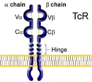
T-cell receptor (TCR)
As mentioned, TCR is the molecule responsible for recognizing antigens in the context of MHC. In 95% of T-cells, TCR is a heterodimer consisting of α and β chain, while in the remaining 5% of T-cells TCRs consist of γ and δ chains. Each chain of the TCR is a member of the immunoglobulin superfamily and is constituted by one N-terminal variable (V) domain, one constant (C) domain, a transmembrane/cell membrane-spanning region and a short cytoplasmic tail at the C-terminal end. The variable domains of the TCR α and β chain have each one three hypervariable or complementary determining regions (CDRs):
CDR3 is the most important because is the specific site for recognizing of processed antigen peptide bound to MHC.
Figure III schematizes TCR structure.
Figure III. TCR structure.
TCR α and β chains are created, as mentioned above, during T-cell development in the thymus, following the so called somatic recombination process between polymorphic genes (constituted by several gene segments) in the germinal line (Davis 1988). These polymorphic genes are located in the chromosome 7 and code for aminoterminal variable domain (V), for a diversity region (D) (only for β chain), and for the junction region (J). The TCR α chain is generated by V-J genes recombination while the β chain is generated by V-D-J genes recombination (Alt 1992). The intersection of these
Recombination processes and final product are visualized in Figure IV for the β-chain rearrangement.
Figure IV. Β-chain rearrangement. A first recombination process occurs
between genes encoding for diversity and junction region. In a second phase DJ segments are rearranged with a variable region V.
TCR diversity is guaranteed either by somatic recombination, either by the combination of a particular α chain with a particular β chain (Lewis 1994): it’s to note that while more of one α chain could be expressed on a T-cell, only one β chain could be expressed in each T-cell (allelic exclusion).
Furthermore during the recombination processes random palindromic P nucleotides are added (due to imprecise junction during recombination) and also T nucleotides are inserted by the action of terminal deossinucleotide transferase (TdT) enzyme. This imprecise junction produces CDR3 segments with diverse lengths in a range of 6-8 amino acids (Pannettier 1995) so that CDR3 lengths are a direct measure of TCR diversity.
Antigen presenting cells (APCs)
APCs are constituted by macrophages, which ingest microbes and particulate Ags, B cells, which take up soluble Ags by binding them with surface Igs, and DCs, which are the most potent and important APCs, having the unique function of antigen presentation (Banchereau 1998). In order to stimulate immunological responses, APCs present Ags trough peptide exposition on MHCs expressed on surface. Furthermore they provide co-stimulatory signals like B7-1 (CD80) and B7-2 (CD86) necessary to prime a naïve T-cell through interaction with CD28 (Chambers 1997).
APCs initiate their action trapping pathogens in peripheral lymphoid organs and presenting them to lymphocytes that continuously circulate from the blood stream to the lymphoid organs and back again, monitoring the Ags presented by APCs.
Migration of cells and cell-cell interactions require many adhesion molecules including selectins, integrins and intercellular adhesion molecules (ICAMs).
Dendritic cells (DCs)
DCs are cells dedicated to the Ags up-take and presentation. Consequently they are the best stimulators of naïve T lymphocytes to undergo clonal expansion (Banchereau 1998, Hart 1997). DCs can be classified into several subsets on the basis of their anatomical distribution, immunological function, and cell-surface marker expression. The main DCs subsets include myeloid DCs (myDCs) and plasmacytoid DCs (pDCs) in the blood, and Langherans
12), whereas pDCs can prime adaptive immunity by producing large amounts of type I interferons (IFNs) (Cella 1999); LCs mainly express langerin (also known as CD207), a C-type lectin involved in cell migration (Valladeau 1999).
DCs develop in the BM from CD34+ monocyte precursor. Until immature, they are specialized in the Ag up-take (Sallusto 1994) while, once activated, they mature and loss the Ag up-take ability becoming specialized in the Ag presentation on MHC. They migrate to the lymphoid tissues, interact with T-cells and B-T-cells, forming the so named “immunological synapse” and trigger the immune response (Dustin ML 2006).
Therefore the efficiency of an immunological response is dependent by a correct antigen presentation process and thereby by the correct expression of MHCs on DCs surface.
Major histocompatibility complex (MHC)
Two types of MHC molecules exist: class I MHC (MHC-I) and class II MHC (MHC-II).
Endogenous antigens (obtained from tumour and virus-infected cells) are presented to CTLs following the loading on MHC-I molecules after a proteolitic process performed by proteasome, followed by transport into the lumen of endoplasmic reticulum (ER) (Townsend 1985, Heemels 1995, Lehner 1996); exogenous antigens (obtained from bacteria or free virus particle) are presented to CD4+ cells following the loading on MHC-II molecules inside acidified endosomes after a proteolitic process performed by phagosome and intracellular vesicles (Cresswell 1994, Germain 1994, Watts 1997, Geuze 1998). MHCs are both polygenic (several MHC-I and -II genes
exist) and polymorphic (there are multiple alleles of each gene) (Klein 1993) and both consist of two polypeptide chains. In humans, the most important class I α chain genes are called HLA-A, -B and –C. MHC-I molecule has 4 domains, of which three are formed by α chain and one by β2-microglobulin
(not encoded within the MHC). There are also three major pairs of MHC-II α- and chain genes, called HLA-DR, -DP and –DQ, and in addition an extra β-chain in the DR-cluster.
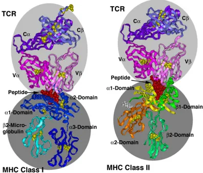
MHC-I and MHC-II general structures and their interaction with TCR molecules are reported in Figure V.
Figure V. MHC-I and –II structures and interaction with TCR. MHCI is
constituted by a α-chain associated with β2-microglobulin. The α1 and α2
Children’ immune system
Children immune system is not fully developed, making them more susceptible of infections. On the other hand the presence of the thymus (which disappears in adults) give them the possibility to obtain prompt immune reconstitution after eventual T-cell deletion, a characteristic that is to be considered when they undergo to various treatment or therapy. Before birth, children immune defences are mainly sustained by humoral arms transmitted from the mother via placenta and, in the first months of life in breast milk. This passive immunity is short-lived and is gradually substituted by longer-lasting immunity acquired following exposure to infection or through immunisation.
Immune system disorders and immunodeficiency
Despite the efficiency of immunological defence, failure of immune system may occur leading to a number of immunological disorders like lymphoproliferative disease, in the case of tumour, autoimmunity and hypersensitivity, when lymphocytes maturation is altered and responses are directed towards self-antigens and own tissues, and immunodeficiency, in the case of defective functioning of one or more immunological component. As a consequence of immunodeficiency, responses to Ags are severely impaired or entirely absent so that patients become very vulnerable to opportunistic infections. It is possible to distinguish two types of immunodeficiencies: primary immunodeficiencies, which are congenital and have genetic origin, and secondary immunodeficiencies, which are acquired: an immune compromised status could be a consequence of chemotherapeutical drugs administration, and could be voluntarily induced in organ transplantation to avoid rejection. However, the most famous acquired immunodeficiency is the Acquired Immune Deficiencies Syndrome (AIDS) caused by HIV-infection.
Pathogenesis of HIV
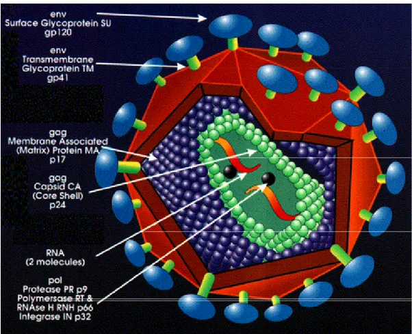
HIV-1 is a complex retrovirus firstly identified in the early ’80 (Barrè-Sinoussi 1983, Gallo 1984, Levy 1984, Popovic 1984), which impairs immune system until to cause death of infected people in absence of therapy (Colebunders 1991). Its structure is schematized in Figure VI and its natural replication cycle is following described.
Natural replication cycle of HIV initiates with the entry of the virus in the host cell, and then by RNA release, retrotranscription and integration in the host genome (Brown 1990). After host T-cells activation, proviral transcription and RNA translation occur, leading to formation of new mature virions able to spread infection in the host (Felber 1989, Hadzopoulou-Cladaras 1989).
Infection results in a primary burst of viral replication (Albert, Gaines 1987) followed by immune adaptive response activation, which does not achieve complete virus eradication, but leads to a marked decrease of initial viraemia. After primary infection most patients enter into an asymptomatic phase which can last for months to more than 10 years (Lifson 1991). During this phase immune system is progressively deleted and individuals become subject for opportunistic infection, developing clinical manifestation of AIDS.
Either B, T and DCs compartments are impaired from HIV that is able to escape immune system through several mechanisms.
Figure VI. HIV-1 structure. HIV is constituted by two copies of
single-stranded RNAenclosed by a conical capsid comprising the viral protein p24. The single-strand RNA is tightly bound to enzymes that are indispensable for the development of the virion, such as reverse transcriptase and integrase. A matrix composed of an association of the viral protein p17 surrounds the capsid, ensuring the integrity of the virion particle. Also enclosed within the virion particle are the viral proteases. An external envelope is formed from host-cell membrane with host proteins and the viral glycoproteins gp120 associated with the transmembrane proteins gp41. p24 and p17 are encoded by gag gene; gp120 and gp41 are encoded by env gene.
B-cell compartment
After primary infection an important defensive action is carried out by B-cells, which product antibodies predominantly raised against structural proteins released from destroyed infected cells and intact virions (Burton 1997, Poignard 1996). However, the genetic variability of HIV permits the virus to escape from the action of neutralizing antibodies. Indeed, as new variants arise, nAbs against those variants develop within three months, but by this time new viral variants resistant to neutralization by those antibodies has already arisen (Wey, Richman 2003). Other escape mechanisms from neutralizing antibodies include glycosylation, and steric and conformational block of receptor binding sites (Johnson 2002, Kwong 1998).
Furthermore, although during asymptomatic phase antibody titers remain elevated (Lang 1989), with disease progression B-cells become hyperactive (Vendrell 1991, Yarchoan 1986) causing hypergammaglobulinemia, increased expression of activation markers, and increased incidence of B-cell lymphomas (De Milito 2004). All these events lead B lymphocytes to undergo to apoptosis (De Milito 2001, Titanji 2003) causing a reduction in their number (Titanji 2006) and an impairment in the responsiveness to mitogens and antigens (Samuelsson 1997, De Milito 2004), specially in the memory subset (De Milito 2004). These alterations could be only partially recovered by current therapies (Titanji 2006).
T-cell compartment
HIV specifically targets T-lymphocytes expressing on their surface CD4+ receptor (Klatzmann 1984) and during primary infection number of CD4+ T-cells strongly, but transiently, decreases (Fauci 1996).
These events are followed by the activation of CD4+ helper T-cells (Kalams 1999), which appear to be critical in containing viral replication through the release of the soluble mediator IL-2 (Kalams 1999), and the cytotoxic CD8+ T-cells (Ariyoshi 1992, Koup 1994), which undergo a clonal expansion and release cytotoxic cytokines (IFN-γ) and granules (granzyme and perforin) (Berke 1994, Shresta 1998). These events lead to the control of viraemia and to the asymptomatic phase (Clark, Daar, Lifson 1991).
On the other hand, as in the case of B lymphocytes, the virus accumulates mutations during infection in order to escape from specific attack of CTLs that recognize specific viral epitopes (Walker 1991, Plata 1987, Riviere 1994). Furthermore, since HIV continuously triggers CTLs response, it causes the clonal exhaustion of specific TCRBV families and the progressive decrease in the CD8+ number (Hoffenbach 1989).
During the course of disease, the persistent release of HIV-1 provokes the progressive disappearance of CD4+ T-cells (Haase 1999), an increase in the frequency of activated cells undergoing apoptosis (Herbeuval JP 2005, Copeland 1996) and the lost of T-cell proliferative capability in response to recall antigens and mitogens (Schulick 1993). Furthermore a shift in T helper function from Th1 to Th2 has been observed (Clerici 1993, Graziosi 1994).
DCs compartment
Current data suggest that both myDCs and pDCs are susceptible of infection by HIV-1 (Smed-Sorensen 2005, Donaghy 2003, Cameron 1992, Patterson 1998, 2001, Macatonia 1990), although only 1-3% of the DCs population can be productively infected with HIV in vitro (Smed-Sorensen 2005) and HIV replication in DCs is 10- to 100-fold lower than in CD4+ cells (Mcllroy 1995). Virus entry may occur through lectins such as DC-specific intercellular adhesion molecule 3 (ICAM3)-grabbing non-integrin (DC-SIGN), langerin, mannose receptor (also known as CD206) and un unidentified trypsin-resistant C-type lectin (Geijtenbeek 2000, Turville 2001, 2002).
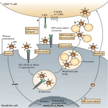
Two types of DC-mediated HIV transmission have been proposed (Wu 2006): trans-infection through formation of so called infectious synapse (Mc Donald 2003, Arrighi 2004, Turville 2004, Garcia 2005) or through HIV-associated exosomes (Wiley 2006), and cis-infection with de novo production and long-term transmission of the virus, after infection and replication in DCs (Nobile 2005, Burleigh 2006). DCs-mediated HIV transmission is schematized in Figure VII.
An important reservoir of HIV is also represented by FDCs of GC: they are not productively infected by HIV, but can trap and maintain large quantities of HIV early after infection (Heath 1995). In addition FDCs can promote the migration of resting CD4+ cells to the germinal-centre microenvironment, favouring their infection (Heath 1995, Smith 2001).
The number of DCs decreases during the course of disease (Knight 1997, Pacanowsky 2001, Barron 2003), but it’s still unclear if they are impaired in
Figure VII. Mechanisms of dendritic-cell-mediated HIV transmission. a)
trans-infection with infectious synapse: DCs transfer captured HIV to target CD4+ T-cells through a complex formed by cell-cell junctions involving DC-SIGN. b) Trans-infection with exocytotic pathway: endocytosed HIV can gain access to endosomal multivescicular bodies, enabling the release of HIV associated with exosomes: they are likely to be transmitted to CD4+ T cells through membrane binding and fusion. c) Cis-infection: after initial exposure of HIV, HIV infection of DCs results in de novo production of viral particles. (Adapted from Nature Rev. Immunol. Wu 2006).
function only at late-stage disease (Hsieh 2003), while others showed that both myDCs and pDCs are significantly less efficient at stimulating allogeneic T-cells (Donaghy 2003, Macatonia 1990), have a reduced maturation ability (Granelli-Piperno 2004) and a decreased expression of the co-stimulatory molecules CD80 and CD86 (Lore 2002).
Paediatric HIV-infection.
Paediatric HIV-1 infection is primarily due to mother-to-child virus transmission (vertical infection), which occurs during pregnancy or after delivery or during breast-feeding (Scarlatti 1996, Ehrnst 1991, Goedert 1991, Oxtoby 1988, Ziegler 1985). Influencing factors for transmission are immunological status of the mother (with inverted correlation between CD4+ number and transmission risk) (Scarlatti 1991), the virus phenotype (Scarlatti 1993), and the presence of neutralizing antibodies in sera mother (Devash 1990, Goedert 1989, Rossi 1989). Caesarean section, the avoided breast-feeding and the use of antivirals during pregnancy reduce the risk for children to be infected (Eur. Coll. Study 1992, Mayaux 1997).
In the 25-30% of children heavy symptoms appear in the first year of life with immunodeficit and strong decrease of CD4+ lymphocytes. In the remaining 70% disease course seems similar to the adults with symptoms variable and arising of similar opportunistic infections. 5-8% of these patients are asymptomatic for more than 7-8 years (Martin 1996).
It’s possible that children with a rapid course of disease are infected during pregnancy, the others at the end of pregnancy or during delivery (Wilfert 1994).
Treatment
Positive results against HIV in vitro were firstly obtained by the use of an antiretroviral drug, the zidovudine (AZT) and subsequent clinical trials
and NNRTI respectively) and Protease Inhibitors (PI) were developed.
In the following years became evident how the combined action of three or more antiretroviral drug led to strong positive effects in HIV-infected patients with also a reduction in the mutational load.
DRUG CLASS ACTION MECHANISM
Zidovudine or
Azidothymidine (ZDV or AZT)
NRTI AZT is a thymidine analogue
Stavudine (d4T) NRTI Stavudine is thymidine analogue. Lamivudine (3TC) NRTI Lamivudine is a cytidine analogue. Abacavir (ABC) NRTI Abacavir is a guanosine analogue Didanosine (ddI) NRTI Didanosine (ddI) is an adenosine
analogue.
Efavirenz (Efv) NNRTI Efavirenz binds to a Reverse Transcriptase pocket
Nevirapin (Nvr) NNRTI Nevirapin binds to a Reverse Transcriptase pocket
Ritonavir (RTV) PI Ritonavir inhibits a liver enzyme that normally metabolizes away protease inhibitors
Indinavir (IDV) PI Indinavir inhibits both HIV-1 and HIV-2 proteases
Nelfinavir (NFV) PI Nelfinavir inhibits both HIV-1 and HIV-2 proteases
Saquinavir (Sqv) PI Saquinavir inhibits both HIV-1 and HIV-2 proteases
Lopinavir (LPV) PI Lopinavir inhibits proteases binding plasma proteins
Tab I. Antiretrovirals. NRTIs inhibit retrotranscription trough competitive
binding to RNA. NNRTIs effect a direct inhibition towards Reverse transcriptase. PIs compete with viral protease for the same binding sites.
Therefore Highly Active Antiretroviral Therapy (HAART), based on combination of two NRTI and one PI, was successful introduced.
Action mechanism of the main antiretroviral drugs are reported in Table I. Nowadays antiretroviral regimens are currently used: they permit to control viral replication and to increase CD4+ counts leading to a general immune reconstitution of T-cell compartment (Chiappini 2006, Hainaut 2000, Romiti 2001)
Therefore, even if in B-cells and DCs positive effects are less evident and partial (Pastori 2002, Montefiori 2001, Titanji 2005, Gompels 1998, Pacanowsky 2001, Chehimi 2002, Barron 2003), introduction of HAART has strongly decreased morbidity and mortality in HIV-infection slowing down disease progression either in adult population (Berrey 2001, Palella 1998), either in paediatric patients (Romiti 2001, Resino 2002).
However, in paediatric HIV-infection a crucial point of discussion is represented by the initiation time of therapy, because there are no immunological markers that could suggest it (Abrams 2003).
Since the higher risk of disease progression and death in the first year of life, an early start of HAART was proposed (Dunn 2003, Faye 2004). On the other hand, some authors suggested that HAART applied from the first weeks of life, because of viral suppression, may reduce the availability of viral antigens for processing and presentation to the T-cell compartment, thus impairing the generation and the expansion of HIV-1 specific memory response during this period of relative immunological immaturity (Scott 2001, Spiegel 1999). Indeed humoral and cellular HIV-specific responses were not detected in
On the contrary a delayed initiation of HAART in the first year of life could favour the priming and the development of HIV-specific T-cell responses. Other discussion points are related to the applicability of alternative regimens to avoid the numerous side effects of HAART.
Indeed, besides the positive effects of HAART, this therapy may lead to several serious adverse events, including diarrhoea, hepatitis, hypercholesterolemia, encephalomyelitis, neutropenia, pancreatitis (Carr, Wanke 1999). Furthermore poor compliance augment risk of viral failure and it’s also possible that the virus develop antiviral resistance due to rapid genomic mutations. These limitations impact negatively on the patient’ quality of life and are particularly worrisome in children, given their long-term expectative of life (McComsey 2004, Arpadi 2001, Jaquet 2000, Babl 1999, Amaya 2002).
As a consequence, in the last years, several HAART simplified regimens were developed (Martinez, Raffi 2000, Negredo 2002) either for adults, either for children.
In the last years, a number of studies are also focused on alternative treatments, including cellular therapies, in order to definitively substitute HAART regimens and to obviate to the most important restriction of HAART: indeed current antiretroviral regimens are not able to eradicate definitively HIV that could persist in latent state inside the host genome (Finzi 1999, Siliciano 2003) thereby making not possible any treatment interruption. In this context of particular relevance are the recent studies focused on DCs manipulation and pulsing to induce a strong CD8+ HIV-specific cytotoxic response maintaining at the same time the control of viral replication and the number of CD4+ lymphocytes (Andrieu 2007), in view of future efficient vaccine development.
Haematological disorders and transplantation: an
other case of acquired immunodeficiency
A typical situation of immune system impairment is a consequence of haematological disorders like as Leukaemia, in which too many abnormal and not functional lymphocytes are produced. Such diseases are treated with chemotherapeutical drugs that kill all leucocytes, leading to a strong impairment of the host immune compartments, and to a high susceptibility to viruses and other pathogens.
Haematopoietic stem cell transplantation (HSCT) is used to favour immune reconstitution of these patients (Sullivan 2000) or for patients with metabolic diseases, autoimmune pathologies (Ikehara 1998) or HIV-associated lymphomas (De Paoli 2006).
Haematopoietic stem cells (HSCs) are pluripotent T-cells that originate since the embryo development and are continuously produced in BM in the entire course of life in physiological conditions (Peault 2003). Bone marrow transplantation (BMT) is the most common type of transplantation, but other sources of HSCs are the Peripheral Blood (PB) and the Cord Blood (CB). Before transplant, BM could be in vitro treated to eliminate T-cells, to avoid the Graft Versus Host Disease (GVHD), or mature erythrocytes, in the case of erithrocytic incompatibility between donor and recipient. BM is infused in a peripheral vein of the host, and the HSCs spontaneously reach the host BM, through the homing process, involving adhesion molecules like as the ICAM (Lee 2004).
this procedure, but there is an elevated risk to re-administer cancerous cells to the patients.
An ideal compatibility condition is represented by the singeneic transplantation: in this case the donor is recipient homozygous twin, but the risk is related to possibility that donor cells lead the same mutations favouring the disease development.
Allogeneic transplantation is the most applied: in this case transplanted cells become from an other person that could be related (if he comes from the same family), or not related (if he is a voluntary donor). Familiar donor could be genotypically identical if he has the same HLA alleles of the donor, or aploidentical if donor and recipient have identical only one out of two chromosomes containing genes coding for HLA. If the donor is not a familiar, transplantation could occur only if donor and recipient are fenotypically identical: therefore is necessary to find HLA-compatible donors through the HLA-typing.
Allogeneic transplantation major risks are correlated to rejection and, especially in children, to GVHD in which donor cells attack host immune system (Kondo 2001). To avoid GVHD before and after transplantation immunosuppressive drugs such as Cyclosporine A (CSA), alone or in association with other drugs, are included in conditioning regimens generally consisting in total body irradiation (TBI), in association with cyclophosphamide (CTX) or other chemotherapeutical agents used to create a place for the donors HSCs, to suppress recipient immune system, and to kill cancerous cells.
Since of the conditioning regimen and GVHD prophylaxis, after transplantation, a number of patients show complications related to the duration of immune-compromised status. Recovery of polymorphonuclear and
dendritic cells is relatively rapid, occurs within weeks and could be favoured by the administration of Granulocyte Colony Stimulating Factor (G-CSF) (Iori 2004). In contrast T lymphocytes reconstitute themselves less rapidly and often present a reduced functionality: functional numbers of T-cells might appear 3 to 4 months after transplantation and full recovery might take 6 to 12 months (Crooks et al 2006). Therefore is necessary to follow carefully patients especially within the two years after transplantation: during this time there is an elevated risk to develop viral and fungal infections, associated with defective cell-mediated immunity, or late bacterial infections, as a result of defective antibody production, respectively treated with antiviral and antifungal drugs, and antibiotics (Crooks 2006). Immunoglobulins are somministered in the case of ipogammaglobulinemy (AIEOP 2003).
Haematopoietic reconstitution is evaluated by emocromocytometric tests. The presence of genetically different cells, the so named chimerism, is evaluated by cytogenetic test like Fluorescence in situ hybridization (FISH) and molecular analyses like Polymerase Chain Reaction (PCR).
Engraftment failure occurs if after 90 days from transplant there is not autologous haematopoietic reconstitution (Iori 2004).
Umbilical cord blood transplantation (UCBT)
Recently UCBT, particularly from unrelated donors, has progressively become a widely employed treatment for patients with both malignant and non malignant haematological disorders (Wagner 1995, Locatelli 1999, Barker 2003).
HSCs after mieloablative therapy (Broxmeyer 1982) and, in 1988 the first UCBT was carried out (Gluckman 1989).
UCBT offers several advantages including simplicity to be obtained, rapid availability of biologic product, absence of risk for donors, reduced likelihood of transmitting infections, less stringent criteria for HLA matching for donor/recipient selection, and, overall, reduced incidence and severity of GVHD (Barker 2003, Madrigal 1997). Indeed T-cells are in immaturity state and seem functionally and phenotipically naïve (CD45 RA+RO-). Furthermore only a small fraction of them show the expression of activation marker HLA-DR and CD25+ (D’Arena 1998), and it is demonstrated that they have a low T-cell mediated cytotoxic capacity, having a higher threshold for IL-2, IFN-γ and TNF-α cytokine stimulation, low reactivity to mitogens as Phytohaemaglutinin (PHA), and reduced ability to generate CTLs in Mixed Leucocyte Reaction (MLR) (Quian 1997, Harris 1992). Finally they show a widely polyclonal TCR (Garderet 1998, Moretta 1999).
Besides advantages, UCBT present also some disadvantages: indeed contains low HSCs number in comparison to BM, a characteristic that, accompanied by immaturity state of cells, could hamper the complete immune recovery and cause a delay in the reconstitution (Gluckman 2000, Thomson 2000, Barker 2001, Moretta 2001, Rocha 2001, Dalle 2004). On the other hand, the stem cells immaturity is a factor that could be determinant in the obtaining of a long-term immune-haematological reconstitution, given the stronger proliferative ability of these cells.
Regarding humoral immunity, it has been shown that B-cells progenitors in UCB are more than in BM (Locatelli 1996, Hoyt 1999), and B subpopulation reconstitute earlier as also Ig sera levels. Also NK show a rapid reconstitution and a reduced cytotoxicity (Moretta 1999, Thomson 2000).
Previous studies on immune reconstitution after UCBT showed prompt and efficient proliferative response to mitogens and recovery of NK functions (Locatelli 1996, Giraud 2000, Thompson 2000, Moretta 2001), while the only few data available on TCR repertoire reconstitution after unrelated UCBT showed a complete TCR repertoire normalization only after two or more years from transplantation (Klein 2001, Talvensaari 2002).
Immune reconstitution analysis
In the following paragraphs a general overview on the main techniques used to analyse patient’ immune reconstitution will be presented, focusing attention on TCRBV spectratyping, that has been largely used for experiments of this thesis.
Cytofluorimetric analysis
In order to verify cell phenotypes and/or to establish cell counts of the various immunological compartments is very useful the cytofluorimetric analysis, through the use of monoclonal antibodies able to specifically bind differentiation, maturation and activation markers, expressed from the various cellular subpopulation.
TCRBV repertoire analysis - Spectratyping
In the context of T-cell immune reconstitution evaluation, one of the most important analysis tools is given by TCRBV repertoire analysis (Roux et al 1996).
Cytofluorimetric assays could be performed to quantify the expression of the various TCRBV families. Alternative molecular analyses are based on Reverse Transcriptase-PCR (RT-PCR) following by cloning and sequencing of TCRBV families, to obtain repertoire clonotype information (Rosemberg 1992, Choi 1989).
But the most useful technique is the spectratyping, which provides information on TCR diversity at the level of CDR3 length of Vβ chain, considering that length heterogeneity is representative of overall sequence heterogeneity. TCR analyses are performed on β chain because it is the only one that is subjected to allelic exclusion.
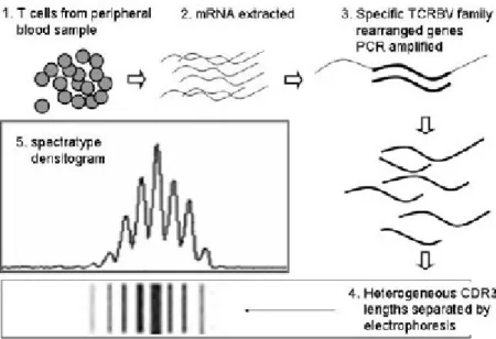
The experimental procedure is briefly schematized in Figure VIII (for major details see materials and methods section).
Figure VIII. Spectratyping experimental procedure. mRNA is extracted from
T-cells (eventually separated into CD4+ and CD8+ cellular subset) and subsequently reverse transcribed and amplified in reverse transcriptase PCR (RT-PCR) using 24 Vβ primers and one Cβ primer. PCR products are heterogeneous CDR3 lengths which are size-separated by electrophoresis. Finally, the quantity of material deposited in each band is quantified by densitometry and the spectratype trace is produced. (Adapted from Kepler, Bioinformatics 2005).
Although there is consistency in the molecular method used to generate TCRBV spectratype data, a wide range of analysis methods exists to describe and quantify TCR diversity. One of the least sensitive, but more commonly used, methods is Visual Scoring, which characterizes TCRBV diversity as oligoclonal or polyclonal (Ferrand 2000, Bour 1999, Kook 2002).
To quantify the complexity of TCR repertoire, Bomberger et al (1998) developed a method in which all peaks greater than 10% of the total peak area are enumerated: distribution is considered normal if there are more than 6 peaks. Bomberger introduced also the Diversity Score, which value is given first calculating the ratio between the sum of the heights of the major peaks to the sum of all peak heights and then dividing this ratio by the number of major peaks present. An overall Complexity Score is given by the sum of all Vβ complexity numbers calculated (Bomberger 1998).
Others have developed various peak area calculation including peak counts for peaks greater than a determined percentage of the maximal peak area relative mean fluorescence divided by the peak area (Dumont-Girard 1998) and peak scoring (Lu 2004, Wu 2000).
One of the most used quantitative methods is that of Gorochov (1998): for this analysis peak areas in each Vβ are compared against the distribution of a normal standard determined by averaging values obtained from CD4+ T-cell component of five normal tissues. The differences between the experimental peak areas and the normal values within each Vβ are averaged and reported as Gorochov values.
Recently various softwares are being developed to facilitate a standardized analysis of TCRBV repertoire data: Immunoscope TM automatically calculates peak area and the Gorochov value (Collette et al 2001, 2003); TC Landscape TM integrates data measuring genetic diversity with data measuring Vβ surface expression (Pilch et al 2002); SPA (He 2005, Kepler 2005) and REPERTOIRE (Long et al. 2006) compare peak areas to normal peak areas which were based upon a theoretical normal Gaussian peak area. By adding the resulting peak amplitude disparities between the sample and the standard peak areas, an individual P value is generated for each Vβ family, and an overall P value is generated by averaging all analysed individual Vβ subfamilies giving equal weight to each analysed Vβ in a sample.
T-cell receptor excision circles (TRECs) analysis
Quantification of T cell receptor excision circle (TRECs) is an other molecular technique that gives information on the thymic output (Kong 1999, Douek 1998, 2000). TRECs are generated during genes rearrangement of V(D)J segments of TCR: they are little circles of excise DNA, which is not integrated into genome. TRECs are stables and do not undergo to cell division, so that are diluted in every cellular division (McFarland 2000). TRECs levels have been shown to correlate with recent thymic emigration and thus thymopoiesis (Douek 1998, Al-Harthi 2000), although also cellular proliferation and apoptosis influence TRECs levels, complicating data interpretation (Ye 2002, Berzins 2002, Hazenberg 2003).
T-cell proliferation
To evaluate T lymphocytes functionality and their ability to respond to common mitogens and common or specific Ags, lymphoproliferative tests are very useful (Romiti 2001), and to evaluate the efficiency of stimulators, MLRs test could be performed using purified DCs versus T-cells
Cause of the low abundance of DCs in vivo (Rissoan 1999), Monocyte-derived Dendritic Cells (MDDCs) are commonly used to model the immunological function of DCs (Steinman R.M. 2003, Romani 1994). CD14+ monocytes from human peripheral blood differentiate into immature DCs after 4-6 days in culture in the presence of IL-4 and granulocyte/macrophage colony-stimulating factor (GM-CSF). These immature DCs have similar characteristics to myeloid DCs and can be converted into mature MDDCs by exposure to various stimuli, including lipopolysaccharide (LPS), Interferon-γ (IFN-γ), tumour-necrosis factor (TNF) and CD40 ligand (CD40L) (Banchereau 2000, 1998).
In the last years the use of MDDCs knew a great expansion, because DCs could be also used for pulsing experiments (Lu et al 2004), in view of cellular therapy, with the finality to prime more potent immunological responses in immune-compromised patients.
AIM OF THE STUDY
Aim of this study was to evaluate the immune reconstitution in different cohorts of paediatric patients with acquired immunodeficiencies.
In particular we studied a cohort of 9 HIV-infected children who started HAART after 3 months of age to analyse if a delayed initiation of therapy, could favour the immune recovery in these children.
Then we investigated in a larger cohort of 19 HIV-1 vertically infected children (9 of which from the precedent study) if the switching from a PI-based HAART to a PI-sparing regimen with 3 NRTI could maintain immune reconstitution obtained with previous HAART.
Finally immune reconstitution was evaluated in a cohort of 6 leukaemia children who underwent to cord blood transplantation.
DESIGN
Immune reconstitution in acquired immunodeficiency children was investigated following 3 main research branches based on the scheme below:
1) Study on delayed (after 3rd month of life) start of PI-based HAART in 9 HIV-infected infants (Follow up: 47, 9 + 18, 5 months).
• Clinical investigations: routine medical/laboratory analyses, virologic evaluation.
• T-cell compartment investigations: CD4+, CD8+, CD45RA+ and CD45RO+ cell counts, T-cell proliferation, TCRBV repertoire, CD8+ citotoxicity.
STUDY N 1 : Delayed start of PI-based HAART in 9
HIV-infected infants
CLINICAL INVESTIGATIONS Medical/Laboratory evaluation Virologic evaluation CD4+, CD8+, CD45RA+, CD45RO+counts T-CELL COMPARTMENT INVESTIGATIONS T-cell proliferation TCRBV repertoire analysis CD8+lymphocytes citotoxicity2) Study on switching from PI-based HAART to 3NRTI regimen in 19 HIV-infected children (Follow up: 96 weeks).
• Clinical investigations: routine medical/laboratory analyses, virologic evaluation
• T-cell compartment investigations: CD4+ and CD8+ counts, T-cell proliferation, TCRBV repertoire analysis.
• Dendritic cell compartment investigations: Phenotype analyses, MLR, DC-pulsing and CD8+ cytotoxicity stimulation (analyses were performed on MDDCs).
STUDY N 2 : Switching from based HAART to
PI-sparing regimen in 19 HIV-infected infants
CLINICAL INVESTIGATIONS Medical/Laboratory evaluation Virologic evaluation CD4+and CD8+ counts T-CELL COMPARTMENT INVESTIGATIONS T-cell proliferation TCRBVrepertoire analysis DCs COMPARTMENT INVESTIGATIONS Phenotype analysis MLR DCs pulsing and IFN-γ production
3) Study on cord blood transplantation in leukaemia children.
• Clinical investigations: routine medical/laboratory analyses, virologic evaluation.
• Cellular investigations: CD4+, CD8+, CD45RA+, CD45RO+, NK, CD19+ cell counts T-cell proliferation, TCRBV repertoire analysis.
STUDY N 3 : Repertoire reconstitution in 6 cord blood
transplanted leukemia children
CLINICAL INVESTIGATIONS Medical/laboratory evaluation HLA typing Cell counts CELLULAR INVESTIGATIONS T-cell proliferation TCRBV repertoire analysis CD4+, CD8+, CD45RA+, CD45RO+, NK, CD19+ cell countsMATERIALS & METHODS
Patients, therapies and clinical investigations
Children were enrolled followings specific criteria in each study. In all cases the institutional review ethical board approved the study and written informed consent was obtained from the patients’ parents or legal guardian.
Periodical controls consisting in complete medical physical examinations and routine laboratory tests including CD4+ and CD8+ T-cell count were performed during follow up in all patients
Study 1
Nine HIV-vertically infected children (pA-pI), who started PI-based HAART between 3 and 8 months of age, were enrolled in the study. All infants were diagnosed as HIV-1 infected on the basis of at least two positive results for the HIV-1 DNA PCR and one viral isolation.
Infants underwent HAART with two or three NRTI plus NFV as a PI or, for the last two patients enrolled under recent guidelines, (Sharland et al 2004) LPV/RTV. Children were followed at Paediatric Hospital “Bambino Gesù”, in Rome.
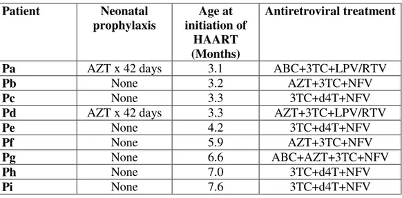
Individual patient characteristics and antiretroviral regimens at baseline are outlined in Table 1.1
Patient Neonatal prophylaxis Age at initiation of HAART (Months) Antiretroviral treatment
Pa AZT x 42 days 3.1 ABC+3TC+LPV/RTV
Pb None 3.2 AZT+3TC+NFV
Pc None 3.3 3TC+d4T+NFV
Pd AZT x 42 days 3.3 AZT+3TC+LPV/RTV
Pe None 4.2 3TC+d4T+NFV
Pf None 5.9 AZT+3TC+NFV
Pg None 6.6 ABC+AZT+3TC+NFV
Ph None 7.0 3TC+d4T+NFV
Pi None 7.6 3TC+d4T+NFV
Table 1.1. Individual patient treatments adopted. AZT: azidovudine; ABC:
Abacavir, 3TC: lamivudine; LPV: Lopinavir; RTV: Ritonavir; d4T: Stavudine.
Study 2
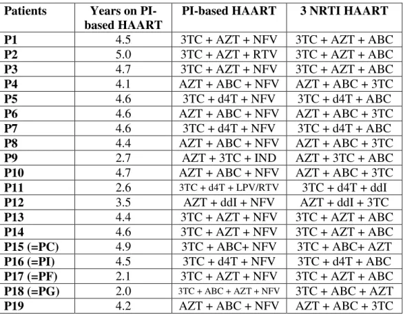
Nineteen HIV vertically infected children (p1-p19), previous treated with a successful stable PI-based HAART for at least 12 months were enrolled in the study. These patients were from 2 to 18 years old, had normal value of CD4+% for age before study entry,and did not have previous NNRTI therapy neither history of virological failure with others regimens. At study entry PI-based HAART was switched to a PI-sparing simplified protocol in which the PI was substituted with a third NRTI. Years of precedent PI-based HAART and drugs combination before and after simplification are summed for each patients in Table 2.1.
Of these children, four were the same of the precedent study: corresponding number are the sequent: pC=p15; pF=p17; pG=p18; pI=p16
Patients Years on PI-based HAART
PI-based HAART 3 NRTI HAART
P1 4.5 3TC + AZT + NFV 3TC + AZT + ABC
P2 5.0 3TC + AZT + RTV 3TC + AZT + ABC
P3 4.7 3TC + AZT + NFV 3TC + AZT + ABC
P4 4.1 AZT + ABC + NFV AZT + ABC + 3TC
P5 4.6 3TC + d4T + NFV 3TC + d4T + ABC
P6 4.6 AZT + ABC + NFV AZT + ABC + 3TC
P7 4.6 3TC + d4T + NFV 3TC + d4T + ABC
P8 4.4 AZT + ABC + NFV AZT + ABC + 3TC
P9 2.7 AZT + 3TC + IND AZT + 3TC + ABC
P10 4.7 AZT + ABC + NFV AZT + ABC + 3TC
P11 2.6 3TC + d4T + LPV/RTV 3TC + d4T + ddI
P12 3.5 AZT + ddI + NFV AZT + ddI + 3TC
P13 4.4 3TC + AZT + NFV 3TC + AZT + ABC
P14 4.6 3TC + AZT + NFV 3TC + AZT + ABC
P15 (=PC) 4.9 3TC + ABC+ NFV 3TC + ABC+ AZT P16 (=PI) 4.5 3TC + d4T + NFV 3TC + d4T + ABC P17 (=PF) 2.1 3TC + AZT + NFV 3TC + AZT + ABC P18 (=PG) 2.0 3TC + ABC + AZT + NFV 3TC + ABC + AZT
P19 4.2 AZT + ABC + NFV AZT + ABC + 3TC
Table 2.1. Individual patient treatments adopted (previous PI-based HAART and PI-sparing regimens). ddi: Didanosine.
Study 3
Six children who underwent UCBT from unrelated donors with a 24 months observation time were enrolled in the study. Children were transplanted at the Haematology and Bone Marrow Transplantation Unit of Paediatric Hospital Bambino Gesù (Rome, Italy) since November 2000. Cord blood units for