1
Alma Mater Studiorum – Università di Bologna
DOTTORATO DI RICERCA IN
SCIENZE MEDICHE SPECIALISTICHE
Ciclo 29 N. MATR. 712017
Settore Concorsuale di afferenza: 06/D6 – NEUROLOGIA Settore Scientifico disciplinare: MED/26 NEUROLOGIA
TITOLO TESI
IN-DEPTH CLINICAL, GENETIC AND NEUROPSYCHOLOGICAL STUDY OF FAMILIAL AND SPORADIC CASES WITH SLEEP-RELATED HYPERMOTOR EPILEPSY (SHE): IDENTIFICATION OF NEW GENES BY WHOLE EXOME
SEQUENCING (WES)
Presentata da: Dr. LAURA LICCHETTA
Coordinatore Dottorato
Relatore
Prof. Gaetano D. Gargiulo
Prof. Paolo Tinuper
2
INTRODUCTION ... 4
I. Sleep-related Hypermotor Epilepsy (SHE) ... 4
I.1.Definition and clinical features ... 4
I.2. Genetics of SHE ... 5
I.2.1. CHRNA4, CHRNB2, CHRNA2 ... 6
I.2.2. CRH ... 7
I.2.3. KCNT1 ... 7
I.2.4. DEPDC5 ... 8
I.2.5. PRIMA1 ... 10
I.3. Neuropsychological features of SHE ... 11
II.FAMILIAL FOCAL Epilepsy WITH VARIABLE FOCI (FFEVF) ... 13
AIMS ... 15
METHODS ... 16
I. Patients recruitment ... 16
I.1. Setting and period ... 16
I.2.Population and inclusion criteria ... 16
II. Clinical study ... 16
III. Genetic study ... 17
III.1. Pre-screening of known genes ... 17
III.2 WES analysis ... 17
III.2.1. DNA sample collection ... 18
III.2.2. WES experiment ... 18
III.2.3. Bioinformatic analysis ... 18
III.2.4. Variants prioritization ... 19
IV. Neuropsychological study ... 20
V. Genotype-phenotype correlation ... 21
RESULTS ... 22
I. Study Population ... 22
II. Genetic study ... 22
II.1. Pre-screening for mutations in known genes ... 22
II.2 WES analysis ... 24
II.2.1 Mutations in known/novel genes ... 24
II.2.2 Mutations in candidate genes ... 28
II.2.3 Variants of unknown Significance (VUS) in known SHE genes ... 30
III. Neuropsychological study ... 32
IV. Genotype-phenotype correlations ... 36
3
REFERENCES ... 45
SUPPLEMENTARY MATERIALS ... 54
Part A – Mutations identified by the preliminary genetic screening ... 54
4
INTRODUCTION
I. SLEEP-RELATED HYPERMOTOR EPILEPSY (SHE)
I.1.DEFINITION AND CLINICAL FEATURES
Sleep-related hypermotor epilepsy (SHE), previously named Nocturnal Frontal Lobe Epilepsy (NFLE), is a distinctive epilepsy syndrome characterized by clusters of paroxysmal motor events occurring predominantly during sleep (Ryvlin et al., 2006a).
SHE is a rare focal epilepsy (FE) with an estimated prevalence of 1.8/100,000 individuals (Vignatelli et al., 2016). It affects individuals of both sexes and any age, with a peak of seizure onset during childhood and adolescence (Scheffer et al., 1994; Provini et al., 1999).
The electro-clinical features of the syndrome were recently revised during an international consensus conference that provided diagnostic criteria developed with three levels of certainty (Panel 1): witnessed (possible) SHE, video documented (clinical) SHE and video-EEG documented (confirmed) SHE (Tinuper et al., 2016). The change in nomenclature emphasized the characteristic aspect of the seizures (hypermotor seizures), the relation with sleep instead of the chronobiological patterns of seizure occurrence and the possible extra-frontal origin of the seizures (Tinuper et al., 2016). In fact, the primary clinical expression of SHE consists of hypermotor events characterized by hyperkinetic features possibly associated with asymmetric tonic/dystonic posturing with or without head/eye deviation. Seizures typically show variable complexity and duration varying from brief stereotyped sudden arousals from sleep to more complex dystonic-dyskinetic seizures and, more rarely, prolonged ambulatory behavior known as “epileptic nocturnal wandering” (Provini et al., 1999; Nobili et al., 2003; Montagna, 1992; Terzaghi et al., 2008). Retained awareness during seizures is common and affected individuals may report a distinct aura.
Seizures occur predominantly during sleep non-REM (NREM) sleep and rarely during REM sleep. Seizures during active wakefulness may also occasionally occur during the patient’s lifetime.
Surface EEG and invasive intracranial stereotactic EEG recordings (SEEG) documented a frontal lobe origin of seizures in most cases (Nobili et al., 2007; Rheims et al., 2008). However, it has been widely documented that ictal discharges may arise from various extra-frontal area including temporal (Nobili et al., 2004; Vaugier et al., 2009), insulo-opercular (Ryvlin et al., 2006b; Dobesberger et al., 2008; Nguyen et al., 2009; Proserpio et al., 2011) and parietal (Montavont et al., 2013; Gibbs et al., 2016) cortices, then propagating to the frontal cortex and resulting in hypermotor seizures.
Etiology is unknown in the majority of patients. Recognized etiologies of SHE are heterogeneous and include acquired injuries, genetic causes and structural anomalies such as focal cortical dysplasia (FCD). Multiple etiologies (structural-genetic) are also possible. Non-specific clinical features distinguished different etiologies (Tinuper et al., 2016) even if SHE due to structural lesions (FCD) usually manifests with early-onset drug-resistant seizures (Nobili et al., 2009) and showed a worse long-term prognosis
5 (Licchetta et al., 2017). In these cases, epilepsy surgery and removal of the epileptogenic zone could represent a highly effective treatment option (Nobili et al., 2007).
Most patients (86%) are sporadic cases while 14% reported a family history for epilepsy. The familial, autosomal dominant form of SHE (ADSHE, previously ADNFLE) accounts for about 5% of cases (Licchetta et al., 2017).
Panel 1 Electro-clinical features and diagnostic criteria of SHE Clinical features
Brief seizures (<2 minutes) with stereotyped motor patterns and abrupt onset and offset The most common clinical expression consists of ''hypermotor'' events
Seizures occur predominantly during sleep; seizures during wakefulness may also occur Electro-clinical features
Interictal and ictal scalp EEG features may be uninformative
Prolonged video–EEG is the best diagnostic test; if negative it does not rule out the diagnosis (seizures may not be recorded, interictal EEG abnormalities may be absent)
Seizures may arise from various frontal as well as from extra-frontal areas Diagnostic criteria
Witnessed (possible) SHE
Presence of seizures consistent with HS, as provided by an eyewitness Video-documented (clinical) SHE
Reliable audio-video documentation of at least 1entire HS (preferably 2), confirmed to be typical by witness
Video-EEG documented (confirmed) SHE
Video-EEG documentation of HS from sleep associated with clear-cut epileptic discharges and/or interictal epileptiform abnormalities
Comorbidities with ID/neuropsychiatric disorders DO NOT represent exclusion criteria
I.2. GENETICS OF SHE
SHE is the first inherited focal epilepsy syndrome and the first epilepsy channelopathy described, as it was initially related to mutations in genes coding for subunits of a ligand-gated cationic channel, the neuronal nicotinic acetylcholine receptor (nAChR) (Steinlein et al., 1995).
Currently SHE has been associated with mutations in several genes encoding proteins with different functions and involved in different biological pathways, such as CHRNA4, CHRNB2, CHRNA2, CRH,KCNT1, DEPDC5 and PRIMA1. Familial SHE usually shows an autosomal dominant pattern of inheritance (ADSHE), except for a single reported family mutated in PRIMA1showing an autosomal recessive inheritance (Hildebrand et al., 2015).
6 I.2.1.CHRNA4,CHRNB2,CHRNA2
The first gene for ADSHE, CHRNA4 (Cholinergic Receptor Nicotinic Alpha 4 Subunit, MIM *118504), encodes the α4 subunit of the nAChR. It was identified in 1995 in the original, large Australian family described by Scheffer and coauthors (Scheffer et al., 1994) where all affected family members studied carried a S280F amino acid exchange (Steinlein et al., 1995).
Subsequently, two homologous genes have been implicated in ADSHE: CHRNB2 (Cholinergic Receptor Nicotinic Beta 2 Subunit, MIM *118507), and CHRNA2 (Cholinergic Receptor Nicotinic Alpha 2 Subunit, MIM *118502) encoding the β2 and α2 subunit of the nAChR, respectively (De Fusco et al., 2000; Aridon et al., 2006).
Neuronal nAChRs are widely distributed in the brain, mainly localized at the presynaptic level where they modulate the release of different neurotransmitters (including gamma-amino butyric acid, glutamate and dopamine) whit variable effects on excitatory/inhibitory pathways. They have a pentameric structure, the most common configuration being (α4)2(β2)3 subunits. Each subunit is composed of the extracellular portion, the cytoplasmic part and four transmembrane regions (TM1-TM4). The second transmembrane domain of the receptor (TM2) shapes the ion channel pore and is the site of most of the pathogenic variants implicated in ADSHE (Kurahashi and Hirose, 2015).
Functional studies of different pathogenic variants provide conflicting results (Weiland et al., 1996; Kuryatov et al., 1997; Steinlein et al., 1997; Bertrand et al., 1998; De Fusco et al., 2000; Phillips et al., 2001; Bertrand et al., 2002): although an increase in acetylcholine (Ach) sensitivity in vitro is typical for most known ADSHE-associated pathogenic variants (De Fusco et al., 2000; Phillips et al., 2001; Bertrand et al., 2002; Leniger et al., 2003; Bertrand 2005; Hoda et al., 2008; Aridon et al., 2006), other variants, including the more recent CHRNA2 mutation have been associated with a loss of function (Conti et al., 2015). Thus, the mechanism whereby the pathogenic variants cause ADSHE is poorly understood. Knock-in mice carrying two CHRNA4 mutations (S252F and +L264) presented abnormal electroencephalography patterns but only some of them presented spontaneous seizures. The seizures could be blocked by picrotoxin, an open channel blocker of the GABAA receptors, and it was therefore suggested that hypersynchronization was mediated by GABAergic neurons (Klaassen et al., 2006). In agreement with this hypothesis, it had been documented that in the hippocampus, nAChRs can modulate the release of the neurotransmitter GABA and could interact with interneurons (Klaassen et al., 2006). Since neuronal nAChRs regulate sleep and arousal oscillations at both the cortical and subcortical level (Lena et al., 2004), altered channels lead to unbalanced excitation/inhibition circuitry within the GABAergic reticular thalamic neurons, thus favoring seizures through the synchronizing effect of spontaneous thalamo-cortical oscillations. Introduction of the α4 mutation S252L in a transgenic rat model (referred as S248L) yielded, however, different conclusions: electrophysiological recordings carried out in brain slices revealed two major abnormalities with a reduced GABAergic synaptic and extrasynaptic transmission and abnormal glutamate release during slow-wave-sleep (Zhu et al., 2008).
7 By now, 14 different mutations in CHRNA4, CHRNB2 andCHRNA2 have been reported in 20 ADSHE pedigrees (Steinlein et al., 2012a; Nobili et al., 2014; Conti et al., 2015) and three sporadic cases (Nobili et al., 2014; Wang et al., 2014). Table 1 shows all the mutations reported so far. Overall, they are found in less than 20% of SHE/ADSHE, reflecting the genetic heterogeneity of the syndrome and the possible role of systems other than the cholinergic one, involved in its pathogenesis (Steinlein et al., 2012a). I.2.2.CRH
CRH (corticotropin-releasing hormone, MIM *122560) was the first gene not belonging to the nAChR subunits family that has been implicated in SHE, even if molecular findings are still not fully convincing. Originally, two different variants were identified in the promoter of CRH: the g.1470G>A polymorphism recurred in three ADSHE pedigrees and two patients without family history and it was shown to increase CRH levels; the g.1166G>C mutation was found only in the proband of a non-compliant family (Combi et al., 2005) and later recognized as non-causative (Combi et al., 2008). In an additional family the g.1470 G>A was shared by the two affected siblings, inherited by the father who was homozygous for the change (Combi et al., 2008) although healthy. One of the affected patients was compound heterozygous for both the variants. Finally, in 2013, a novel heterozygous exonic missense mutation was detected in an additional ADSHE family; in vitro assay in this case showed decreased CRH concentrations (Sansoni et al., 2013). CRH encodes for a neurotransmitter/neuromodulator widely distributed throughout the central nervous system that acts in extrahypothalamic circuits to integrate a multisystem response to stress that controls numerous behaviors such as sleep and arousal (Combi et al., 2005). The authors suggested that altered (decreased/increased) CHR levels cause increased susceptibility of seizures through excessive sleep fragmentation and brain hyperexcitability (Combi et al., 2005).
I.2.3.KCNT1
Further insight into the biology of SHE come only starting from 2012, when combining genome-wide linkage analysis with novel Next Generation Sequencing (NGS) techniques, Heron and coauthors identified a novel gene for SHE, KCNT1 (Potassium Sodium-Activated Channel Subfamily T Member 1, MIM *608167), encoding a subunit of the sodium-activated potassium channel (Heron et al., 2012). KCNT1 is expressed in the neurons of the frontal cortex (Bhattacharjee et al., 2002) and assemble with KCNT2 to form heterotetrameric channel complexes composed of a small amino-terminal domain, a transmembrane domain containing six TM segments and a large intracellular carboxy-terminal domain containing tandem regulators of K+ conductance (RCK) domains and an NAD+binding domain. Its activity contributes to the slow hyperpolarization that follows repetitive firing, regulates the rate of bursting and enhances the accuracy with which action potentials lock incoming stimuli (Bhattacharjee and Kaczmarek, 2005; Brown et al., 2008).
Mutations in KCNT1were detected in three ADSHE families with 100% of penetrance and a sporadic case, all with early-onset refractory seizures, possible ID and psychiatric or behavioral problems including psychosis, catatonia and aggression (Heron et al., 2012).
8 Simultaneously, de novo gain-of-function mutations in this gene were identified in six out of 12 unrelated individuals with Malignant Migrating Focal Seizures of Infancy (MMFSI) (Barcia et al., 2012), a rare early onset epileptic encephalopathy characterized by refractory, polymorphous focal seizures and arrest of psychomotor development within the first six months of life (Coppola et al., 1995). All the mutations initially described in both ADSHE and MMFSI were clustered around the RCK and NAD+binding domains of the large C-terminal cytoplasmic region, which also interacts with a protein network, including fragile X mental retardation protein (FMRP). Functional study documented that KCNT1 mutations cause a constitutive hyperactivation of the channel that impairs its gating and suppress its subconductance states whit effect on ion currents; moreover, they may also alter the conformation of the C-terminal region and its ability to interact with developmentally relevant proteins (Barcia et al., 2012). Differences in the increase in current amplitude caused by the different mutations seemed to explain the different phenotypes associated with KCNT1 mutations (Milligan et al., 2014).
In the last few years, KCNT1 has been implicated in wide spectrum of focal/multifocal epilepsy and early onset epileptic encephalopathies, in addition to the “classical” ADSHE and MMFSI phenotypes (Shimada et al., 2014; Møller et al., 2015; Ohba et al., 2015; Rizzo et al., 2016).
Some of the variants recurred in several patients, suggesting the presence of mutational “hot spots” in KCNT1 (Møller et al., 2015). Specific mutations (p.G288S and p.R398Q) can lead to either ADSHE or MMFSI, even within the same family, indicating that genotype–phenotype correlations are not straightforward (Kim et al., 2014; Møller et al., 2015).
I.2.4.DEPDC5
In 2013, mutations in DEPDC5 (DEP Domain Containing 5, MIM *614191) were implicated in familial focal epilepsy with variable foci (FFEVF) (Dibbens et al., 2013), as well as and in a variable percentage (12.5%-37%) of heterogenous familial FEs, including ADSHE (Ishida et al., 2013; Picard et al., 2014). In particular, DEPDC5 loss-of-function mutations were found in the 13% of a series of 30 families with ADSHE presentation (Picard et al., 2014). Electro-Clinical assessment reveal features comparable to those families from the literature (onset in childhood, clusters of brief hypermotor seizures with rare secondarily generalization, breathless feeling experienced by some of the patients, few abnormalities on interictal and ictal EEG, co-occurrence of intellectual disability (ID) and/or psychiatric features), except for a higher rate of drug resistance and of daytime seizures compared to classical phenotype (Picard et al., 2014). However, given the limited number of the affected members, it could be argued whether these pedigrees represent in fact small FFEVF families (Picard et al., 2014).
DEPDC5 encodes a 1603 amino acids protein which bears a DUF domain of unknown function and a DEP domain that participate in G-protein signaling (Chen and Hamm, 2006). DEPDC5, along with NPRL2 (NPR2-Like, GATOR1 Complex Subunit, MIM *607072) and NPRL3 (NPR3-Like, GATOR1 Complex Subunit, MIM *600928), is a component of the GATOR1 complex (Gap Activity TOward Rags 1), a negative regulator of the mammalian target of rapamycin (mTOR) complex1 (mTORC1) (Bar-Peled et al., 2013). Most of the DEPCD5 variants described, including those described in SHE
9 patients, are loss of function mutations (LOF: nonsense, splice site and frameshift indels), with impact on the protein product and consequent hyperactivation of mTORC1 pathway (van Kranenburg et al., 2015). The hypothesized pathogenic mechanism in these cases is a reduction of the GAP activity of GATOR1 complex towards its targets RAG GTPases proteins, causing a loss of inhibition of the RAG complex, which in turn leads to the recruitment of mTORC1 complex at the lysosomal membrane and its activation (Bar-Peled et al., 2013; van Kranenburg et al., 2015).
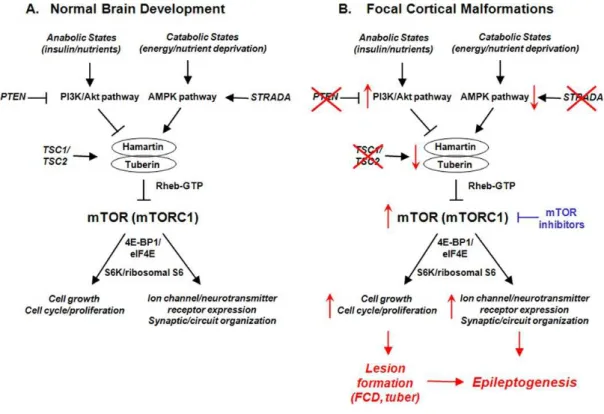
In neurons, mTOR covers a wide range of cellular function ranking from the regulation of the neuronal soma size to axon pathfinding and regeneration, dendrite arborization, dendritic spine morphology and synaptic (Tang et al., 2002; Swiech et al., 2008; Takei and Nawa, 2014; Lasarge and Danzer, 2014). Dysregulation of mTOR pathway causes a number of pathological conditions (grouped under the term “mTORopathies”), including a range of malformation of cortical development (MCD) and neurodevelopmental diseases associated with severe mental retardation and epilepsy such as tuberous sclerosis (Curatolo and Moavero, 2013; Lipton and Sahin, 2014), as shown in Figure 1.
Fig. 1 Role of the mTOR pathway in pathogenesis and epileptogenesis of corticalmalformations
From Wang M, Exp Neurol 2013, 244:22-6.
A. Physiological functions regulated by the mTOR pathway (primarily by mTORC1) during normal brain development, via activation of protein synthesis mechanisms (i.e. S6K/ribosomal S6 protein, eukaryotic initiation factor eIF4E pathways). In turn, mTORC1 is regulated by upstream signaling pathways (i.e. PI3K/Akt or AMPK pathway) in response to different physiological conditions and stimuli.
B. Hyperactivation of the mTOR pathway due to mutations in upstream regulators (e.g. TSC1 or TSC2 in TSC; STRADA gene in PMSE syndrome) led to focal MCD and tubers by abnormally increased cell growth and proliferation. The gross structural lesions themselves, as well as non-structural molecular and cellular changes in ion channel expression and synaptic organization may promote epileptogenesis in these disorders.
10 In line with this evidence, so far a wide number of germline and somatic (brain-only) mutations of DEPDC5 leading to mTORC1 hyperactivation have been associated with a range of lesional and non-lesional FEs. The MCD detected in some family members include different types of FCDs, subtle band heterotopias and hemimegalencephaly, with the predominant pattern being FCD type IIb (Scheffer et al., 2014; Lal et al., 2014; Martin et al., 2014; Scerri et al., 2015; D'Gama et al., 2015; Baulac et al., 2015).
Additional insights into the role of DEDCD5 in FCD-related FE derive from the rat model recently published (Marsan et al., 2016). The heterozygous rats exhibited an altered cortical neuron excitability and firing patterns and cortical cytomegalic dysmorphic neurons and balloon-like cells strongly expressing phosphorylated rpS6, indicative of mTORC1 upregulation. These neuropathological abnormalities are reminiscent of the hallmark brain pathology of human FCD.
I.2.5.PRIMA1
In a two-generation Australian family of Italian origin affected with SHE and ID, Hildebrand and coauthors identified by WES analysis a homozygous mutation in PRIMA1 (Hildebrand et al., 2015). This gene encodes a transmembrane protein that anchors acetylcholinesterase (AChE), the enzyme hydrolyzing Ach to membrane rafts of neurons. The c.93+2T>C mutation identified leads to knockout of PRIMA1, with reduction of AChE and accumulation of acetylcholine at the synapse, as shown in PRIMA1 knockout mice. The authors concluded that, similarly to the gain of function mutations of the genes coding for nAChR subunits, the enhanced cholinergic responses are the likely cause the severe SHE and ID in this family. However, apart from this single pedigree, this findings has not been replicated yet, as no other mutations were identified in a confirmation cohort of hundreds of SHE probands (Hildebrand et al., 2015).
In summary, ADSHE is a heterogeneous genetic syndrome. Overall, mutations in the genes identified so far cumulatively explain 20% of families and fewer than 5% of sporadic cases (Kurahashi and Hirose, 2015), clearly pointing to additional genetic factors. The penetrance of the mutations identified is incomplete, except for mutations in KCNT1.
By now, the small number of SHE families suitable for traditional approaches (i.e. linkage studies) prevented the fast discovery of novel genes. However, in the last few years, the advent of Next Generation Sequencing (NGS) technologies, such as Whole Exome Sequencing (WES) has completely revolutionized gene hunting, allowing the application of this technology and the discovery of novel epilepsy genes in nuclear pedigrees or even in sporadic patients.
The high intrafamilial variability and the overlapping features of the clinical manifestations, together with the rarity of ADSHE and the differences in study designs have hampered the definition of a genotype–phenotype correlation.
11 I.3. NEUROPSYCHOLOGICAL FEATURES OF SHE
Intellect is usually preserved in SHE patients, as briefly alluded by some large series of both SHE and ADSHE cases (Ryvlin et al., 2006a). However, no systematic studies have evaluated the neuropsychological profile of representative cohorts of SHE patients yet. Data available are scant and contradictory and derive mainly from selected populations with ADSHE (Ryvlin et al., 2006a). ADSHE was originally proposed as paradigm of a benign FE, occurring in patients with normal intelligence (Scheffer et al., 1995), as emphasized by the majority of reports (Nakken et al., 1999; Saenz et al., 1999). However, only few years later, several studies reported cognitive deficit, behavioral and psychiatric problems in some members of ADSHE pedigrees without specific molecular diagnosis (Khatami et al., 1998; Picard et al., 2000) or turned negative for mutations in nAChRs genes (Derry et al., 2008).
In some families found mutated in CHRNA4, in-depth and long-term clinical/psychiatric assessment revealed psychiatric comorbidity and ID. Firstly, in the Japanese pedigree with the S252L mutation Hirose and Ito described the occurrence of atypical features, namely mild ID and hyperactivity in two members of the third generation (Hirose et al., 1999; Ito et al., 2000). Moreover, long-term follow-up of the two siblings later revealed autistic disorder with profound ID (Miyajima et al., 2012). In 2003, Magnusson and colleagues described schizophrenia, recurrent psychosis and unspecified psychiatric disturbances in six out of the 11 members of the family carrying the 776ins3 mutation (Steilein et al., 1997; Magnusson et al., 2003), previously reported as of normal intelligence (Nakken et al., 1999). Following these reports, a number of studies described cognitive disabilities in ADSHE families carrying different mutations of the known genes. Specific mutations of CHRNA4 (S248F, S252L, 776ins3), CHRNB2 (V287M, I312M) and CHRNA2 (I297F) have been associated with ID, psychological or behavioral problems, including impulsivity, aggression, and hyperactivity, as shown in Table 1 (Steinlein et al., 2012a; Conti et al., 2015). The important effort of providing a genotype-phenotype correlation, revealed that the same molecular change could be associated with extremely variable endophenotypes, from severe ID to normal cognitive performance. This highlighted the need to extend in-depth neuropsychological assessments also in patients without apparent deficits.
A couple of small case-series systematically assessed the frequency and degree of neurocognitive disorders in CHRNA4/CHRNB2-mutated patients using a comprehensive battery of neuropsychological tests. Picard and coauthors studied 11 patients from four unrelated ADSHE pedigrees carrying different mutations of CHRNA4 (S248F, S252L, T265I) and CHRNB2 (V287L) (Steinlein et al., 2000; De Fusco et al., 2000; Rozycka et al., 2003; Leniger et al., 2003) including three families were neuropsychological problems were not previously reported (De Fusco et al., 2000; Rozycka et al., 2003; Leniger et al., 2003). All patients, regardless of the type of mutations or overt seizures had some degree of cognitive dysfunction: (i) borderline IQ/ID in the 45% of them, (ii) decreased performances in inhibitory task and/or verbal fluency in all them and (iii) deficit in verbal/non-verbal memory in 91% (Picard et al., 2009). These finding proved that neuropsychological disorders in ADSHE were underestimated.
12 Tab. 1 Neuropsychological findings in families and sporadic cases with mutations in CHRNA4, CHRNB2 and CHRNA2
Abbreviations AA aminoacid; ID: Intellectual Dysability; Psy: psychiatric disorders; Psych: psychological disturbances; Beh: behavioral problems; Memory: memory deficit; Memory v: verbal memory; Memory nv: non-verbal (visual) memory; ASD: autism spectrum disorder; S: sporadic case.
Grey lines reported cases with neuropsychological/psychiatric disturbances. Families studied by extensive neuropsychological assessment are underlined.
MUTATIONS FAMILIES REPORTED
GENE AACHANGE/DOMAIN FUNCTIONAL N ORIGIN NEUROPSYCHOLOGY REFERENCES
S248F/ TM2 Gain ↑ sensitivity to Ach (Bertrand 2002) 4 British-AU# - Steinlein 1995
CHRNA4 Loss faster desensitization, slower recovery Spanish - Saenz 1999
(Weiland 1996; Kuryatov 1997) Norwegian* ID, beh, psy Steinlein 2000 ↓ affinity to Ach, low currents (Bertrand 1998) Scottish Psych McLellan 2003
S252L/ TM2 Gain ↑ R sensitivity to Ach 4 Japanese ID, ASD Hirose 1999; Ito 2000; Miyajima 2012 (Bertrand 2002) Lebanese Low average intellect Phillips 2000
Korean ID, beh (hyperactivity) Cho 2003
Polish* - Rozycka 2003
1 Italian (S) - Sansoni 2012
776ins3/ TM2 Gain ↑ R sensitivity to Ach
(Steinlein 1997; Bertrand 1998, Bertrand 2002)
1
Norwegian Psy: Schizophrenia, recurrent psychosis
Steinlein 1997, Nakken 1999, Magnusson 2003
T265I/ TM2 Gain ↑ R sensitivity to Ach 1 German* - Leniger 2003
R336H/ Intracel loop 2 NA 1 Chinese - Chen 2008
c.823A>T NA 1 Chinese (S) - Wang, 2014
CHRNB2 V287L/ TM2 Gain ↑ ACh-evoked current, retarded desens, 1 Italian* - De Fusco 2000, Gambardella 2000
V287M/ TM2 Gain ↑ sensitivity to Ach (Phillips 2001, (Bertrand 2002)
2 Scottish Psych Phillips 2001, McLellan 2003
Spanish - Dıaz-Otero 2008
L301V/ TM3 Gain ↑ R sensitivity to Ach 1 TurkishCypriot Hoda, 2008
V308A/ TM3 Gain ↑ R sensitivity to Ach 2 Scottish Hoda, 2008
English
I312M/ TM3 Gain ↑ R sensitivity to Ach (Bertrand 2005) 2 English ID, memory Bertrand 2005
Korean Memory (v and nv) Cho 2008
V337G/ TM3-intrac
loop NA 1 Chinese (S)
-
Liu 2011
CHRNA2 I279N/TM1 Gain ↑ R sensitivity to Ach 1 Italian - Aridon 2006
I297F/TM2 Loss ↓ current density 1 Italian ASD Attention deficit
13 The impairment of executive functions as well as learning and memory, suggested a fronto-temporal dysfunction, differing from previous studies on frontal lobe epilepsies were memory seemed preserved (Picard et al., 2009). The authors hypothesized that neuropsychological deficits in ADSHE may be a consequence of seizures/interictal EEG abnormalities (despite the rarity of scalp anomalies and the focal seizures that remain localized to the frontal lobe) or fragmentation of NREM sleep, whose role in memory consolidation is well known (Tucker et al., 2006). However, they pointed to a contribution of nAChR subunits genes mutations, given the role of these receptors and nicotine in sustained and selective attention, automatic response inhibition and working memory (Poltavski and Petros, 2006; Meinke et al., 2006; Bacciottini et al., 2000; Levin et al., 2002).
The second case series evaluated a well-defined group of nine ADSHE members from the original Australian pedigree with the S248F mutation of CHRNA4 and normal intelligence. The mutation group was compared to an age-, gender-, and education-matched control group. ADSHE patients showed significantly worse scores in task requiring flexible adaptation and verbal fluency (stroop test, trail making test B, controlled oral word association) while intellectual abilities were preserved. Deficits in verbal memory correlated with disease-related factors, or medications (Wood et al., 2010).
Identification of additional mutations and novel genes for ADSHE enriched genotype-phenotype correlations and at present, neuropsychological and psychiatric comorbidities have been definitively related also to mutations in KCNT1 (Heron et al., 2012; Derry et al., 2008) and DEPDC5 (Picard et al., 2014; Picard et al., 2000).
Conversely, the majority of sporadic cases affected by SHE do not seem to present with gross cognitive disturbance, even though many of these patients complain of chronically disrupted sleep and daytime sleepiness (Ryvlin et al., 2006a). No more specific data on this population are available, although it represents the largest part of SHE patients. Several neuropsychological studies evaluated the impact of frontal lobe epilepsy on cognition and behavior, but they may include other epilepsy syndromes, with different types of frontal seizures occurring in wakefulness (Helmstaedter et al., 1996; Upton and Thompson 1997 (a,b); Exner et al, 2002; McDonald et al., 2005; Riva et al., 2005; Risse, 2006; Centeno et al., 2010; Braakman et al., 2011; O'Muircheartaigh and Richardson, 2012; Rayner et al., 2015; Patrikelis et al., 2016). However, these studies cannot be representative of SHE, where typical ictal manifestations are mostly exclusively sleep-related and may originate from extra-frontal areas with secondary involvement of frontal structures.
II.FAMILIAL FOCAL EPILEPSY WITH VARIABLE FOCI (FFEVF)
FFEVF is an unusual epilepsy syndrome characterized by focal seizures originating from different cortical regions in different affected family members and multifocal EEG abnormalities.
FFEVF was originally described in a large Australian family (Scheffer et al., 1998); seven further pedigrees were later reported: three were French-Canadian (Xiong et al., 1999; Berkovic et al., 2004), two from Spain (Berkovic et al., 2004; Morales Corraliz et al., 2010), two Dutch (Callenbach et al., 2003; Klein et al., 2012). All these families showed an autosomal dominant inheritance with penetrance between 50% and 80% and
14 marked intrafamilial variation in severity (Xiong et al., 1999; Callenbach et al., 2003; Berkovic et al., 2004; Morales Corraliz et al., 2010; Klein et al., 2012). Structural MRI studies were reported as unremarkable. Patients usually have normal intellect but ID, psychiatric disorders or autism spectrum disorder have been reported (Xiong et al., 1999; Callenbach et al., 2003; Klein et al., 2012).
Affected family members can present with frontal, temporal (mesial and lateral) occipital, parietal or multifocal seizures starting by the third decade (Klein et al., 2012). Although the heterogeneous seizure pattern within the members of the same pedigree, seizure semiology is constant in individual subjects (Berkovic et al., 2004). In some of the original FFEVF pedigrees, nocturnal, sleep-related seizures with a frontal lobe semiology are the most common phenotypes, leading to consideration of the diagnosis of ADSHE (Berkovic et al., 2004). The clinical overlap between FFEVF and ADSHE may lead to misdiagnosis, in particular in smaller pedigrees where the wide intrafamilial phenotypic variability of FFEVF might not be appreciated.
Linkage studies confirmed chromosome 22q12 as the solitary locus for all the eight FFEVF pedigrees originally reported (Klein et al., 2012).
In 2013 Dibbens and colleagues performed WES analysis in one Australian (A1) and one Dutch (D1) FFEVF family (Callenbach et al., 2003, Klein et al., 2012), identifying in each family a novel heterozygous nonsense mutation in the DEPDC5 gene, including c.21C>G (p.Tyr7*) in family A1 and c.1663C>T (p.Arg555*) in family D1. Mutations in the same gene where subsequently identified in additional five out of the six FFEVF families previously linked to chromosome 22q (S1, S2, F1-3), confirming DEPDC5 as the major gene for FFEVF (Dibbens et al., 2013). Of the eight large families with FFEVF, only one (family A in Klein et al., 2012) did not have a DEPDC5 mutation. The three French-Canadian families had the same deletion mutation (c.488_490delTGT; p.F164del), suggesting a shared ancestor.
This disorder is difficult to recognize in small families due to the low number of affected individuals and because the pedigrees are too small to demonstrate a clear autosomal dominant inheritance. The sequencing of the gene by high-resolution melt curve analysis (HRM) in other 82 unrelated probands of families with at least two affected members, identified DEPDC5 mutations in ten (12.2%), confirming a major role of this gene in familial FE (Dibbens et al., 2013).Later, the evidence that some affected members had structural cortical alterations (i.e. bottom of the sulcus dysplasia, a form of FCD type II B), implying mutations in this gene in MCD (Scheffer et al., 2014).
15
AIMS
We conducted an accurate clinical, neuropsychological and genetic characterization of a large cohort of patients with SHE, negative for mutations in the genes coding for the nAChRs, in order to:
(i) identify new genetic determinants for ADSHE/FFEVF by WES analysis;
(ii) estimate the frequency of mutations in KCNT1 and DEPDC5,recently implicated in ADSHE;
(iii)assess the impact of SHE on neuropsychological functioning, characterizing a possible profile of impairment;
(iv)correlate genetic findings with clinical and neuropsychological data.
The application of innovative tools for gene discovery (Whole Exome Sequencing – WES) allowed us to include in the genetic analysis small pedigree and sporadic patients studied by a trio approach.
16
METHODS
I. PATIENTS RECRUITMENT
I.1. SETTING AND PERIOD
The study was carried out over 2012-2016 at the IRCCS, Institute of Neurological Sciences of Bologna and the Medical Genetics of Sant’Orsola Hospital, following the approval by the Human Research Ethics Committee of Bellaria Hospital, Bologna (Prot. N 945/CE; cod CE: 13084).
The study was been supported by a no-profit association (Telethon foundation, GGP13200).
I.2.POPULATION AND INCLUSION CRITERIA
We included individuals of any age and gender, diagnosed with video/video-EEG documented SHE according to reliable diagnostic criteria, with/without a positive family history for FE.
The study population derives mainly from a larger cohort of SHE patients that, referred to the Epilepsy and Sleep centers of our Institute since 1980 for sleep-related motor events, were finally diagnosed with SHE. All patients still followed up in our clinic and consecutively attending the Epilepsy center for a control visit between September 2012 and April 2016 were asked to participate in the study. Consenting patients negative for mutations in the AChRs genes were prospectively enrolled after obtaining specific written consent. We included also newly-diagnosed cases referred and diagnosed in our Institute since 2012. Additional cases were referred by other Italian epilepsy Centers thanks to the collaboration with the Italian League against Epilepsy (Lega Italiana Contro l’Epilessia, LICE), patron of 47 Epilepsy Centers located in 15 Italian regions. We selected patients fulfilling the following inclusion criteria:
(i) personal history of sleep-related paroxysmal motor events suggestive of hypermotor seizures;
(ii) video-polysomnographic (VPSG) recording of at least one major event (asymmetric tonic seizures/hyperkinetic seizures/epileptic nocturnal wandering) or of two stereotyped minor events (paroxysmal arousals).
For all cases the diagnosis of SHE was confirmed by three experts in epileptology and sleep disorders (P. Tinuper, F. Bisulli and F. Provini) and conformed to the new diagnostic criteria, based on level of certainty (Tinuper et al., 2016).
We enrolled both sporadic and familial cases; the latter were defined as patients having at least one relative within three degrees of relatedness affected with SHE or other FE, including both ADSHE and possible FFEVF families.Biological samples (blood or saliva samples) were collected for DNA extraction and analysis from each index case and all the affected and unaffected consenting relatives available.
II. CLINICAL STUDY
All probands underwent a comprehensive evaluation including videopolygraphic monitoring for recording of at least one hypermotor sleep-related seizure. For each patient we reviewed the clinical history and the
17 neurophysiological and neuroradiological documentation. All data were collected in an ad hoc database, collecting:
- demographic data (age, gender, educational level);
- family history of intellectual deficit and psychiatric disorders;
- age at seizures onset, seizures semiology, presence of seizures on wakefulness, specific auras, bilateral convulsive seizures and status epilepticus;
- seizures frequency at the onset and during the last year; past and current antiepileptic treatment, compliance and response to therapy;
- interictal/ictal EEG abnormalities (specifying the distribution); - presence of brain MRI abnormalities.
Clinical and neurophysiological assessment was extended to all the available affected relatives, to define the individual clinical phenotype and then, the familial epilepsy syndrome (ADSHE/FFEVF).
All patients were studied by a detailed and direct clinical interview, routine EEG (if necessary, video-EEG recording) and targeted brain 3-T MRI acquisitions following a specific “epilepsy protocol”.
Following the pedigree reconstruction, we drew each pedigree.
III. GENETIC STUDY
III.1. PRE-SCREENING OF KNOWN GENES
Since 1995, available patients from the historical cohort of SHE patients followed up in our Institute had been screened for mutations in CHRNA4, CHRNB2 and CHRNA2. This analysis, conducted over the years at the Genetic Unit of the Meyer Hospital in Florence was in part performed by denaturing High Performance Liquid Chromatography (dHPLC) assay.
Additionally, in 2012 a subpopulation of patients underwent a preliminary screening for mutations in KCNT1 and DEPDC5 by high resolution melt curve analysis (HRM) at the Epilepsy Research Centre, University of Melbourne.
III.2 WES ANALYSIS
Genetic study was conducted by Dr. T. Pippucci and S. Baldassari, at the Genetic Unit, Policlinico Sant'Orsola Malpighi, University of Bologna.
WES analysis was performed in
(i) familial cases, including at least one affected relative (when available);
(ii) sporadic cases, in part analyzed by a trio approach (affected proband and healthy parents) in order to discover possible de novo mutations (DNMs).
18 Fig. 2 WES experiment workflow
III.2.1.DNA SAMPLE COLLECTION
DNA samples for WES analysis were extracted from peripheral blood collected specimens using the commercial kit QIAamp DNA Blood Mini Kit (QIAGEN), which is based on the usage of columns containing membranes that enhance a selective DNA adsorption and a final purified DNA elution.
III.2.2.WES EXPERIMENT
WES experiments were performed at Beijing Genomics Institute (BGI Tech Solutions, Hong Kong, www.bgi.com). Two different enrichment methods were used: NimbleGen SeqCap EZ (Roche), a BGI exome capture (BGI). Both kits cover more than 20,000 genes in the human genome and are based on oligonucleotide DNA probes that capture the target exome. The generated libraries have been sequenced as 91 bp (base pair) paired-end reads on the Illumina HiSeq2000 platform (Illumina) at BGI, requiring a mean coverage per sample of 50X for the exomes enriched with NimbleGen SeqCap EZ and 100X for the exomes enriched with BGI exome capture.
III.2.3.BIOINFORMATIC ANALYSIS
Bioinformatic analysis workflow is shown in Fig. 2. FASTQ format sequencing reads were received from BGI and processed for quality control following a pipeline developed at the Medical Genetics Unit of the Policlinic Sant’Orsola-Malpighi in Bologna. The reads were aligned against the hg19 reference genome with BWA. Single nucleotide variants (SNVs) and small indels were called using GATK Unified Genotyper, and GATK VariantFiltrationWalker was used to filter out variants by quality, according to specific parameters. Multi-sample variant calling for SNVs and indels has also been performed, to improve variant calling, using the GATK package utility HaplotypeCaller (www.broadinstitute.org/gatk/): variants flagged as “PASS”, meaning that they are reliable calls, were considered in the downstream steps of the analysis workflow. These were then annotated against the NCBI RefSeq (www.ncbi.nlm.nih.gov/RefSeq)and UCSC KnownGene (www.genome.ucsc.edu) databases with ANNOVAR (Wang et al., 2010), or against the Ensembl database (www.ensembl.org) using Variant Effect Predictor (www.ensembl.org/info/docs/tools/vep/index.html)and
19 organized in a structured query language-based database by Gemini (www.gemini.readthedocs.org/en/latest/) (Paila et al., 2013).
Once annotated, the variants were filtered. We retained all rare variants affecting coding sequences of the targeted exome by filtering out all SNVs and InDels observed in dbSNP137(www.ncbi.nlm.nih.gov/SNP/),
1000genomes (www.broser.1000genomes.org/),Exome Variant Server(www.evs.gs.washington.edu/EVS/) and CNVs overlapping with those observed in Database of Genomic Variants(projects.tcag.ca/variation). The whole filtering procedure retained only rare coding variants with functional effect on the protein coding sequence and CNVs.
III.2.4.VARIANTS PRIORITIZATION
The variants prioritization was performed considering the following criteria: presence or absence of the variant in public databases, pathogenicity prediction by in-silico tools, conservation of the mutated site in other vertebrates, expression and function of the mutated gene in the brain, association of the mutated gene with other neurological diseases. The analysis was divided into 4 steps: the first including variants with highest pathogenicity scores, the last including variants with the lowest pathogenicity scores.
(iii)LOF mutations, including nonsense or splicing affecting single nucleotide variants (SNVs) and frameshift indels, not described in public databases.
(iv)Missense mutations, not reported in public databases, predicted to be damaging by Polyphen2 (www.genetics.bwh.harvard.edu/pph2), SIFT (www.sift.jcvi.org)and CADD (>15,
www.cadd.gs.washington.edu)scores and affecting conserved sites (GERP++ score >4,
www.mendel.stanford.edu/SidowLab/downloads/gerp) and rare LOF mutations with a frequency lower than 1% in an internal exome database.
(v) Missense mutations, not reported in public databases, and predicted to be damaging by at least one in-silico predictor among Polyphen2, SIFT and CADD, and affecting conserved sites (GERP++ score >2). (vi)Missense mutations, reported with a low frequency in public databases, and predicted to be damaging by
at least one in-silico tool among Polyphen2, SIFT and CADD, with a GERP++ score between 0 and 2. In each mutational class, the most candidate variants were selected according to the available information on the expression or function of the mutated gene: to this purpose, different databases have been queried, including Pubmed (www.ncbi.nlm.nih.gov/pubmed), Braineac (www.braineac.org), Omim (www.omim.org) and STRING (www.string-db.org). Pubmed and Omim were used to assess the function of the gene of interest and its possible associated diseases. Braineac is a database of brain expressed genes, distinguishing for brain regions, and was interrogated to evaluate the expression pattern of the gene of interest in the brain. Finally, the protein-protein interactions involving the protein encoded by a mutated gene were analyzed using the STRING database.
Sanger sequencing was subsequently applied to validate the prioritized variants in the proband and to determine the segregation in the family.
20
IV. NEUROPSYCHOLOGICAL STUDY
This was a cross-sectional study carried out over 2013 and 2016 at the Neurological Clinic Unit of IRCCS, Institute of Neurological Sciences of Bologna.
All the patients recruited underwent an assessment of intellectual functioning and cognitive status by: - Wechsler Adult Intellectual Scale (WAIS) or Stanford-Binet Intelligence Scale (SB, for patients <16
years);
- Raven's progressive Matrices;
- Mini-Mental State Examination (MMSE).
ID was defined when IQ score was <70; MMSE was considered pathological when corrected scores where <23.8.
Patients aged >16 years with normal cognitive functioning (IQ scores >70; MMSEc scores >23.8) carried on with an extensive, standardized neuropsychological battery. These additional neuropsychological measures were selected in order to explore a range of frontal and extra-frontal functions, schematically sampled in the following cognitive domains:
- language: semantic and phonemic fluency;
- verbal and non-verbal memory: Rey Auditory Verbal Learning Test (RAVLT), forward verbal span, verbal supra span + 2, paired-associated words learning (for verbal memory); Rey-Osterrieth complex figure (ROCF) immediate recall; Visual-Spatial supra span + 2, corsi-Block-Test (for visual memory); - visuospatial ability (ROCF copy);
- attention and executive functioning: Trail Making test A, Trail Making test B (for attention, shifting and flexibility); backwards Verbal Span (for working memory); Wisconsin Card Sorting Test (WCST) (executive function); Stroop test (executive function, response inhibition).
The final score was calculated after adjustment for age and education.
Neuropsychological testing was conducted by a single expert neuropsychologist (R. Poda) at the neuropsychological Service of our Institute.
All the tests were administered to each patient in a standardized order, over a single session held in the morning and lasting between 1 and 2 hours.
A paired clinical assessment, performed on the same day as neuropsychological testing, was focused on seizure frequency and antiepileptic drugs taken at that stage, in addition to the other variables collected in the clinical database. For patients hospitalized at the time of the study, a VPSG recording and questionnaire evaluating daytime somnolence in the days immediately close were also available.
All neuropsychological data were collected in an ad hoc database.
Statistical analysis was performed using statistical package Stata SE, version 14.0.
For descriptive statistics, continuous variables were presented as mean ± standard deviation, while categorical variables as absolute and relative frequency (%). Performances of SHE patients were evaluated with respect to age adjusted normative data of healthy controls.
21 Fisher’s exact test was used to highlight possible associations between each neuropsychological test with clinical features, comparing variables among groups. All p-values were based on 2-sided tests; p<0.05 was considered significant.
To further investigate the impact of disease severity on cognitive functioning, we used the non-parametric Wilcoxon Rank-Sum test, comparing the distribution of the scoring for each neuropsychological test between groups categorized according clinical variables.
V. GENOTYPE-PHENOTYPE CORRELATION
All anatomo-electro-clinical data were correlated with genetic findings in order to highlight differences on epilepsy phenotype related to mutations in a specific gene. In particular, we paid particular attention on particular clinical features (such as age at onset, presence of MCD or other cerebral structural lesions, subjective symptoms preceding the seizure onset, presence of seizure during wakefulness, ID and psychiatric comorbidity) in order to disclose possible key elements to direct genetic diagnostic tests. Binomial Exact test was used to calculate 95% confidence intervals (95%CI).
Similarly, the results of the systematic neuropsychological study were correlated with genetic findings in order to disclose possible differences in mutated and not-mutated cases.
22
RESULTS
I. STUDY POPULATION
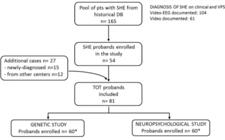
A total of 81 patients (M/F=45/36; mean age at enrollment available in 75 cases: 35.4±14.6, range: 2-69 years) were enrolled in the genetic and/or the neuropsychological study.
The flow-chart in Figure 3 provides details on patient recruitment. Briefly:
- 54 probands belonged to the historical cohort of 165 SHE patients diagnosed and followed up at our Institute since 1980;
- 15 were new-diagnosed cases, referred to our Institute since 2012;
- 12 patients were referred from other Italian Epilepsy Centers, by means the LICE.
Thirty-nine probands underwent both WES analysis and neuropsychological assessment, while 21 had only genetic study (in total, 60 probands) and another 21 had only neuropsychological assessment (in total, 60 patients).
Fig. 3 General recruitment flow-chart
*Consider overlapping of 39 patients who underwent both genetic and neuropsychological study.
II. GENETIC STUDY
II.1. PRE-SCREENING FOR MUTATIONS IN KNOWN GENES
Overall, 76 probands from the original cohort underwent preliminary genetic analysis. Details are reported in Table 2.
Seventy patients were analyzed for mutations in at least one of the genes coding for the nAChR subunits by dHPLC (27 cases) or by Sanger sequencing (43 cases): no mutations of these genes were detected.
A subsample of 43 probands underwent molecular analysis of KCNT1 and/or DEPDC5 performed at the Epilepsy Research center of Melbourne University. This study led to the identification of a mutation in KCNT1
23 and three in DEPDC5 reported in the original publications on these genes (Heron et al., 2012; Dibbens et al., 2013; Scheffer et al., 2014; Ricos et al., 2016).
The pedigrees (indicated as P) and more detailed clinical data on these families (F) and sporadic cases (S) are provided as Supplementary materials, part A.
Tab. 2 Preliminary screening for mutations in SHE known genes
Abbreviations dHPLC: high performance liquid chromatography; HRM: high resolution melt curve analysis; fam: familial; spo: sporadic case.
KCNT1 - F1 The p.Y796H mutation of KCNT1 (NM_020822.2) segregated in four affected members of an ADSHE pedigree (P1) with 100% of penetrance and a more severe phenotype: earlier age at onset (mean 5.5±2.1 years) compared with classical ADSHE, refractory seizures in two patients, recurrence of ID and psychiatric/behavioral problems (Heron et al., 2012, family B). The family was also reported in a previous publication (Phillips et al., 1998, as family C).
DEPDC5 - F2 Of the three mutations of DEPDC5 (NM_001242896.1), the c.279+1 G>A insertion was first identified in the proband of a FE family (P2, IV.2) but not in her affected mother (III.2), suggesting gonadic mosaicism in this individual (Dibbens et al., 2013, Family I). A few months later an additional individual (V.1) developed frontal lobe seizures at 4 years of age. Interictal EEG showed frequent epileptiform abnormalities over the left frontal field (Supplementary Figure 1). He carried the familial c.27911G>A mutation and a left frontal subtle band heterotopia adjacent to the dysplastic cortex at brain MRI (Supplementary Figure 2). The other affected relatives had normal MRI. This was one of the first pieces of evidence implying mutations of DEPDC5 in MCD (Scheffer et al., 2014, family C).
DEPDC5 - F3 The p.T329Lfs*7 DEPDC5 frameshift mutation was identified in nine individuals of a four-generation FFEVF pedigree with five affected members available for genetic testing (penetrance 55%). SHE was the most common phenotype running in the family (P3, individuals III.6, IV.1, IV.2, IV.4). Affected members showed a wide range of disease severity: the proband (IV.2) had sleep-related seizures well controlled by low doses of carbamazepine, while several individuals (III.6, III.8, IV.1, IV.4) had seizures refractory to pharmacological treatment. Two of them (III.6, III.8) underwent pre-surgical work up but were excluded from surgery because of lack of a clear epileptogenic focus (III.6) or documentation of independent seizures arising from both left and right fronto-temporal regions (III.8). Brain MRI disclosed MCD in
GENES N# Pts
TYPE OF ASSAY MUTATION FAM/ REFERENCES
Sanger dHPLC HRM SPO
CHRNA4 62 36 26 - -
CHRNB2 60 34 26 - -
CHRNA2 65 38 27 - -
KCNT1 43 - - 43 p.Y796H Fam Heron 2012 (fam B); Phillips 1998
DEPDC5 37 - - 37 c.279+1 G>A p.T329Lfs*7 p.R165Yfs*14 Fam Fam Spo
Dibbens 2013 (fam I), Scheffer 2014 (fam C) Ricos 2016 (fam 28)
24 individuals IV.1 (who showed FCD, Supplementary Figure 3) and IV.4 (who had a suspected gyral abnormality of the right middle frontal gyrus; not shown). Moreover, severe psychiatric and behavioral problems recurred in affected individuals II.1, III.8, IV.2, IV.4.
DEPDC5 - S1 An additional frameshift mutation p.R165Yfs*14 was identified in a sporadic case who underwent invasive pre-surgical assessment because of frequent seizures refractory to AEDs (P4, II.1). Despite a bilateral limbic Stereo-EEG (SEEG) exploration (Supplementary Figure 4), no definitive epileptogenic focus was identified in this patient. The SEEG electrical pattern was suggestive of focal cortical dysplasia (Supplementary Figure 5) but tailored brain MRI (1.5 T) was unrevealing. The mutation was inherited by her healthy mother (I.2).
II.2 WES ANALYSIS
WES was performed in 60 probands with SHE (M/F:34/26) and 76 affected/not affected relatives, for a total of 136 individuals (Figure 4).
The proband sample includes:
- ten familial cases: five belonging to ADSHE pedigrees and five with a family history for other epilepsy, in some cases compatible with FFEVF;
- 50 sporadic cases, 31 of whom were studied as trios (the healthy parents were also sequenced to evaluate the potential impact of de novo mutations in the pathogenesis of SHE).
Thirty-nine probands were recruited form the historical pool, nine have been diagnosed and enrolled since the beginning of the study (2012) while 12 were referred by other colleagues for WES analysis.
Mean age at epilepsy onset (available on 53 probands) was 11.79±8.77 years (range: 0-42 years). Fourteen probands had a positive brain MRI: ten with MCD (FCD in eight, dysplastic hemimegalencephaly in one, abnormal cortical gyration in one), one patient had mesial temporal sclerosis, three with gross brain abnormalities.
WES analysis identified:
- a number of mutations in known genes and a novel gene responsible for SHE, NPRL2; - 16 novel candidate genes, mainly identified as DNMs by trio analysis;
- several variants of unknown significance (VUS), eight in genes already implicated in SHE. Supplementary Table 1 lists the DNMs identified in the trio cohort.
Figure 4 summarizes all WES findings that are detailed below. II.2.1MUTATIONS IN KNOWN/NOVEL GENES
DEPDC5 - S2 One novel splice donor site variant (c.193+1 G>A, NM_001136029.2) was identified in the male proband of pedigree 5 (P5). The patient had SHE with rare seizures in wakefulness controlled by medication. There was a history of sleep-related epileptic events in the paternal branch, but no definite clinical information was available. The trio study in this case highlighted that the variant was inherited from the healthy mother. The RNA sample of the proband was not available to confirm the effect of the variant on the DEPDC5 transcript, but the variant was considered to be likely pathogenic.
25 Fig. 4 Summary of WES study results
NPRL2 - F4 WES analysis performed in two out of the four affected members of a FFEVF family (P6) allowed the identification of a heterozygous missense change (p.L105P) of NPRL2 (NM_006545.4) coding for one interactor of DEPDC5. This variant is predicted to be deleterious by all in silico prediction tools used, affects a conserved aminoacid residue and has never been reported in public databases. The proband (II:3) and her son (III:3), were both affected with typical SHE, experiencing clusters of hypermotor seizures exclusively from sleep. Accurate phenotyping suggested an extra-frontal (insular) onset of the seizures in the proband who reported a painful sensation of the left arm, sometimes followed by contraction of facial muscles or auditory hallucinations preceding the motor events.
Segregation analysis by Sanger sequencing confirmed that the variant was present in four other family members, two of whom definitely affected (penetrance of 66%): the proband’s father (I:1) who suffered from focal (possible temporal) seizures both from sleep and wakefulness; one healthy sister (II:2); another sister with unconfirmed seizures during childhood (II.1) and her son (III:1) who recently experienced a few sleep-related events described as bilateral convulsive seizures.
This family was published in a collaborative study (Ricos et al., 2016) describing mutations of NPRL2 and This family was published in a collaborative study (Ricos et al., 2016) describing mutations of NPRL2 and NPRL3 in FE for the first time. The study confirmed the role of GATOR1 components in the pathogenesis of FE and established that mutations in this complex account for about 9.5% of all genetic FEs (Ricos et al., 2016).
26 KCNT1 - S3 In a female patient (P7) studied as a trio we identified a de novo missense change of KCNT1 (NM_020822.2), p.A934T, which affects a highly conserved residue in the cytoplasmic C-terminal domain of the protein.
This mutation has been already reported as pathogenic in a French patient affected with MMFSI (Barcia et al., 2012) and is published in public databases ClinVar and dbSNP with the code rs397515403 (http://www.ncbi.nlm.nih.gov/clinvar/; http://www.ncbi.nlm.nih.gov/SNP/).
The proband of our trio had a different phenotype, characterized by bilateral asymmetric tonic seizures occurring in clusters up to 40/night since the age of 9 years. Her epilepsy showed a spontaneous remitting- relapsing pattern of evolution, without a clear-cut pharmacoresistance. The patient also had a congenital profound sensorineural hearing loss and a cognitive delay with predominant language impairment.
Genetic analysis in this patient also highlighted the CHRNA4 p.D104N change, inherited by her father who had a history of arousal parasomnias during childhood and “agitated” sleep-related events of uncertain etiology between 30 and 40 years of age. This variant has been considered of unknown significance (see later). Finally, given the comorbidity with congenital hypoacusia (unlikely to be ascribable to the phenotypic spectrum of this gene), we conducted an additional analysis aimed at discovering possible causes of this specific disorder. The patient proved to be compound heterozygous for two mutation in TMPRSS3 (MIM 605511), involved in an autosomal recessive form of non-syndromic congenital hearing loss. Both mutations were inherited by the healthy parents. In particular, the first mutation (p.P277L, NM_032404), reported as pathogenic in the public databases Omim and ClinVar (rs28939084), was inherited by the mother. The second is a deletion p.H70Tfs*X19 (NM_001256317) reported in ExAc database with a frequency of 5/10000) and was inherited by her father.
CHRNA4 - F5 WES analysis identified three mutations in CHRNA4 the first of which had been missed by the previous dHPLC analysis.
The novel heterozygous variant p.G307V (NM_000744.6) was found in two sisters with typical SHE of family 5 (P8). The variant was confirmed by Sanger sequencing and the segregation analysis revealed that it was inherited from the healthy father (penetrance of 66%). This change is predicted to be damaging by all the in silico tools (Table 3) and affects a conserved aminoacid located close to the third transmembrane helix (Figure 5). The change in the aminoacid sequence in this region may alter the protein structure.
Tab. 3 Features of the novel mutation in CHRNA4
hg19_coordinate transcript N_change AA_change zyg N_ PhyloP predictions freq_ExAC
chr20: 61981843 NM_000744.6 c.G920T p.G307V het 0.998 D,D -
Abbreviations N_change: nucleotidic change; AA_change: aminoacidic change; zyg: zygotes; N_PhyloP: normalized PhyloPhen*; freq_Exac: allele frequency from Exac (Exome Aggregation Consortium) database; het: heterozygous; D: damaging.
*PhyloPhen (evaluates conservation among different species);
Predictive tools included SIFT (whose prediction is based on the degree of conservation of amino acid residues in sequence alignments derived from closely related sequences) and Polyphen (predicts the possible impact of amino acid substitutions on the stability and function of human proteins using structural and comparative evolutionary considerations).
27 Extracellular domain
CHRNA4 - S4 The missense variant p.S284L (rs28931591) of CHRNA4 is a hotspot mutation associated with a CpG hypermutable site in the TM2 domain of the protein. It corresponds to the well-known CHRNA4 p.S252L mutation, reported since 1999 in four ADSHE families and one sporadic case (Hirose et al., 1999; Phillips et al., 2000; Cho et al., 2003; Rozycka et al., 2003; Sansoni et al., 2012). Cognitive deficit has been reported in some affected family members (see Table 1). Similarly, our patient (P9) showed a particularly severe SHE phenotype with early onset refractory seizures and ID. There was a positive family history for NREM parasomnias in both parents and his sister, however the mutation was de novo. This patient carried also a de novo mutation in a candidate gene, MIOS, as detailed below.
CHRNA4 - S5 Finally, a novel missense change p.S284W was identified in the female proband of pedigree 10 (P10) affected with refractory SHE. The variant is predicted to be damaging for protein function, and affects the same aminoacidic residue that has been found mutated in different SHE patients, including our sporadic case of pedigree 9 (P9). The segregation analysis revealed that the variant is not present in the patient’s healthy father and healthy brother, reinforcing the pathogenic role of the mutation, although the segregation in the healthy mother could not be assessed.
Fig. 5 Schematic representation of CHRNA4 (NM_000744.5). The mutations found are indicated by red stars.
Domain AA Extracellular 29-242 TM1 243-267 Intracell loop 1 268-274 TM2 275-293 Extracell loop 1 294-308 TM3 309-330 Intracell loop 2 331-600 TM4 601-619
SCN1A - S6 A missense change p.P824H of SCN1A (NM_001165963) was identified in a sporadic case referred to us as affected by SHE for genetic study (P11). The mutation has never been reported so far, especially in association with the SHE phenotype, but evaluation of the patient’s whole clinical history suggests that this variant could underlie the pathogenesis of his epilepsy. In fact, the patient had a history of recurrent, prolonged febrile/afebrile seizures and status epilepticus starting from age nine months, slow regression of cognition after seizure onset, and polymorphic seizures (hemiclonic seizures, temporal seizure with loss of awareness, atypical absences) refractory to antiepileptic medications (of note, the clinical worsening induced by lamotrigine and other sodium blockers and the efficacy of valproate and topiramate treatment). However, the sleep-related paroxysmal motor events from age 13 years are not commonly reported in SCN1A-related epilepsy phenotype.
28 II.2.2MUTATIONS IN CANDIDATE GENES
PIK3R3 Three out of the five affected individuals of an ADSHE family were sequenced. The analysis, focused on shared heterozygous variants according to an AD inheritance model, led to the identification of a shared, novel candidate variant p.N110S in PIK3R3 (NM_001303428.1) that affects a conserved aminoacid residue and is predicted to be damaging by Polyphen2, Sift and Cadd scores.
Moreover, an additional missense change p.R16M in this gene was identified in a male patient of a trio, inherited from the healthy father. The variant is predicted to be damaging by Polyphen2 and Cadd scores, and is not reported in public databases. PIK3R3 encodes a regulating subunit of the phosphoinositide-3 kinase (PI3K), which is highly expressed in the brain cortex and interacts with PIK3R1 and PIK3CA to regulate mTOR, whose deregulation has already been described in FE. To date, pathogenic germline and mosaic mutations in multiple PI3K/AKT pathway genes have been associated with focal MCD manifesting with FE (Lee et al., 2012; Jansen et al., 2015; D’Gama et al., 2015). None of the reported patients showed structural lesions at conventional 1.5 T brain MRI, but a targeted high-resolution MRI was not available.
PIK3CA A missense variant p.R770Q (NM_006218.3) of PIK3CA was identified in a male proband of a trio, inherited by his mother. The patient had refractory SHE of unknown etiology. Given the persistence of highly frequent hypermotor seizures from sleep, following invasive SEEG recordings he underwent a left orbito-frontal lesionectomy-cortectomy. Histological analysis of the resected brain specimen disclosed a FCD type IIb.
The aminoacidic change found is predicted to be damaging by Sift and Cadd scores and affects a conserved residue. However, it has been reported in four alleles in ExAC database, and was therefore classified as a VUS. PIK3CA encodes a catalytic subunit of PI3K, which interacts with the regulatory subunit encoded by PIK3R3 and PIK3R1. Interestingly, somatic mutations in this gene have been associated with abnormalities of brain development including FCD, as reported above.
PIK3C2B A de novo missense change in PIK3C2B (p.E1294Q; NM_002646.3)has been identified in the male proband of a trio with unconfirmed family history for sleep-related paroxysmal attacks which were reported in his father. The proband had documented hypermotor seizures arising exclusively from sleep, well controlled by antiepileptic medication. Brain MRI did not show structural lesions.
The protein encoded by this gene belongs to the PI3K family and consecutively considered a strong candidate gene that has not yet been reported.
The same patient carried also a novel nonsense mutation of AMBRA1 p.Q276* (NM_001300731.1) that was inherited from the father. AMBRA1 is a gene intolerant to missense and LOF mutations according to ExAC database. The encoded protein regulates autophagy and the development of the nervous system, controlling the protein turnover (Fimia et al., 2007); it is expressed ubiquitously and has a high expression in neuronal cells of the brain cortex. Impaired autophagy has already been implicated in the pathogenesis of neurological diseases, including epilepsy, emphasizing the possible causative role of the identified mutation.
MTOR A novel missense change in MTOR, p.L2354M (NM_004958.3) was identified in the male proband of a trio, inherited from the healthy mother. The variant is predicted to be damaging by Polyphen2, Sift and Cadd