Department of Biomolecular Sciences
PhD program in Sciences of Life, Health and Biotechnologies
Curriculum in Biology of cells and organisms
XXXI course
Venous lower limb microenvironment: an open window on the
Chronic Venous Disease
SSD BIO/12
Supervisor PhD Candidate
Professor Ferdinando Mannello Lidia Croce
INDEX
Preface ... 3
Chapter 1. Introduction ... 5
1.1 Epidemiological, diagnostic and therapeutic background of the Chronic Venous Disease ... 5
1.2 The structural and hemodynamic players of the Chronic Venous Disease ... 15
1.4 The cellular and biochemical players of the Chronic Venous Disease ... 25
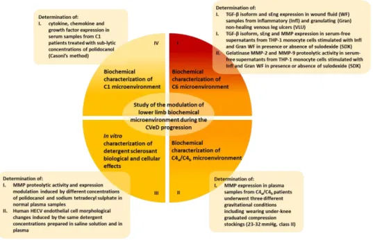
Chapter 2. Thesis aim ... 43
Chapter 3. Materials and methods ... 46
3.1 Biochemical characterization of C6 microenvironment: TGF-β isoform, sEng and MMP expression profile ... 46
3.1.1 Patient recruitment and sample collection ... 46
3.1.2 Cell culture and treatments ... 47
3.1.3 Multiplex suspension immunomagnetic assay ... 48
3.1.4 Zymography assay ... 50
3.1.5 Statistical analysis ... 54
3.2 Biochemical characterization of C4a/C4b microenvironment: MMP expression profile ... 54
3.2.1 Patient recruitment and sample collection ... 54
3.2.2 Multiplex suspension immunomagnetic assay ... 56
3.2.3 Statistical analysis ... 56
3.3 In vitro characterization of detergent sclerosant biological and cellular effects ... 57
3.3.1 Sample collection and treatment ... 57
3.3.2 Zymography assay ... 58
3.3.3 SDS-PAGE assay ... 59
3.3.4 Cell culture and treatment ... 60
3.4 Biochemical characterization of C1/C2 microenvironment: cytokine, chemokine and growth factor expression profile ... 61
3.4.1 Patient recruitment and sample collection ... 61
3.4.2 Multiplex suspension immunomagnetic assay ... 62
Chapter 4. Results ... 64
4.1 Biochemical characterization of C6 microenvironment ... 64
4.1.1 Demographic data ... 64
4.1.2 TGF-β isoform and sEng expression profile in wound fluid ... 64
4.1.3 TGF-β isoform, sEng and gelatinase expression profile in WF-stimulated THP-1 co-treated with sulodexide ... 65
4.1.4 Proteolytic activity profile of MMP-2 and MMP-9 in WF-stimulated THP-1 co-treated with sulodexide ... 68
4.2 Biochemical characterization of C4a/C4b microenvironment ... 69
4.2.1 Demographic data ... 69
4.2.2 MMP expression profile in plasma samples from healthy volunteers and C4a/C4b patients ... 70
4.3 In vitro characterization of detergent sclerosant biological and cellular effects ... 72
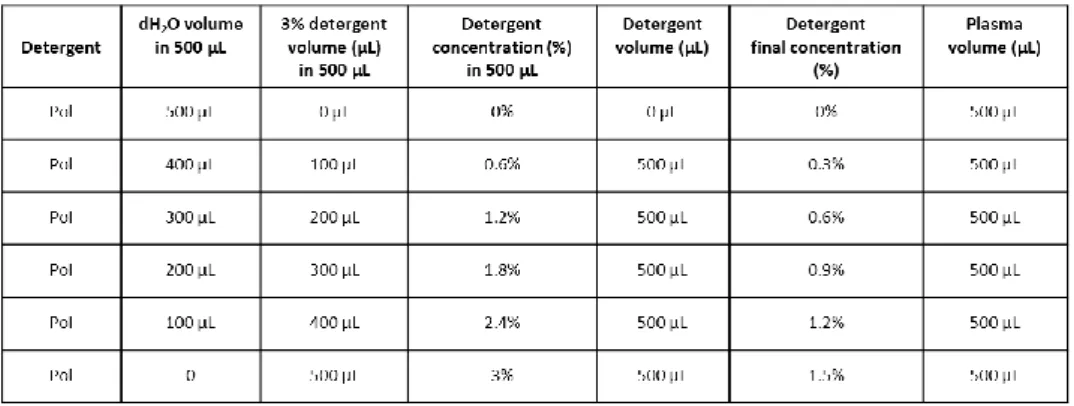
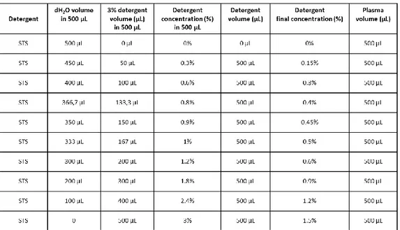
4.3.1 Proteolytic activity profile of MMP-2 and MMP-9 in normal plasma samples treated with polidocanol and sodium tetradecyl sulphate ... 72
4.3.2 Electrophoretic profile of plasma protein content after the treatment with polidocanol and sodium tetradecyl sulphate ... 74
4.3.3 Cellular morphological changes in HECV endothelial cells induced by polidocanol and sodium tetradecyl sulphate treatment ... 75
4.4 Biochemical characterization of C1/C2 microenvironment ... 79
4.4.1 Demographic data ... 79
4.4.2 Inflammatory mediator expression profile in serum samples from C1/C2 patients ... 80
Chapter 5. Discussion ... 86
Chapter 6. Conclusions ... 108
Preface
The title of this thesis might mislead by inducing to think that it will be only a careful examination of the numerous factors involved in the onset of the Chronic Venous Disease (CVeD) and in its progression, but the words “open window“are far from being trivial. Different points of view can justify their use, starting from that historical which enables us to dredge up that the Greek physicians Hippocrates (460-377 before Christ) and Galeno (130-210 before Christ), with their first observations and theories about the CVeD and the physiology of venous circulation, have marked the begin, or rather the opening of a research season which nowadays goes on1. Figures as Vassaseus (1544),
who described venous valves and their function, Harvey (1628), a pioneer of modern medicine, who revolutionized the Galenic theory by clarifying the venous valve role in blood flow maintenance, Brodie, (1846) who described symptoms and signs of Chronic Venous Insufficiency (CVI)and later Linton, who founded the pathophysiological theory for CVeD on the concept of ambulatory venous hypertension1,2, have begun to give voice
to the still current need for improving the knowledge about the most deep mechanisms of the widespread venous disorders, although this goal is still long way off. In effect, questions as, are there further CVeD risk factors besides those well-known? What are the deep cellular and molecular mechanisms triggering the switch among the CVeD stages? What is their timing? What are the real moderators of the biochemical interactions characterizing the venous disorders? What criteria should be used to consider a therapeutic treatment as successful? These might represent some of the
partially opened questions which inspire the efforts to find suitable answers to lean out of that open window consciously.
The favourable implication of this positive disposition consists in the contribution to a knowledge shift from the only CVeD pathophysiology framework to the details which could make the difference in developing of targeted therapeutic approaches.
Chapter 1
Introduction
1.1 Epidemiological, diagnostic and therapeutic background of the Chronic Venous Disease
In the centuries, numerous clinical studies have endeavoured to elucidate the multifaceted magnitude of the macro- and microcirculatory impairments and the potential molecular mechanisms responsible for the multiple symptoms and signs of CVeD. Although these efforts are certainly praiseworthy also due to their implications in the unbated research of the suitable therapeutic options, they represent only one side of the medal. In fact, it should not be neglected the socio-economic and psychological impact that the different manifestations of venous disorders and their consequent management have on the patient quality of life. The costs of diagnosis, therapeutic treatments and the loss of working days represent only some aspects of the heavy burden bore by the worldwide Western country population affected by telangiectasies (80%), varicose veins (from 20% to 64%), the more advanced stages of CVeD edema, hyperpigmentation, lipodermatosclerosis (5%) and active or healed ulcers (from 1% to 2%)1,3
. This prevalence rates should be certainly considered with relation to some of
well-known risk factors, as age, gender, ethnicity, obesity, familiar history, in order to understand their real significance1,4. In this regard, the correlation between the
increased appearance of varicose veins and chronic venous insufficiency (CVI) and the older age and female gender remains one of the most confirmed epidemiological evidence of the progression from the CVeD asymptomatic signs to the most sever clinical
manifestations5,6,7. In particular, the second of the above-mentioned factors is strictly
correlated with the pregnancy, which seems to have its weight in venous disorder appearance8. Furthermore, the obesity (BMI >30 Kg/m2) and a CVeD familiar positive
history might also contribute to some hemodynamic disfunctions, despite the genetics role remains unclear in several respects nowadays3,9,10. This risk factor summary might
be enriched by considering other factors, as oral contraceptive assumption, low fibre intake or constipation, smoking, hypertension, prolonged orthostatism (> 4 hours daily) and physical activity, whose potential relationship with CVeD should be clarified in some respects6. However, the population ethnicity might be considered a persistent
background in both the worldwide spread of the progressive CVeD manifestations and in the risk factor contribution to these11. In this regard, several recent studies have
interestingly noticed that the CVeD is one of the diseases with the major prevalence in the western world population compared to that Asian (Tab. 1.1.1)3,12,13.
Tab. 1.1.1 Clinical and general prevalence of CVeD in the main world geographical areas3. The tables show data collected during the period 2009-2013 and relating to the different CVeD manifestations in worldwide scale
Although it is fairly quick, this epidemiologic review enables to set the CVeD in a framework of implications highlighting that it certainly represents a serious clinical problem. Nevertheless, the increased pain, the reduced physical function and mobility, which often accompanied the most advanced CVeD stages, contribute to convert the venous disorders in a real psycho-social burden often characterized by depression and social isolation feelings9. These uneasiness sensations might be defined only as the tip
of an iceberg represented by a series of health dimensions, as physical functionalities and limitations, emotional problems, pain, vitality, mental and general health perception, which are evaluated through different Quality-of-Life (QoL) assessments. They consist in questionnaires which are often specific for well-defined CVeD manifestations (e.g. AAVQ for varicose veins, CIVIQ for the CVI early manifestations) and so prone to poorly flexibility, but their features enable them to represent a mirror of the CVeD whole spectrum severity useful to elaborate an accurate evaluation of patient clinical condition and its therapeutic outcomes14,15,16. Sure enough, the QoL
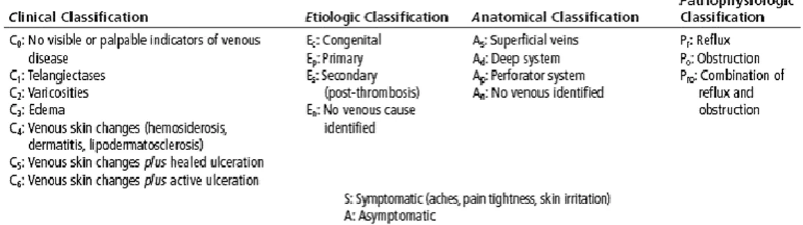
measurements are affected by the patient sociocultural context besides their aging, the disease perception and the presence of comorbidities, thus they might be considered a promising instrument halfway through the population epidemiologic characterization and the effective diagnosis of CVeD symptoms and signs. In this regard, the chances of mastering the management of venous disorders is also related to the opportunity of recognizing their hallmarks from the first appearance, by a thorough disease classification. The Clinical-Etiology-Anatomy-Pathophysiology classification (CEAP) lives up to this expectation, though it provides only a descriptive approach consistent with the original aim of refining the CVeD description and ensuring a straightforward
scientific and clinical communication. This remark enables to get to the heart of the CEAP classification, which defines the CVeD as a series of morphologic and functional venous disorders, rather than as disease, through its four above-mentioned points of view (Tab. 1.1.2)1,17. In particular, the first provides a clinical focus on the CVeD
manifestations by arranging them in seven worldwide adopted sequential classes (C0-C6) in order to explain the potential venous disorder progression from the absolute absence of disease signs (C0) to the appearance of the first structural and functional abnormalities, as telangiectasies and varicose veins (C1-C2), followed by the arising of chronic venous insufficiency corresponding to the development of edema, skin changes, eczema, lipodermatosclerosis and healed or active venous ulcers (C3-C6)17.
Tab. 1.1.2 The complete clinical, etiologic, anatomical and pathophysiologic (CEAP) classification of the CVeD. The table shows the clinical, etiological, anatomical and pathophysiological features of the CVeD
manifestations
However, it should be borne in mind that this progression through these worldwide adopted CEAP clinical classes representing the whole spectrum of CVeD is prone to different influencing factors, which might lead to arrest it in one of them. As above-anticipated, the etiological, anatomical and pathophysiological CEAP classification might be considered from three further points of view which enable to pin the CVeD down by specifying its primary or secondary aetiology, the exact affected venous system
(superficial, deep or perforator veins) and the pathologic events at the origin of the disorder (obstruction, reflux or other pathophysiologic processes) (Tab. 2)17.
Overall, these information doubtless contribute to build a reliable characterization of the clinical CVeD manifestations, but they are lacking in considering the appearance of some key symptoms and in pointing out the disorder severity1. This deficiency is often
filled by resorting to the venous severity scores (VSS), which are suitable to report also faint changes in disease symptoms and severity. However, their limitations, as the poor reliability of the patient-reported symptoms, the existence of comorbidities and surveys of CVeD not specific symptoms, make them complementary instruments with CEAP classification and QoL assessments in evaluating both the clinical picture and outcomes14. Although all these aforementioned diagnostic instruments are affected by
clinicians or patient disorder perceptions, they still provide the basis for a more thorough investigation of the venous system general state and potential functional disorders by more or less invasive diagnostic tools. The cornerstone in the CVeD symptom detection consists in the Duplex Ultrasonography (DUS) which offers the considerable advantage of observing the vein anatomy and the presence of valvular incompetence or venous obstructions by combining ultrasound imaging and pulsed wave Doppler. Its use has supplanted the outdated handheld Continuous Wave Doppler (CW Doppler) mainly due to its weak reliability in venous anatomical and hemodynamic impairment detection18,19. Furthermore, the DUS consists in a non-invasive diagnostic
approach and its high accuracy is increased by the frequent concurrent performance with colour flow imaging which quickens the visualization of also deep venous system incompetence20. These assets have led to overtake invasive diagnostic tools, as
phethysmography and air-plethysmography (APG) based on venous volume and pressure measurements, or the further imaging technique phlebography21,22. Moreover,
the DUS diagnostic results might be enhanced by the acquisition of three-dimensional vein structure processed by the alternative methods Computed Tomography (CT) and Magnetic Resonance Venography (MRV) in order to determine a careful CVeD diagnosis and choose the most suitable treatments23. This latter aspect, which represents the
desirable result of the whole diagnostic effort, might consist in different therapeutic options. In this regard, the compression therapy constitutes the undisputed gold-standard among the numerous conservative therapeutic strategies24. Its non-invasive
approach, consisting in the exercise of a controlled pressure on both lower limb superficial and deep venous systems, and its versatility are the effective advantages that allow it to work on the calf pump functionality to restore the physiological ambulatory pressure25,26,27. The main consequence of this mechanical action is the prevention or the
management of venous hypertension, which represents one of the first pathophysiological processes at the origin of the different CVeD manifestations. Numerous garments, as elastic stockings, tights, and elastic or non- bandages commercially available have physical properties able to provide a proper graduated or an intermittent pneumatic compression (Tab. 3)28,29. In particular, the stiffness, which
represents a direct correlation between the increase of pressure applied and the leg circumference increase (mmHg/cm), and the number of layers constituting the compression devices determine specific resting and walking pressure values suitable for treating the different CVeD manifestations26. Multilayer elastic compression stockings
recommended in the active treatment of varicose veins, lipodermatosclerosis and edema due to their ability to apply a pressure which decreases from the ankle up to the thigh and results higher during the walking than the resting position (Tab. 1.1.3)28,30,31.
Tab. 1.1.3 Pressure values (mmHg) and respective compression classes of stockings used in different world countries28. The table shows the pressure values applied by compression garments to a hypothetical cylindrical ankle; (1 mmHg = 1,333 hPa)
In this regard, the wanted effect is comparable with a strong massage on the calf during the patient deambulation able to reduce pain and swelling. These functional characteristics, based on the La Place’s law stating that the pressure applied around a cylinder is directly proportional to the radius of the cylinder one, make them useful also in maintenance treatment of lower limb healed ulcers and lymphoedema, even if elastic bandages are preferred in these cases25,32,33.
As previously mentioned, non-elastic bandages, as the Unna’s boot impregnated with zinc oxide paste or adjustable velcro straps, and intermittent pneumatic compression devices, obtained through inelastic cuff intermittently pumped up by a pump which produces different pressure degree in established time intervals, complete the range of the available compression approaches1,25. Furthermore, other non-invasive CVeD
treatments consist in leg physiotherapy, which seems to improve the calf muscle functionality by selected exercises, leg elevation, which is recommended especially in C2-C6 treatment by enhancing the microcirculation and reducing edema, and the leg massage, which is mainly aimed to ameliorate the tissue edema by making deep
massages around the ulcer area before wearing the elastic stockings or bandages34,35.
Nevertheless, a noteworthy common feature of these numerous CVeD non-invasive therapeutic approaches often consists in their concurrent use with those invasive. In this regard, a real cornerstone in the CVeD treatment is the well-known surgery. Different surgical strategies are developed over the time from the varicose vein ablation through the High Ligation with Stripping (HL/S) often associated with the ambulatory phlebectomy, the Ambulatory Selective Varices Ablation under Local anaesthesia (ASVAL), a less invasive phlebectomy technique aimed to preserve the undamaged segments of saphenous trunk, or the Cure conservatrice et Hémodynamique de l’Insuffisance Veineuse en Ambulatoire (CHIVA), improving the superficial venous system hemodynamics, up to the innovative Transilluminated Powered Phlebectomy (TIPP) consisting in large varicose vein cluster ablation with a considerably reduced number of incisions36,1. All these treatments are effectively followed by the
recommended wearing of compression devices with proper pressure levels in order to ameliorate the post-operative outcomes by giving relief from pain and other potential complications. Moreover, this represents the same clinical procedure followed after performing the superficial venous system treatment by the sclerotherapy37,38. This
consists in a therapeutic strategy for the chemical ablation of varicosities, venules or telangiectasias by the injection of liquid or foam sclerosant agents which damage the venous endothelial lining due to their close contact with it and induce the collagen and smooth muscle basal layer exposition accompanied by vasospasms and the treated vessel transformation in a fibrous cord39. However, it should not be neglected that this
chemical agents and the blood circulating cells which take away them and determine their dilution, also defined consumption40,41,42. This interaction is delayed by the more
efficient blood displacement induced by foam than liquid sclerosant by extending the treatment effects43. In fact, the chemical nature of the sclerosants and their physical
state (liquid or foam) result decisive for the treatment outcome44,45. The best known
sclerosant polidocanol and sodium tetradecyl sulphate have respectively non-ionic and anionic nature which determines their membrane solubilization and protein denaturing properties on circulating and non- components following their injection in the damaged vessel46,47. Furthermore, several studies have interestingly highlighted time-dependent
pro-coagulant effects at low concentrations of both the sclerosant agents unlike those anti-coagulant revealed at increased concentrations48,49,50. The sclerotherapy is only
one of a series of alternative and less invasive approaches developed for the saphenous vein incompetence treatment also including the Endovenous Laser Ablation (EVLA), the Endovenous Thermal Ablation (EVTA), the Radiofrequency Laser Ablation (RFA) and the more recent Mechanochemical Ablation (MOCA) and cyanoacrylate glue injection1,51,36.
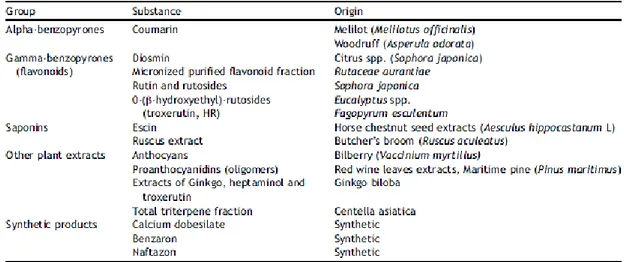
These strategies are equally aimed to induce endothelial damages through different percutaneous procedures by causing the treated vessel occlusion. Thus, all these treatment features might enable to set them halfway between the aforementioned invasive clinical approaches and the controversial pharmacological strategies. In this regard, drugs as alpha-benzopyrones (Coumarin), gamma-benzopyrones (flavonoids and their micronized purified fraction or MPFF), saponines (escin, ruscus extract), other plant extracts (anthocyans, proantocyanidins) and calcium dobesilate, benzarone,
naftazon are overall defined as natural or synthetic venoactive drugs (VAD), which differ from the nonvenoactive drugs (pentoxifylline, acetylsalicylic acid) (Tab. 1.1.4)52,53,13.
Tab. 1.1.4 Classification of the main venoactive drugs (VAD)52
The firsts are able to increase the venous tone also by exploiting the noradrenaline pathway, to reduce the venous permeability and the inflammation by counteracting the leucocyte-endothelium interactions, the second act by limiting the white cell and platelet activation with consequent anti-inflammatory effects. Furthermore, this action spectrum encompasses also the therapeutic effects of some glycosaminoglycans (e.g. Sulodexide) which contribute to restore the endothelial physiological functionality and to reduce inflammatory processes54,55,13. Clearly, all these effects make the medical
approaches fit for each CVeD stage treatment often in association with the compression therapy.
1.2 The structural and hemodynamic players of the Chronic Venous Disease
The occurrence of visible dilated blue or red/purple dermal veins, corresponding to reticular veins or telangiectasies (C1), with a smaller diameter (<4 mm) than the palpable and tortuous varicose veins (>4 mm) (C2) up to the unsightly skin changes and ulcerations represent some of the signs of the primary and secondary CVeD56,17,57. The
choice of drawing the attention to these CVeD clinical signs is consistent with the perspective of considering them mirror of break points in the structural and mechanical integrity of the intricate lower extremity vascular system. Three different venous components, the superficial, deep and perforating veins, provide the correct blood distribution in the supplied districts through their specific localization56,58. However,
their role runs down far from being simple and passive conduits since they have structural features actively related to the main hemodynamic events responsible for the physiologic drainage of lower limbs, besides for the blood return to the heart. In detail, the set of reticular veins lining the dermis, which accompany the Great Saphenous Vein (GSV) and the Small Saphenous Vein (SSV) with their numerous tributaries and a series of associated nerves, represent the abovementioned superficial venous system implicated in the skin microcirculation56,58,59. This function is also consistent with their
spatial organization in a superficial sub-compartment, known as saphenous compartment, in which the GSV and the SSV are confined between the saphenous and the muscular fascia56. The ascending GSV course starts from the dorsal pedal venous
arch up to the joining with the common femoral vein in the thigh through the saphenofemoral junction (SFJ) after crossing the medial malleolus and the tibia. Tributary veins, principally organized in the Leonardo’s arch, enable the GSV to drain
numerous ankle, tibial and anterior or posterior calf veins. Moreover, the GSV is also connected with the SSV and collects blood from the superficial external pudendal, epigastric and circumflex iliac veins by spilling it into the femoral vein. The small or short saphenous vein equally rises up from the dorsal pedal arch by passing behind the malleolus and proceeds posteriorly up to the calf, where it crosses the gastrocnemius muscles and joins the popliteal vein through the saphenopopliteal junction (SPJ)60,56.
The functionality of this intricate system of lower limb superficial veins is sustained by the equally complex arrangement of the deep vein system. They might be described according with their anatomical localization in foot, calf and thigh deep veins. In particular, the two plantar veins give origin to both the saphenous veins and are organized in a calcaneus plexus involved in the ambulatory pressure control besides in ejecting blood in the paired posterior tibial veins. These last with the corresponding anterior tibial veins, the peroneal, soleal and gastrocnemius veins consist in the deep calf venous system. The soleal and gastrocnemius veins, which are connected with the popliteal vein, give rise to muscular venous plexuses essential for the calf pump function. The tibial and soleal veins are usually connected with the peroneal veins to form the popliteal vein, which goes up the thigh and becomes the femoral vein after running into the thigh adductor magnus muscle. The popliteal and femoral veins represent a fundamental deep check-point in the preservation of the calf pump functionality through their valvular competence59. Furthermore, the femoral vein runs through most
of the thigh up to the inguinal region where it joins the deep femoral vein and the GSV to form the common femoral vein besides to accompany the femoral arteries and receive numerous muscle tributary veins. The common iliac vein, which takes origin
from the connection between the external (derivative of the common femoral vein) and the internal iliac vein, and the pelvic vein, which is organized in a series of venous plexuses interconnecting deep and superficial veins with pelvis, visceral and parietal districts, enrich the deep thigh venous system59,61.
The perforating veins exactly located in four anatomical districts, consisting in the foot, the medial and lateral calf and the thigh, represent “the glue” of the whole lower limb drain system by just favouring blood exchange between deep and superficial veins cited until now56,59. However, the collecting station of the whole blood circulation through
this venous network lies in the Inferior Vena Cava (IVC) which takes origin from the deep common iliac vein and gathers blood from lumbar, renal, inferior phrenic, right gonadal and hepatic veins before heading for the heart through the diaphragm and pericardium59.
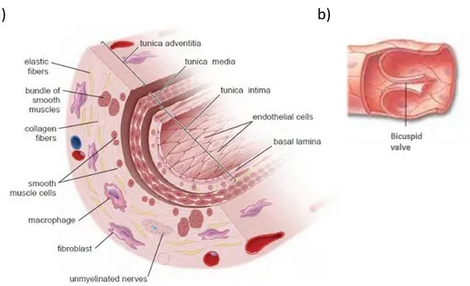
Structural and mechanical features of these intricate vascular systems represent the basis for the correct physiologic blood drainage in lower limbs and their competence ensures the awaited results of this transport process. In this regard, the valvular systems, variously distributed in the lower vascular architecture, have a noteworthy role in association with some essential pressure phenomena and compressive mechanisms mainly induced by muscles variously distributed in lower extremities. They consist in bicuspid and unidirectional venous valves showing half-moon-shaped cusps which are characterized by a thin collagen and endothelial layer consistent with the vein wall structural composition2,62. In particular, each cusp has the margin attached to the vein
wall which appears thicker in the point of contact than the opposite free (Fig. 1.2.1b). These valve components, whose length is strictly related to the respective venous
calibre, present two different sides named luminalis and parietalis. The first, consisting in the cusp side directly exposed to the lumen of the vessel and, thus, to the blood flow, is made up of an internal and fairly thick elastic layer covered by medial connective tissue and an external endothelial lining disposed along the vessel major axis, whereas the second, representing the cusp side facing the vein wall of the valve sinus, is characterized by a depleted connective and muscular layer covered by endothelial cells transversally stretched. The mechanical implication of this variable endothelial cell orientation accompanied by the presence of plentiful smooth muscle cells and elastic fibres in specific valve portions (e.g. the join between the valve cusps and the vein wall) consists in the acquisition of properties useful in tackling blood flow fluctuations62. In
this regard, the very existence of the venous valves and their open-and-closure mechanism passively regulated by a transvalvular pressure gradient determine the fragmentation of the blood column in multiple segments with controlled pressure1,57.
The reversal of this gradient triggers the valve closure after a physiological quick reflux (< 0.5 sec)59,63,64. These events occur continuously in each valvular system to generally
favour the blood drainage from the superficial to the deep venous system despite their variable distribution progressively less abundant by raising along the leg56. Nonetheless,
the competent valvular functionality is further sustained by the structural arrangement of the vein wall three layers which spread to the bicuspid valves (Fig. 1.2.1a). In particular, the most internal tunica intima is mainly characterized by the presence of endothelial cells which rest on their basement membrane followed by an intimal elastic lamina whose fibres stretch out in the valvular cusps accompanied by some muscular bundles. This thin monolayer, endowed with anti-thrombogenic or pro-coagulant
properties dependent on endothelium stimulations, is sequentially followed by the tunica media organized into three superimposed smooth muscle layers accompanied with scattered collagen and elastin fibres in addition to an adrenergic innervation65.
These consistent venous muscular and elastic components take part of the valvular portions subjected to the major mechanical stress in order to develop an appropriate resistance besides to prevent an excessive venous dilatation62. The last and thickest
venous wall tunica consists in the adventitia which is mainly composed of collagen fibres which sustain the just mentioned functional aim56.
a) b)
Fig. 1.2.1 Representation of the intimate venous wall and bicuspid valve structure. The figure in a) shows
the three overlapped layers (tunica intima, tunica media, tunica adventitia) and their cellular components; the figure in b) shows the structural organization of the venous bicuspid valve
Thus, the evidenced structural features should be counted as the mechanical requirements for providing both the unidirectional valve role of preventing the blood reflux occurrence and the physiologic vascular tone and compliance1,66. These last
aspects, closely related to the contractile ability of the muscular component and the adaptation of vascular capacitance to the blood flow entity, in addition to the valvular competence, the hydrostatic, ambulatory and dynamic pressures and the respiration contribution represent the main physiological forces influencing the venous blood return to the right atrium18,67. Although the respiratory airflow might seem the least
pertinent factor among those affecting the lower limb circulation, this involuntary act makes its indirect contribution to the physiologic process. In fact, the inspiration determines a consistent reduction of the thoracic cavity pressure accompanied by a consequent increased blood flow in the upper portion of trunk and the opposite effect on the abdominal pressure with a correspondent decrease in lower extremities blood outflow. The expiration reverses these anatomical and pressure adjustments leading to a heavy blood return from the lower body districts59,68. In line with the essential need to
provide the correct occurring of this last physiologic process, noteworthy assets might be also credited to the dynamic pressure. It directly depends on the heartbeat propagation firstly in the arteries followed by the other vascular systems with a progressive decrease related to different factors, as the arterial precapillary vasoconstriction (12-18mmHg in capillary venous side). However, this pressure is affected by the influence of the lower still atrial pressure (4-7mmHg), which induces the presence of an effective gradient favouring the blood return to the heart1. This flow
modulation, mainly predominant in supine position, is heavily influenced by the other two mentioned pressure systems. The ambulatory pressure, as its name implies, is closely related to the well-organized contractile activity of the principal foot, calf and thigh muscles during the daily ambulation. In this regard, it should be noted that the
functional competence of these real pumping systems also depends on the elliptical geometry typical of the calf and foot veins which enhances the venous capacitance by minimizing the effect of blood volume and pressure changes. Their flaccid appearance at low pressure value alternates with more circular sectional geometry consistent with an increased blood volume are a representative confirm of this their property59.
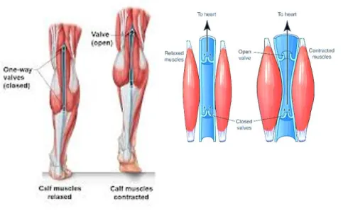
Therefore, the high capacitance and low resistance of vessels and the prevalent activity of the muscular component make the calf pump the main driving force of the blood return to the heart (Fig. 1.2.2)69. Its ejection fraction (EF) of about 65% is tightly
associated with the ambulatory pressure, which still consists in a gradient involving the thigh and the distal leg districts and exactly generated by the calf muscle activity70. In
particular, the contraction determines the increase of venous pressure and the consequent centripetal blood expulsion from the deep popliteal and femoral veins to the superficial GSV (pressure of about 140 mmHg), whereas induces a pressure fall around 25-30 mmHg in the lower leg and foot. The calf pump relaxation reverses this effect with a consequent centrifugal blood flow directed from the GSV, characterized by high pressure, to deep venous system56,69.
Fig. 1.2.2 Representation of the venous calf pump. The figure schematically shows the structural
organization of the calf pump muscles and the mechanical effects induced on the retrograde blood return
The very existence of this horizontal flow depends on the venous perforating system, which spreads the pressure changes between the other two venous systems due to its medial position. Furthermore, the physiologic muscular activity of the calf pump is also able to indirectly affect the retrograde blood return to the right atrium by influencing the hydrostatic pressure71. This consists in an additional blood pressure component
arisen from the gravitation force effects on the lower limb veins during the quiet standing position. Interestingly, it can be defined as the vertical distance between the heart, which corresponds to the pressure zero line, and a specific body anatomical district by depending on the subject height. The hydrostatic pressure generally increases of about 0.8mmHg/cm as proceeding from the right atrium to the ankle, where it amounts to 80-100mmHg67. Although it is not subjected to variations both in deep and
superficial venous systems at the resting position, however the ambulation induces the calf pump activation, which determines the drop of the high hydrostatic pressure to
about 25-30mmHg in the calf and foot during its muscular contraction. The pump relaxation causes a slow pressure increase to the initial value instead67.
On the basis of this general focus on the roles of each dynamic variable involved in the regulation of the physiological blood circulation in distal leg and the vital retrograde blood flow, there needs to point out the essentiality of the valvular competence as common denominator18. In this regard, the previously hinted open-and-closure
mechanism of the venous valve leaflets (about 20 times/min in standing position) determines a pulsatile blood flow which is characterized by two dynamic components consisting in the directed and the vortical flows. The first is mainly sustained by the ambulation effects on foot and calf muscle functionality, which accelerate the flow by reducing the pressure applied on the luminal side of the valve cusps, while the second is consequent to the previous by preventing the blood stasis in the valvular parietalis side and favouring its exposition to the shear stress effects (Fig. 1.2.3a)72. Furthermore,
the low pressure of the blood directed jet compared to that of the vortical flow is essential to determine the valve leaflets closure, despite the appearance of the previously mentioned quick reflux57.
b)
Fig. 1.2.3 Schematic representation of the shear stress change effects57. The figure in a) shows the laminar and steady shear stress effects ensuring the endothelium and venous wall integrity; the figure in
b) shows the venous wall damages with specific secretive responses to the low and irregular shear stress
The shear stress is a further physiological consequence of the fluid dynamics in the blood vessels73. In effects, its relevance might be attributed both to the direct contact with the
endothelial glycocalyx, which transduces its laminar or low and irregular turn by triggering different cellular response pathways, and to the high leucocyte responsiveness to its variations (Fig. 1.2.3a,b)74,75. These additional details enable to
understand the real significance of venous valve functional impairment often associated to leaflet structural damages or vein wall alterations (e.g. hypertrophic region with dysregulated collagen synthesis, smooth muscle and elastin fibres scatter)57,76.
Consequently, incompetent venous valves become unable to fragment the blood column and to tackle its high pressure induced by muscle pump activity in deep venous system by abolishing the two opposite poles of the previously said pressure gradients. The immediate result of the knock-on effect sparked off by these last functional and structural disturbances, which are reflected in hemodynamic perturbation, consists in hypertension development characterizing the different CVeD manifestations76,77.
1.3 The cellular and biochemical players of the Chronic Venous Disease
Despite controversial opinions about the right sequence between the venous valve and wall impairment occurrence have followed one another over the years, it might be intuitive thinking that a series of preliminary alterations of the vein wall structure induce vessel dilation and, thus, valve closure failure. The extracellular matrix (ECM) depletion in laminin and elastin concentration accompanied by compromised deposition of collagen by the vascular smooth muscle cells and fibroblasts are effectively the structural premises of venous hypertension52,57,78. In this regard, the pointed-out
imbalance between the collagen I and III ratio in favour of the first seems to be coherent with the venous loss of elasticity and distensibility characterizing this pathological condition79. Hence, the vein walls weak and stiff become unable to handle the whole
spectrum of physiological pressure and hemodynamic fluctuations with the mechanical breakdown of the valvular unidirectional checkpoints. The consequent abolition of the previously discussed pressure gradients and the general increase of pressure in all districts of lower extremities force to shift the focus on their effects on the venous endothelial layer. This acts as a sort of condenser capable of gathering the physiological mechanical stress induced by the very blood flow with all its dynamic components and transducing them in proliferative, secretive and transcriptional stimuli in the nearby cells79,80. However, the real transducer is the glycocalyx consisting in some
glycosaminoglycans (e.g. heparan and chondroitin sulphate, hyaluronan), proteoglycans and glycoproteins which line the luminal side of the vascular endothelial layer73. It
represents an effective interface between the shear stress effects and the overhanging cellular lining, besides a selective permeability barrier able to provide the positive
regulation of coagulation and fibrinolysis processes and the necessary hindrances to the preliminary inflammatory events81. For all these reasons, the venous wall homeostasis
can be guaranteed until the glycocalyx integrity is preserved. In fact, the decrease of shear stress, induced by the hemodynamic alterations characterizing the venous hypertension, firstly results in a dropped synthesis of glycocalyx components by the vascular endothelial cells73,82. Thus, the depletion of this functional interface consists in
the starting point of a vicious circle of maladaptive events favouring the hypertension perpetuation in the different CVeD manifestations. The endothelial cells, directly exposed to the widespread hemodynamic dysregulation, strengthen the current pronounced venous dilatation by the increased synthesis of nitric oxide (NO) through the stimulation of the inducible nitric oxide synthase (iNOS) rather than of the constitutive and Ca2+-dependent endothelial enzyme isoform (eNOS). In effect, the
increased release of NO, prostacyclin (PGI2), endothelin-1 (ET-1) and endothelium-derived hyperpolarizing factor (EDHF) is coherent with the overbalanced vasodilation compared to the vasoconstriction mainly revealed in warped varicose vein segments (C2)57,79. The correlated venous distension is also sustained by the smooth muscle cell
relaxation induced by the enhanced release of acetylcholine as further consequence of shear stress alteration79. Furthermore, the NO overexpression appears increasingly
related to the interesting vascular distension mechanism sustained by the up-regulated matrix metalloproteinases (MMP)83,84. In fact, these enzymes are able to favour the
hyperpolarization of the varicose vein vascular smooth muscle cells by involving some protease-activated receptors upregulated in endothelial and muscle cells and platelets, besides an hyperpolarizing factor and the hypoxiainduced transcription factor1A and
-2A (HIF-1A and HIF--2A), which inhibit the cellular Ca2+-intake to reduce the
contraction85,86.
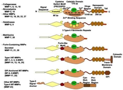
Of note, the high responsiveness of MMPs to the hemodynamic impairments and their extensive involvement in the numerous vascular remodelling processes mirror their variable expression by the different cellular components of the venous wall. They consist in a superfamily of 26 zinc-dependent endopeptidases (only 23 expressed in human) whichare synthesized as pre-pro-MMPs, although the signal peptide is lost during the translational process (Tab. 1.3.1)87,88. They share three constant structural features
consisting in the sequence homology with the collagenase 1 or MMP-1, the pro-domain cysteine switch motif PRCGXPD essential to maintaining the enzyme inactive by the interaction with the Zn2+ ion of the catalytic domain, and the Zn2+-binding motif
characterized by three histidine residues in the conserved sequence HEXGHXXGXXH, a conserved glutamate and a methionine residue placed in a downstream sequence (XBMX) from the catalytic domain and essential as support of its structure89. In addition,
the repeated type II fibronectin domain, the linker-domain located between the catalytic site and the hemopexin domain and this last are differently distributed among the family members (Fig, 1.3.1).
Fig. 1.3.1 Schematic representation of MMP structure89. The figure represents the main MMP domains consisting in the pro-peptide domain characterized by the cysteine switch motif PRCGXPD and associated with the signal sequence, the catalytic domain containing the Zn2+-binding sequence HEXGHXXGXXH and two or three Ca2+ ions with stabilisation function, the linker domain and the hemopexin domain; three type II fibronectin repeats in the catalytic site are showed in the gelatinase structure. Further alternative structures are associated to other MMP family members
These are usually set in six different groups on the basis of their substrates, including the collagenases MMP-1, -8 and -13 which, as their name suggests, mainly interact with the collagen I, II and III by releasing fragments of variable length; the gelatinase-A or MMP-2 and -B or MMP-9 cleaving the gelatine (consisting in denatured collagen), collagen and laminin through their repeated fibronectin II domain in the catalytic site; the stromelysin-1 or MMP-3 characterized by more efficient proteolytic activity on different collagen types, other ECM structural components (elastin, fibronectin, laminin) and numerous proteins than the stromelysin-2 or MMP-10; the matrilysin-1 or MMP-7 and -2 or MMP-26, which lack hemopexin domain and interact with limited collagen types (collagen IV and X) and further non-ECM substrates; the membrane-type MMPs
(MT-MMPs) consisting in the type-I transmembrane proteins MT1-MMP (MMP-14), which mainly digests collagen I, II and III and is involved in the proMMP-2 activation, and the MT2-, MT3- and MT4-MMP (MMP-15, -16, -24) accompanied by the glycosylphospatidylinositol (GPI)-anchored proteins MT5- and MT6-MMP (MMP-17, MMP-25); and the remaining stromelysin-3 (MMP-11) presenting different sequence and substrate specificity compared to the MMP-3, the metalloelastase (MMP-12) cleaving the elastin and other ECM structural components, and the MMP19, 20 , 22, -23, -28, some of which characterized by different domain organization or recently identified, are just defined as the other MMPs (Tab. 1.3.1; Fig. 1.3.1)86,90. Additional
metalloproteinase families consisting in ADAMs (A Disintegrin And Metalloproteinases) and ADAMTS (A Disintegrin and Metalloproteinase with Thrombospondin motifs), which are similarly known to exercise their Zn2+-dependent catalytic activity on collagen and
Tab. 1.3.1 The MMP family members86. The table summarizes the MMP family members associated with their tissue expression and their different substrates.
The passage from the pro- to the active MMP is marked by the interruption of Cys-Zn2+
coordination and the removal of the hemopexin domain representing the starting point of the MMP proteolytic activition94. The enzyme is then ready to interact with its
substrate through the Zn2+ ion which gives rise to a nucleophilic attack mediated by a
carbonyl oxygen atom of the substrate by releasing a water molecule. This process is primarily due to the presence of a water molecule accompanying the conserved glutamate residue, which determines the penta-coordination of the catalytic Zn2+ ion
with the three histidine, methionine and substrate oxygen atoms. Furthermore, the MMP-11, -21 and -28 as well as the MT-MMPs contain a furin domain comprised between the pro-peptide and the catalytic site, which is intracellularly cleaved by the endopeptidase furin following the recognition of a specific sequence in C-terminus of their pro-peptide89,95. These pro-MMP activation mechanisms can involve either
enzymatic family members, as demonstrated by the MMP-3 and all the MT-MMPs (with the only exclusion of the MT4-MMP), or different treatments including heat, pH decrease and chemical agents (4-aminophenylmercuric acetate, mercury chloride, sodium dodecyl sulphate, reactive oxygen species) by determining structural interferences86,96. Interestingly, the proMMP-2 activation requires the concurrent
involvement of the MT1-MMP (MMP-14) and the tissue inhibitor of matrix metalloproteinases 2 (TIMP-2) on the cell membrane. The TIMP-2-proMMP-2 complex involving the C-terminal domains can interact through the TIMP-2 N-terminal domain with the MT1-MMP anchored on the cell surface. Thus, the pro-MMP2 bound to the
membrane might become substrate for another MT1-MMP molecule, which determines its activation. However, it might also occur an early interaction between TIMP-2 and MT1-MMP on the cell surface, which inhibited binds the hemopexin domain of proMMP2 to expose it to a free MT1-MMP which performs the activation84,86,95. The
mention of this TIMP-2 function allows to make remarks on the other three tissue MMP inhibitors TIMP-1, -3 and -4. Each one is characterized by a C-terminal domain and a N-terminal domain responsible for the MMP inhibition through the direct interaction with the protease catalytic site97,98. TIMPs are specifically distributed among the different
venous wall layers, where exercise their activity on all MMPs, despite some peculiarities consisting in the TIMP-1 lack of MT1-MMP interaction, the just mentioned TIMP-2 role in the MMP-2 activation, the TIMP-3 ADAM inhibition and the TIMP-4 activity mainly localized in cardiovascular systems. Moreover, they are endowed with additional functions, as the pro-angiogenic TIMP-1 properties, the endothelial cell proliferation inhibition by TIMP-2 and TIMP-3, which may prevent cellular migration and angiogenesis by interacting with VEGF receptor86,99. Further endogenous factors able to interfere
with the MMP and other different endopeptidase catalytic activity are the α2
-macroglobulins provided of four identical subunits with Zn2+-binding domain
fundamental to their function. In particular, they determine MMP-complex formation which is degraded by endocytosis mechanisms89.
This overall picture enables to understand both the modulation of MMP targeted activity on the main ECM components (e.g. collagens, elastin, fibronectin, vitronectin, tenascin, laminin), which explains the intensive involvement in the physiologic and pathologic venous wall remodelling, and the protease further interactions with factors,
which differentially determine the CVeD functional impairments100,101,102. In these
functions as well as in the cell proliferation, migration and differentiation control, the MMP/TIMP balance is an essential requirement to provide the venous physiological homeostasis. In fact, the loss of this mutual counterbalance and the significant venous pressure increase effects on MMP transcriptional and post-translational processes might be considered determinant in the appearance of both hypertrophic and atrophic segments of varicose veins (C2)103,104. Interestingly, this evidence is confirmed by the
pressure stress on vascular endothelial and smooth muscle cells, which induces them to synthesize large amount of MMP-2 following the transcription factor activator protein-1 (AP-protein-1) activation by the reactive oxygen species (ROS) release96. This event is
exacerbated by the endothelial mechanical and inflammatory activation, which determines an hyperexpression of the NADP(H) oxidases79. A similar MMP-2
concentration increase is also associated to other CVeD manifestations including hyperpigmentation(C4a), lipodermatosclerosis or atrophie blanche (C4b). They share a
common hypoxic microenvironment accompanied by a huge oxidative stress and hemosiderin deposition, which enhance the MMP and the vascular endothelial growth factor (VEGF) expression by determining the appearance of typical clinical signs 57,105. In
addition to the 2 widely present in all venous wall layers, the increase of the MMP-1, mainly synthesized by fibroblasts, endothelial cells and smooth muscle cells, and the MMP-9, highly expressed by endothelial and muscle cells, influences the proliferation and migration of venous smooth muscle cells in varicose veins (Tab. 1.3.1)106. In this
regard, the proteolytic degradation of the ECM components is one of the immediate structural assets offered to the varicose vein dedifferentiated muscle cells, which are
then induced to migrate. The removal of some adhesion sites and the exposition of binding sites for different factors enhanced by the MMP increased secretion potentiate the migration process79. This mechanical implication is accompanied by an effective
smooth muscle cell phenotype switch from contractile to proliferative and secretive which materializes through increased ECM-muscle cell interactions and the release of growth factors in the hypertrophic regions of varicose veins65. Furthermore, MMP-3, -7
and -13, equally up-regulated in this pathological condition, might contribute to the venous wall weakening by determining the ECM component degradation. Of note, the decreased TIMP release and the consequent uncontrolled MMP-3 proteolytic activity are responsible of some collagen III post-translational modifications inducing its deposition imbalance with the collagen I in thinned and tortuous atrophic regions of varicose veins79,84,107. Bearing in mind all these MMP proteolytic activity implications and
the wide cellular expression of these enzymes in the vascular system, it might result expectable to associate their involvement also with the other CVeD stages likewise characterized by ECM alterations and diffused inflammation. In effects, this condition is further sustained by the MMP effects on the activated endothelial cells interacting with similarly activated leucocytes and the modulation of some inflammatory mediators, as cytokines, chemokines and growth factors108. In this regard, one of the most
representative process might consist in the sequence of the four wound healing time-dependent phases (hemostasis, inflammation, granulation and re-epithelialization). The initial inflammatory events and the ECM turnover, facilitating keratinocyte and fibroblast migration, pave the way to the progressive granulation tissue formation and the angiogenesis stimulation by leading to the endothelial basement membrane and
skin barrier restoration109,110. The matrilysin MMP-7, expressed by vascular endothelial
and muscle cells, takes part to this process through both the activation of the latent tumor necrosis factor-α (TNF-α) stimulating chemoattractant mechanisms and macrophage infiltration, and the alterations of some cell-cell and ECM-cell interactions to promote cellular migration107. Similarly, the MMP-12, mainly secreted by
macrophages (Tab. 1.3.1), might be involved in the inflammatory processes by triggering the TNF-α activation, besides in the reparative phases due to its ECM elastin, fibronectin, collagen VI and laminin degradation111,112,113. The restoration of the tissue integrity in
the final phases of the wound closure is also favoured by the up-regulation of the collagenases MMP-1, MMP-8 and MMP-13 which are expressed in endothelial cells, smooth muscle cells and fibroblasts and are determinant mainly in promoting the proliferation and migration of these last during the ECM remodelling (Tab. 1.3.1)114,115.
An undebated role in the prolonged inflammatory state is attributed to increased concentrations of the MMP-2 and MMP-9 during the healing process, although their proteolytic activity are strictly moderated by their inhibitors TIMP-1 and TIMP-299,116,117.
Thus, their loss might induce the reduction of the angiogenesis and the impairment in the correct ECM and tissue deposition characterizing the wound chronicity.
Furthermore, as reported on several occasions during the chapter, the switch from a laminar and pulsatile to an irregular and low shear stress is comparable to squeezing a trigger which sparks off self-reinforcing inflammatory events, besides structural impairments (Fig. 1.2.3b). In effects, this hemodynamic force acts as a physiological modulator of the circulating leucocytes, which undergo their superficial integrin CD11b/CD18 (Mac-I) proteolytic shedding and cytoskeleton F-actin depolymerization
with consequent pseudopod retraction, if exposed to a laminar blood flow. The shear stress drop is then a key event in determining the leucocyte recruitment on the vascular endothelial layer through the integrin increased expression, the pseudopod sprouting and the loss of the cellular spherical shape to give rise to contact regions118,119,120.
Although the integrin CD11b/CD18 up-regulation is an effective requirement to the leucocyte migration and subsequent penetration in the vascular endothelial layer, however further adhesion molecules, as the endothelial E-selectin, the intercellular adhesion molecule-1 (ICAM-1), the vascular adhesion molecule-1 (VCAM-1), and the leucocyte L-selectin, the lymphocyte function-associated antigen-1 (LFA-1) and the very late activation antigen-4 (VLA-4), are determinant in this respect57,110,121. Interestingly,
the reduced expression of the CD11b/CD18 on leucocyte cellular surface and the correspondent increase of soluble L-selectin plasmatic levels might be representative of cellular migration into the endothelium110,79. The generalized activation state of the
endothelium is also prompted by the shear stress-induced glycocalyx damages, which unmask the adhesion molecules promoting leucocyte docking and inflammation advance57. These events are further exacerbated by the previously mentioned sharp
reduction of endothelial NO synthesis by the eNOS, which physiologically plays an essential anti-inflammatory role, besides inhibiting smooth muscle cell proliferation and stimulating their relaxation. The nitric oxide depletion is counterbalanced by the endothelial increased levels of the pro-inflammatory agent angiotensin II (AngII), which stimulates the ROS production and the adhesion molecule and inflammatory cytokine release (Fig. 1.2.3b)57,122. Furthermore, the compromised and activated endothelium
activator inhibitor-1 (PAI-1), factor VIII (FVIII) and Von Willebrand factor (Vwf), which might be considered endothelial disfunction markers. The high concentration of these last along with D-dimer, IL-6, IL-8, monocyte chemoattractant protein 1 (MCP-1) and C reactive protein (CRP) revealed in the lower limb circulating blood might be associated with a fibrinolytic profile characterizing the CVeD79. The presence of these just
mentioned cytokines mirrors the heavy secretion of inflammatory mediators which act as constant background of the whole spectrum of the hemodynamic and structural impairments approached so far. In this regard, the cytokines consist in a multifaceted family of glycoproteins and peptides able to induce their effect on the basis of the timing and the environment of their release, the potential presence of inhibitor or synergistic factors, the cellular distribution of their receptors and the balance between inflammatory and anti-inflammatory family members (Tab. 1.3.2). Cytokines as, interleukin (IL) -4, -10, -13, interferon-α (IFN-α) and variously the transforming growth factor-β (TGF-β) are known to counteract inflammatory reactions induced by tumour necrosis factor-α (TNF-α), other interleukins as IL-1, -6, -8, -11, -12, -19, IFN-β and IFN-γ, TGF-β, macrophage inflammatory protein-1α and -1β (MIP-1α, MIP-1β), the regulated on activation, normal T-expressed and secreted (RANTES), the platelet factor-4 (PF-4) and the monocyte chemoattractant protein-1, -2, -3 (MCP-1, -2, -3)122. This initial
distinction, manly based on the general functional roles of cytokines, enables to refer to their specific organization in subfamilies including the numerous interleukins, the tumour necrosis factors (TNF-α and -β), the interferons (IFN-α, -β, -γ), the colony stimulating factors (CSF) distinct in granulocyte (G-CSF), monocyte (M-CSF) and granulocyte-monocyte (GM-CSF), the transforming growth factors (TGF-β1, - β2, -β3)
accompanied by the bone morphogenetic proteins (BMPs), the activins and the inhibins, the chemokines (IL-8/CXCL8, PF-4/CXCL4, MIP-1α/CCL3, MIP-1β/CCL4, RANTES/CCL5, MCP-1/CCL2, MIP-2/CCL8, MIP-3, MIP-4), and other members(Tab. 1.3.2).
Tab 1.3.2 Cytokine, chemokine and growth factor family members122. The table summarizes some of the most representative cytokines, chemokines and growth factors and their corresponding cellular sources and targets, receptors and functions
Interestingly, the chemokines, so named in reference to their chemotactic properties, are distinct in further 4 groups characterized by the presence or not of a conserved cysteine (C) residue in the N-terminus accompanied by a variable region (X). These consist in XC group, whose members (XCL1, XCL2) are involved in lymphocyte chemoattraction, CC group acting on monocytes, CXC group including ELR+-CXC
members with a N-terminus sequence glutamic acid-leucine-arginine (ELR motif), which interact with neutrophils, and ELR--CXC members lacking in the ELR sequence and mainly
acting on lymphocytes, and CX3C group including the fractalkine122,123. The pleiotropic
cellular types is consistent with their variable release by macrophages and T cells, which synthesize different inflammatory and anti-inflammatory interleukins (IL-1, -6, -10, -12) besides TGF-β, TNF-α, IFN-γ and some chemokines (MCP-1/CCL2, MCP-4/CCL13, IL-8/CXCL8), platelets, which store in their secretion granules numerous cytokines and chemokines (IL-1β, PF4/CXCL4, MIP-1/CCL3, RANTES/CCL5), and other non-inflammatory cells as the vascular endothelial and smooth muscle cells secreting IL-1α, IL-1β and TNF-α (Tab. 1.3.2). However, the efficacy of their biological functions is strongly tied to the interaction with specific receptors which often represent the starting point of an activation cascade culminating in the synthesis of new products or in mechanisms of transcriptional modulation. Four receptor structures are located on the target cellular surface to mediate cytokine effects by binding them through their extracellular domains. In particular, the hematopoietin receptor, which is characterized by a dimeric or trimeric structure with some conserved cysteine residues in the extracellular domain, interacts with different interleukins, as IL-2 and IL-7, and with the GM-CSF, the IFN receptors equally presenting some conserved cysteine residues, the TNF receptors, and the chemokine receptors which are G-protein coupled and typically characterized by seven transmembrane domains122. Numerous ILs, IFNs and CSFs,
binding their aforementioned receptors, give rise to their dimerization activating the JAK-STAT signalling pathway, which involves the Janus kinases (JAK) and the signal transducer and activator of transcription proteins (STAT) recruitment. Alternatively, the inflammatory IL-1 and IL-18 with the TNFs exploit the NF-kB signalling to influence the expression of some adhesion molecules (E-selectin, VCAM-1, ICAM-1), the iNOS and some MMPs besides other cytokines and growth factors determinant in the
inflammatory process. Instead, the TGF-βs induce numerous endothelial alterations and inflammation mediator expression through the Smad-signalling pathway (Tab. 1.3.2). The existence of strict regulation systems, as specific inhibitors or phosphatases, is required to ensure the appropriate balancing of these transductional mechanisms. In this regard, the increased concentrations of the basic fibroblast growth factor (bFGF) in the activated endothelium as well as of the TNF-α, IFN-γ, CSFs, MCPs, different interleukins and growth factors are evidence of these mediator negative feedback system disturbances favouring the uncontrolled inflammatory state which characterizes numerous CVeD manifestation. In effects, the association of TGF-β1 enhanced expression with the varicose veins (C2) is consistent with its ability to stimulate elastin and collagen synthesis and to down-regulate the TIMP expression to determine the stiffening of the vein walls57,124. The role of TGF-β1 in promoting the ECM remodelling
and cellular migration is also compatible with the early phases of the wound healing process, in which the reorganization of vascular basement membrane and some angiogenic stimulator release occur109. Furthermore, the pro-inflammatory cytokine
induces the production of bFGF exercising its mitogenic function principally on fibroblasts and vascular smooth muscle cells in varicose veins. For their part, muscle cells are stimulated to produce IL-6 and the chemokine MCP-1 in presence of thrombin by promoting monocyte recruitment122. Furthermore, TGF-β1, as well as TNF-α and IFN-γ,
seems to be involved in the up-regulation of the iNOS especially in the varicose vein tortuous segments122,125. These cytokines are also correlated with the endothelial
increased permeability, which characterizes the whole CVeD. In particular, TNF-α determines a loosening of the endothelial junctions by inducing the up-regulation of the
vascular endothelial growth factor (VEGF), bFGF, IL-1α and IL-6, which stimulate the MMPs expression, and iNOS which increases the NO levels79,122,126. This structural
alteration is detectable from the first CVeD sign appearance sustained by a pronounced venous hypertension and progressively worsened by the extravasation of some macromolecules, as fibrinogen, red blood cell degradation fragments and proteins stimulating leucocyte migration and trapping in the fibrin cuff meanwhile accumulated in the interstitial space57,109. The consequent edema and the hemosiderosis occurrence
generate a microenvironment heavily hypoxic which represents an undebated stimulator factor for MMP, pro-inflammatory cytokine and growth factor synthesis characterizing the different CVI manifestations105,106,127. The same TNF-α induced by the
urokinase-type plasminogen activator (u-PA) and accompanied by IL-17 and -19 seems to enhance the MMP-9 expression in varicose veins 79,122. However, a similar effect is
produced by TNF-α and IL-1β on MMP-1, -2, -3, -9, -13 and MT1-MMP to maintain the inflammatory state in chronic wound healing. The up-regulation of MMPs is in line with the general chemotactic role of the IL-8, which is also able to stimulate angiogenesis, besides the proliferation and migration of keratinocytes during the wound closure128.
Differently, the inflammatory events are moderated by IL-10, which prevents the endothelial cell-leucocyte interactions and inhibits the pro-inflammatory cytokine release. High expression of this interleukin is consistent with the containing of the inflammatory phase to determine the tissue restoration during the healing process122,112,126. Overall, an interesting implication of the activation of this complex
biochemical machine regulated by effective chain reactions involving different cellular types and numerous mediators consists in its stimulation of nociceptors located
between the endothelial and smooth muscle cells of the venous wall by determining the onset of pain52.
Certainly, this last detail as well as the different aspects approached until now is consistent with the aim of this introductive focus on CVeD to provide instruments suitable to properly contextualising the implications of the evidences which are presenting.