UNIVERSITÀ DEGLI STUDI DELLA TUSCIA DI VITERBO
DIPARTIMENTO DI SCIENZE MMFFNN E AGRARIA
CORSO DI DOTTORATO DI RICERCA IN BIOTECNOLOGIE VEGETALI - XXII Ciclo
BIO/04
IMPROVING THE NUTRIENT COMPOSITION OF
CHLAMYDOMONAS REINHARDTII TO ENHANCE THE PRODUCTION OF
PHOTOSYNTHETIC ACTIVE METABOLITES: A MOLECULAR STUDY ON THE PHOTOSYSTEM II COMPLEX.
Coordinatore: Prof. Stefania Masci
Tutor: Dott. Giuseppina Rea
I
1.1. The oxygenic photosynthesis ... 1
1.1.2. Main features of the structure and function of PSII ... 1
1.1.3 Photosynthetic pigments. Chlorophylls and carotenoids... 3
1.1.4 Carotenoids and plastoquinone biosynthesis... 5
1.1.5 The PSII D1 core protein... 9
1.1.6 Effect of stressful light and temperature conditions on thylakoid structure... 11
1.1.7 Adaptation response of photosynthetic apparatus to high light condition ... 13
1.2 Correlation between human health and photosynthetic compounds ... 15
1.2.1 Applications of plant-derived phytochemicals... 17
1.2.2 Carotenoid benefits for human health ... 19
1.2.3 Enrichment of nutraceutical contents in foodstuff ... 22
1.3 Chlamydomonas reinhardtii as biological farm ... 24
1.4 Chlamydomonas reinhardtii mutants ... 27
1.4.1 C. reinhardtii NPQ mutants ... 27
1.4.2 C. reinhardtii D1 mutants... 30
2. PROJECT AIM ... 31
3 MATERIAL AND METHODS ... 32
3.1 Growth conditions and cell count... 32
3.2 Chlamydomonas reinhardtii strains ... 32
3.2.1 D1 strains... 32
3.2.2 NPQ strains... 33
3.3 Antenna size determination ... 33
3.4 Fluorescence measurements ... 34
3.4.1 Chlorophyll fluorescence... 34
3.4.2 Oxygen evolution analyses... 34
3.5 Quantitative and qualitative analyses of photosynthetic pigments... 35
3.5.1 Pigment standards and HPLC system... 35
3.5.2 Carotenoids and chlorophylls extraction ... 35
3.5.3 Pigments analyses... 36
3.6 Chlamydomonas nucleic acids purification... 36
3.6.1 Genomic DNA purification ... 36
3.6.2 RNA extraction and cDNA synthesis... 36
II
3.8 Polymerase chain reaction (PCR)... 38
3.8.1 Oligonucleotides design ... 38
3.8.2 PCR standard conditions ... 38
3.8.3 RNA-Retrotranscription (RT) and Real-Time PCR (RT-PCR) ... 39
3.9 Elicitation experiments: high light and high temperature treatments... 40
4. RESULTS AND DISCUSSION... 41
4.1 Selection of the strains and experimental set up... 41
4.2 Characterization of C. reinhardtii strains under physiological conditions... 45
4.2.1 Growth curve ... 45
4.2.2 Chlorophyll fluorescence... 47
4.2.3 Oxygen evolution capacity ... 50
4.2.4 PSII antenna size ... 52
4.2.5 Accumulation of photosynthetic antioxidant pigments... 54
4.3 Elicitation strategies to improve antioxidant photosynthetic pigment accumulation... 56
4.3.1 Superimposition of high light and high temperature on D1 strains... 56
4.3.2 HL/HT effects on the chlorophyll content and PSII photochemistry in D1 strains ... 58
4.3.3 Antioxidant photosynthetic pigment accumulation during HL/HT treatment on D1 strains ... 59
4.4 Comparative analyses of time-course gene expression profiles in chlamy-domonas mutants by Real-Time RT PCR in response to high light and high temperature treatments... 61
4.4.1 Expression analyses of psbA and psbD genes in D1 mutants in response to high light and high temperature treatments... 63
4.4.2 Expression analyses of genes involved in carotenogenisis in D1 mutant strains in response to high light and high temperature treatments... 65
4.4.3 Expression analyses of genes involved in plastoquinone biosynthetic pathway in D1 mutant strains in response to high light and high temperature treatments... 69
4.5 C. reinhardtii as biological farm ... 71
5. CONCLUSIONS ... 75
1 1. INTRODUCTION
1.1. The oxygenic photosynthesis
The oxygenic photosynthesis is the most important chemical process in biology by which plants, algae, and some bacteria and protistans, harvest light and convert it into readily utilizable energy (Kargul and Barber, 2008). In oxygenic photosynthesis, water is used as electron donor to reduce CO2 to carbohydrates, generating molecular oxygen as a secondary
product of the reaction. The whole process can be separated in a light dependent and a light independent reactions. Light reactions are triggered by the absorption of sunlight by photosynthetic pigments (chlorophylls and carotenoids) and are catalyzed by two separate macromolecular complexes: photosystem II (PSII) and photosystem I (PSI). A light-induced electron flow generates reducing equivalents (NADPH2) and the proton motive force that
drives the production of ATP (Mitchell, 1961). Oxygen is released by the oxygen evolving complex (OEC) that is constituted by proteins and a cluster of manganese ions. It is located on the lumenal side of the thylakoid membrane and it is associated to both the reaction centre and the cytochrome-b6f complex, a dimeric integral membrane protein complex that mediates
electron transport between PSII and PSI (Figure 1).
In photosynthetic eukaryotes, both light and dark reactions take place in a specialised organelle called chloroplast.
1.1.2. Main features of the structure and function of PSII
PSII is a multisubunit chlorophyll–protein complex that drives electron transfer from water to plastoquinone using energy derived from light. PSII is embedded into the thylakoid membranes as well as all the complexes that catalyze the light reactions. Its native form is surrounded by the light-harvesting complex (LHCII complex), consisting of more than 30 proteins and thus called the PSII–LHCII super complex (Figure 1).
At the centre of this complex is the reaction centre (RC), which is composed of the D1 and D2 heterodimer (encoded by the chloroplast psbA and psbD genes, respectively). This heterodimer binds cooperatively to the primary chlorophyll donor P680 and to several small polypeptides (Lucinski and Jackowsky, 2006). D1 and D2 bind all cofactors involved in PSII mediated electron transport: Tyr161 (YZ), pheophytin a (Phe), a plastoquinone tightly bound
to the binding pocket on D2 (QA), a plastoquinone loosely bound to D1 (QB), the Mn cluster
[Mn]4 of OEC, and the non-haem iron, along with two peripheral chlorophylls a and one
2 Two core antenna proteins, CP43 and CP47 surround the RC as several additional small polypeptides. In addition, three and two all trans-β-carotenes are assigned on CP43 and CP47, respectively, and there might be more carotenes on the core antenna complexes (Barber, 2006). On the lumenal side of PSII, at least three extrinsic polypeptides, PsbO, PsbP, and PsbQ, as well as several other low molecular weight subunits function as an oxygen evolving enhancer in chlorophytes (Minagawa and Takahashi, 2004). Moreover, six individual proteins (Lhcb1-6) binding an array of chlorophyll a, chlorophyll b, lutein, violaxanthin, zeaxanthin and antheraxanthin molecules form the peripheral LHCII complexes (Lucinski and Jackowsky, 2006).
The electron flow through PSII begins with the release of an electron from an excited P680 molecule (Figure 1). When the primary donor P680 is excited by light energy captured by antenna pigments, the primary charge separation takes place between P680 and the intermediate acceptor Phe; this reaction generates the radical pair (P680+/Phe-). Reduced Phe
transfers an electron to the primary acceptor QA to generate QA-, and subsequently reduces the
secondary acceptor QB. The non-heme iron (Fe2+) laying between QA and QB is essential for
mediating the electron transfer from QA to QB, but is not directly involved in the redox
reaction (Lucinski and Jackowsky, 2006).
The second photochemical reaction coupled with two stromal protonations generates doubly reduced QB, QBH2 (a plastoquinol), which is released from the binding pocket and diffuses
freely in the lipid bilayer of thylakoid membrane toward the cytochrome b6f complex. On the
donor side, P680+ is reduced by the immediate electron donor, YZ, and the resulting neutral
radical YZ• is reduced by an electron from a cluster of four manganese atoms (Mn-cluster)
involved in oxygen evolution. A mutation of Tyr161 on D1 abolishes oxygen-evolving activity in chlamydomonas (Minagawa et al., 1996). The manganese cluster accumulates four oxidizing equivalents to split two water molecules into one oxygen molecule and four protons. This linear electron transfer reaction in PSII catalyzes the light-induced water– plastoquinone oxidation–reduction with a high quantum yield. In addition to the main linear electron transfer, it has been proposed that there is a low quantum yield cyclic electron transfer around PSII, which may protect PSII against photoinhibition by preventing over-reduction of QA and QB on the acceptor side and accumulation of long-lived P680+ on the
donor side (Diner and Rappaport, 2002).
According to the Q-cycle theory (Hope, 2000; Crofts 2004; Allen 2004; Osyczka et al., 2005), only one of the two electrons that PQH2 can donate, goes to the FeS protein, whereas the other
re-3 oxidation of a second PQH2 molecule, both hemes become reduced and a PQ molecule on the
stromal side of cyt b6f is doubly reduced. The PQ2--molecule picks up two protons from the
stromal side and diffuses to the lumen side of the cyt b6f complex. In this way the Q-cycle
increases the number of protons released into the lumen per transferred electrons.
Figure 1. Schematic model of photosynthetic electron transfer chain including structural information on the organization of the protein complex involved. (Adapted from Jon Nield, Mechanistic and Structural
Biology, SBCS, Queen Mary, University of London, 2007-2009).
1.1.3 Photosynthetic pigments. Chlorophylls and carotenoids
As outlined before, two main classes of pigments are responsible for light absorption, charge separation and energy transfer toward the RC in both photosystems: chlorophylls and carotenoids. The different photosynthetic pigments can be distinguished by their absorption spectra (Figure 2A).
Chlorophylls
The basic component of all different types of chlorophylls is a porphyrin (a cyclic tetrapyrrole) in which the four nitrogen atoms of the pyrroles coordinate a magnesium atom. A fifth ring and the phytyl, a chain of 20 carbon atoms responsible for their hydrophobicity, are also present. Different chlorophylls are distinguished from their substitutions: in higher plants, two types of molecules are present, differing in a substituent in the second pyrrhole ring: a methyl for the chlorophylls a (Chl a), an aldehyde for the chlorophylls b (Chl b) (Figure 2B). The characteristic ability of chlorophylls to absorb light in the visible region is due to the high number of conjugated double bonds present in these molecules (Figure 2B).
4 LCHII is the main thylakoid component accounting for more than half the total chlorophyll and intrinsic protein of most plant thylakoids (Thornber, 1986). Plants and green algae have a mobile pool of chlorophyll a/b-binding proteins that can switch between being light harvesting antenna for PSI or PSII, in order to maintain an optimal excitation balance (Kargul and Barber, 2008). The marked adaptation of the pigment composition and content of higher plant thylakoids from plants grown under different light intensities is well established. Obligate shade plants have much more chlorophyll per chloroplast and lower Chla/Chlb ratios (~2.0 – 2.4) compared to sun plants (~2.8 – 3.6) (Anderson, 1986).
In C. reinhardtii, the core complex of PSII contains between 40 and 50 Chl, with a Chl a/Chl
b ratio >14 (Hobe et al., 2003).
A) B)
Figure 2. Chlorophylls. A) Absorption spectra of chlorophylls. B) Carotenoids structure of chlorophyll a and b.
Carotenoids
Carotenoids are among the most widespread natural pigments and fulfil a variety of functions, playing essential roles in organisms performing oxygenic photosynthesis. Carotenoids are polyisoprenoid compounds containing 40 carbon atoms. They possess a long chain of conjugated double bonds in the central part of the molecule, and variable end groups: different level of hydrogenation and introduction of oxygen-containing functional groups create a large family of over 600 natural compounds (Green and Durnford, 1996). In higher plants, two different classes are found into thylakoids: (i) carotenes (e.g. β-carotene), which are hydrocarbones with linear structure and with cyclic groups in one or both extremities, and (ii) xanthophylls (e.g. lutein, zeaxanthin), which are oxygenated derivatives of the first group. In higher plants, the carotenoids normally associated with thylakoid membranes are α- and β-carotene (bound especially to the core complex of both photosystems) (Green and Durnford, 1996) and the xanthophylls lutein, zeaxanthin, violaxanthin and neoxanthin (bound to antenna complexes) (Formaggio et al., 2001; Caffarri et al., 2001). Carotenoids are non-covalently
5 bound to the protein complexes, probably involving hydrophobic interactions (Gastaldelli et al., 2003).
Carotenoids have at least three main roles in photosynthesis: a) structure stabilisation and assembly of protein complexes in the thylakoid membrane; b) light absorption and excited state energy transfer to the chlorophylls (Green and Durnford, 1996); c) protection against photo-oxidative damages (Havaux and Niyogi, 1999).
Carotenoids composition of thylakoid is not constant: it can undergo modifications during long-term acclimation of plants to stressing condition, as well as rapidly changes following fluctuations of solar light intensity (Green and Durnford, 1996). The xanthophyll cycle (Figure 3) involves the three xanthophylls violaxanthin, antheraxanthin and zeaxanthin, and consists in a light-dependent, reversible de-epoxidation of violaxanthin to zeaxanthin via the intermediate antheraxanthin; the former reaction is catalysed by violaxanthin deepoxidase (VDE), a lumenal enzyme activated by acidification of the luminal compartment (Green and Durnford, 1996).
Figure 3. Biosynthetic pathway of β-carotene-derived xanthophylls in higher plants. The arrows between pigments denote enzymatic conversion caused by xanthophyll cycling. Enzymes involved are also reported.
Carotenoids involved in the xanthophyll cycle are localized in the peripheral antenna proteins of PSII (Ruban et al., 1999), not in the core complex and inner antenna of PSII. Upon de-epoxidation, the newly synthesized zeaxanthin is distributed among LHCII and minor antennae.
1.1.4 Carotenoids and plastoquinone biosynthesis
In vascular plants and algae carotenoids are usually confined to the plastid, which is also the site of their synthesis. Most carotenoids are bound to membrane proteins associated with the
6 photosynthetic apparatus, and to some extent with plastid envelope membranes (Derguini et al., 1991). Among green algae, the ability to accumulate extraplastidic carotenoids under unfavourable conditions is frequently encountered. For example, astaxanthin of the green alga
Haematococcus pluvialis is located in cytoplasmic lipid globules (Boussiba, 2000). The
prominent carotenoids in green algae and vascular plants are β-carotene, lutein, 9-cis-neoxanthin, and violaxanthin (Figure 4), being rapidly converted to antheraxanthin and zeaxanthin under high light conditions, as reported above.
The carotenogenic enzymes are encoded by nuclear genes in C. reinhardtii. Biosynthesis of carotenoids (Figure 4) is initiated with the formation of isopentenyl-diphosphate (IPP), called “active isoprene.” The subsequent reaction sequences are often divided into four stages: (a) stepwise condensation of isoprene units to form the first carotenoid, phytoene, (b) extension of the π-electron system by sequential desaturation resulting in lycopene formation, (c) cyclization reactions that generate the carotenes, and (d) synthesis of xanthophylls by the introduction of the oxygen functions (Grossman et al., 2004). Sequential addition of three molecules of IPP, results in the formation of the C20-compound geranylgeranyl pyrophosphate (GGPP) (Figure 4). This chain elongation is catalyzed by the enzyme GGPP synthase. GGPP is not only an intermediate in carotenogenesis, but also a substrate for the formation of a variety of other cellular components, including the phytol moiety necessary for the synthesis of Chl, tocopherols and phylloquinone and, in the case of vascular plants but not algae or bacteria, the hormone gibberellin. In chloroplasts and mitochondria, polyprenyl transferases synthesize the long chain isoprenoid component of plastoquinone and ubiquinone, respectively, by adding isoprene units to GGPP (Figure 4).
Head-to-head condensation of two molecules of GGPP by phytoene synthase (PSY) results in formation of phytoene, the first carotenoid of the pathway. This reaction and all subsequent steps are linked to plastid membranes as the carotenoid substrates are hydrophobic and membrane associated (Schledz et al., 1996; Bonk et al., 1997). As PSY catalyzes the first committed step of carotenoid biosynthesis, it was expected to represent a key target for regulatory control. Experimental evidence supports this expectation. Over-expression of the
psy gene in tomato plants resulted in dwarf plants because elevated phytoene production in this strain caused a severe reduction in gibberellin synthesis (Fray et al., 1995). Similarly, the carotenoid content of tomato fruits increased substantially in concert with the fruit-specific expression of a bacterial psy gene (Fraser et al., 2002). In vascular plants, psy mRNA increases in red light in a phytochrome-dependent manner (Von Lintig et al., 1997). In H.
7 increased transcript accumulation in high light could be prevented by the addition of the herbicide 3-(3,4-dichlorophenyl)-1,1-dimethylurea (DCMU), which blocks electron transport at PSII (Steinbrenner and Linden, 2001). In C. reinhardtii, psy mRNA increases following exposure of cells to blue light, suggesting that the gene may be under the control of a blue light photoreceptor such as PHOT1 or CRY1 (Bohne and Linden, 2002).
Figure 4. Schematic representation of carotenoids (blue) and plastoquinone (red). The carotenoid and
plastoquinone biosynthetic pathways in Chlamydomonas reinhardtii chloroplast. In green the mainly enzymes are reported too.
Phytoene contains only three conjugated double bonds and consequently shows no visible absorbance. In vascular plants and green algae, two desaturases sequentially introduce additional conjugated double bonds into phytoene, thereby extending the π-electron system and shifting the absorbance to longer wavelengths. These enzymes are structurally and functionally related and likely have evolved from a common ancestor by gene duplication (Sandmann, 2002). The initial desaturation by phytoene desaturase (PDS) introduces two double bonds into the molecule, resulting in the formation of ζ -carotene. Two additional desaturation steps catalyzed by ζ -carotene desaturase (ZDS) yield lycopene.Both PDS and
8 ZDS contain an amino-terminal conserved dinucleotide binding motif and use FAD as a redox cofactor. Electrons generated by the reaction are transferred from FADH2 to plastoquinone,
and from there can be funnelled either into the photosynthetic electron transfer chain or to O2
via a plastid terminal oxidase (Carol and Kuntz, 2001). Similar to psy, expression of pds is under photosynthetic redox control in H. pluvialis (Steinbrenner and Linden, 2003), but is induced by blue light in C. reinhardtii (Bohne and Linden, 2002).
Following cyclization of lycopene the carotenogenesis pathway splits, forming the α- and β-branches of carotenes (Figure 4). Introduction of β-ionon rings at both ends of the linear lycopene molecule results in the formation of the symmetric β,β-carotene, termed β-carotene. This reaction is catalyzed by a single enzyme, lycopene β-cyclase (LCYB). The concerted action of LCYB on one side of the lycopene molecule and a second cyclase on the opposite end results in formation of β,ε-carotene, more commonly known as α-carotene. In most plants, the enzymatic capacity of the second cyclase, designated lycopene ε-cyclase (LCYE), is restricted to the introduction of a single ε-ionon ring per lycopene (Cunningham and Gantt, 1998). The α-carotene molecule is the precursor of lutein and loroxanthin, whereas violaxanthin, zeaxanthin, and neoxanthin are synthesized from β-carotene. The genes encoding both lyc-β and lyc-ε are nuclear and have been cloned from a number of vascular plants (Cunningham et al., 1996; Hirschberg, 1998). The encoded proteins share significant similarity and the genes probably evolved as a consequence of a duplication (Cunningham and Gantt, 2001; Sandmann, 2002).
In photosynthetic tissues of vascular plants, the introduction of oxygen functions into carotenoids is limited to hydroxylations and epoxidations. Hydroxylation takes place at the C3-atoms of the ionon rings of both α- and β-carotene, resulting in the formation of lutein (β,ε-carotene-3,3-diol) or zeaxanthin (β,β-carotene-3,3-diol), respectively. The β-ionons can be oxidized by carotene β-hydroxylase (CHYB), a nonheme di-iron protein with four predicted trans-membrane helices (Bouvier et al., 1998; Sun et al., 1996).H. pluvialis is the
only green alga from which a putative chy-β homolog was cloned, and the encoded protein was shown to be capable of hydroxylation of β-ionon rings as well as their 4-keto derivatives (Linden, 1999). Like the other carotenogenic genes, expression of chy-β from H. pluvialis is under redox control (Steinbrenner and Linden, 2001; Steinbrenner and Linden, 2003). Violaxanthin is synthesized from zeaxanthin via the intermediate antheraxanthin by the stepwise introduction of epoxy-groups at the C5, C6 double bonds of the two ionon rings. Epoxidation by zep is restricted to β-ionon rings carrying a hydroxyl group at the C3 position (Bouvier et al., 1996).The zep gene from C. reinhardtii was identified by searching an EST
9 library for clones encoding polypeptides with similarity to vascular plant zep; strains of this alga with point mutations in the zep gene were impaired in zeaxanthin epoxidation (Baroli et al., 2003).In vascular plants, violaxanthin de-epoxidase (VDE) catalyzes the reverse reaction of the ZEP enzyme, the de-epoxidation of violaxanthin to form zeaxanthin via antheraxanthin. Although not necessary for carotenoid biosynthesis, the reaction is part of the photoprotective xanthophyll cycle involved in the generation of hydroxy-carotenoids that function in the dissipation of excess absorbed light energy within the light-harvesting apparatus (Niyogi, 1999). The vde gene was identified in a number of vascular plants (Bugos et al., 1998; Bugos and Yamamoto, 1996; Emanuelsson, 2003; Zhang et al., 2003), and a VDE mutant of C.
reinhardtii (npq1) was isolated by screening for mutants with reduced non-photochemical
quenching (NPQ) (Niyogi et al., 1997).Loroxanthin, a xanthophyll present in C. reinhardtii but not in vascular plants, is derived from lutein by hydroxylation of the methyl group at C9 of the polyene chain (Britton, 1998). The mechanism of this reaction and the nature of the putative loroxanthin synthase (LSY) are not known.
Surprisingly, although there are no reports on the presence of astaxanthin in C. reinhardtii, its genome contains an open reading frame with strong similarity to the carotene β-ketolase (BKT) from H. pluvialis; this enzyme catalyzes the introduction of keto groups at C4 and C4 of β-carotene as a step toward the biosynthesis of astaxanthin (Britton, 1998; Lotan and Hirschberg, 1995). This putative bkt gene is located next to chy-β and expressed since it is represented in cDNA libraries. A biochemical analysis of the protein product of this gene will establish whether it has ketolase activity or has acquired a different catalytic function.
1.1.5 The PSII D1 core protein
As described above, the scaffold of the PSII reaction centre is formed by two protein subunits, D1 and D2, each composed of five transmembrane α-helices (named from A to E) (Satoh, 1993) with their N- and C-termini exposed to the stromal and lumenal sides, respectively (Xiong et al., 1996). Moreover, other two helices on the stromal (in DE loop, between helices IV and V) and lumenal side (in CE loop, between III and IV helices) are present (Figure 5). The D1 protein acts both as a structural and multifunctional component of the reaction centre, mediating both photosynthetic electron transport and oxygen evolution (Aro et al., 2005; Barber, 2006).
D1 is synthesized as a 33.5–34 kD precursor on chloroplast ribosomes anchored on the non-appressed stromal lamellae and processed at the carboxy terminus (Edelman and Mattoo,
10 2008) as a part of a maturation process that is required for the assembly of the manganese-cluster into PSII (Diner et al., 1988; Satoh and Yamamoto, 2007).
Mature D1 is assembled into a native PSII complex on the stromal lamellae (Ghirardi et al., 1990, 1993) and then translocated to grana lamellae, where it becomes functional (Mattoo and Edelman, 1987).
Due to continuous damage in the light through inevitable generation of harmful reactive oxygen species, D1 is continuously degraded and re-synthesized to maintain photosynthetic electron transport, making D1 the protein with the highest turnover rate in the chloroplast (Mattoo et al., 1984; Jansen et al., 1999; Kanervo et al., 2005). After damage, the D1 protein is promptly degraded by proteolytic enzymes or forms specific aggregates with nearby polypeptides such as the D2 or CP43 protein (Aro et al., 1993; Yamamoto, 2001). Two families of proteases are involved in catabolism of mature D1 protein, which are the independent serine endoprotease Deg/Htr family (Huesgen et al., 2005) and the ATP-dependent zinc metalloprotease FtsH family (filamentation temperature sensitive H) (Adam et al., 2006).
Figure 5. The membrane folding pattern of mature spinach D1 protein. Roman numerals I-V indicate the
membrane-spanning helices (A-E). Two minor helices, on the stromal and lumenal sides, are reported. The putative positions of the bound cofactors and histidine residues on the proposed trans-membrane helices are shown. Sequence data were obtained from SWISSPROT database.
11 1.1.6 Effect of stressful light and temperature conditions on thylakoid structure
Light and temperature stressful conditions determine significant modifications in the structures of thylakoids and PSII complexes, resulting in photosynthesis impairment. High light and high temperature have an evident effect on the PSII activity. During the heat stress, the QB plastoquinone is destabilized and released from its binding pocket. A conformational
modification was expected to take place around the QB site in the DE-loop of the D1 protein,
but the exact nature of the structural change is not known. Simple physical perturbation by heat of the stroma-exposed DE-loop is possible, but another possibility is that the DE-loop was chemically modified from the temperature stress, which leads to the conformational change in the loop structure (Yamamoto et al., 2008).
It was also shown that the oxidation rate of the reduced primary electron acceptor of PSII is significantly affected by the fluidity of the thylakoid membranes, suggesting the importance of lateral diffusion of the lipids in the functioning of the acceptor side of PSII (Yamamoto et al., 1981). If the fatty acids of the lipid around the QB site are unsaturated, moderate heat
stress may cause lipid peroxidation which may subsequently damage the DE-loop of the D1 protein. Complete recovery of the heat-damaged PSII was not observed, probably because the release of QB plastoquinone at the acceptor-side of PSII and of the extrinsic proteins and Mn
at the donor-side are irreversible processes. At higher temperatures, the repair system including the proteases is also damaged, this could be the reason for the irreversible inhibition of PSII (Murata et al., 2007).
Thylakoid membrane un-stacking is a stress-induced phenomenon related to structural modification of lipids and proteins assembly (Figure 6).
Figure 6. A model of thylakoid un-stacking under light or heat stress. PSII, PSI, cytochrome b6f, ATPase,
and FtsH complexes are shown by symbols with pale green, dark green, blue, red and purple colour, respectively. Un-stacking of the thylakoids was depicted in an exaggerated manner. Actually stress-induced un-stacking takes place only partially and the stacked membrane region which is resistant to the stresses exists as a ‘‘grana core’’ (Adapted from Yamamoto et al., 2008).
12 It has been shown that thylakoid stacking is regulated by electrostatic interactions on the membrane surfaces, which is determined by the distribution of surface electric charge over the membranes (Barber, 1982). LHCII seems to be crucial for thylakoid stacking. It is well known that under long-term light stress, LHCII decreases its antenna size. Destabilization of chlorophylls and degradation of the apoproteins could occur during this process (García-Lorenzo et al., 2005).The irreparable consequence of the stress-induced un-stacking of the thylakoids assayed in vitro suggests that the surface properties of the thylakoid membranes changed drastically and irreversibly during the stress. Probably important for the irreversible membrane un-stacking are the changes in the distribution of protein complexes such as LHCII in the thylakoids as well as the structural changes of the individual proteins in PSII and changes in the lipid environments of the thylakoids.
It is well established that the oxidation of water and reduction of plastoquinone during the light reactions of photosynthesis cause an accumulation within PSII of oxidizing radicals, including reactive oxygen species (ROS) (Powles, 1984; Asada, 1996; Foyer and Noctor, 2003; Apel and Hirt, 2004).
In detail, the rate of ROS accumulation in PSII increases with light intensity (Hirayama et al., 1995) and was linked by many investigators to photoinhibition (Adir et al., 2003), “a light-dependent irreversible inactivation of PSII reaction centre activity, which can be restored only
via the degradation and synthesis of the Dl protein’’ (Tyystjärvi and Aro, 1996).
Photoinhibition reduces the photosynthetic capacity, when absorbed light energy exceeds the ability of the organism to repair the photodamage by ex novo synthesis of PSII proteins.
Two mechanisms have been proposed to account for the photoinhibition of PSII, namely acceptor-side and donor-side photoinhibition (Figure 7) (Barber and Andersson, 1992; Aro et al., 1993; Yamamoto, 2001).
Damage to the oxidizing side of the PSII reaction centre via impaired electron donation from the oxygen-evolving complex (Callahan et al., 1986), and/or damage to the reducing side via blocked electron flow from QA to QB (Vass et al., 1992), was singularly, or sequentially (Song
et al., 2006), implicated in eliciting ROS (Figure 7). PSII-derived ROS were theorized to trigger D1 protein degradation by changing the conformation of the protein and rendering it susceptible to protease (Aro et al., 1993). ROS were also suggested to act directly on the D1 protein, oxidizing amino acids close to the redox active components of PSII (Sharma et al., 1997). The idea that ROS may be involved in D1 degradation was based on evidence that scavengers of oxygen-free radicals inhibit light-dependent degradation of the D1 protein while increasing the photosynthetic efficiency of Spirodela plants(Sopory et al., 1990).
13 Figure 7. Mechanism of the photoinhibition of PSII. (A) The acceptor-side photoinhibition of PSII. (B) The
donor-side photoinhibition of PSII. In the acceptor-side photoinhibition, excess illumination with visible light induces 1O
2, which damages the D1 protein, while in the donor-side photoinhibition, the cationic radicals formed
at the donor-side of PSII by the illumination damage the D1 protein (Yamamoto et al., 2008).
1.1.7 Adaptation response of photosynthetic apparatus to high light condition
Photosynthetic organisms evolved a particular sensitivity to light changes in the environment. Under high illumination, when light is a factor in excess, the turnover rate of the reaction centres and electron transport speed are rate limiting. In these conditions, excess excitation energy from absorbed photons cannot be used for the oxidation of water (photoinhibition) and the oxidized intermediates, generated through light absorption, may led to close bio-molecules damaging. To reduce photodamage, photosynthetic organisms dynamically regulate light-harvesting and carbon fixation to balance the absorption and light utilization via reversible short- and long-term physiological responses referred to as photo-acclimatation. Hence, in order to assure optimal efficiency of photosynthesis under both strong and weak illumination, protecting mechanisms must be up and down regulated.
Algae and plants have evolved non-photochemical quenching (NPQ) mechanisms to dissipate excess absorbed light energy as heat (Horton et al., 1996; Gilmore, 1997; Müller et al., 2001; Holt et al., 2004). NPQ, measured as non-photochemical quenching of chlorophyll a (Chl a) fluorescence, consists of at least three components (Horton and Hague 1988; Müller et al., 2001). The main element, called qE, reflects the de-excitation of singlet excited Chl (1Chl*) in the PSII antenna by a feedback regulatory system that involves: (i) an increase of proton gradient (ΔpH) across the thylakoid membrane by an accelerate photosynthetic electron transport due to a higher sunlight absorption than CO2 fixation plant’s capacity; (ii) a
14 synthesis of high-light-induced xanthophylls such as zeaxanthin and (iii) of the LHCII superfamily proteins (Müller et al., 2001). Indeed, when light absorption exceeds photosynthetic capacity, the degree of the ΔpH increases due to a reduction of H+ conductance
ATP synthase (Kanazawa and Kramer, 2002). The decrease in lumen pH in excessive light activates the inter-conversion of specific xanthophyll pigments that are mostly bound to LHC proteins. A low pH in the thylakoid lumen activates violaxanthin de-epoxidase (VDE), which converts violaxanthin to antheraxanthin and zeaxanthin via the xanthophyll cycle (Yamamoto et al., 1962) (Figure 1). This enzyme has a pH optimum of approximately 5.2, suggesting that it is located in the thylakoid lumen, which becomes acidic as the cells photosynthesize (Yamamoto et al., 1999). Lumen acidification facilitates attachment of the enzyme to the thylakoid membranes and access to violaxanthin (Hager and Holocher, 1994), its membrane-associated substrate. A different enzyme, zeaxanthin epoxidase (ZEP), catalyzes the epoxidation reactions that complete the violaxanthin cycle.Because of its pH optimum of 8, ZEP is thought to be located on the stromal side of the thylakoid membrane and to be constitutively active (Müller et al., 2001). The level of zeaxanthin is therefore determined by the activity of VDE and ZEP. Moreover, several LHC proteins, associated with PSII have been implicated in qE. In particular the minor LHC proteins CP29 and CP26 were suggested to be involved in qE based on the relative enrichment of associated xanthophyll cycle pigments (Bassi et al., 1997). The ΔpH mediated induction or reversal of qE is very quick, enough to cope with natural light intensity modifications, occurring within seconds to minutes of a variations in light intensity.
Mutants affecting in H+ translocation, by electron transport of the cytochrome b6/f complex
(Munekage et al., 2001), to plastocyanin (Shikanai et al., 2003), and PS I cyclic (Munekage et al., 2002), limit acidification of the thylakoid lumen and finally inhibit qE. Mutants that are deficient in qE (called npq mutants) were isolated first in C. reinhardtii and later in
Arabidopsis thaliana by using video imaging of Chl fluorescence quenching (Niyogi et al.,
1997, 1998; Shikanai et al., 1999; Peterson and Havir, 2000).
The zeaxanthin implication in qE was demonstrated by a strong correlation between zeaxanthin concentration and qE in leaves (Demmig-Adams et al., 1990) and thylakoids (Gilmore and Yamamoto, 1991), by inhibition of VDE and by analysis of C. reinhardtii and
A. thaliana mutants, that are defective in the xanthophylls cycle (Niyogi et al., 1997, 1998). In
particular, zeaxanthin, is a key component in the activation of thermal dissipation of excitation energy in excess (Havaux and Niyogi, 1999; Holt and al., 2005).
15 In tobacco seedlings (Nicotiana tabacum L. cv Samsun) was observed a different transcript expression levels between zep and vde genes under low and moderate light conditions. In particular, high transcript levels of zep and vde and relative pigments production were found in early stages of chloroplast development after 5 hours of illumination with moderate light intensities. These data indicated that also moderate light intensities are sufficient to induce lumen acidification hence activation of the VDE enzyme (Woitsch and Romer, 2003).
Recently, it was demonstrated that heat induces isomerization of violaxanthin and zeaxanthin (Niedzwiedzki et al., 2005; Milanowska and Gruszecki, 2005).
1.2 Correlation between human health and photosynthetic compounds
Human health is determined by the interaction of several factors such as genetic traits, eating-habits and life-styles that in turn, are all influenced by the environment and its changes. Currently, several disciplines are bringing to light the complex pathways by which environmental factors influence health patterns. Environmental changes arising from urbanization, population increases, industrial and agricultural activities have resulted in thermal fluctuations, reduced air, water and soil quality, and increased exposure to radiation and persistent chemical pollutants that can trigger various disease processes. In particular, environmental degradation and chemical overload coming from agriculture practices have been linked to diseases such as respiratory and cardiovascular problems, neurological and physiological disorders, and increased incidence of cancers (Valko et al., 2007).In addition to the built environment, several life style choices such as smoking habits, decrease of physical activity and consumption of alcohol and drugs can have profound impacts on our health and have been associated with the above mentioned pathologies. Among lifestyles, dietary factors greatly affect human well-being. Poor eating-habits leading to inadequate intake of calories can negatively affect our health and have been unequivocally and causally associated with the risk of obesity, cardiovascular disease, type 2 diabetes, stroke, cancers and neurodegenerative disorders (Mattson, 2008).
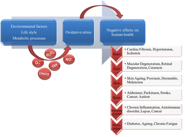
Another harmful health risk factor common to the development of these and other chronic pathologies (Maritim et al., 2003; Andersen, 2004; Singh and Jialal, 2006; Ward and Croft, 2006; Bez and Yau, 2008; Pacher and Szabo, 2008; Manos et al., 2009) is oxidative stress (Figure 8), a process resulting from an imbalance between excessive production of reactive oxygen species and/or nitrogen species and limited action of antioxidant defences (Spitz et al., 2004).As oxidative stress is believed to play a major role in the ageing process and in several diseases, there is considerable public interest in the antioxidative effects of dietary factors
16 (Lesgards et al., 2002; Mattson, 2008). Indeed, with proper nourishment, the body can, on its own, make sufficient quantities of antioxidant enzymes and substrates for those enzymes. These can facilitate the quenching of excess free radicals by antioxidants. An enhancement in dietary intake of antioxidants and phytochemicals with related functions can counteract oxidizing species and potentially restore a healthy cellular redox balance (Vattem et al., 2005).
Environmental factors Life style Metabolic processes
Oxidative stress Negative effects on human health
NO•
Heart
• Cardiac Fibrosis, Hypertension, Ischemia
Eyes
• Macular Degeneration, Retinal Degeneration, Cateracts
Skin
• Skin Ageing, Psoriasis, Dermatitis, Melanoma
Brain
• Alzheimer, Parkinson, Stroke, Cancer, Autism
Immune system
• Chronic Inflammation, Autoimmune disorder, Lupus, Cancer
Multi-organ
• Diabetes, Ageing, Chronic Fatigue
Figure 8. The main factors and protagonists in the development of oxidative stress, and the possible negative outcomes to human health.
However, in recent years, following the achievement of the human genome sequencing, the exploitation of molecular biology tools revealed that specific antioxidant compounds modulate various cellular functions, acting not only as radical scavengers, but also as regulators of genes and enzymes expression. This approach gives rise to the new sciences of nutrigenomics and nutrigenetics which consider the relationship between specific nutrient or diet and gene expression and determine how genetic variability affect the response to diet, respectively (Knasmüller et al., 2008). In this scenario, nutraceutics, which are food or part of a food providing medical or health benefits, acquire a new additional significance for the development of novel food and personalized diets.
17 Nowadays, the consumption of midday and/or midnight snack food is becoming a dominant eating-habit, despite its close correlation with several metabolic disorders. Eating small amounts of food on a regular basis is considered healthy, but often snacks available in the market provide excess calories and fats and little or no nutrient value to the diet. Moreover, most snacks contain preservatives, sweeteners and flavouring that can have negative effects on health.
There is no doubt that a healthy diet must include proper food selection, keeping track of meal times and regulation of the amount of food intake. The limits imposed by modern life-styles on eating-habits create a necessity to develop an adequate food management rather than simply body-weight control. One of the most popular eating diet plans suggests the 40:30:30 ratios for carbohydrates, proteins and fats that a person should consume. With this ratio of components, the body is able to balance insulin and glucagon, as well as to realize a more effective internal metabolism. Currently, experts from the manufacturing sector are concerned with the development of modern alimentary strategies aimed at those who believe in health through correct nutrition and food supplementation.
1.2.1 Applications of plant-derived phytochemicals
Plants produce a big number of chemicals that are important for their function and development. Some of these compounds are primary metabolites, which include proteins (aminoacids), carbohydrates, fats, nucleic acids etc. Besides these primary chemicals, the plants also produce secondary metabolites, which are specific to some taxonomic groups (families, genera) (Krzyzanowska et al., 2009).
Compared to the main and most abundant molecules found in plants, these secondary metabolites are defined by their low abundance, often less than 1-5% of the dry weight. They are formed under environmental pressure and have no recognized role in the maintenance of fundamental life processes in plants; but, they contribute mainly to protect plants against several environmental stresses. This group includes classes of compounds able to promote public health, with different biological activities (Table 1), which in traditional medicine were used for centuries to cure or protect from diseases.
A correlation between diet and health has been known since ancient time. However, numerous epidemiological studies confirm the correlation, especially regarding the frequency of some diseases incidence in relation to the intake of particular nutrients. In particular, a low consumption of fruit, vegetables and whole grain was often associated to disease.
18
ACTIVE METABOLITE (dietary sources)
STRUCTURE MAIN NUTRACEUTIC
ACTIVITY
Carotenoids 1 (carrot, tomato, spinach, broccoli, kale)
antioxidant, anticancer,
immunostimulation, vision, protection
Cysteine derivates 2, 3 (garlic, onion)
antioxidant, antibacterial, anti– arteriosclerosis, anticancer, antibiotic, lipid lowering
Flavones 4,5 (cereal, herbs, citrus fruit, tea)
antioxidant, antibacterial, anticancer, anticolitis
Isoflavones 6 (pea, bean, soybean, salvia)
antioxidant, anti -arteriosclerosis
Phenolic compounds 7 (cichorium, coffee, lettuce, red wine)
antioxidant, cardio protective, antihypertensive
Plastoquinones 8 (green vegetables and fruits)
antioxidant, anticancer
Polyphenolic acids 9 (rosemary, sage, oregano, basil, lemon balm)
antioxidant, antibacterial
Ferulic acids and derivatives 10
(apple, orange, peanut, pineapple)
antioxidant, cardio protective, immunostimulation
Phytoalexin11, 12 (grapes, pine nuts, peanuts, wine)
antioxidant, cardio protective, anti-arteriosclerosis, anticancer
Table 1. Secondary metabolites, dietary sources, structures and related functional activities.
(1Mares-Perlman et al., 2002, 2Cai et al., 2007, 3Ariga and Seki, 2006, 4Fiamegos et al., 2004, 5Cermak, 2008, 6Romani et al., 2003, 7Vagi et al., 2005, 8Kruk et al., 2006, 9Tepe, 2008, 10Yongyue et al., 2005, 11Prokop et al.,
2006, 12Baur and Sinclair, 2006).
zeaxanthin alpha-carotene allicin genistein caffeic acid plastoquinone rosmarinic acid curcumin resveratrol luteolin
19 Several studies indicate that populations consuming high levels of plant derived foods, namely fruits-including berries, nuts, vegetables, whole-grains, legumes, seeds, various types of tea and a bewildering array of spices, but also low in red meat and animal fats, have low incidence rates of various cancers (Krzyzanowska et al., 2009; Whale et al., 2009).
Such diets are exemplified by the classical Mediterranean diet of Southern Europe and the highly vegetarian diets of South East Asian populations (Block et al., 1992; Messina et al., 1994).
The research for natural, plant-derived phytochemicals that express anticancer properties, is not restricted to food plants. Plant secondary metabolites are a very important industrial and economic source of many drugs, flavors, insecticides, fragrances and dyes. Pharmaceutical companies are currently isolating and assessing the anticancer potential of numerous phytochemicals from all regions of the world, in order to discover more efficient treatments for various types of cancer (Krzyzanowska et al., 2009).
The technology of large-scale plant cell culture is feasible for the industrial production of plant-derived fine chemicals, but there are still several limitations.
The natural content of the valuable compounds in a plant is often very low, or production is often impossible due to a lack of raw material. Some compounds can only be isolated from rare plants. On the other hand, chemical synthesis of these compounds is often not technically or commercially feasible because of their highly complex structures (Oksman-Caldentey and Inzé, 2004).
1.2.2 Carotenoid benefits for human health
Carotenoids are synthesised by plants, algae, fungi, yeasts and bacteria. Animals and humans cannot synthesise them and are dependent on the dietary sources (Fraser and Bramley, 2004; Stahl and Sies, 2005).Different 600 carotenoids have been identified in nature; however, only about 40 are present in a typical human diet. Human plasma and tissues contain only 20 carotenoids, which are represented mainly by β-carotene, lycopene, lutein, β-cryptoxanthin and α- carotene (Fraser and Bramley, 2004; Rao and Rao, 2007).
They are present in various types of food, but the major sources of dietary carotenoids include orange and yellow fruits and vegetables as well as green leafy vegetables. Smaller amounts can be extracted from milk and foods containing dairy fat, egg yolks, sea fish, and carotenoids added as colorants to foods during processing (Rock, 1997).
Carotenoids are important components of the human diet because they have been linked to a multitude of health benefits. They play an important role in the cell communication and
20 protection against photo-oxidative processes by acting as singlet molecular oxygen, as well as, peroxyl radical scavengers and can interact synergistically with other antioxidants (Reddy et al., 2003). In mammals, some of them can be metabolised to retinol and function as vitamin A precursors. Positive effects between high dietary consumption and tissue concentration of carotenoids and reduced risk of chronic diseases such as age-related macular degeneration were also observed. There is also strong evidence showing that a rich diet in carotenoids prevents cardiovascular diseases and certain cancers like lung, colon, breast, and prostate cancer (Rao and Rao, 2007;Reddy et al., 2003; Palace et al., 1999).
Initially, most of this attention was directed towards β-carotene, also known as provitamin, the precursor of vitamin A, an essential nutrient for human health, responsible for the promotion of growth, cellular differentiation, morphogenesis, embryonic development, and visual function. Some carotenoids can be converted into active forms of this vitamin and, in doing so, may prevent vitamin A deficiency (Krzyzanowska et al., 2009).
Recently, the attention, is increased also for lycopene (the red pigment of tomatoes) as well as zeaxanthin and lutein (the yellow pigments responsible for the colour of corn and eggs). In the case of lycopene, there is some evidence for a protective role against prostate cancer (Ellinger et al., 2006). For lutein and zeaxanthin, considerable evidence (Mares-Perlman et al., 2002) is available for a role of these xanthophylls in the protection against age-related vision loss, such as age-related macular degeneration (AMD) (Seddon et al., 1994) and cataracts (Chasan-Taber et al., 1999; Brown et al., 1999).In addition, the important role for zeaxanthin and lutein in the protection against cancer and heart disease emerged (Mares-Perlman et al., 2002).
In particular, zeaxanthin and lutein are synthesized by plants (as well as photosynthetic microbes) and especially when these plants are grown in high light or under otherwise stressful conditions (Demmig-Adams and Adams, 1996; Demmig-Adams and Adams, 2006). Zeaxanthin, as mentioned above, protects against the formation of potentially destructive reactive oxygen species in leaves exposed to intense sunlight alone or moderate levels of sunlight in the presence of environmental conditions un-favourable for plant growth (Demmig-Adams and Adams, 2002). This photoprotective process is necessary for plant survival and reproductive success (Külheim et al., 2002); oxygenic photosynthesis could not exist without it. In addition, zeaxanthin serves in photoprotection via a second, not fully understood, mechanism that involves a direct inhibition of the oxidation of fatty acids of biological membranes (lipid peroxidation) (Havaux and Niyogi, 1999; Havaux et al., 2004). Epidemiological studies have identified inverse links between zeaxanthin/lutein and a wide
21 range of human diseases (Mares-Perlman et al., 2002; Demmig-Adams and Adams, 2002; Sajilata et al., 2008). However, the underlying mechanisms for these apparent protective effects remain poorly understood.
Zeaxanthin/lutein protect the human eye from damage by intense light. A breakthrough in the understanding of the function of retinal zeaxanthin was made when it was shown that zeaxanthin prevents programmed cell death of retinal photoreceptor cells in an animal model (Thomson et al., 2002a; Thomson et al., 2002b). On the other hand, studies with human cancer cell lines provided evidence that lutein can stimulate programmed cell death of human breast cancer cells (Sumatran et al., 2000) and leukemic cells (Müller et al., 2002). Lutein furthermore selectively induces programmed cell death in mouse tumour cells, but decreases programmed cell death in cancer-fighting immune cells (blood leukocytes) of tumour-bearing mice (Chew et al., 2003). Up to now is not known how xanthophylls exert these remarkable and beneficial roles, including opposite effects on programmed cell death in different cell types.
For sure the zeaxanthin can modulate the oxidation of fatty acids (lipid peroxidation) in plants (Havaux and Niyogi, 1999; Havaux et al., 2004) as well as in humans (e.g. in epithelial cells of the eye’s lens) (Chitchumroonchokchai et al., 2004); indeed, zeaxanthin protects lipids against destructive reactive oxygen in vitro (Wrona et al., 2003; Wrona et al., 2004).
As aforementioned, age-related blindness (with photoreceptor death) and cancer (with run-away cell proliferation) involve apparently opposite problems, i.e. either too little or too much programmed cell death. Current studies suggest that several dietary factors possess the capacity to ameliorate both of these contrasting conditions (Demming Adam et al., 2009). Thus far, phenolics (Youdim et al., 2002; Surh, 2003) the polyunsaturated fatty acids (of fish oil) (Seo et al., 2005), and zeaxanthin/lutein (Maccarrone et al., 2005) have all been shown to possess the notable ability to ‘work both ways’, i.e. triggering programmed cell death of unwanted cells while aiding in the survival of needed cells. This makes these food-derived compounds potentially highly desirable nutraceuticals.
As reviewed above, both zeaxanthin and lutein have roles in protecting human health. However, there is great disparity in how much zeaxanthin versus lutein can be obtained from leafy green plant foods. Indeed, the plant green parts typically contain high levels of lutein and maintain a few zeaxanthin amounts, because they carefully adjust the level of zeaxanthin in response to the light intensity. The level of zeaxanthin is finely controlled by a set of biochemical reactions, before mentioned. Human consumption of high levels of zeaxanthin is highly desirable, because zeaxanthin needs to be preferentially accumulated and incorporated
22 into the parts of the mammalian retina exposed to high irradiance (Landrum et al., 2001), and thus appears to be even more important in human diets than the more readily available lutein (Demming Adams et al., 2009). For this reason, arrest of the xanthophyll cycle in green leaves in the state of zeaxanthin may be a desirable trait to incorporate into crops that provide green leafy foods. Such a retention of zeaxanthin can be accomplished e.g. by knocking out or silencing the enzyme/gene (zeaxanthin epoxidase of the xanthophyll cycle) responsible for zeaxanthin conversion to violaxanthin and/or by overexpressing enzymes in earlier portions of the carotenoid biosynthetic pathway (Demming Adams et al., 2009).
As a generalization, over-expression of antioxidants in plants and algae may (i) make these organisms more resistant to abiotic stresses, while (ii) possibly increasing their susceptibility to some pests/pathogens, and (iii) likely also affecting growth, development, and reproduction. It may be feasible to simultaneously decrease crop losses due to environmental stress (like drought, heat, or frost), while enhancing the nutritional quality of food plants (Demming Adams et al., 2009).
Xanthophyll cycle mutants that accumulate zeaxanthin have already been produced in model plants and algae (Niyogi, 2000), and these traits can be transferred to crop plants. Due to the need for leaves to photosynthesize efficiently, overexpression of zeaxanthin in fruit, rather than leaves, is attractive. Tomato fruit with an increased zeaxanthin content has recently been engineered via two other manipulations (overexpression of lycopene-cyclase and beta-carotene hydroxylase) (Dharmapuri et al., 2002). Zeaxanthin-rich potato has also been produced (Romer et al., 2002) as well as zeaxanthin-accumulating E. coli (Albrecht et al., 1999).
1.2.3 Enrichment of nutraceutical contents in foodstuff
Molecular engineering of crop plants has offered a number of tools to markedly enhance intracellular concentrations of some of the beneficial nutrients. Some of these include increases in: protein level in potato; lysine in corn and rice; carotenoids (β-carotene, phytoene, lycopene, zeaxanthin and lutein) in rice, potato, canola, tomato; folates in rice, corn, tomato and lettuce; vitamin C in corn and lettuce; polyphenolics such as flavonol, isoflavone, resveratrol, chlorogenic acid and other flavonoids in tomato; anthocyanin levels in tomato and potato; iron and zinc in transgenic rice (Matoo et al., 2009). The concept of developing nutritionally functional food requires: (1) the understanding of the mechanisms of prevention and protection; (2) the identification of the biologically active molecules, and (3) the demonstrated efficacy of these molecules with human subjects.
23 Databases are growing with information on the content of phytonutrients in edible vegetables and fruits as well as with transgenic technology developments for enhancing the nutritional content of vegetable crops via engineering of specific metabolic pathways. Metabolic pathway engineering approaches have demonstrated the power of genetic manipulation in enhancing the content of nutrients beneficial for human health in transgenic crops; as well as key genes to inhibit production of allergenic proteins or toxins in crops is highly sought (Matoo et al., 2009).
Therefore, genetic engineering was used to produce β-carotene in rice endosperm. Daffodil (Narcissus pseudonarcissus) psy gene, which encodes phytoene synthase and produces the first carotenoid phytoene, a key precursor of β-carotene, was expressed in rice under a CaMV 35S (constitutive) or Gt1 (endosperm-specific) promoter (Burkhardt et al., 1997). This study catalyzed efforts to engineer the carotenoid biosynthetic pathway in rice endosperm using daffodil psy gene and bacterial (Erwinia uredevora) phytoene desaturase crt1 gene (Ye et al., 2000; Beyer et al., 2002). Transgenic rice seeds were yellow in colour due to the accumulation of carotenoids mainly β-carotene and, to some extent, lutein and zeaxanthin. Several studies have shown that psy gene product catalyzes a limiting and regulatory step in carotenoid biosynthesis. The maximum carotenoid accumulation was achieved with maize
psy, increasing the total carotenoid level to 37 µg/g, some 23-fold higher than a previous
report on the Golden Rice (Paine et al., 2005).
Transformation of canola seed with bacterial phytoene synthase (crtB) gene using seed-specific napin promoter resulted in orange coloured seed with a 50-fold higher (1000-1500 µg/g FW) total carotenoid level than the wild type (33 µg/g FW) (Shewmaker et al., 1999). Canola seeds coexpressing crtI and crtB gene accumulated lycopene (29 µg/g FW) and β-carotene (857 µg/g FW) (Ravanello et al., 2003).In a seed-specific manner, bacterial crtB gene expression in flaxseeds resulted in 7.8 to18.6-fold increase in the carotenoid levels (Fujisawa et al., 2008).
crtB gene was also used in a tuber-specific manner to increase carotenoids, violaxanthin,
lutein and carotene, in transgenic potato (Ducreux et al., 2005). Downstream from the β-carotene pathway are important photoprotective compounds whose levels have also been successfully manipulated via genetic intervention. Using the fruit-specific pds promoter to drive the expression of lcy-β and chy-β genes from Arabidopsis and pepper, respectively, about 100-fold increase in β-cryptoxanthin and zeaxanthin was achieved (Dharamapuri et al., 2002).
24 Ketocarotenoids are rare in plants but are strong antioxidants. They are chemically synthesized and used as dietary supplements and pigments in aquaculture and nutraceutical industry. Ketocarotenoid biosynthesis pathway was engineered in carrot by introducing an algal Haematococcus pluvialis β-carotene ketolase gene under the control of constitutive, ubiquitin or rolB promoter (Jayraj et al., 2008). This resulted in 70% conversion of total β-carotene to ketocarotenoid that accumulated to a level of 2,400 µg/g root dry weight. These studies showed that these transgenic carrots are suitable for biopharming ketocarotenoid production for functional food, nutraceutical and aquaculture industries.
Phytonutrients are now recognized as important determinants of human health. This has catalyzed investigations into broader aspects of plant-based nutraceuticals. These include: elucidation of biochemical pathways to identify the rate-limiting steps; engineering metabolic pathways to direct the intermediary metabolism flux towards a particular nutrient (nutraceutical); testing efficacy of either an isolated and purified nutraceutical or a crop engineered with an enhanced nutrient level in animal and human models; testing crops silenced for health-detrimental factors including allergens; comparing bioavailability of an individual nutrient (nutraceutical) fed either as a food supplement or in the form of a fortified food (Matoo et al., 2009). A common goal of such studies is to enable dietary intervention in human health to combat monogenic or polygenic diseases.
Progress in merging agriculture with preventive medicine will depend on intense collaborative research between physicians, nutritionists and plant biologists so that planning strategies are rationally designed and the developments in genetic technology successfully applied for better crop engineering (Matoo et al., 2009).
1.3 Chlamydomonas reinhardtii as biological farm
Eukaryotic microalgae have recently gained interest as biological farms, because they are attractive alternatives to bacterial, yeast, plant and other cell-based systems currently in use (Pulz and Gross, 2004; Walker et al., 2005; Spolaore et al., 2006; Mayfield et al., 2007). Over the last decades there has been considerable progress in genetic engineering technologies for algae. Biotechnology companies now apply these techniques to alter metabolic pathways and express valuable proteins in different cell compartments. Particularly, the eukaryotic unicellular alga Chlamydomonas reinhardtii (Figure 9), sometimes called “green yeast” (Goodenough, 1992; Rochaix, 1995), portraits an important niche in cell biology eukaryotic world and nowadays become a model organism widely used in laboratories thanks to the availability of complete genome sequence, including chloroplast and mitochondrial genomes,
25 annotated databases for any searching or cloning requirements and successful high-level expression of recombinant proteins in the chloroplast compartment (Mayfield et al., 2007; Manuell et al., 2007). Besides, C. reinhardtii belongs to the group of green algae included in the category of organisms with a GRAS status (Generally Regarded As Safe) granted by the FDA. Thus, enhancing food with edible algae like chlamydomonas engineered or not to (over)produce functional ingredients has the potential to become an important factor in food and feed technologies.
A) B)
Figure 9. Eukaryotic unicellular alga Chlamydomonas reinhardtii A) A single C. reinhardtii cell (from
http://www.veda.cz/article.do?articleId=26215). B) Schematic diagram of C. reinhardtii cell structure (from website: http://universe-review.ca/R10-34-anatomy2.htm#classification).
Moreover, C. reinhardtii has been extensively used as an experimental system for studying flagellar motility, function of the basal body, as well as formation and function of chloroplasts, exploiting the powerful of microbiology and genetic engineering techniques (Lefebvre and Silflow, 1999).
Chlamydomonas has interesting biotechnological features including a rapid growth in both liquid and solid media; a sexual cycle, that can be precisely controlled; genetic aspects which enable the generation and characterization of a wide collection of mutants with lesions in structural and metabolic regulator genes and a flexible metabolism being able to growth both phototrophycally and heterotrophycally (Grossman et al., 2003).
Microalgae have long been used as food or food additives. For human nutrition, edible microalgae like Arthrospira species (“Spirulina”) and Chlorella are marketed as tablets, capsules and liquid or added to e.g. noodles, breads and candies to improve their nutritive and health values. Other major commercial strains used as food ingredients are Dunaliella and
Aphanizomenon flos-aquae, whose extracts exhibit health promoting effects (Spolaore et al.,
26 Safety has recently been demonstrated with the marine microalgae Odontella aurita certified in 2002 as novel food by the French company INNOVALG. While the exploitation of products derived from natural microalgae has a long history, the generation of transgenic microalgae for biotechnological applications has just started to become an attractive system for expressing foreign proteins or other high-value compounds with e.g. antioxidant, colorant, provitamin or therapeutic properties. Nowadays, genetic engineering of algae by introducing and controlling foreign genes has been developed to an extent that allows to exploit these organisms as bioreactors for the production of high-value compounds (Walker et al., 2005). While agrobacterium-mediated transformation is now the standard method for nuclear transformation, the biolistic approach using accelerated particles (particle gun) turned out to be the preferential procedure for organelle transformation.
The organellar genomes, specifically those of plastids, are particularly attractive for genetic engineering purposes. Not only do chloroplasts naturally produce high amounts of protein, but they are also uniparentally inherited and integrate properly flanked foreign genes via homologous recombination into their genome. As opposed to plant nuclear genomes, these properties provide distinct advantages of chloroplast transgenes with respect to bio-safety and epigenetic stability (Maliga, 2004). Genetic engineering of chloroplasts has made particular progress with genes conferring agronomical valuable traits like e.g. resistance to herbicides (Daniell et al., 1998; Lutz et al., 2001), to fungal and bacterial diseases (DeGray et al., 2001) or to insects (Kota et al., 1999; McBride et al., 1995). Chloroplasts have also been employed to overproduce biopharmaceuticals (Daniell et al., 2002; Staub et al., 2000). Chloroplast genetic engineering is currently most advanced in higher plants, particularly in tobacco (Maliga, 2004). While higher plants offer several advantages over expression systems in other organisms, there are distinct drawbacks like e.g. the length of time necessary for their generation or concerns about containment of transgenic plants in the environment even in trans-plastomic lines (Ellstrand, 2001; Ellstrand, 2003; Ruf et al., 2007). The use of microalgae like C. reinhardtii provides interesting alternatives: from vector construction to lab-scale culture volumes of the transgenic strain it takes about 6 weeks and cells can be easily contained in photobioreactors (Johanningmeier et al., 2009).
C. reinhardtii was the first organism for which stable chloroplast transformation was reported
(Boynton et al., 1988). Early selection methods used cloned chloroplast genes to rescue photosynthetic mutants or chloroplast gene constructs that confer resistance to herbicides (Przibilla et al., 1991) or antibiotics (Newman et al., 1991), later on transformant selection
27 was mainly based on the expression of bacterial markers conferring antibiotic resistances (Goldschmidt-Clermont, 1991; Bateman and Purton, 2000).
Recent progress in engineering C. reinhardtii chloroplasts is the result of 20 years research on developing transformation techniques, improving codon usage and finding efficient promoters and un-translated regions (UTRs) for boosting foreign gene expression.
Today several transgenes have been successfully expressed in C. reinhardtii chloroplasts. Although most of them are intended to be used as reporters and thus serve basic research purposes, now several proteins for pharmaceutical applications appear in the list. In particular, the expression of a bioactive mammalian protein was achieved, whose level is estimated to be above 5% of total cellular protein (Mayfield et al., 2007). In the near future it appears possible that such transgenic chlamydomonas strains could be ingested orally without the need for extensive purification of the bioactive compound. This perspective is supported by experiments, in which chlamydomonas cells expressing an epitope of a pathogenic bacterium infecting salmonids were fed to trout. An immune response was observed when transgenic cells were added to the fish food (Patent application US020030022359) (Griesbeck et al., 2006).
The use of microalgae as green cell factories assures a significantly faster generation of stable transgenic lines and is often coupled with high yields, lower costs and rapid, controllable growth in simple media. Microalgae are not hosts to major pathogens and recombinant strains can be grown in full containment, excluding the risk of contaminating natural populations. Moreover, methods exists which allow to construct transgenic algae without employing antibiotic resistance genes (Debuchy et al., 1989; Ferris, 1995) thus minimizing any consumer-based concerns regarding the transfer of marker genes from a food product into the cells of the body.
1.4 Chlamydomonas reinhardtii mutants 1.4.1 C. reinhardtii NPQ mutants
A set of C. reinhardtii mutants carrying single and double mutation within carotenoid biosynthetic genes were, included in this study (kindly provided by Prof Niyogi, University of California, Berkeley, Department of Plant and Microbial Biology, U.S.A.). These mutants (lor1, npq2 and npq2 lor1) are impaired in the biosynthetic steps that lead to the synthesis of xanthophylls and α-carotenes derivatives (Figure 10).