I Facoltà di Medicina e Chirurgia
Scuola di Dottorato di Ricerca in Epato-gastroenterologia sperimentale e clinica XXIX Ciclo – Curriculum di Epatologia sperimentale e clinica
Coordinatore Prof. P. Onori
Biliary complications in liver transplantation:
role of the hepatic ischaemia-reperfusion injury
Complicanze biliari nel trapianto di fegato:
ruolo del danno da ischemia-riperfusione
Supervisore Dottoranda
INDEX
ABSTRACT 6
INTRODUCTION 11
AIM OF THE STUDY 14
SCIENTIFIC BACKGROUND 15
Extended criteria donors – donation after circulatory death 15 Hepatic ischemia reperfusion injury and systemic inflammatory response 18
Local complications: the bile duct 21
Biliary complications in liver transplantation 25 Remote organ complications: the kidney in liver transplant setting 28 Acute kidney injury post liver-transplantation 29
METHODS 35
Clinical data considered 35
Evaluation of bile duct histology 38
Evaluation of cholangiocyte apoptosis 39
Evaluation of cholangiocyte proliferation 40
Statistical analyses 41 RESULTS 42 DISCUSSION 45 TABLES 51 FIGURES 60 REFERENCES 69
ABSTRACT
Introduction
Liver transplantation is currently the only effective therapy for patients with end stage acute or chronic liver failure. The increasing request of organs has led to the more extensive use of the so-called marginal donors, in particular donors after circulatory death (DCD).
Within this model of donation, a more severe degree of ischaemia-reperfusion injury (IRI) is occurring, that seems to play a role on the pathogenesis of local and remote organ complications.
This research will focus on the influence of liver graft injury on the pathogenesis of local and remote organ complications, evidencing the role of IRI leading to biliary complications and development of systemic inflammatory response associated with the occurrence of acute kidney injury (AKI).
Aim of the study was to assess the role of IRI in two different models of ischaemia, DCD and donation after brain death (DBD), in liver transplanted grafts, on the pathogenesis of local and remote organ complications.
We evaluated the development of biliary complications and its association with the degree of donor liver graft bile duct injury after liver reperfusion.
Moreover, the development of AKI after liver transplantation was considered, as consequence of IRI and systemic inflammatory response.
Methods
Retrospective single-centre study of adult patients who underwent liver transplantation at University Hospital Birmingham (UHB) National Health Service (NHS) Foundation Trust from January 2007 to December 2014.
Characteristics of recipient at transplant, recipient renal and liver function in the immediate pre-transplant period, donor and graft variables, intra-operative parameters, indicators of initial graft function and renal function in the post-transplant period were considered.
Primary end points were the occurrence of biliary complications, in particular ischaemic cholangiopathy (IC), and development of AKI. Secondary end point was the evaluation of IRI damage on the basis of transaminases, bilirubin and INR over the first week post-transplant and the appearance of bile duct retrieved after liver reperfusion on histological examination.
Severity of donor bile duct injury was assessed and scored on the basis of biliary epithelial cell loss, mural stroma necrosis, inflammation, peribiliary vascular plexus (PVP) damage, arteriolonecrosis, thrombosis, periluminal and deep peribiliary glands (PBGs) damage.
Cholangiocyte apoptosis in periluminal and deep PBGs was evaluated by quantitative terminal deoxy-nucleotidyl transferase dUTP-mediated nick-end labeling (TUNEL) analysis on bile duct sections. Cholangiocyte proliferation was studied in bile duct sections by PCNA immunohistochemical expression.
Results
One thousand and 60 liver transplant recipients (813 from DBD and 247 from DCD) were considered.
Recipients from DCD had higher ALT and AST in the first 7 days after transplant, compared to DBD. The occurrence of biliary complications was higher in DCD liver transplant recipients (85/247; 34%) compared to DBD (166/813; 20%) (p<0.001), in particular IC incidence was significantly higher in DCD.
The incidence of AKI was 59.3% (629/1060 recipients) and was significantly higher in DCD (160/247, 64.8%) compared to DBD (469/813, 57.7%) recipients (p=0.047).
Sixty-two patients comparable with the entire cohort, had the bile duct sample available for histological evaluation. A significantly higher number of DCD patients presented necrosis >50% of the bile duct wall [DCD 14/28 (50%), DBD 9/34 (26.5%) p=0.056], PVP damage [DCD 8/28 (29%), DBD 3/34 (9%); p=0.053] and periluminal PBGs damage [DCD 20/28 (71%), DBD 14/34 (41%); p=0.016]. These features defined the occurrence of severe histological injury, that was significantly more frequent in DCD liver transplant patients [15/28 (53.6%)] compared to DBD [7/34 (20.6%)] (p=0.007). A significant increased apoptosis and decreased proliferation was evidenced in both periluminal (Tunel assay p=0.029; PCNA expression p=0.029) and deep PBGs (Tunel assay p=0.002; PCNA expression p=0.006) from bile duct sample with severe histological injury.
Discussion
A more severe degree of IRI is occurring within DCD, as evidenced by greater graft dysfunction and increasing peak perioperative transaminases, likely related to the added donor warm ischaemia time.
The IRI seems to play a role on the pathogenesis of local and remote organ complications, evidencing the role exerted in DCD leading to biliary complications and development of systemic inflammatory response associated with the occurrence of AKI.
This study also shows an early picture of microscopic damage at the level of the bile duct soon after reperfusion of liver graft during transplantation. Bile duct samples retrieved from DCD grafts expressed more severe injury at the histological level, as evidenced by the increased incidence of mural stroma necrosis, PBP damage and PBG damage, defining the new feature of severe histological injury.
Bile ducts with severe histological injury showed increased apoptosis and reduced proliferation, as evaluated by Tunel assay and PCNA expression, both on periluminal and deep PBG cholangiocytes.
The higher incidence of IC development in DCD strongly suggests a relation between the occurrence of severe histological injury and alteration in bile duct repair mechanisms, raising hypothesis to further evaluate those mechanisms leading to the development of bile duct non-anastomotic strictures.
INTRODUCTION
Over the past 40 years, orthotopic liver transplantation has evolved from an experimental procedure to become the treatment of choice for patients with end-stage liver failure (ESLD). The first few liver transplants by T.E. Starzl in 1963 were not successful but showed the feasibility of the surgical technique and four years later he reported patient survival beyond one year. These early attempts at liver transplantation were in high-risk patients and the problems of controlling hemorrhage and coagulopathy and poor graft function resulted in significant intra-operative and early postoperative mortality. In these early series, the one-year survival after liver transplantation in both adults and children was only 19%.
Prednisolone and azathioprine were the primary immunosuppressive drugs, but some regimens included combinations of cyclophosphamide, antilymphocyte globulin, splenectomy, total body irradiation and thymectomy. One-year survival after liver transplantation, however, remained poor and was only 30% in 1978.
The introduction of cyclosporin in 1981 transformed clinical practice because of the improved outcome following liver transplantation. The National Institute of Health Consensus Conference in 1983 concluded that liver transplantation for end-stage liver disease deserved broad application and should be considered a therapeutic option rather than an experimental procedure.
Liver transplantation is currently the only effective therapy for patients with end stage acute or chronic liver failure. Advances in immunosuppressive therapy, organ
preservation, surgical and anaesthetic techniques and improved management of post-transplantation complications have led to further improvement in survival. Currently patients with chronic liver disease undergoing liver transplantation can anticipate a one-year survival of 85% and a 5-year survival of greater than 75%.
Nevertheless, the procedure of liver transplantation is still associated with a series of significant complications. Transplantation of the liver leads to hepatic-ischaemia reperfusion injury (IRI), the damage occurring for the duration of several phases: during warm ischemia time, during cold preservation throughout and following the retrieval of the liver and within warm reperfusion at implantation to the recipient. Hepatic ischemic-reperfusion injury is a complex process that results in apoptosis or necrosis of hepatic cells and activation of the inflammatory response. An inflammatory response associated with the expression of pro-inflammatory cytokines has been reported in consequence of liver reperfusion in both animals and humans (Colletti et al, 1996; Bellamy et al, 1997; Monbaliu et al, 2007). Numerous cell types and molecular mediators act in a complex communication networks involving the immune and non-immune systems to initiate and propagate IRI (Abu-Amara et al, 2010).
Two different models of donation are nowadays available, donation after circulatory death (DCD) and donation after brain death (DBD), that are characterized by different degree of IRI in relation to retrieval modalities.
This research will focus on the influence of liver graft injury on the pathogenesis of local and remote organ complications, evidencing the role of IRI leading to biliary
complications and development of systemic inflammatory response associated with the occurrence of acute kidney injury (AKI).
AIM OF THE STUDY
Aim of the study was to assess the role of ischaemia-reperfusion injury in two different models of ischaemia, DCD and DBD, in liver transplanted grafts, on the pathogenesis of local and remote organ complications.
We evaluated the development of biliary complications and its association with the degree of donor liver graft bile duct injury after liver reperfusion.
Moreover, the development of acute kidney injury after liver transplantation was considered, as consequence of IRI and systemic inflammatory response.
SCIENTIFIC BACKGROUND
Extended criteria donors – donation after circulatory death
Liver transplantation has become victim of its own success, with the increasing number of patients awaiting a transplant not faced with the same increasing of organs donation. The shortage of donor organs prompted the development of newer techniques like transplantation of reduced-size organs for small children, of split livers (one donor organ split into two segmental liver grafts) and living-related liver transplantation. However, this has not satisfied the increasing discrepancy between resources and needs, leading to the use of the so-called marginal donors, which for
reasons of age or non-ideal systemic and/or graft conditions, question the real fitness
of the donor.
Within this group a substantial portion comprises donors after circulatory death (DCD). Donors after circulatory death are patients with brain injury incompatible with recovery whose condition does not meet formal criteria for brain stem death and whose cardiopulmonary function ceases before organs are retrieved. In the 1960’s DCD were the main source of allografts until the Harvard definition and criteria for brain death were published in 1968. Following the introduction of brain stem death criteria, interest in DCD declined because of the better results obtained with organs from heart beating donors. Renewed interest in DCD occurred because of the current organ shortage, improvements in organ preservation, immunosuppression and surgical
techniques, and changing neurosurgical practice in the management of traumatic head injuries.
Donation after circulatory death can be categorised into 2 groups: controlled and uncontrolled. Controlled donation takes place when the death occurs within an intensive care/hospital setting and there is a planned withdrawal of therapy by the patient’s medical team. Uncontrolled donation occurs outside the hospital or within the emergency room and is unpredictable.
The several reports regarding liver transplantation from DCD have shown a progressive reduction in the incidence of primary non-function in recent years due to a shift towards controlled donation, better donor selection and shorter warm and cold ischaemic times.
The long-term outcome for liver transplantation from DCD donors has been considered inferior to organs from DBD donors (Selck FW, et al. 2008; Yamamoto S, et al. 2010). Recent reports suggest comparable outcomes for DBD and DCD livers after transplantation (Dubbeld J, et al. 2010; Fujita S, et al. 2007). It is also becoming apparent that the outcome of DCD livers depends on a number of donor and recipient factors and technical factors in relation to the organ retrieval and implantation (Mathur AK, et al. 2010). An important role has been recognised for duration of both primary warm and cold ischaemia time (pWIT and CIT, respectively), which are associated with recipient complications, specifically for ischaemic cholangiopathy, graft failure and recipient mortality (Taner CB, et al. 2012; Hong JC, et al. 2011).
Currently, DCD account for about 26% of livers transplanted in United Kingdom (Annual Report on Liver Transplantation 2015/16 – NHS Blood and Transplant).
Patients are considered for liver donation provided they have good liver and renal function and after explicit consent is obtained from relatives. A minimum waiting period of 5 minutes after cardiac arrest is observed before starting the retrieval procedure following the recommendations of the Institute of Medicine. The transplant team must not be involved in decisions regarding the modality and logistics of withdrawal and these are undertaken by the intensive care physician and according to local hospital policies. Similarly, the transplant team plays no part in the declaration of death.
Suitability of the DCD livers for transplantation is initially assessed at retrieval. Factors considered in making the decision include the time between withdrawal of therapy and death and the period of warm ischaemia. Retrieval of the liver is not considered if cardiac arrest does not occur within one hour of withdrawal because of damage resulting from prolonged periods of hypotension. During the donor procedure, the liver is assessed macroscopically for the presence of fatty change, perfusion quality and physical characteristics.
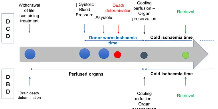
The organ donation procedure occurs in accordance to the UK National standards and local guidelines. After death certification and the obligatory no-touch period of 5 minutes a rapid access of the vessels is gained and cannulation performed. Usual retrieval process follows, with cold preservation of the liver graft until the implantation into the recipient. (Figure 1)
Hepatic ischemia reperfusion injury and systemic inflammatory response
The ischemic phase, lacking oxygen supply, leads to depletion of adenosine triphosphate (ATP) in Kupffer cells (KCs), hepatocytes and sinusoidal endothelial cells (SECs), with a consequent loss of intracellular ion homeostasis, oedema, swelling and cell death (Selzner M, et al. 2007).
A series of danger-associated molecular patterns (DAMPs) are released by necrotic cells or secreted by stressed or injured cells, with the ability to initiate and propagate the inflammatory response binding to pattern recognition receptors expressed on cells of the innate immune system. During IRI, DAMPs are released, which are reported to interact with toll-like receptors (TLRs) and receptors for the advanced glycation end products (RAGE), resulting in cytokines release and neutrophils (polymorphonuclear monocytes - PMNs) and leukocyte recruitment and activation with extension of liver injury (Abu-Amara M, et al. 2010).
Concurrent activation of the complements system occurs, leading to liver damage directly mediated by lysing of hepatic cells by the membrane attack complex or by activating KCs and recruiting and triggering PMNs (Arumugam TV, et al. 2004; Fondevila C, et al. 2008). Complement activation is also involved in remote organ damage secondary to IRI (Inderbitzin D, et al. 2004).
The activation of KCs, the liver-resident macrophages, occurs during both the ischemic and the reperfusion phases, leading to release of reactive oxygen species (ROS), tumor necrosis factor (TNF)- and interleukin (IL)-1 (Llacuna L, et al. 2009). These mediators work to recruit and activate PMNs s and CD4+ lymphocytes, to stimulate SECs to express cell-surface adhesion molecules for PMNs and platelets adhesion and to trigger hepatocytes to release ROS. The same mediators are responsible for the direct damage of SECs and hepatocytes (Hanschen M, et al. 2008; Taniai H, et al. 2004; Nakano Y, et al.2008). Sinusoidal endothelial cells dysfunction is further enhanced by the depletion of nitric oxide resulting in a vasoconstrictive state. Neutrophils accumulate in the swollen and constricted hepatic sinusoids early in response to inflammatory mediators, and the adhesion to target cells leads to fully degranulation with release of proteases and myeloperoxidase and production of ROS. These can diffuse into cells generating an intracellular oxidant stress resulting in disruption of cellular calcium homeostasis, damage to mitochondria and necrotic cell death, while proteases produce cell killing and proinflammatory mediators (Jaeschke H, 2006).
CD4+ T lymphocytes accumulate early in the liver after IRI. These have an important role in recruiting PMNs and secreting interferon- (IFN-), which stimulates KCs to release TNF- and IL-1 and hepatocytes to release chemokines (Caldwell CC, et al. 2007).
Tumor necrosis factor- holds the central role in the pro-inflammatory cytokine pathways activated by the IRI. It is identified as crucial inducer of apoptotic cell death
of hepatocytes, associated with the increase of aspartate aminotransferase (AST) as sensitive marker of hepatocyte injury (Rudiger HA and Clavien PA, 2002). TNF- activates specific receptors on hepatocytes, TNF-receptor 1 (TNF-R1), connected to a cytoplasmatic region called "death domain," which activates specific caspases with subsequent selective release of mitochondrial proteins such as cytochrome C, leading to morphological changes of cellular structure as well as degradation of DNA and finally to cell death (Yuan J, 1997). TNF- also mediates the recruitment of PMNs to the liver as well as the release of reactive oxygen species (ROS) from hepatocytes (Schwabe RF and Brenner DA, 2006). It is principally released by activated KCs and acts both locally in a paracrine manner and remotely as a decisive player of remote organ damage (Colletti L, et al. 1990).
Allograft damage resulting from the transplantation process, commonly defined as preservation injury, is a major contributor to primary allograft dysfunction and is reported to be related with the presence of sinusoidal neutrophilia and hepatocellular necrosis in post-reperfusion biopsy (Gaffey MJ; et al. 1997). Primary non-function (PNF) of the liver graft is the most feared complication of severe preservation injury representing a life-threatening problem, but early allograft dysfunction is also associated with >10-fold increase in the risk of death (Olthoff KM, et al. 2010). The extent of hepatocellular damage is assessed according to AST levels and peak AST well correlates with the histological grade of hepatic preservation injury (Hilmi I, et al. 36,37).
The systemic release of pro-inflammatory cytokines such as TNF-, IL-1, IL-6 and IL-8, subsequent to reperfusion is possibly involved together with the cellular damage in hemodynamic changes, described as post-reperfusion syndrome, occurring after liver reperfusion.
Post-reperfusion syndrome is defined in its severity by the effects on cardiovascular parameters (Paugam-Burtz C, et al. 2009) and it is associated with a lower early survival and an increased post-operative renal failure (Ramsay M, 2008).
These changes in cytokines concentrations and hemodynamic, responsible for the systemic inflammatory response triggered by IRI, resemble those seen in sepsis syndrome (Bellamy MC, et al. 1997).
Donation after circulatory death is characterized by more severe degree of IRI as evidenced by greater graft dysfunction and increasing peak perioperative aspartate aminotransferase. The key mediator of this greater graft dysfunction is hypothesized to be the added donor warm ischemic time. Warm ischemia duration correlates with the postoperative systemic inflammatory response (Leithead JA, et al. 2012).
Local complications: the bile duct
The biliary tree is a complex network of conduits that begins with the canals of Hering and progressively merges into a system of interlobular, septal, and major ducts which then coalesce to form the extrahepatic bile ducts, which finally deliver bile to
the gallbladder and to the intestine. Bile ducts run in parallel with a branch of the portal vein and with one or two branches of the hepatic artery, giving rise to a close anatomic association classically represented in the liver microarchitecture by the portal triad. (Carruthers JS and Steiner JW, 1961).
According to the ductal diameter, the human intrahepatic bile duct can be classified into: small bile ductules (diameter <15 μm), interlobular ducts (15–100 μm), septal ducts (100–300 μm), area ducts (300–400 μm), segmental ducts (400–800 μm), and hepatic ducts (>800 μm) (Carruthers JS and Steiner JW, 1961).
Cholangiocytes line the bile ducts, showing a morphological heterogeneity that is strictly associated with a variety of functions performed at the different levels of the biliary tree. (Alpini G, et al. 1997).
In addition to rerouting bile into the intestine, cholangiocytes are actively involved in absorption and secretion to produce the final bile composition. More recently, cholangiocytes lining the smaller bile ducts have been demonstrated to directly react to several insults with specific properties such as the ability to undergo limited phenotypic changes, the ability to participate in the inflammatory reaction to liver damage and the ability to behave as liver progenitor cells (Sell S, 2001).
The small ductules are lined by four to five cholangiocytes, morphologically characterized by a cuboidal shape, with a basement membrane, tight junctions between adjacent cells and microvilli projecting into the bile duct lumen Cholangiocytes become larger in size and more columnar in shape with the progressive enlargement of the ductal system. (Benedetti A, et al. 1996).
In addition to morphological heterogeneity, cholangiocytes display ultrastructural, antigenic, functional and proliferative heterogeneous responses (Alpini G, et al. 1996; Benedetti A, et al. 1996 Kanno N, et al. 2000; Glaser SS, et al. 2009).
Recent studies of cholangiocytes at the ultrastructural level have shown phenotypic differences between those lining small and large bile ducts. Small cholangiocytes possess cuboidal shape and show a high nucleus to cytoplasm ratio (Benedetti A, et al. 1996). This aspect is typical of poorly differentiated cells in which it occurs an intense synthesis of RNA messenger but lower post-transcriptional activity. Conversely, large cholangiocytes have columnar shape and are characterized by a relatively small nucleus and abundant cytoplasm (Alpini G, et al. 1996; Kanno N, et al. 2000; LeSage G, et al. 2000). A rich Golgi apparatus is detectable between the apical pole and the nucleus, whereas rough endoplasmic reticulum is scarce in the smallest ductules, and increased only slightly in the large ducts (Benedetti A, et al. 1996).
Both small and large cholangiocytes express cytokeratin-7 and -19, alkaline phosphatase, whereas only large cholangiocytes the secretin receptor, cystic fibrosis transmembrane conductance regulator (CFTR) and chloride bicarbonate anion exchanger 2 (Cl-/HCO-3 AE2) and respond to secretin with enhanced secretory and proliferative activities (Alpini G, et al. 1998; Alpini G, et al. 1997).
This morphological heterogeneity is associated with a variety of different functions performed by cholangiocytes at different levels of the biliary tree: beyond funneling the bile into the intestine, cholangiocytes also are actively involved in bile production
by performing both absorbitive and secretory functions as well as in regenerative/reparative processes.
Cholangiocytes in the adult liver are quiescent (LeSage GD, et al. 1999), since they express factors such as cyclin dependent kinase inhibitors p27, bcl2 and BclxL that are responsible for this mitotically dormant state (Harnois DM, et al. 1997). However, in pathological states cholangiocytes become mitotically activated both in experimental cholestasis (e.g., BDL and A feeding) (Alpini G, et al. 1988) and in human cholangiopathies such as PBC and PSC classified as “vanishing bile duct syndrome” (Desmet VJ, et al. 1998).
In contrast to hepatocytes, the biliary epithelium is specifically nourished by terminal branches of the hepatic artery, which constitute a complex vascular system called the peribiliary vascular plexus (PVP), which is crucial for maintaining integrity and function of the biliary epithelium (Kono N and Nakanuma Y, 1992). They originate from the terminal branches of the hepatic artery and deliver blood to the sinusoids into the portal vein (Gaudio E, et al. 1996). This specific vascular supply, lacking in canals of Hering and terminal cholangioles, accounts for the prevalent involvement of the interlobular bile ducts in case of ischemic injury due to obstruction of hepatic artery branches less than 200 mm in diameter, featuring the ‘‘ischemic cholangiopathy’’ (Deltenre P and Valla DC, 2006).
The function of the intrahepatic biliary epithelium is linked to its vascular supply sustained by the PVP (Gaudio E, et al. 2006), since alterations of intrahepatic bile duct mass are associated with architectural changes in the PVP (Gaudio E, et al.
2006/2). The PVP stems from the hepatic artery, nourishes the biliary tree, and sustains a countercurrent of substances reabsorbed from bile toward parenchymal cells (Gaudio E, et al. 1996). After BDL, the increase in intrahepatic bile duct mass is followed by a parallel growth of the PVP, which is fundamental in sustaining the enhanced nutritional and functional demands of the proliferating biliary epithelium (Gaudio E, et al. 2005). Nevertheless, the proliferation of the PVP occurs only after the hyperplasia of the biliary epithelium (Gaudio E, et al. 1996), suggesting a cross-talk mechanism between cholangiocytes and endothelial cells, an interaction that mediates the adaptive changes of these cells during liver damage (Gaudio E, et al. 2006).
Biliary complications in liver transplantation
Biliary complications continue to be one the main causes of morbidity and mortality after liver transplantation, with stenosis and fistulae being the most common problems (Buck DG and Zajko AB. 2008).
Bile duct strictures post liver transplant can be classified on the basis of localization as anastomotic strictures and non-anastomotic strictures (NAS). These have different aetiology and time of development after transplant (Buis CI, et al. 2007).
Anastomotic strictures are mainly related to the surgical technique of the anastomosis (choledocho-choledochostomy or choledohco-jejunostomy), while NAS recognize a multifactorial aetiology not entirely defined yet.
Non-anastomotic strictures were originally reported after hepatic artery thrombosis (HAT), leading to biliary duct ischemia and necrosis. The typical characteristics of NAS include stenosis, dilatations and intra-duct biliary stones (Buis CI, et al. 2007).
The same features (biliary strictures and dilatations) have been reported in the absence of HAT and defined ischemic cholangiopathy (IC).
The incidence of IC after liver transplant is variable, depending on different studies considering different timings of follow-up and definitions, and is widely reported to account for 1 to15% of transplant complications (Buis CI, et al. 2006; Sharma S, et al. 2008).
Ischemic cholangiopathy may involve both intra- and extra-hepatic biliary tree, usually develop over the first few weeks to 1 year after transplantation and strongly influence graft and patient survival.
The underlying pathogenic mechanism of IC is not yet completely understood. Reported risk factors for IC include donor older age, DCD, longer cold and warm ischaemia times, steatosis of transplanted organ, high viscosity preservation solution and prolonged use of dopamine in the donor (Jay CL, et al. 2011; DeOliveira MD, et al. 2011).
Three types of injury have been hypothesized to cause IC including ischemia-reperfusion injury to the transplanted organ, the immune-mediated damage in patients
with immunological primary diseases (i.e. primary sclerosing cholangitis and autoimmune hepatitis) and a cytotoxic injury due to hydrophobic biliary salts.
Ischaemia-reperfusion plays an important role and the cholangiocytes lining the biliary ducts are very sensitive to ischaemic injury. To date, there is no universally accepted classification on the histological degree of bile duct damage post-liver transplant. Different reports have been published about the timing between transplantation and the occurrence of significant biliary damage. Noack et al. reported an increased vulnerability of cholangiocytes during the reperfusion phase (Noack K, et al. 1993), while Brunner et al. showed the prominent damage of bile ducts in DBD donors occurring after cold storage (Brunner SM, et al. 2013). Boenerth et al, conversely, did not demonstrate bile duct necrosis after organ retrieval and cold storage in an animal DCD transplant model.
A higher incidence of IC has been reported in transplants from DCD donors compared to donation after DBD, probably due to the longer and complex ischemic damage in this model of donation. Recently op den Dries et al failed to highlight differences in the degree of biliary injury in terms of biliary epithelial injury, mural stromal necrosis, intramural bleeding, peribiliary glands (PBGs) injury and inflammation, in DBD and DCD livers developing IC. They only showed a significantly higher percentage of injury of the PVP in the DCD group (Op den Dries S, et al. 2014).
Inadequate perfusion of PVP during the retrieval process has been suggested as one of the causes of IC development, because the small arterial plexus surrounding the
biliary tree could be poorly washed by some high viscosity solutions leading to micro-thrombotic complications during the reperfusion phase that exacerbate the ischemic damage to the biliary cells (Polack WG and Porte RJ, 2006).
Recently it has been demonstrated that ischaemia-reperfusion injury induces a functional damage of bile ducts as established by increased apoptosis of large bile duct in liver sections and increased expression of angiogenic factors as compensatory mechanisms in response to biliary damage (Mancinelli R. et al, 2015).
Remote organ complications: the kidney in liver transplant setting
Liver transplant recipients demonstrate a spectrum of renal dysfunction that affect the patient outcome. Portal hypertension, and the resulting circulatory and neuro-humoral derangement, is associated with a progressive functional renal impairment. This first becomes evident in early compensated cirrhosis, and evolves in parallel with advancing disease. Pre-ascitic cirrhotic patients have impaired renal sodium metabolism, with reduced natruiresis in the standing position and following a saline load. Later, sodium excretion is reduced further, there is a positive sodium balance, and ascites develops. With more advanced circulatory changes, there is a progressive fall in total renal blood flow, which is eventually accompanied by a fall in glomerular filtration rate when intra-renal compensatory mechanisms fail. (Arroyo V, et al. 1996)
Many aetiologies of liver disease may also involve kidneys in patients undergoing liver transplantation, such as hepatitis C, hepatitis B, and adult polycystic liver and kidney disease. Twenty-five percent of listed patients have diabetes mellitus and 15% have hypertension (Leithead JA, et al. 2009). Furthermore, episodes of temporary acute kidney injury are frequent in this group (Cabezuelo JB, et al. 2006).
The importance of renal dysfunction in liver transplant patients is well highlighted in the Model for End-stage Liver Disease (MELD) score, a validated measure of the risk of mortality in patients with end-stage liver disease, based on bilirubin levels, international normalized ratio (INR) and serum creatinine (Kamath PS, et al. 2001). MELD is used to prioritize the sickest liver transplant candidates in many countries worldwide since 2002 and a higher MELD score has been reported to be associated with the development of AKI (O’Riordan A, et al. 2007).
Acute kidney injury post liver-transplantation
AKI is a frequent complication of liver transplantation. Reported incidence rates vary widely, largely reflecting differing definitions of renal dysfunction.
Recent publications using standardised criteria suggest that approximately one third of all liver transplant recipients develop AKI, and one quarter require renal replacement therapy (O’Riordan A, et al. 2007; Leithead JA, et al. 2012).
The well recognised clinical consequences of AKI during the immediate post-operative period are electrolyte and acid base disturbance, and fluid overload.
AKI after liver transplantation is multi-factorial in origin. Pre-existing renal dysfunction probably plays an important role; chronic kidney disease is a consistent risk factor for the development of AKI in other settings, possibly as a result of haemodynamic dysfunction and altered renal autoregulation, and an increased pre-disposition to renal injury (Chawla LS and Kimmel PL. 2012).
Intra-operative events play a key role in driving renal injury at the time of transplantation. The classical liver transplant surgery involved resection of the recipient retro-hepatic vena cava with secondary marked haemodynamic instability; a dramatic fall in cardiac pre-load and renal perfusion, and splanchnic and renal congestion (Fonouni H, et al. 2008). The more recently employed piggyback technique preserves venous continuity and has been associated with a reduced incidence of acute renal dysfunction (Sakai T, et al. 2010). However, it does not avoid splanchnic hyperaemia as a result of portal vein clamping. In one small randomised controlled trial, the additional use of a temporary portocaval shunt was related to a lesser fall in cardiac output and greater urine output during the anhepatic phase. Yet, there was no convincing beneficial effect on post-operative renal function. The literature does not currently support veno-venous bypass on top of piggyback cava-preserving alone in the minimisation of renal injury (Sakai T, et al. 2010).
Intra-operative hypotension and need for inotropes are common predictors of AKI after liver transplantation (O’Riordan A, et al. 2007). The greatest intra-operative haemodynamic derangement typically occurs at the time of graft reperfusion, and post reperfusion syndrome, the extreme manifestation, is associated with a marked increase in the frequency of severe renal impairment (Paugam-Burtz C, et al. 2009). Similarly, blood transfusion requirements have been associated with AKI in many observational studies (Chen J, et al. 2011), probably reflecting the severity of surgical haemorrhage. Alternatively, excessive transfusion may play a causal role and increase blood losses.
The administration of a calcineurin inhibitor further compromises renal perfusion and function. Tacrolimus and cyclosporine cause an acute, dose-dependent, renal vasoconstriction and fall in glomerular filtration rate. Such effects have been attributed to an imbalance between vasoconstricting and vasodilating substances including endothelin and prostaglandins that are also implicated in the pathogenesis of hepatorenal syndrome (Arroyo V, et al. 1996). It has therefore been postulated that the greater haemodynamic and neuro-humoral derangement of cirrhotic patients may result in an increased susceptibility to the nephrotoxic effects. Delayed and lower dose peri-operative tacrolimus has been demonstrated to be beneficial for short term post-transplant renal function.
Despite the identification of multiple risk factors for AKI after liver transplantation it is clear when comparing the incidence of AKI in different populations and time periods that other factors are contributing. For example, in an American study of patients transplanted in the early 1990s with a mean baseline estimated glomerular
filtration rate of 78 ml/min/1.73m2, who received predominantly cyclosporine, the incidence of AKI stage 1-3 AKI was 36% (Iglesias JI, et al. 2010). In an Irish study of patients transplanted 1993 to 2004 with a pre-operative prevalence of stage 3-5 chronic kidney disease of 35%, mean MELD score of 31, and mean 2 week tacrolimus and cyclosporine trough levels of 8.7 ng/mL and 150 ng/mL respectively, 37% of patients developed at least RIFLE class Injury (O’Riordan A, eta al. 2007). Moreover, in a contemporary cohort of our transplant population, even with comparatively low MELD scores, better pre-transplant renal function and renal sparing immunosuppression being administered to a quarter of patients, the incidence of AKI was similar (Leithead JA, et al. 2012). Detailed donor data is lacking in many such studies. However, given that graft quality has evolved in recent years in parallel with the discrepancy between supply and demand for liver transplantation, and that donor characteristics differ significantly between countries, this suggests that the liver graft itself may be playing a role in the aetio-pathogenesis of AKI.
The systemic cytokine release responsible of the SIRS seems to be the common pathway for the multiple organ dysfunction of sepsis and other inflammatory disorders (Bone R. 1996). Acute kidney injury occurring in the context of inflammatory disease states such as sepsis, pancreatitis and trauma has largely recognised its aetiology in the occurrence of a systemic inflammatory response that mediates renal damage.
The leading paradigm that AKI occurring in sepsis is due to renal ischemia associated with a decline in global renal blood flow as it was reported in hypodynamic shock has recently been reviewed (Laffey JC, et al. 2002). Immunologic factors have
been accounted in mediating renal injury during sepsis, with TNF-, IL-1 and IL-6 involved as major players (Cunningam PN, et al. 2002; Jo SK, et al. 2002).
In the setting of severe acute pancreatitis, cytokines are reported as main inflammatory mediators participating in renal failure. Different studies proved an increase of inflammatory cytokines linked to the severity of acute pancreatitis and TNF- has been primary involved in inducing renal injury through different mechanisms (Hirota M, et al. 2000).
An intense and long-lasting inflammatory response has been reported in major burn trauma patients, with a persistent and marked increased levels of TNF, IL-8, IL-6 and monocyte chemoattractant protein-1. The activation of an inflammatory response is reported to precede the acute increase in plasma creatinine concentration in patients with major burns, suggesting for this a role in the development of kidney failure (Steinvall I, et al. 2008).
A strong association between SIRS and acute renal dysfunction was also reported in patients with non-paracetamol-induced acute liver failure (Leithead JA, et al. 2009) Evidence supporting the association between IRI and the development of AKI are strongly supported by the recent literature reporting increased incidence of renal dysfunction following graft preservation injury (Leithead JA, et al. 2012; Cabezuelo JB, et al. 2006; Glanemann M, et al. 2004; Bilbao I, et al. 1997).
Graft dysfunction was a recognised risk factor for development of acute renal failure during the first week post-OLT in multivariate analysis (Cabezuelo JB, et al. 2006). The incidence of haemodialysis was reported to be significantly higher in
patients with severe preservation injury, with an increased risk of more than 6 folds for haemodialysis in these patients compared to patients with minor hepatic injury (Glanemann M, et al. 2004). A positive correlation between post-transplant
preservation injury and renal insufficiency and need for postoperative dialysis was also reported (Bilbao I, et al. 1997).
Hepatic ischemia reperfusion injury plays a critical role in the pathogenesis of AKI after liver transplantation from DCDs also. The greater injury of these organs following the prolonged warm ischemic time was accompanied by a 1.7-fold increased incidence of AKI (Leithead JA, et al. 2012).
METHODS
This is a retrospective single-centre study of adult patients (age>16 years) who underwent liver transplantation at University Hospital Birmingham (UHB) National Health Service (NHS) Foundation Trust from January 2007 to December 2014. The project was approved by University Hospitals Birmingham NHS Foundation Trust's Clinical Audit Registration and Management System (CARMS-11695) and by the Human Biomaterials Resource Centre, College Medical & Dental Sciences, University of Birmingham (Application Number: 14-196).
Clinical data considered
Characteristics of recipient at transplant (age; gender; race; BMI; aetiology of end stage liver disease; presence of hepatocarcinoma – HCC; hepatitis C virus – HCV, hepatitis B virus – HBV and cytomegalovirus – CMV infections; comorbidities such as diabetes mellitus and hypertension; time on the waiting list); recipient renal and liver function in the immediate pre-transplant period (serum creatinine, glomerular filtration rate – GFR, serum sodium, renal replacement therapy; MELD score, International Normalized Ratio – INR, serum bilirubin and albumin); renal function in the post-transplant period (daily serum creatinine and need for renal replacement therapy within one week post-operatively); donor and graft variables (type of donation – DBD vs. DCD; cause of death; donor age, gender, race, BMI, CMV infection; functional donor warm ischaemia time, graft cold ischaemia time, rewarming
ischaemia time, donor risk index); intra-operative parameters (red blood cell, plasma and platelet transfusions; use of inotropes; duration of transplant); indicators of initial graft function (alanine aminotransferase – ALT, aspartate aminotransferase – AST, INR and bilirubin within one week after surgery) were considered.
Baseline recipient characteristics were extracted from the hospital administrative databases at the time of admission; baseline bloods were extracted based on the last measurement before transplantation.
Functional donor warm ischaemia time (dWIT) is exclusively related to DCD donors and was defined as the time from the decline of patient’s systolic arterial pressure below 50 mmHg and/or arterial oxygen saturation below 70% to cold perfusion. It includes the mandatory 5-minute period of waiting, after asystole, before donor incision.
Graft cold ischaemia time (CIT) was defined as the time from the cold perfusion to the liver out of ice before the implantation in the recipient.
Rewarming ischaemia time corresponds to the recipient warm ischaemia time (rWIT) and was defined as the time from liver out of ice to hepatic artery reperfusion into the recipient.
Donor risk index (DRI) was calculated as: exp[(0.154 if 40≤ age <50) + (0.274 if 50≤ age <60) + (0.424 if 60≤ age <70) + (0.501 if 70 ≤ age) + (0.079 if cause of death = anoxia) + (0.145 if cause of death = cardiovascular accident) + (0.184 if cause of death = other) + (0.176 if race = African American) + (0.126 if race = other) + (0.411
if DCD)+(0.422 if partial/split)+(0.066 ((170–height)/10))+(0.105 if regional share)+(0.244 if national share)+(0.010×cold time)]. (Feng S, et al. 2006)
Primary end points were the occurrence of biliary complications (cholangitis, leak, anastomotic strictures, and IC in particular) and development of AKI. Secondary end point was the evaluation of ischemia-reperfusion injury damage on the basis of transaminases, bilirubin and INR over the first week post-transplant and the appearance of bile duct on histological examination.
IC was defined as non-anastomotic biliary stricture in the presence of a patent hepatic artery confirmed on magnetic resonance cholangiopancreatography (MRCP) or endoscopic retrograde cholangiopancreatography by one or two consultant specialist radiologists.
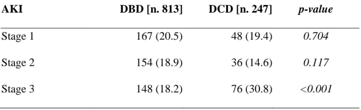
Acute kidney injury was defined according to the recent KDIGO Guideline (Kidney Disease: Improving Global Outcomes Acute Kidney Injury Work Group 2012) as an increase in serum creatinine (sCr) by ≥ 0.3 mg/dl within 48 hours or an increase in sCr to ≥ 1.5 times baseline within the first 7 days after transplantation and classified as: Stage 1, increase ≥ 0.3 mg/dl or increase of 1.5-1.9 fold from baseline; Stage 2, increase of 2-2.9 fold from baseline; Stage 3, increase > 3-fold from baseline or increase in sCr to ≥ 4.0 mg/dl or initiation of renal replacement therapy.
Glomerular filtration rate was estimated by the Modification of Diet in Renal Disease (MDRD) Study equation: [186 × sCr (mg/dL)−1.154 × age (years)−0.203 × 1.212 (if black) × 0.742 (if female)] (Gonwa TA, et al. 2004).
Evaluation of bile duct histology
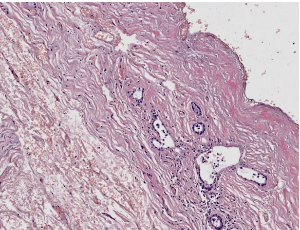
A series of consecutive patients with sample of bile duct available, were selected. The bile duct sample was retrieved after portal and arterial reperfusion of the liver, before the procedure of biliary anastomosis.
Bile duct samples were immediately preserved in 10% paraformaldehyde, then embedded in paraffin, sectioned (3-4 m thick) and stained with hematoxylin-eosin (H&E) for histological evaluation.
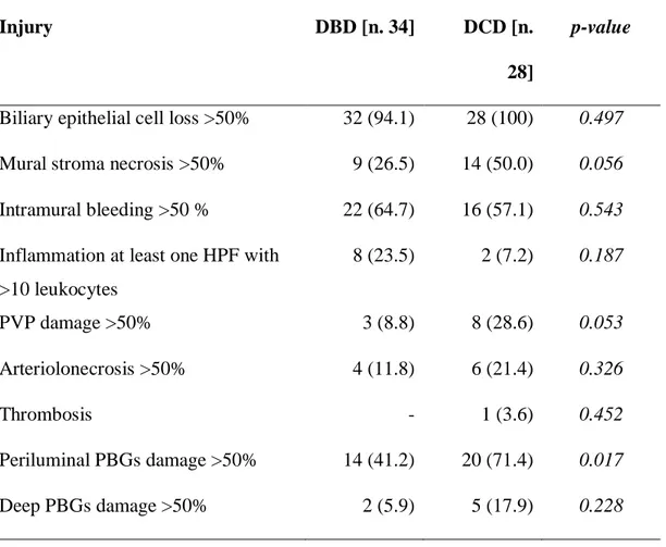
Severity of donor bile duct injury was assessed and scored on the basis of the following criteria, modified by Hansen et al (Hansen T, et al. 2012) and op den Dries et al (op den Dries S, et al. 2014):
- Biliary epithelial cell loss (absence of the epithelial lining, leading to denuded subepithelial stroma) – no epithelial cell loss, <50% of bile duct with absent epithelial lining; >50% of bile duct with absent epithelial lining.
- Mural stroma necrosis (loss of nucleated cells in duct wall) – <25% of bile duct circumference; 25-50% of bile duct circumference; >50% of bile duct circumference.
- Intramural bleeding (erythrocyte extravasation into the bile duct wall) – ≤50% of the bile duct wall; >50 % of the bile duct wall.
- Inflammation (occurrence of leukocytes anywhere in the bile duct wall) – no inflammation, at least one high-power field (HPF) with >10 leukocytes; at least one HPF with >50 leukocytes.
- Peribiliary vascular plexus (loss of endothelial cells or subendothelial oedema) – no injury; ≤50 % vessels damaged; >50 % vessels damaged.
- Arteriolonecrosis (complete loss of vital cells in the wall of arteriole/small artery) – no injury; ≤50 % vessels damaged; >50 % vessels damaged.
- Thrombosis – absent; present.
- Periluminal PBGs (detachment of the cells from the basement membrane and/or disappearance of epithelial cells from the glands) – no signs of injury; ≤50% detachment/loss of cells; >50% detachment/loss of cells.
- Deeper PBGs, located at the junction of bile duct wall stroma and muscular layer (detachment of the cells from the basement membrane and/or disappearance of epithelial cells from the glands) – no signs of injury; ≤50% detachment/loss of cells; >50% detachment/loss of cells.
Evaluation of cholangiocyte apoptosis
Cholangiocyte apoptosis in periluminal and PBGs was evaluated by quantitative terminal deoxy-nucleotidyl transferase dUTP-mediated nick-end labeling (TUNEL) analysis (Apoptag; Chemicon, Billerica, MA) on bile duct sections. Sections were analyzed in a coded manner using BX-51 light microscopy (Olympus, Tokyo, Japan) with a video cam (Spot Insight; Diagnostic Instrument, Sterling Heights, MI) and processed with an Image Analysis System (Delta Sistemi, Rome, Italy).
Evaluation of cholangiocyte proliferation
Cholangiocyte proliferation was studied in bile duct sections by proliferating cell nuclear antigen (PCNA) immunohistochemical expression. Sections were deparaffinized and endogenous peroxidase activity was blocked by a 30-min incubation in methanolic hydrogen peroxide (2.5%). Later, the endogenous biotin was blocked by a biotin blocking system (code X0590; Dako, Copenhagen, Denmark) according to the instructions supplied by the vendor. Sections were then hydrated in graded alcohol and rinsed in 1 PBS (pH 7.4) before applying the selected primary antibody. Sections were incubated overnight at 4°C with PCNA polyclonal antibodies (Santa Cruz Biotechnology, Milan, Italy). The following day, samples were rinsed with PBS for 5 min, incubated for 20 min at room temperature with secondary biotinylated antibody (LSAB Plus system; Dako, Milan, Italy) and then with Dako ABC (LSAB Plus system; Dako), and finally developed with 3,3=-diaminobenzidine. To confirm the specificity of immunoreaction, negative controls were performed for all immunoreactions. We measured the percentage of PCNA-positive small and large cholangiocytes (36).
All the available periluminal and deep PBGs were evaluated for both apoptosis and proliferation. An easy cell counting method was applied, as previously reported by Bologna-Molina et al (Bologna-Molina R, et al. 2011). Briefly, a 6x 6 fixed grid is placed over the picture of the slide taken at fixed magnification, i.e. x40, and the cell counting is started in the top left frame and finished in the top right frame following a fixed path. Numbers of both negative and positive cells were counted manually in each
image and scheduled separately. The resulting data were calculated as follows: % positive cells = positive nuclei cells ⁄ total cells nuclei x 100.
Statistical analyses
Continuous variables were tested for normality, non-normal variables have been summarised using medians and inter-quartile range with the Mann-Whitney test used for comparisons, and normal variables are described using mean and standard deviation and comparisons tests using t-tests. Chi-squared or Fishers' exact tests were used to make comparisons for categorical variables.
Data were analyzed using the SPSS 24 package. p <0.05 was considered statistically significant unless otherwise stated.
RESULTS
Recipient, donor and graft characteristics from pre- to post-transplant
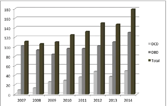
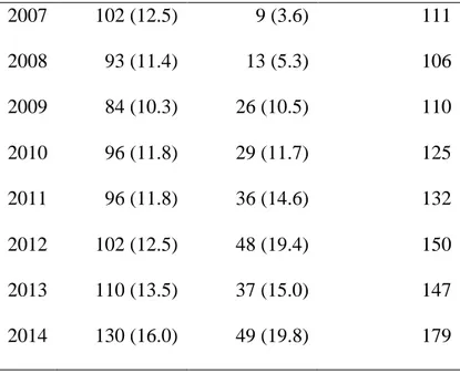
From the entire cohort of 1150 patients, 1060 liver transplant recipients (813 from DBD and 247 from DCD) were considered. Ninety patients were excluded from the analysis because of acute hepatic failure (n. 66), living donor (n. 7), combined liver-kidney (n. 16) transplantation, previous history of liver-kidney transplant (n. 1). The distribution of liver transplants performed at UHB over time is reported in Table 1 and in Figure 2.
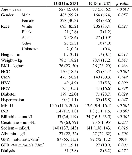
The baseline characteristics of DBD vs. DCD recipients are reported in Table 2. DCD recipients, compared to DBD, had significantly better hepatic and renal functions before LT, as showed by significantly low median MELD score (12.6 vs. 15.5;
p<0.001), serum bilirubin level (34 vs. 53; p<0.001), INR (1.3 vs. 1.4; p<0.001) and
sCr (75 vs. 79; p=0.033) levels.
Furthermore, DCD had a higher DRI than DBD (2.70 vs. 1.87; p<0.001) but, after exclusion of DCD status from the formula, there was a reversal of the trend towards a significant higher DRI in the DBD group (1.87 vs. 1.79; p=0.015) (Table 3).
In addition, DBD liver recipients had a significantly longer CIT (8.4 vs. 7.1;
p<0.001) and rWIT (39 vs. 37; p=0.0157) compared to DCD (Table 3).
Recipients from DCD had higher postoperative peak ALT and AST compared to DBD (Table 4).
The occurrence of biliary complications was higher in DCD liver transplant recipients (85/247; 34%) compared to DBD (166/813; 20%) (p<0.001), in particular incidence of IC was significantly higher in DCD (Table 5).
The incidence of AKI was 59.3% (629/1060 recipients) and was significantly higher in DCD (160/247, 64.8%) compared to DBD (469/813, 57.7%) recipients (p=0.047). The classification of AKI according to KDIGO Guideline is reported in Table 6. There were no significant differences in the incidence of AKI stages 1 and 2, whereas DCD showed a significantly higher incidence of AKI stage 3 compared to DBD (p<0.001).
Evaluation of bile duct histology
Sixty-two patients had the bile duct sample available for histological evaluation. These were comparable in terms of age (p=0.085), pre-transplant liver and renal function (INR p=0.327; bilirubin p=0.147; creatinine p=0.802; MELD score
p=0.702), liver graft warm (p=0.150) and cold ischaemia time (p=0.166) with the
entire cohort population.
Severity of donor bile duct injury was assessed and scored as reported in Table 7. A significantly higher number of DCD patients presented necrosis >50% of the bile duct wall [DCD 14/28 (50%), DBD 9/34 (26.5%) p=0.056], PVP damage [DCD 8/28 (29%), DBD 3/34 (9%); p=0.053] and periluminal PBGs damage [DCD 20/28 (71%), DBD 14/34 (41%); p=0.016].
No differences have been detected in terms of biliary epithelial cell loss, intramural bleeding, inflammation, arteriolonecrosis, thrombosis and deep PBGs damage.
Severe histological injury was defined when mural stroma necrosis >50%,
periluminal PBGs injury >50% and PVP damage >50% occurred (Figure 3). Sever
histological injury was significantly more frequent in DCD liver transplant patients
[15/28 (53.6%)] compared to DBD [7/34 (20.6%)] (p=0.007).
Evaluation of cholangiocytes apoptosis and proliferation
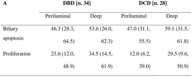
Livers transplanted from DCD demonstrate increased apoptosis and reduced proliferation at the level of both periluminal and deep PBGs, even if this not reach statistical significance (Table 8A; Figures 4, 5).
Otherwise, a significant increased apoptosis and decreased proliferation was evidenced in both periluminal (Tunel assay p=0.029; PCNA expression p=0.029) and deep PBGs (Tunel assay p=0.002; PCNA expression p=0.006) from bile duct sample with severe histological injury (Table 8B). This trend is maintained within DCD and DBD separately, but the small number of patients in the two groups reduced the statistical power of the results (Figures 6-9).
DISCUSSION
Over the past 40 years, orthotopic liver transplantation has evolved from an experimental procedure to become the treatment of choice for patients with end-stage liver failure. In the past decades, advances in immunosuppressive therapy, organ preservation, surgical and anesthetic techniques and better management of post-transplantation complications have led to further improvement in survival of patients receiving organ transplantation.
The increasing request of organs has led to the more extensive use of the so-called
marginal donors, in particular donors after circulatory death.
Within this model of donation, a more severe degree of ischaemia-reperfusion injury is occurring, as evidenced by greater graft dysfunction and increasing peak perioperative transaminases, likely related to the added donor warm ischaemia time.
The IRI seems to play a role on the pathogenesis of local and remote organ complications, leading in particular to biliary complications and development of systemic inflammatory response associated with the occurrence of acute kidney injury. Ischaemic injury in organs from DCD accumulated during the cold storage phase is exacerbated and multiplied by the initial phase of warm ischaemia, leading to an increased exposure to IRI and higher incidence of delayed graft function. Multiple pathways are implicated in IRI including cellular cascade systems, oxygen free radicals, and activation of T-cell lymphocytes.
Hepatic IRI acts locally, inducing hepatic tissue repair characterized by proliferation of hepatocytes, removal of necrotic tissue and restoration of the hepatocellular and hepatic microvascular architecture. Cholangiocytes lining the biliary ducts are very sensitive to ischaemic injury, in particular increased vulnerability of cholangiocytes during the reperfusion and prominent damage of bile ducts occurring after cold storage have been described (Noack K, et al. 1993; Brunner SM, et al. 2013). Recently, increased apoptosis and reduced proliferation of large bile duct and altered expression of biliary angiogenic factors have also been demonstrated (Mancinelli R, et al. 2015).
A higher incidence of ischaemic-cholangiopathy has been reported in transplants from DCD donors compared to donation after DBD, probably due to the longer and complex ischemic damage in this model of donation. An important role has been hypothesized for the inadequate perfusion of PVP during the retrieval process, because the small arterial plexus surrounding the biliary tree could be poorly washed by some high viscosity solutions leading to micro-thrombotic complications during the reperfusion phase that exacerbate the ischemic damage to the biliary cells (Polack WG and Porte RJ, 2006). Moreover, the injury occurring at the PBGs seems to play a role in the development of biliary complications post-liver transplantation (op den Dries S, et al.2014).
Donor warm ischemia duration also correlates with the postoperative systemic inflammatory response that could be responsible of remote organ damage, in particular acute kidney injury.
The systemic release of pro-inflammatory cytokines such as TNF-, IL-1, IL-6 and IL-8, subsequent to reperfusion of the liver and responsible of the development of SIRS, seems to be the common pathway for the occurrence of renal damage that leads to acute kidney injury.
Evidence supporting the association between IRI and the development of AKI are strongly supported by the recent literature reporting increased incidence of renal dysfunction following graft preservation injury. In particular the association between post-transplant graft dysfunction and AKI was reported (Cabezuelo JB, et al. 2006) as well as the increased risk of haemodialysis in patients with severe preservation injury (Glanemann M, et al. 2004). The greater injury of liver transplantation from DCD following the prolonged warm ischemic time, was accompanied by a 1.7-fold increased incidence of AKI (Leithead JA, et al. 2012).
In our study we evaluated the role of two different models of ischaemia, DCD and DBD, in liver transplanted grafts, on the pathogenesis of local and remote organ complications.
We retrieved clinical and laboratory data from a single high-volume transplant centre in UK, where DCD transplantations account for a quarter of the total transplant volume.
The retrieval of bile duct samples before the duct to duct anastomosis in the recipient for histological assessment has also recently become a standard of care. Therefore, it has been possible to reassess the histological features and complete the immunohistochemistry study for apoptosis and proliferation at the bile duct level.
The incidence of bile duct complications, in particular the development of IC, was increased in DCD liver transplantation, as previously reported. The occurrence of AKI was also increased in transplants from DCD compared to DBD.
Interesting results came from the histological evaluation of bile duct samples. Biliary epithelial cells loss is almost complete in the majority of bile ducts, suggesting the high degree of IRI suffered from these. Intramural bleeding >50% was also a common feature present in more than 50% of patients, irrespective of type of donation.
Bile duct samples retrieved from DCD grafts expressed more severe injury at the histological level, as evidenced by the increased incidence of mural stroma necrosis, PVP damage and periluminal PBGs damage, defining the new feature of severe
histological injury.
The evaluation of apoptosis and proliferation of PBGs cholangiocythes showed increased damage as well as reduced reactivity in line with the damage: bile ducts with
severe histological injury showed increased apoptosis and reduced proliferation as
evaluated by Tunel assay and PCNA expression, both on periluminal and deep PBGs. The same trend was confirmed analysing apoptosis and proliferation in DCD and DBD separately, but the statistical significance was not reached probably in relation to the small number of samples for each model.
Two patients only within the 62 group of patients with bile duct histology developed IC, therefore no statistical analysis was conducted. Otherwise both patients presented the severe histological injury of the bile duct.
The model of increased IRI in DCD livers seems to confirm the relation with local and remote organ damage as evidenced by the more severe damage of bile duct samples and the increased incidence of AKI.
The higher incidence of IC development in DCD strongly suggests a relation between the occurrence of severe histological injury and alteration in bile duct repair mechanisms.
This study shows an early picture of microscopic damage at the level of the bile duct soon after reperfusion of liver graft during transplantation, raising hypothesis to further evaluate those mechanisms leading to the development of bile duct non-anastomotic strictures.
A series of factors are reported to stimulate cholangiocytes proliferation in different cholangiopaties such as primary sclerosing cholangitis, primary biliary cirrhosis, polycystic liver disease and cholangiocarcinoma. During these pathologies, cholangiocytes, which in normal condition are in a quiescent state, begin to proliferate acquiring phenotypes of neuroendocrine cells, and start secreting different cytokines, growth factors, neuropeptides, and hormones to modulate cholangiocytes proliferation and interaction with the surrounding environment, trying to reestablish the balance between proliferation/loss of cholangiocytes for the maintenance of biliary homeostasis (Franchitto A, et al. 2012). Further studies are needed to define the role of long-term cholangiocyte reaction to the ischemia-reperfusion injury after liver transplantation in the development of biliary complications.
A continue cross-talk is also established between biliary cholangiocytes and endothelial cells of the PVP (Gaudio E, et al. 1996), with an interaction that mediates the adaptive changes of these cells during liver injury.
Altered expression of biliary angiogenic factors (Vascular endothelia growth factors and Angiopoietins) has been demonstrated in animal models of IRI induced damage of intrahepatic cholangiocytes (Mancinelli R, et al. 2015), therefore future research will focus on the evaluation and measurement of angiogenic factors and related receptors after liver reperfusion, to better address the actual knowledge of bile duct reaction in liver transplantation.
TABLES
Table 1. Liver transplants performed at Queen Elizabeth University Hospital Birmingham from 2007 to 2014. Year DBD [n. 813] DCD [n. 247] Total [n. 1060] 2007 102 (12.5) 9 (3.6) 111 2008 93 (11.4) 13 (5.3) 106 2009 84 (10.3) 26 (10.5) 110 2010 96 (11.8) 29 (11.7) 125 2011 96 (11.8) 36 (14.6) 132 2012 102 (12.5) 48 (19.4) 150 2013 110 (13.5) 37 (15.0) 147 2014 130 (16.0) 49 (19.8) 179
Table 2. Baseline characteristics of DBD vs. DCD liver transplant recipients. DBD [n. 813] DCD [n. 247] p-value Age – years 52 (42, 60) 57 (50, 62) <0.001 Gender Male 485 (59.7) 164 (66.4) 0.057 Female 328 (40.3) 83 (33.6) Race White 693 (85.2) 206 (83.4) 0.523 Black 21 (2.6) 3 (1.2) Asian 70 (8.6) 27 (10.9) Other 27 (3.3) 10 (4.0) Unknown 2 (0.2) 1 (0.4) Height – m 1.7 (0.1) 1.7 (0.1) 0.612 Weight – kg 78.5 (18.2) 78.4 (17.2) 0.543 BMI – kg/m2 26 (23, 30) 26 (23, 29) 0.966 HCC 150 (18.5) 85 (34.4) <0.001 CMV 473 (58.2) 149 (60.3) 0.549 HBV 40 (4.9) 13 (5.3) 0.009 HCV 85 (10.5) 41 (16.6) 0.828 Diabetes 179 (22.0) 71 (28.7) 0.029 Hypertension 90 (11.1) 39 (15.8) 0.047 MELD 15.5 (11.5, 20.7) 12.6 (9.4, 16.4) <0.001 INR 1.4 (1.2, 1.8) 1.3 (1.1, 1.5) <0.001 Bilirubin – umol/L 53 (26, 119) 34 (16.5, 63.5) <0.001 Creatinine – umol/L 79 (63, 99) 75 (61, 95) 0.033 Sodium – mEq/L 140 (137, 143) 141 (138, 143) 0.016 Albumin – g/L 27 (22, 32) 27 (22, 32) 0.794 GFR – ml/min/1.73m2 87 (65, 115) 92 (72, 112) 0079 GFR <60 ml/min/1.73m2 155 (19.1) 27 (10.9) 0.003 Dialysis 31 (3.8) 8 (3.2) 0.675
Values are expressed as number (percentage) or median (interquartile range), as appropriate.
BMI: body mass index; CMV: cytomegalovirus; DBD: donation after brain death; DCD: donation after circulatory death; HBV: hepatitis B virus; HCC: hepatocellular carcinoma; HCV: hepatitis C virus; GFR glomerular filtration rate; INR:
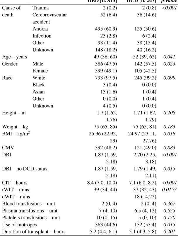
Table 3. Donor, graft and intra-operative parameters of DBD vs DCD liver transplantation. DBD [n. 813] DCD [n. 247] p-value Cause of Trauma 2 (0.2) 2 (0.8) <0.001 death Cerebrovascular accident 52 (6.4) 36 (14.6) Anoxia 495 (60.9) 125 (50.6) Infection 23 (2.8) 6 (2.4) Other 93 (11.4) 38 (15.4) Unknown 148 (18.2) 40 (16.2) Age – years 49 (36, 60) 52 (39, 62) 0.041 Gender Male 386 (47.5) 142 (57.5) 0.023 Female 399 (49.1) 105 (42.5) Race White 793 (97.5) 245 (99.2) 0.099 Black 3 (0.4) 0 (0.0) Asian 13 (1.6) 1 (0.4) Other 0 (0.0) 1 (0.4) Unknown 4 (0.5) 0 (0.0) Height – m 1.7 (1.62, 1.76) 1.71 (1.62, 1.79) 0.208 Weight – kg 75 (65, 85) 75 (65, 81) 0.183 BMI – kg/m2 25.96 (22.92, 29) 24.97 (23.11, 27.76) 0.018 CMV 392 (48.2) 121 (49.0) 0.883 DRI 1.87 (1.59, 2.18) 2.70 (2.25, 3.18) <0.001 DRI – no DCD status 1.87 (1.59, 2.18) 1.79 (1.49, 2.11) 0.015 CIT – hours 8.4 (7.0, 10.0) 7.1 (6.0, 8.2) <0.001 rWIT – mins 39 (34, 44) 37 (32, 43) 0.0157 dWIT – mins 18 (14,22)
Blood transfusions – unit 2 (0, 4) 2 (0, 4) 0.367
Plasma transfusions – unit 7 (4, 10) 6.5 (4, 12) 0.525
Platelets transfusions – unit 10 (0, 15) 5 (0, 10) 0.170
Use of inotropes 363 (44.6) 132 (53.4) 0.015
Values are expressed as number (percentage) or median (interquartile range), as appropriate.
BMI: body mass index; CIT: cold ischaemia time; CMV: cytomegalovirus; DBD: donation after brain death; DCD: donation after circulatory death; DRI: donor risk index; dWIT: donor warm ischaemia time; rWIT: recipient warm ischaemia time.
![Table 5. Biliary complications in DBD vs. DCD liver transplant recipients. DBD [n. 813] DCD [n](https://thumb-eu.123doks.com/thumbv2/123dokorg/2897771.11835/56.892.143.729.211.430/table-biliary-complications-dbd-dcd-liver-transplant-recipients.webp)