Central
Bringing Excellence in Open Access
JSM Gastroenterology and Hepatology
Cite this article: Grossi L, Ciccaglione AF, Marzio L (2017) Spontaneous Opening of LES in Achalasia: an Uncommon on-Line Documentation by High-Resolution Manometry. JSM Gastroenterol Hepatol 5(1): 1075.
*Corresponding author
Laurino Grossi, School of Gastroenterology, Department of Medical, Oral and Biotechnological Sciences, School of Gastroenterology c/o Digestive Sciences Unit, G. d’Annunzio University of Chieti-Pescara, Ospedale Spirito Santo, Via Fonte Romana 8, 65124 Pescara, Italy, Tel: 39 085 425260; Fax: 39 085 4295547; Email: Submitted: 13 December 2016 Accepted: 19 January 2017 Published: 20 January 2017 Copyright © 2017 Grossi et al. OPEN ACCESS Keywords • Achalasia
• Lower esophaeal sphincter • Esophageal motility • High resolution manometry • Dysphagia
Case Report
Spontaneous Opening of LES in
Achalasia: an Uncommon
on-Line Documentation by
High-Resolution Manometry
Laurino Grossi*, Antonio Francesco Ciccaglione, and Leonardo
Marzio
Department of Medical, Oral and Biotechnological Sciences, University of Chieti-Pescara, Italy
Abstract
The case reported is related to a 72 years old female patient, with a long-lasting clinical history of dysphagia and retrosternal discomfort. Endoscopic and radiologic exams were strongly suggestive for achalasia and high resolution manometry was scheduled to confirm the diagnosis. A type I achalasia was documented, with absent peristalsis throughout the esophageal body and not-relaxing lower esophageal sphincter. Throughout the procedure a progressive increase of the intraesophageal pressure after the subsequent wet swallows was recorded, the patient experiencing concomitant retrosternal discomfort. Once spontaneous opening of the LES region occurred not related to deglutition the esophageal pressure suddenly decreased, with significant symptoms relief by the patient. The manometric images seem to offer a clear documentation of the motor abnormalities underlying clinical pattern in end stage type I achalasia.
ABBREVIATIONS
LES: Lower Esophageal Sphincter; HRM: High Resolution Manometry; IRP: Integrated Relaxation Pressure; EGJ: Esophago-gastric Junction.
INTRODUCTION
Achalasia represents a primary motor disorder of the esophagus characterized by an incomplete or absent relaxation of the lower esophageal sphincter (LES) associated with alterations of esophageal body peristalsis [1,2]. The disease has an estimated incidence of 0.4-0.6/100000 with a prevalence of 8/100000 per year and usually affects people aged between 30-60 yrs, sometimes with a significant diagnostic delay due to the low intensity of symptoms in early stages. The main clinical feature is progressive dysphagia either for solids and liquids, regurgitation of undigested food, occasionally chest pain during meals which may lead to reduced food intake with weight loss. Clinical impact of the disease can be quantified using the Eckardt score [3]. Diagnosis of achalasia is a combination of endoscopic (dilated esophagus with undigested food, saliva in the lumen without mucosal stricturing) and radiological (aperistalsis, esophageal dilatation with “bird-beak” appearance of the LES region at X-ray esophagogram) findings; usually manometry is required to confirm the disease and, in the last years, also to distinguish
different subtypes of motor abnormalities. High Resolution Manometry (HRM) is a recently developed procedure allowing a better visualization of the amplitude and propagation of intraluminal motor waves as compared to traditional manometry by using a seamless isobaric colour representation [4]. HRM can distinguish different types of esophageal motility in achalasia, according to the most recent Chicago classification [5] : type I when failed peristalsis is present, type II when post-deglutitive panesophageal pressurization is documented and type III when some preserved signs of high-amplitude distal peristalsis are evident. The analysis of the LES pattern is performed utilizing the Integrated Relaxation Pressure (IRP), which represents the mean pressure recorded in the 4 seconds with the lowest LES pressure, during the 10-sec window after deglutition. An IRP value greater than 15 mmHg is indicative of an incomplete or absent LES relaxation suggesting an outflow obstruction at the esophago-gastric junction (EGJ). The identification of subtypes seems also useful as a prognostic factor, since therapeutic strategies appear more effective in type II achalasia, potentially representing a still “compensated” disease as compared to the absent motility present in type I; type III has an intermediate position in such prognostic evaluation [6].
The case presented is related to a patients with type I achalasia. The interest relies on the opportunity given by the
Central
Bringing Excellence in Open Access
Grossi et al. (2017) Email:
JSM Gastroenterol Hepatol 5(1): 1077 (2017)
2/4
manometric evaluation of documenting “on-line” the esophageal and LES pressure pattern exactly at the onset and subsequent relief of symptoms during the exam.
CASE PRESENTATION
A 72 years old female was admitted to our Unit because of progressive dysphagia over the years for both solids and liquids, frequent retrosternal discomfort during feeding, episodic vomit with consequent reduced food intake and weight loss of about 7 kg in the last 4 months (Eckardt score 8). Upper GI endoscopy and X-ray timed barium esophagogram had documented an esophageal dilatation with narrowing at the esophago-gastric junction and no evidence of organic stenosis. Because of suspect achalasia the patient was therefore referred to our motility lab for an esophageal high-resolution manometry (Manoscan 360, Sierra Instruments, CA, USA). Figure (1) shows that the first water deglutitions (5-ml water each) elicited no peristaltic activity throughout the esophageal body and that the LES had an increased basal tone (35 mmHg) with a complete absence of postdeglutitive relaxation (integrated relaxation pressure, IRP, >15 mmHg). These features were strongly representative of type I achalasia [7]. Throughout the procedure such a motility defect determined a progressive increase of the intraesophageal pressure following the subsequent wet swallows (Figure 2, left). At the same time the patient experienced increasing discomfort at the retrosternal level. Once the LES opening occurred, not swallow-related the esophageal pressure suddenly decreased with concomitant significant symptoms relief by the patient (Figure 2, right) as the luminal content reasonably passed into the stomach. Pressure values of esophageal body and LES surrounding the LES opening are summarized in Table (1). The exam was successfully ended without further problems, the patient received diagnosis of type I achalasia and was offered the option of undergoing laparoscopic Heller miotomy or pneumatic dilatation [8]. She preferred to
be scheduled for endoscopic pneumatic dilatation and after one session she experienced a significant improvement of her clinical conditions. One year of follow-up evidenced no clinical recurrence and no need of further therapies.
DISCUSSION
Diagnosis of achalasia is often delayed because the initial symptoms are not specific and may mimic other conditions either organic or behavioural, from esophagitis and Gastroesophageal Reflux Disease [9] to eating disorders [10]. One particular condition described by patients with achalasia is the feeling of food impaction inside the esophagus, often associated with retrosternal discomfort or pain and the need to drink liquids to overcome the sense of retained bolus. This istinctive behaviour leads to a progressive increase of the intraluminal pressure until reaching a critical threshold inside the esophagus able to overcome the barrier represented by the not relaxing LES. This phenomenon has been described for decades during radiologic barium esophagogram and is known as Hurst phenomenon [11] but it has been rarely demonstrated during HRM. Some evidences are present in literature on radiologic and manometric evaluation of motor pattern in patients with achalasia [12] but those cases have been studied using the standard manometry, which have less resolution power as compared to HRM, particularly in the analysis of LES pattern [13]. Furthermore, high resolution manometry allows not only a precise diagnosis of the disease, but also gives a visual direct representation of the motor pattern during symptoms occurrence. Our case demonstrates what could really happen in a patient with type I achalasia. The interesting finding is that by the time the patient started referring the onset of her usual retrosternal discomfort, the HRM tracing revealed a concomitant progressive increase in the intraluminal pressure, with no peristaltic activity. The opening of the esophago-gastric junction and the fall in intraesophageal body were associated
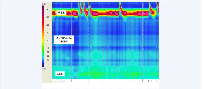
Figure 1 HRM tracing at the beginning of recording. From top to bottom Upper Esophageal Sphincter (U.E.S.), Esophageal Body and Lower Esophageal Sphincter (L.E.S.) reveal that after swallow, no peristaltic activity was present throughout the esophagus and no LES relaxation. This motor pattern is referrable to type I achalasia.
Central
Bringing Excellence in Open Access
Grossi et al. (2017) Email:
JSM Gastroenterol Hepatol 5(1): 1077 (2017)
3/4
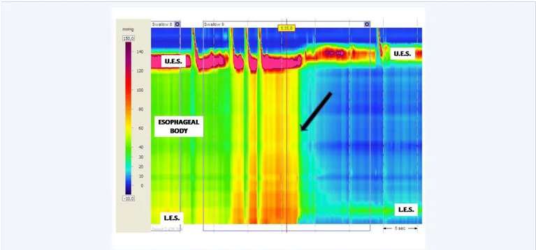
Figure 2 Throughout the procedure a progressive increase of the intraesophageal pressure following the subsequent wet swallows is clearly documented in the left half of Figure. At the same time the patient experienced increasing discomfort at the retrosternal level. It seems likely that the progressive entrapment inside the esophageal lumen of water and secretions determined a further dilatation and elongation of the esophageal body, with LES actually displaced toward the very bottom of the tracing(left side). Once the intraluminal pressure reached a critical threshold (black arrow), a spontaneous opening of the LES region occurred, not related to deglutition; the esophageal pressure suddenly decreased (right side of the Figure), with concomitant significant symptoms relief by the patient. After about 5 seconds the LES tone returned to the basal value and both UES and LES came back to the baseline position (right side). Table 1 indicates the pressure values surrounding the LES opening.
Table 1: Manometric values in the four seconds before and after the LES spontaneous opening. Pressure values of esophageal body and LES are expressed in mmHg. Note the progressive increase in the intraesophageal body pressure and the fall after LES opening, as documented by the values in the lower row. The manometric corresponding image is showed in Figure 2.
Time(sec) -4 -3 -2 -1 0 1 2 3 4
Esophageal body
(mmHg) 85 90 90 95 50 30 20 20 20
L.E.S.
(mmHg) 40 41 40 38 5 4 3 4 4
to the resolution of symptoms. The most likely explanation of our manometric finding is that, once the pressure reached a critical threshold, a sudden not swallow-related opening of the LES region occurred, the intraluminal content moved into the stomach with emptying of esophageal lumen and return of esophageal body pressure to basal levels. It should be considered that the intraluminal content was not only consistent of the water swallowed during the exam, but also included a significant amount of secretion and saliva whose production is known to be enhanced in achalasia [14]. Another further possibility to justify the LES opening could be the occurrence of a transient lower esophgeal sphincter relaxation (TLESR), a relaxation not related to swallowing, frequent in gastroesophageal reflux disease, but demonstrated also in some cases of achalasia [15]. However TLESRs occurring in achalasia patients are often incomplete and associated to a characteristic hypercontractility of the LES region. Furthermore such relaxations are usually a reflex elicited by gastric fundus distention, which is clearly uncommon in achalasia, therefore in our opinion what we found was unlikely to be a TLESR. In effect the behaviour of LES in achalasia is still under deep investigation in literature, especially after the introduction
of HRM, and it is known that a wide range of options are possible, from the complete absence to the persistence of post-deglutitive inhibition [16]. It remains that regardless of the underlying mechanism, our manometric recording was able to directly correlate the clinical feature and the esophageal pressure profile of a patient, giving the opportunity to visually confirm what in the last decades only radiological exams could detect. In conclusion this case reinforces the role of High Resolution Manometry in the diagnosis and visual characterization of all the subtypes of achalasia.
ACKNOWLEDGEMENTS
There has been no significant financial support for this work potentially influencing its outcome. The equipment used for this case (Manoscan 360, Sierra Instruments, CA, USA) is property of G. d’Annunzio University. No specific drug was used for this study.
CONFLICT OF INTEREST
The Authors declare that there are no conflicts of interest associated with the publication of the manuscript and that there
Central
Bringing Excellence in Open Access
Grossi et al. (2017) Email:
JSM Gastroenterol Hepatol 5(1): 1077 (2017)
4/4
has been no significant financial support for this work that could have influenced its outcome. The equipment used for this case is property of G d’Annunzio University.
CONTRIBUTION OF THE AUTHORS TO THE
MANUSCRIPT
Laurino Grossi and Antonio Francesco Ciccaglione performed the manometric exam and interpreted the results; Laurino Grossi was the author involved in the conception and drafting of the manuscript and Leonardo Marzio gave the final approval of the version to be submitted.
REFERENCES
1. Vaezi MF, Richter JE. Diagnosis and management of achalasia. American College of Gastroenterology Practice Parameter Committee. Am J Gastroenterol. 1999; 94: 3406-3412.
2. Vaezi MF, Pandolfino JE, Vela MF. ACG clinical guideline: diagnosis and management of achalasia. Am J Gastroenterol. 2013; 108: 1238-1249. 3. Eckardt AJ, Eckardt VF. Treatment and surveillance strategies in
achalasia: an update. Nat Rev Gastroenterol Hepatol. 2011; 8: 311-319.
4. Clouse RE, Staiano A. Topography of the esophageal peristaltic pressure wave. Am J Physiol. 1991; 261: G677-684.
5. Kahrilas PJ, Bredenoord AJ, Fox M, Gyawali CP, Roman S, Smout AJ, et al. The Chicago Classification of Esophageal Motility Disorders, v3.0. Neurogastroenterol Motil. 2015; 27: 160-174.
6. Boeckxstaens G, Zaninotto G. Achalasia and esophago-gastric junction outflow obstruction: focus on the subtypes. Neurogastroenterol Motil. 2012; 24: 27-31.
7. Bredenoord AJ, Fox M, Kahrilas PJ, Pandolfino JE, Schwizer W, Smout
AJP, et al. Chicago classification criteria of esophageal motility disorders defined in high resolution esophageal pressure topography. Neurogastroenterol Motil. 2012; 24: 57-65.
8. Boeckxstaens GE, Annese V, des Varannes SB, Chaussade S, Costantini M, Cuttitta A, et al. Pneumatic dilation versus laparoscopic Heller’s myotomy for idiopathic achalasia. N Engl J Med. 2011; 364: 1807-1816.
9. Kessing BF, Bredenoord AJ, Smout AJ. Erroneous diagnosis of gastroesophageal reflux disease in achalasia. Clin Gastroenterol Hepatol. 2011; 9: 1020-1024.
10. Stacher G, Wiesnagrotzki S, Kiss A. Symptoms of achalasia in young women mistaken as indicating primary anorexia nervosa. Dysphagia. 1990; 5: 216-219.
11. Hurst A. The treatment of achalasia of the cardia: so called “cardiospasm”. Lancet 1927; 1: 618.
12. Amaravadi R, Levine MS, Rubesin SE, Laufer I, Redfern RO, Katzka DA. Achalasia with complete relaxation of lower esophageal sphincter: radiographic-manometric correlation. Radiology. 2005; 235: 886-891. 13. Herbella FA, Patti MG. Can high resolution manometry parameters
for achalasia be obtained by conventional manometry? World J Gastrointest Pathophysiol. 2015; 6: 58-61.
14. Boyce HW, Bakheet MR. Sialorrhea: a review of a vexing, often unrecognized sign of oropharyngeal and esophageal disease. J Clin Gastroenterol. 2005; 39: 89-97.
15. Kwiatek KA, Post J, Pandolfino JE, Kahrilas PJ. Transient lower esophageal sphincter relaxation in achalasia: everything but LES relaxation. Neurogastroenterol Mot. 2009; 21:1250-1255.
16. Kahrilas PJ, Boeckxstaens G. The spectrum of achalasia: lessons from studies of pathophysiology and high-resolution manometry. Gastroenterol. 2013; 145: 954-965.
Grossi L, Ciccaglione AF, Marzio L (2017) Spontaneous Opening of LES in Achalasia: an Uncommon on-Line Documentation by High-Resolution Manometry. JSM Gastroenterol Hepatol 5(1): 1075.