The Influence of the
1-(3-Trifluoromethyl-Benzyl)-1H-Pyrazole-4-yl Moiety on the
Adenosine Receptors Affinity Profile of
Pyrazolo[4,3-e][1,2,4]Triazolo[1,5-c]
Pyrimidine Derivatives
Stephanie Federico1*, Sara Redenti1, Mattia Sturlese2, Antonella Ciancetta2,
Sonja Kachler3, Karl-Norbert Klotz3, Barbara Cacciari4, Stefano Moro2, Giampiero Spalluto1*
1 Dipartimento di Scienze Chimiche e Farmaceutiche, Università degli Studi di Trieste, Trieste, Italy, 2 Molecular Modeling Section (MMS), Dipartimento di Scienze del Farmaco, Università degli Studi di Padova, Padova, Italy, 3 Institut für Pharmakologie und Toxicologie, Universität Würzburg, Würzburg, Germany, 4 Dipartimento di Scienze Chimiche e Farmaceutiche, Università degli Studi di Ferrara, Ferrara, Italy
*[email protected](SF);[email protected](GS)
Abstract
A new series of pyrazolo[4,3-e][1,2,4]triazolo[1,5-c]pyrimidine (PTP) derivatives has been developed in order to explore their affinity and selectivity profile at the four adenosine recep-tor subtypes. In particular, the PTP scaffold was conjugated at the C2 position with the 1-(3-trifluoromethyl-benzyl)-1H-pyrazole, a group believed to confer potency and selectivity toward the human (h) A2Badenosine receptor (AR) to the xanthine ligand
8-(1-(3-(trifluoro-methyl)benzyl)-1H-pyrazol-4-yl)-1,3-dimethyl-1H-purine-2,6(3H,7H)-dione (CVT 6975). Interestingly, the synthesized compounds turned out to be inactive at the hA2BAR but they
displayed affinity at the hA3AR in the nanomolar range. The best compound of the series
(6) shows both high affinity (hA3AR Ki= 11 nM) and selectivity (A1/A3and A2A/A3> 9090;
A2B/A3> 909) at the hA3AR. To better rationalize these results, a molecular docking study
on the four AR subtypes was performed for all the synthesized compounds. In addition, CTV 6975 and two close analogues have been subjected to the same molecular docking protocol to investigate the role of the 1-(3-trifluoromethyl-benzyl)-1H-pyrazole on the bind-ing at the four ARs.
Introduction
Adenosine is an ubiquitous modulator, which exerts its functions through interaction with four G protein-coupled receptors classified as A1, A2A, A2Band A3adenosine receptors (ARs).
[1,2] While A1, A2Aand A3ARs are stimulated by low concentrations of adenosine, the A2B
OPEN ACCESS
Citation: Federico S, Redenti S, Sturlese M, Ciancetta A, Kachler S, Klotz K-N, et al. (2015) The Influence of the 1-(3-Trifluoromethyl-Benzyl)-1 H-Pyrazole-4-yl Moiety on the Adenosine Receptors Affinity Profile of Pyrazolo[4,3-e][1,2,4]Triazolo[1,5-c] Pyrimidine Derivatives. PLoS ONE 10(12): e0143504. doi:10.1371/journal.pone.0143504 Editor: James Porter, University of North Dakota, UNITED STATES
Received: June 11, 2015 Accepted: November 5, 2015 Published: December 1, 2015
Copyright: © 2015 Federico et al. This is an open access article distributed under the terms of the
Creative Commons Attribution License, which permits unrestricted use, distribution, and reproduction in any medium, provided the original author and source are credited.
Data Availability Statement: All relevant data are within the paper and its Supporting Information files. Funding: This work was supported by: Italian Ministry for University and Research, PRIN2008: protocol number 200834TC4L_002 (SM) and protocol number 200834TC4L_004 (GS SF);http://prin.miur.it/ index.php?pag=2008. The funders had no role in study design, data collection and analysis, decision to publish, or preparation of the manuscript.
subtype requires higher concentrations of the natural ligand. [3] In recent years, numerous potent and selective ligands (agonists and antagonists) for the A1, A2A, A2Band A3AR subtypes
have been discovered. [4,5] Some of them served as pharmacological tools and demonstrated potential therapeutic applications in various pathological processes. For example, human (h) A1AR agonists are useful against cardiac arrhythmias and pain, while antagonists are beneficial
in kidney failure. Antagonists for the hA2AAR were widely studied for the treatment of
Parkin-son’s disease and other neurodegenerative disorders. [3–5] To date, the physio-pathological role of the hA3AR is controversial. This AR subtype is involved in cancer and inflammation,
although it is not yet clear whether blocking or activating this receptor would be therapeutic for these conditions. [6] Identification of potent and selective ligands for the hA2BAR is
diffi-cult as compared with the other receptor subtypes. In addition, its characterization as a low-affinity adenosine receptor led to the erroneous belief that the A2BAR was not of substantial
physiological relevance. [7–9] As a consequence, the hA2BAR is the least characterized among
the ARs, although in the last years, significant advances have been made in the understanding of the molecular pharmacology and physiology of the hA2BARs. [7–9] This subtype is highly
expressed during inflammatory and hypoxic conditions and, in these circumstances, adenosine reaches very high extracellular concentrations necessary to activate the hA2BAR subtype. [9]
In fact, the hA2BARs seem to be implicated in many different patho-physiological conditions
such as: asthma, [10–14] intestinal functions, vascular tone and permeability, ischemic precon-ditioning, inflammation, angiogenesis and sickle disease. [9,15–20] Therefore, hA2BAR
antag-onists have been proposed as anti-asthmatic and/or anti-inflammatory agents. [4,5] In particular, most of the so far developed hA2BAR antagonists are xanthine derivatives or
ana-logues. [21] In 2006 a very interesting compound named CVT 6975 (8-(1-(3-(trifluoromethyl) benzyl)-1H-pyrazol-4-yl)-1,3-dimethyl-1H-purine-2,6(3H,7H)-dione (1,Fig 1) proved to be one of the most potent A2BAR antagonists ever reported. [22] In the non-xanthine family,
some adenine [23], 7-deazapurines [24], triazolo-triazine, [24] quinazoline, [25], triazinobenzi-midazole [26], pyrimidine [27] and pyrazine [28] derivatives have been reported as hA2BAR
antagonists, but few of the synthesized compounds (e.g. pyrimidines) showed both significant potency and selectivity for this receptor subtype.
In one of our research programs aimed at the identification of new non-xanthine hA2BAR
antagonists, we investigated variously substituted pyrazolo[4,3-e][1,2,4]triazolo[1,5-c]pyrimi-dine (PTP) derivatives. [29–31] A large number of derivatives with different substitutions at the N7, N8 and N5 positions (seeFig 2for numbering of the PTP ring system) have been pre-pared, but unfortunately none of the synthesized compounds showed both affinity and selectiv-ity at the hA2BARs. [29–31] One of the most interesting compounds developed is derivative 2
(Fig 1), which displays good affinity for the hA2BAR (Ki= 165 nM) but it shows a greater
affin-ity towards the hA3AR (Ki= 0.81 nM). [32] Taking into account these experimental
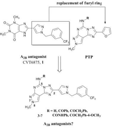
observa-tions, we decided to synthesize new derivatives by replacing the furan moiety in position C2 of the PTP scaffold with the 1-(3-trifluoromethyl-benzyl)-1H-pyrazole-4-yl moiety present in the CVT 6975 (1) with the aim to better explore the role of this position in the receptor recognition and possibly obtain new A2BAR antagonists with non xanthinic structure (Fig 2).
Materials and Methods
Chemistry
General. Reactions were routinely monitored by thin-layer chromatography (TLC) on sil-ica gel (precoated F254 Merck plates). Flash chromatography was performed using Merck 60– 200 mesh silica gel. Infrared spectra (IR) were measured on a Perkin Elmer 257 instrument.
1H-NMR were determined in CDCl
3or DMSO-d6solutions with a Varian Gemini 200
Competing Interests: SM participates in the European COST Action CM1207 (GLISTEN).
spectrometer, peaks positions are given in parts per million (δ) downfield relative to the central peak of the solvent; J values are given in Hz. Melting points were determined on a Buchi-Tot-toli instrument and are uncorrected. All reported products showed IR and1H NMR spectra in agreement with the assigned structures. Electrospray mass spectra were recorded on a ESI
Fig 1. CVT6975 (1) and the pyrazolo[4,3-e][1,2,4]triazolo[1,5-c]pyrimidine derivative 2 as potent A2B
and A3AR antagonists, respectively.
doi:10.1371/journal.pone.0143504.g001
Fig 2. Rational for the design of the target compounds 3–7. doi:10.1371/journal.pone.0143504.g002
Bruker 4000 Esquire spectrometer and compounds were dissolved in methanol. Elemental analyses were performed by the microanalytical laboratory of Dipartimento di Scienze Chi-miche e Farmaceutiche, University of Trieste, and they were within ±0.4% of the theoretical values for C, H and N. Light petroleum ether refers to the fractions boiling at 40–60°C. Organic solutions were dried over anhydrous magnesium sulfate.
Synthesis of 1-(3-trifluoromethyl-benzyl)-1H-pyrazole-4-carboxylic acid hydrazide (10). Pyrazole ester derivative 9 (5.7 g, 20 mmol) was dissolved in absolute ethanol (30 mL) and an equivalent amount of hydrazine monohydrate (0.96 mL, 20 mmol) was added and the resulting mixture was stirred at reflux for three days. The solvent was then removed in vacuo and the resi-due crystallized from EtOAc to afford the corresponding hydrazide 9 as a white solid (mp 95°C) in quantitative yield. IR (KBr): 3445–2960, 1690, 1650, 1610, 1480 cm-1;1H NMR (CDCl3)δ: 3.65
(bs, 2H); 5.21 (s, 2H); 6.51 (bs, 1H); 7.21–7.58 (m, 4H); 7.66 (s, 1H); 7.77 (s, 1H). Anal. Calcd. for C12H11N4OF3(MW 284.24): C, 50.71; H, 3.90; N, 19.17. Found: C, 50.53; H, 3.85; N, 19.18.
Synthesis of 8-methyl-2-[1-(3-trifluoromethyl-benzyl)-1H-pyrazol-4-yl]-8H-pyrazolo [4,3-e][1,2,4]triazolo[1,5-c]pyrimidin-5-ylamine (3). Pyrazole 8 (2.4 g, 20 mmol) was dis-solved in diphenyl ether (30 mL) and hydrazide 10 (6.25 g, 22 mmol, 1.1 eq) was added. The mixture was heated at 260°C using a Dean-Stark for the azeotropic elimination of water pro-duced in the reaction. After 2.5 h, the mixture was poured onto hexane (200 mL) and cooled. The precipitate of crude pyrazole-triazole derivative 11 was filtered off, and utilized for the next step without further purifications. The crude residue, was dissolved in N-methyl pyrroli-done (40 mL), cyanamide (3 eq, 60 mmol, 2.5 g) and p-toluen sulfonic acid (1.5 eq, 30 mmol, 5.7 g) were added and the mixture was heated at 160°C for 4 h. Cyanamide (3 eq, 60 mmol, 2.5 g) was added again and the solution was heated overnight. Then the solution was diluted with EtOAc (80 mL) and the precipitate (excess of cyanamide) was filtered off; the filtrate was con-centrated under reduced pressure and washed with water (3 x 30 mL). The organic layer was dried (Na2SO4) and evaporated under vacuum. The residue was purified by flash
chromatogra-phy (EtOAc/Methanol 9.5:0.5) to afford the desired compound 3 in a good overall yield (60%) as a yellow solid. Mp 125°C (EtOAc-light petroleum); IR (KBr): 3330–2960, 1640, 1605, 1550, 1450 cm-1;1H NMR (DMSO-d6)δ: 3.87 (s, 3H); 5.20 (s, 2H); 5.72 (bs, 2H); 7.18–7.41 (m, 4H);
7.84 (s, 1H); 7.96 (s, 1H); 7.97 (s, 1H). ES-MS: (MH+) 414.4. Anal. Calcd. for C18H14N9F3
(MW 413.36): C, 52.30; H, 3.41; N, 30.50. Found: C, 52.13; H, 3.44; N, 30.38.
Synthesis of 1-{8-Methyl-2-[1-(3-trifluoromethyl-benzyl)-1H-pyrazol-4-yl]-8H-pyrazolo [4,3-e][1,2,4]triazolo [1,5-c]pyrimidin-5-yl}-3-phenylurea (4). Amino compound (3) (206 mg, 0.5 mmol) was dissolved in freshly distilled dioxane (15 mL) and phenyl isocyanate (130μL, 1 mmol, 2 eq) was added. The mixture was refluxed under argon for 18 hours. Then the solvent was removed under reduced pressure and the residue was purified by flash chroma-tography (EtOAc-light petroleum 8:2) to afford the desired compound 4 in a good yield (88%) as a white solid, mp 180°C (EtOAc-light petroleum); IR (KBr): 3355–2980, 1705, 1655, 1600, 1530 cm-1;1H NMR (CDCl3)δ: 3.97 (s, 3H); 5.22 (s, 2H); 7.01–7.42 (m, 9H); 7.88 (s, 1H); 7.98
(s, 1H); 7.99 (s, 1H); 8.30 (bs, 1H); 10.93 (bs, 1H). ES-MS: (MH+) 533.1. Anal. Calcd. for C25H19N10OF3(MW 532.48): C, 56.39; H, 3.60; N, 26.30. Found: C, 56.53; H, 3.62; N, 26.48.
General procedure for acylation of amino group at the 5 position (5–7). Amino com-pound 3 (206 mg, 0.5 mmol) was dissolved in freshly distilled dioxane (15 ml) and the appro-priate acyl chloride (1 mmol) and triethylamine (1 mmol, 140μL) were added. The mixture was refluxed under argon for 18 hours. Then the solvent was removed under reduced pressure and the residue was dissolved in EtOAc (30 ml) and washed twice with water (15 ml). The organic phase was dried on anhydrous Na2SO4and concentrated under reduced pressure. The
residue was purified by flash chromatography (EtOAc-light petroleum 8:2) to afford the desired compounds (5–7).
N-{8-Methyl-2-[1-(3-trifluoromethyl-benzyl)-1H-pyrazol-4-yl]-8H-pyrazolo[4,3-e][1,2,4] triazolo[1,5-c]pyrimidin-5-yl}-benzamide (5): Yield 85%, pale yellow solid; mp 115°C (EtOAc-light petroleum); IR (KBr): 3240–2995, 1685, 1635, 1570, 1525 cm-1;1H NMR (CDCl3)δ: 3.96
(s, 3H); 5.22 (s, 2H); 7.08–7.55 (m, 9H); 7.85 (s, 1H); 7.94 (s, 1H); 7.97 (s, 1H); 9.41 (bs, 1H). ES-MS: (MH+) 518.2. Anal. Calcd. for C25H18N9OF3(MW 517.47): C, 58.03; H, 3.51; N, 24.36.
Found: C, 57.92; H, 3.44; N, 24.18.
N-{8-Methyl-2-[1-(3-trifluoromethyl-benzyl)-1H-pyrazol-4-yl]-8H-pyrazolo[4,3-e][1,2,4] triazolo[1,5-c]pyrimidin-5-yl}-phenyl-acetamide (6): Yield 85%, white solid; mp 190°C (EtOAc-light petroleum); IR (KBr): 3240–2990, 1675, 1605, 1580, 1525 cm-1;1H NMR (CDCl3)δ: 3.96
(s, 3H); 4.27 (s, 2H); 5.20 (s, 2H); 7.03–7.37 (m, 9H); 7.82 (s, 1H); 7.94 (s, 1H); 7.97 (s, 1H); 8.82 (bs, 1H). ES-MS: (MH+) 518.2. Anal. Calcd. for C26H20N9OF3(MW 531.49): C, 58.76; H,
3.79; N, 23.72. Found: C, 58.93; H, 3.65; N, 23.88.
2-(4-Methoxy-phenyl)-N-{8-Methyl-2-[1-(3-trifluoromethyl-benzyl)-1H-pyrazol-4-yl]-8H-pyrazolo[4,3-e] [1,2,4]triazolo[1,5-c]pyrimidin-5-yl}-phenyl-acetamide (7): Yield 77%, yellow solid; mp 178°C (EtOAc-light petroleum); IR (KBr): 3245–2975, 1680, 1615, 1570, 1515 cm-1;
1H NMR (CDCl
3)δ: 3.97 (s, 3H); 4.18 (s, 3H); 4.25 (s, 2H); 5.21 (s, 2H); 7.03–7.37 (m, 6H);
7.57 (d, 2H, J = 9); 7.83 (s, 1H); 7.96 (s, 1H); 7.98 (s, 1H); 8.78 (bs, 1H). ES-MS: (MH+) 562.2. Anal. Calcd. for C27H22N9O2F3(MW 561.52): C, 57.75; H, 3.95; N, 22.45. Found: C, 57.53; H,
3.86; N, 22.33.
Biology
All pharmacological methods followed the procedures as described earlier. [33–36] In brief, membranes for radioligand binding were prepared from CHO cells stably transfected with human AR subtypes in a two-step procedure. In a first low-speed step (1,000 x g) cell fragments and nuclei were removed. The crude membrane fraction was sedimented from the supernatant at 100,000 x g. The membrane pellet was resuspended in the buffer used for the respective binding experiments (50 mM Tris/HCl buffer pH 7.4 for hA1and hA2AAR; 50 mM Tris/HCl,
10 mM MgCl2, 1 mM EDTA, pH 8.25 for hA3AR), frozen in liquid nitrogen and stored at
-80°C. For the measurement of adenylyl cyclase activity only one high speed centrifugation of the homogenate was used. The resulting crude membrane pellet was resuspended in 50 mM Tris/HCl, pH 7.4 and immediately used for the cyclase assay.
For radioligand binding at human A1ARs 1 nM [3H]CCPA was used (KD= 0.61 nM),
whereas 10 nM [3H]NECA were used for A2A(KD= 20.1 nM). The high-affinity agonist [3H]
HEMADO (KD= 1.1 nM) was used for A3AR binding at a concentration of 1 nM. Non specific
binding of [3H]CCPA was determined in the presence of 1 mM theophylline, in the case of [3H]NECA and [3H]HEMADO, 100μM R-PIA was used. Ki-values from competition
experi-ments were calculated with the program SCTFIT. [33–35]
Radioligand binding at human A2BARs is problematic as no high-affinity radioligand is
commercially available for this subtype. Therefore, inhibition of NECA-stimulated adenylyl cyclase activity (stimulation with 5μM of NECA, EC50= 2.4μM) was determined as a
measure-ment of affinity of compounds. The procedure was carried out as described previously with minor modifications. [33–35] Membranes were incubated with about 150,000 cpm of [α-32P] ATP for 20 min in the incubation mixture as described without EDTA and NaCl. [33–35]
Molecular modeling
The models for human A1, A2A, A2Band A3ARs were retrieved from the Adenosiland platform
(http://mms.dsfarm.unipd.it/Adenosiland) [36], selecting the crystallographic structure of hA2AAR, in complex with the high affinity antagonist ZM241385 (PDB access code: 4EIY;
resolution 1.8 Å), as template for homology modelling. [37] The numbering of the amino acids follows the arbitrary scheme by Ballesteros and Weinstein: each amino acid identifier starts with the helix number, followed by the position relative to a reference residue among the most conserved amino acids in that helix, to which the number 50 is arbitrarily assigned. [38]
Ligand conformations were built using the MOE-builder tool implemented in the MOE suite [39] and were subjected to a MMFF94x energy minimization until the rmsd conjugate gradient was<0.05 kcal mol–1Å–1. All antagonists were docked into the hypothetical TM binding site of the hA3AR model derived from the centre of mass of the ZM241385 inhibitor.
The workflow of the molecular docking approach used in the present work has been previously reported. [40] Molecular docking study was performed employing the docking tool of the GOLD Suite v5.2.1. [41] For each compound, 25 independent docking runs were performed and searching was conducted within a user-specified docking sphere with the Genetic Algo-rithm protocol and the GoldScore scoring function. The resulting docking complexes (ligand and side-chain residues within 4.5 Å) were subjected to a MMFF94x energy minimization until the rms conjugate gradient was<1 kcal mol–1Å–1.
Analyses of docking poses and quantitative analysis for nonbonded intermolecular interac-tions (H-bonds, hydrophobic, electrostatic) were calculated and visualized using MOE suite. [39] To estimate the electrostatic contributions, atomic charges for the ligands were calculated with MOPAC-2012 [42] and the PM3/ESP methodology, whereas partial charges for the pro-tein amino acids were computed with the AMBER99 force field. The validation of the proposed docking protocol has been previously published. [43] Physicochemical and ADME properties of references and newly synthesized compounds were calculated with StarDropTM, version 6.0. [44]
Results and Discussion
Chemistry
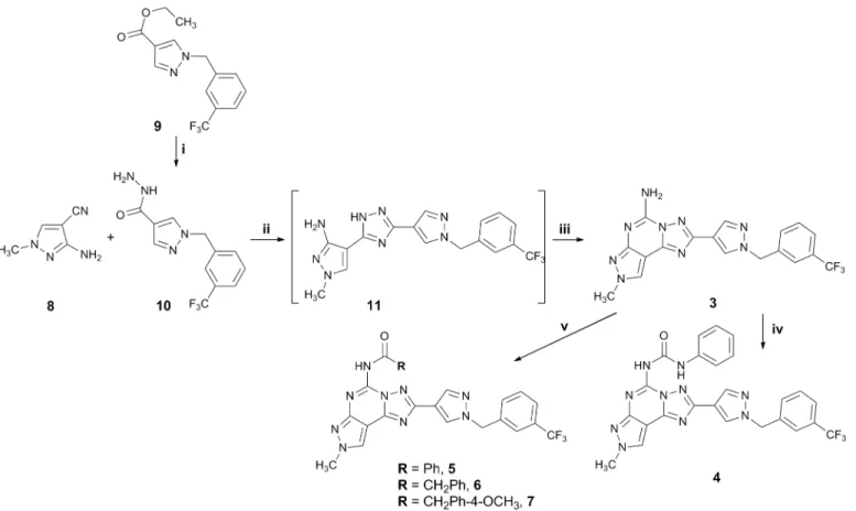
Compounds 3–7 were prepared following the general synthetic strategy summarized inFig 3. They were synthesized according to a well-known procedure for the synthesis of the PTPs, pre-viously reported. [45–47]
Reaction of the N2-methyl-4-cyano-5-amino pyrazole 8 with the hydrazide derivative 10 (prepared from the corresponding ester 9 [22] by reaction with hydrazine monohydrate in absolute ethanol at reflux for three days) in diphenyl ether at 260°C led to the aminotriazole 11, which was in turn converted, without any further purification, into the final compound 3 by reaction with cyanamide in the presence of p-toluen sulfonic acid. Amido derivatives 5–7 were obtained by reaction of the amino compound 3 with the appropriate acyl halide in diox-ane at reflux in the presence of triethylamine, while the ureido derivative 4 was obtained by reaction of the amino compound 3 with phenyl isocyanate in dioxane at reflux (Fig 3).
Biological activity
Newly synthesized compounds (3–7) were tested at the human A1, A2A,A2Band A3ARs
expressed in CHO (Chinese Hamster Ovary) cells. [3H]CCPA ([3H]2-Chloro-N6 -cyclopentyla-denosine) (hA1AR), [3H]NECA ([3H]5'-N-ethylcarboxamidoadenosine) (hA2AAR) and [3H]
HEMADO ([3H]2-(1-Hexynyl)-N-methyladenosine) (hA3ARs) were used as radioligands in
binding assays. Inhibition of NECA-stimulated adenylyl cyclase activity in CHO cells express-ing hA2Breceptors was determined as a measurement of affinity of compounds at the hA2BAR
(Fig 4). [33–35]
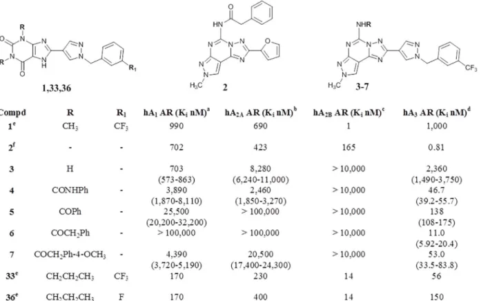
As clearly depicted inFig 4, surprisingly all novel CVT-like compounds do not show appre-ciable activity at the hA2BAR. On the contrary they have shown an unexpected high affinity,
with some in the nanomolar range, towards the hA3AR subtype. InS1 Figrepresentative
com-petition curves for the hA3selective agonist [3H]HEMADO (total binding) from single
experi-ments for compounds 6 and 4, respectively, are reported. The affinity at the hA3AR seems to
be more related to the substitution at the N5 position than to the substituent present at the C2 position. In fact, the N5-unsubstituted derivative 3, proved to be almost inactive at all four ARs, while, the introduction of a substituent at the N5 position (4,7) induced a recovery of affinity at the hA3AR with good levels of selectivity versus the other receptor subtypes. In
par-ticular, the introduction of an arylcarbamoyl moiety, which has been demonstrated optimal substituent for the hA3AR, [29–32,45–48] led to compound 4, which shows a Kivalue of 46.7
nM with a 50–70 fold selectivity versus the other receptor subtypes. The most potent com-pound of this series (6) is the derivative bearing a phenylacetyl moiety at the N5 position which shows a Kivalue of 11.1 nM and high selectivity (hA1/hA3and hA2A/hA3>9090; hA2B/hA3
>909) over the other receptor subtypes. Introduction of a methoxy group on the para position of phenyl ring (7) leads to a reduction of affinity of about 5 fold (hA3AR Ki= 53.0 nM) with a
consequent reduction of selectivity, while the replacement of phenylacetyl group with a benzoyl moiety (5) considerably reduces both affinity and selectivity at the hA3AR.
Regarding the C2 position, in a previous work it has been demonstrated that the substitu-tion of the furyl ring at the C2 posisubstitu-tion of the PTP scaffold with a phenyl moiety gives deriva-tives which maintained high affinity at the hA3AR and showed improved selectivity at this AR
subtype. [48] Moreover, with this work we have demonstrated that at the hA3AR bigger
sub-stituents than five or six-membered aromatic rings, such as the long chain of
1-benzyl-1H-Fig 3. Synthesis of desired compounds 3–7. Reagents: i: NH2NH2.H2O, EtOH, reflux, 3 days; ii: Ph2O, 260°C, 2.5 h; iii: NH2CN, pTsOH, 160°C, 4 h; iv:
PhNCO, dioxane, reflux; v: RCOCl, Et3N, dioxane, reflux.
pyrazole, are also tolerated. Obviously, this short series of compounds does not allow us to out-line a structure activity relationship (SAR) analysis. In addition, the obtained results are consis-tent with the well-known SAR of PTP derivatives at the hA3AR, that is: at the N5 position a
substitution on the amino group is preferred and, in particular, phenylacetamido moieties are better than benzamido moieties; whereas, at the C2 position, the substitution of the furyl ring enhances selectivity versus the other receptor subtypes. [29–32,45–48] It is worth to remember that although no A3AR antagonist has still reached clinical trials, there are several
pharmaco-logical evidences that suggest their potential use for the treatment of asthma, chronic obstruc-tive pulmonary disease (COPD), neurodegeneraobstruc-tive diseases, stroke, glaucoma and cancer. [6] Consequently there is an ongoing interest in the development of novel potent and selective antagonists for the A3AR subtype.
Computational studies
To better rationalize these unexpected experimental results, a molecular docking study on the four hAR subtypes was performed. The workflow of the molecular docking approach used in the present work has been previously reported. [40,49] First of all, the role of the 1-(3-trifluoro-methyl-benzyl)-1H-pyrazole-4-yl moiety in receptor recognition has been investigated starting from the analysis of the hypothetical binding mode of CTV 6975 (1) and two other structurally similar analogues 36 and 33 [22] against all four AR subtypes (Fig 5A). In particular, com-pound 36 differs in the 1,3-disubstitution of the xanthine core, where the methyl groups of
Fig 4. Structures and binding profile of reference (1,2,33,36) and synthesized compounds (3–7).aDisplacement of specific [3H]-CCPA binding at hA1
ARs expressed in CHO cells, (n = 3–6).bDisplacement of specific [3H]-NECA binding at hA2AARs expressed in CHO cells.cKivalues of the inhibition of
NECA-stimulated adenylyl cyclase activity in CHO cells expressing hA2BARs.dDisplacement of specific [3H]-HEMADO binding at hA3ARs expressed in
CHO cells. Data are expressed as geometric means, with 95% confidence limits.edata from ref. [22].fdata from ref. [32].
CVT 6975 (1) are replaced by two propyl groups. Compound 33 shares the same substituents on the xanthine core with 36 with the trifluoromethyl group being replaced by a fluorine atom. Both compounds retain a notable affinity for the hA2BAR (Ki= 14 nM) but a lower selectivity
towards the other subtypes (36: hA1AR Ki= 170 nM; hA2AAR Ki= 400 nM; hA3AR Ki= 150
nM, 33: hA1AR Ki= 170 nM; hA2AAR Ki= 230 nM; hA3AR Ki= 56 nM). [22]
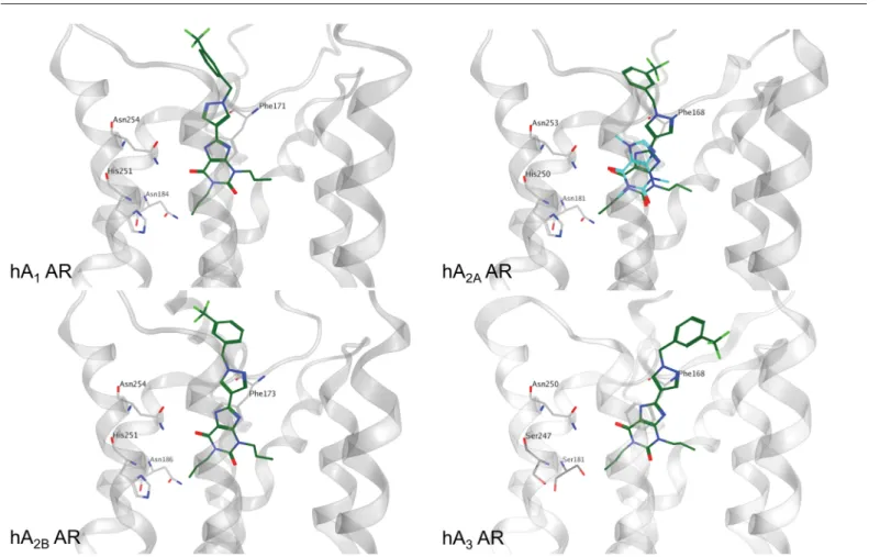
Molecular docking results suggest for the three CVT analogues (1, 36, 33) an univocal binding mode into the putative orthosteric pocket of the hA2BAR with the xanthine core faced to the key
residue Asn254 (6.55) and sided by Phe173 (EL2), Val250 (6.51) and Ile276 (7.39) (Fig 5B). The 1-benzyl-1H-pyrazole moiety whether substituted with trifluoromethyl or fluorine atom interacts with Met272 (7.35) and with residues located in EL2, namely Leu172-Glu174. As an example, the hypothetical binding mode of compound 36 at the hA2BAR is reported inFig 5B.
Interest-ingly, the docking results of compound 36 at the hA1, hA2Aand hA3ARs confirm a similar
rec-ognition pathway to that observed for the A2BAR (Fig 5B). In particular, the xanthine core is
positioned closely to that observed for two other xanthinic ligands, caffeine and XAC, in their crystallographic bound state at the hA2AAR (PDB codes 3RFM and 3REY, respectively). [50]
Moreover, molecular docking results suggest a crucial role of the 1-benzyl-1H-pyrazole moiety in increasing the binding affinity against all the receptor subtypes, while the nature of the
Fig 5. Binding mode of xanthine-based compounds at the four AR subtypes. Compound 36, in dark green, was selected as reference to show the proposed binding mode at the four AR subtypes. The crystallographic coordinates of caffeine, in magenta, bound to hA2AAR are reported superimposed to
the binding mode of compound 36. The xanthine core of compound 36 is oriented in a similar manner to the crystallographic data. Residues particularly important in the binding are reported as light grey sticks.
1,3-disubstitution at the xanthine core seems to play a role in determining the ligand selectivity profile. In fact, the presence of larger substituents in 1,3 positions, such as propyl groups, seems to be well tolerated at the hA1, hA2Aand hA3ARs while at the hA2BAR the 1,3-dimethyl
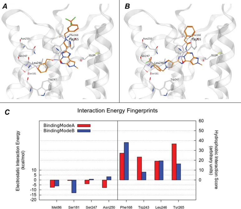
substi-tution seems to guarantee the best shape complementarity in the orthosteric binding pocket. Secondly, all newly synthesized analogues were subjected to the same docking protocol. In particular, at the hA3AR all compounds 4–7 showed an appreciable receptor complementarity
even if two distinct plausible binding modes are detected (hereafter named A and B) analyzing all docking poses. In the binding mode A, the 1-benzyl-1H-pyrazole moiety is placed similarly to the CVT-like compounds previously described, with these specific interacting residues: Gln167 (EL2), Leu264 (7.35), Tyr265 (7.36). The substituent at the N5 position is inserted in an accessory pocket defined by Ser181 (5.42), Leu246 (6.51) and Ser247 (6.52). This pocket is located deeper in the TM bundle, under the Asn250 (6.51), and it is delimited by TM3 and TM5 and by Trp243 (6.48). Interestingly, this accessory pocket is probably the most relevant structural difference among the hypothetical binding sites of the AR subtypes. In particular, the Ser247 (6.52) in the hA3AR is not conserved in the other subtypes, replaced by an histidine
in the other subtypes. This mutation significantly perturbs the shape and the of the hA3AR
orthosteric binding pocket compared to those of all the other subtypes. More interestingly, this histidine has been reported to participate in both agonist and antagonist binding to the hA2A
AR by mutagenesis studies. [51,52] Also Ser181 (5.42) is a peculiarity of the hA3AR being
replaced by conserved asparagine in the other AR subtypes.
However, in the hypothetical binding mode A the Asn250 (6.55) residue is not directly involved in the interaction with the compounds (4–7), although a water-mediated interaction could not be excluded. As an instance of binding mode A the pose of most potent analogue (6) is shown inFig 6A, while the superposition of the receptor complex for 36 and 6 is reported inS4 Fig.
In the binding mode B, the position of the substituent at the C2 and N5 positions is inverted, while the orientations of the PTP core and of the methyl at N8 position are retained in the same region occupied in the binding mode A (Fig 6BandS4B Fig). The two distinct poses show similar interactions networks as demonstrated by comparing the Interaction Energy (IE) plots where the per-residue electrostatic and hydrophobic contributions are calculated (S4C Fig). Even a more quantitative comparison of the IE contributions for the most important resi-dues reveals similar profile (Fig 6C). On the contrary, all analogues when docked into hA1,
hA2Aand hA2BARs did not show a stabilized binding pose comparable to those observed for
the hA3AR. In particular compounds 4–7, that are characterized by large substituents in both
C2 and N5 positions, seem not to be able to occupy the accessory pocked that we have previ-ously described characterizing the orthosteric binding site of the hA3AR. Also in this case, the
analysis of the Interaction Energy (IE) plots supports the hypothesis of the complementary lacking when compounds 4–7 interact with hA1, hA2Aand hA2BARs. Again, the most relevant
difference can ascribed to the missing interaction with the residues delimiting the accessory cavity, as shown inS5 Fig.
The physicochemical and ADME (absorption, distribution, metabolism, and excretion) properties of the newly synthesized compounds (3–7) were calculated in silico and compared to those calculated for CVT-like derivatives and reference hA3AR antagonists recently
reviewed by Borea et al. (S1 Table). [53] The PTP scaffold in comparison to the xanthine core shows a deterioration of the pharmacokinetic (PK) profile. In particular, the partition coeffi-cient cLogP (4.49 and 2.87 for compound 6 and CVT 6975 (1), respectively) and the solubility are affected. Nevertheless, compound 6 and CVT 6975 (1) present similar ADME properties. The limitations in the pharmacokinetic profile is a common issue for the selective hA3AR
antagonists already reported in literature. With only few exceptions, the gain in selectivity over the other human ARs subtypes is associated with a PK profile deterioration.
Conclusions
We have presented a novel series of pyrazolo[4,3-e][1,2,4]triazolo[1,5-c]pyrimidines bearing a 1-(3-trifluoromethyl-benzyl)1H-pyrazol-4-yl moiety at the C2 position in order to explore the effect on affinity and selectivity profiles at the four ARs. In particular, the 1-(3-trifluoromethyl-benzyl)1H-pyrazol-4-yl group when attached at the 8 position of xanthine derivatives, such as CVT-6975 (1), is known to confer high affinity for the hA2BAR. Curiously, the synthesized
Fig 6. Binding mode of compound 6 at the hA3AR. (A) (B) Hypothetical binding mode A and B of newly synthetized compounds to hA3AR. The most
potent derivative, 6, was selected as example and is represented as orange stick. Subsets of hA3AR residues, involved in the binding, are coloured in light
grey. (C) The electrostatic and hydrophobic contributes to interaction energy calculated for the residue mostly involved in the binding are reported compound 6 in the conformation reported in panel A (in red) and B (in blue). Electrostatic energy values are expressed in kcal mol–1, whereas hydrophobic scores are expressed in arbitrary hydrophobic units.
compounds were inactive at the hA2BAR but demonstrated activity as potent and selective
antagonist of the hA3AR. A molecular docking study was performed in order to rationalize the
influence of a 1-(3-trifluoromethyl-benzyl)-1H-pyrazole branch. We observed that only the hA3AR has the topological features to accommodate this new series in agreement with
experi-mental data. Interestingly, the ability to host such a big moiety can be ascribed to an accessory binding pocket present in the hA3AR, which is formed by specific unconserved residues. Two
possible binding modes were proposed in which the substituents in C2 and N5 are reciprocally inverted. Further studies will be needed to validate which one is predominant or if they may co-exist. Although derivatives more potent at the hA3AR, and with a better drug-like
proper-ties than compound 6, have been already reported in literature, our derivative shows high selec-tivity for hA3AR over the other ARs subtypes (S1 Table), which is comparable to other
selective antagonists already reported. [53] More interestingly, the high affinity obtained by introducing long chains, such as a benzyl-1H-pyrazole, at the C2 position, opens to new possi-bilities in the development of new derivatives bearing PTP or PTP-derived simplified scaffolds. In addition, in the binding mode A, the chain at the C2 position is directed to the solvent exposed area of the hA3AR binding pocket, suggesting a new anchoring point for further
derivatization (e.g. with fluorophore for detection purposes).
Supporting Information
S1 Fig. Competition of compounds 6 and 4 for A3receptor binding.Both compounds show
high affinity binding to hA3ARs as shown by competition for the A3selective agonist [3H]
HEMADO. Representative curves (total binding) from single experiments with Kivalues of 17
and 39 nM for compounds 6 and 4, respectively, are reported. (PDF)
S2 Fig. Competition of compound 3 for A1receptor binding.Compound 3 shows in a
radi-oligand competition assay with the A1selective radioligand [3H]CCPA a Kivalue of 764 nM.
The curve shows total binding to hA1ARs from a representative single experiment.
(PDF)
S3 Fig. Competition of compound 3 for A2Areceptor binding.Compound 3 shows in a
radi-oligand competition assay with the nonselective radiradi-oligand [3H]NECA a Kivalue of 6820 nM.
The curve shows total binding to hA2AARs from a representative single experiment.
(PDF)
S4 Fig. Hypothetical binding modes of compound 6 superimposed to compound 36 at the hA3AR and electrostatic and hydrophobic contributions maps for compound 6.The
hypo-thetical binding modes (A and B respectively indicated) of compound 6 are reported super-posed to the coordinates of compound 36 to reveal the similarity in the accommodation of the common 1-(3-Trifluoromethyl-benzyl)-1H-pyrazole residue. The coordinates of compound 36 in B are obtained from a secondary docking solution. (C) Per residue electrostatic interaction energy map and per residue hydrophobic interaction score map. The maps are calculated for a selected pose of compound 6 inside the hA3AR binding site. Electrostatic energy values are
expressed in kcal mol–1, whereas hydrophobic scores are expressed in arbitrary hydrophobic units.
(TIF)
S5 Fig. Comparison of the contribution to the docking score of the key residue for the bind-ing of compound 6 to hA3AR according molecular docking studies.The contributes to
in panels A, B, C and D respectively. In panel D, the profiles of the two predominant binding modes for hA3AR, A (red) and B (blue), are showed. In Panel E the location of residues Met86,
Ser181, Ser247 and Asn250 (in cyan) and Phe168, Trp243, Leu246 and Tyr265 (in green) in the A3AR and the corresponding residues in the others AR subtypes is indicated by the ball
representation of alpha Carbon atoms. (TIF)
S1 Table. Selectivity profile and predicted physicochemical and ADME properties of refer-ences and newly synthesized compounds (3–7).
(DOCX)
Acknowledgments
S.M. is very grateful to Chemical Computing Group for the scientific and technical partnership. S.M. participates in the European COST Action CM1207 (GLISTEN).
Author Contributions
Conceived and designed the experiments: SF K-NK AC SM GS. Performed the experiments: SF SR MS SK. Analyzed the data: SF MS AC SK BC. Contributed reagents/materials/analysis tools: GS SM K-NK. Wrote the paper: SF MS BC GS.
References
1. Chen JF, Eltzsching HK, Fredholm BB. Adenosine receptors as drug targets: what are the challenges? Nat Rev Drug Discov. 2013; 12:265–286. doi:10.1038/nrd3955PMID:23535933
2. Fredholm BB, IJzerman AP, Jacobson KA, Klotz KN, Linden J. International Union of Pharmacology. XXV. Nomenclature and classification of adenosine receptors. Pharm Rev. 2011; 53:527–552. 3. Fredholm BB. Adenosine receptors as drug targets. Exp Cell Res. 2010; 316:1284–1288. doi:10.1016/
j.yexcr.2010.02.004PMID:20153317
4. Muller CE, Jacobson KA. Recent developments in adenosine receptor ligands and their potential as novel drugs. Biochim Biophys Acta. 2011; 1808:1290–1308. doi:10.1016/j.bbamem.2010.12.017
PMID:21185259
5. Fredholm BB, IJzerman AP, Jacobson KA, Linden J, Muller CE. International Union of Basic and Clini-cal Pharmacology. LXXXI. Nomenclature and classification of adenosine receptors: an update. Pharm Rev. 2011; 63:1–34. doi:10.1124/pr.110.003285PMID:21303899
6. Cheong SL, Federico S, Venkatesan G, Mandel AL, Shao YM, Moro S, et al. The A3 adenosine recep-tor as multifaceted therapeutic target: pharmacology, medicinal chemistry, and in silico approaches. Med Res Rev. 2013; 2:235–335.
7. Kalla RV, Zablocki J. Progress in the discovery of selective, high affinity A2B adenosine receptor antag-onists as clinical candidates. Purinergic Signal. 2009; 5:21–29. doi:10.1007/s11302-008-9119-x
PMID:18568423
8. Baraldi PG, Tabrizi MA, Fruttarolo F, Romagnoli R, Preti D. Recent improvements in the development of A2B adenosine receptor agonists. Purinergic Signal. 2009; 5: 3–19. doi: 10.1007/s11302-009-9140-8PMID:19184536
9. Aherne CM, Kewley EM.The resurgence of A2Badenosine receptor signaling. Biochim Biophys Acta.
2011; 1808:1329–1339. doi:10.1016/j.bbamem.2010.05.016PMID:20546702
10. Feoktistov I, Biaggioni I. Adenosine A2B receptors evoke interleukin-8 secretion in human mast cells. An enprofylline-sensitive mechanism with implications for asthma. J Clin Investig. 1995; 96:1979– 1986. PMID:7560091
11. Ryzhov S, Goldstein AE, Matafonov A, Zeng D, Biaggioni I, Feoktistov I. Adenosine-activated mast cells induce IgE synthesis by B lymphocytes: an A2B-mediated process involving Th2 cytokines IL-4
and IL-13 with implications for asthma. J Immunol. 2004; 172:7726–7733. PMID:15187156
12. Zhong H, Wu Y, Belardinelli L, Zeng D. A2Badenosine receptors induce IL-19 from bronchial epithelial
13. Zhong H, Belardinelli L, Maa T, Feoktistov I, Biaggioni I, Zeng D. A2Badenosine receptors increase
cytokine release by bronchial smooth muscle cells. Am J Respir Cell Mol Biol. 2004; 30:118–125. PMID:12855406
14. Zhong H, Belardinelli L, Maa T, Zeng D. Synergy between A2Badenosine receptors and hypoxia in
acti-vating human lung fibroblasts. Am J Respir Cell Mol Biol. 2005; 32:2–8. PMID:15472138
15. Colgan SP, Fennimore B, Ehrentraut SF. Adenosine and gastrointestinal inflammation. J Mol Med. 2013; 91:157–164. doi:10.1007/s00109-012-0990-0PMID:23296303
16. Headrick JP, Ashton KJ, Rose’Meyer RB, Peart JN. Cardiovascular adenosine receptors: expression, actions and interactions. Pharmacol Therapeut. 2013; 140: 92–111.
17. Eltzschig HK, Bonney SK, Eckle T. Attenuating myocardial ischemia by targeting A2Badenosine
recep-tors. Trends Mol Med. 2013; 19:345–354. doi:10.1016/j.molmed.2013.02.005PMID:23540714
18. Feoktistov I, Biaggioni I. Role of adenosine A2Breceptors in inflammation. Adv Pharmacol. 2011;
61:115–144. doi:10.1016/B978-0-12-385526-8.00005-9PMID:21586358
19. Kong T, Westerman KA, Faigle M, Eltzschig HK, Colgan SP. HIF-dependent induction of adenosine A2Breceptor in hypoxia. FASEB J. 2006; 20:2242–2250. PMID:17077301
20. Zhang Y, Dai Y, Wen J, Zhang W, Grenz A, Sun H, et al. Detrimental effects of adenosine signaling in sickle cell disease. Nat Med. 2011; 17:79–86. doi:10.1038/nm.2280PMID:21170046
21. Kalla RV, Zablocki J. Progress in the discovery of selective, high affinity A2B adenosine receptor antag-onists as clinical candidates. Purinergic Signal. 2009; 5:21–29. doi:10.1007/s11302-008-9119-x
PMID:18568423
22. Kalla RV, Elzein E, Perry T, Li X, Palle V, Varkhedkar V, et al. Novel 1,3-disubstituted 8-(1-benzyl-1H-pyrazol-4-yl) xanthines: high affinity and selective A2B adenosine receptor antagonists. J Med Chem. 2006; 49:3682–3692. PMID:16759111
23. Harada H, Asano O, Hoshino Y, Yoshikawa S, Matsukura M, Kabasawa Y, et al. 2-Alkynyl-8-aryl-9-methyladenines as novel adenosine receptor antagonists: their synthesis and structure-activity relation-ships toward hepatic glucose production induced via agonism of the A2Breceptor. J Med Chem. 2001;
44:170–179. PMID:11170626
24. deZwart M, Vollinga RC, Beukers MW, Sleegers DF, von Frijtag Drabbe Kunzel JK, de Groote M, et al. Potent antagonists for the human adenosine A2Breceptor. Derivatives of the triazolotriazine adenosine
receptor antagonist ZM241385 with high affinity. Drug Dev Res. 1999; 48:95–103.
25. Webb TR, Lvovskiy D, Kim SA, Ji XD, Melman N, Linden J, Jacobson KA. Quinazolines as adenosine receptor antagonists: SAR and selectivity for A2Breceptors. Bioorg Med Chem. 2003; 11:77–85. PMID:
12467710
26. Taliani S, Pugliesi I, Barresi E, Simorini F, Salerno S, La Motta C, et al. 3-aryl-[1,2,4]triazino[4,3-a]ben-zimidazol-4(10H)-one: a novel template for the design of highly selective A2B adenosine receptor antagonists. J Med Chem. 2012; 55:1490–1499. doi:10.1021/jm201177bPMID:22257095
27. Vidal B, Nueda A, Esteve C, Domenech T, Benito S, Reinoso RF, et al. Discovery and characterization of 4'-(2-furyl)-N-pyridin-3-yl-4,5'-bipyrimidin-2'-amine (LAS38096), a potent, selective, and efficacious A2B adenosine receptor antagonist. J Med Chem. 2007; 50:2732–2736. PMID:17469811
28. Eastwood P, Esteve C, González J, Fonquerna S, Aiguadé J, Carranco I, et al. Discovery of
LAS101057: A Potent, Selective, and Orally Efficacious A2BAdenosine Receptor Antagonist. ACS Med
Chem Lett. 2010; 2:213–218. doi:10.1021/ml100249ePMID:24900298
29. Pastorin G, Da Ros T, Spalluto S, Deflorian F, Moro S, Cacciari B, et al. Pyrazolo[4,3-e]-1,2,4-triazolo [1,5-c]pyrimidine derivatives as adenosine receptor antagonists. Influence of the N5 substituent on the affinity at the human A3and A2Badenosine receptor subtypes: a molecular modeling investigation. J
Med Chem. 2003; 46:4287–4296. PMID:13678407
30. Baraldi PG, Cacciari B, Romagnoli R, Klotz KN, Spalluto G, Varani K, et al. Pyrazolo[4,3-e]1,2,4-tria-zolo[1,5-c]pyrimidine derivatives as adenosine receptor ligands: A starting point for searching A2B
adenosine receptor antagonists. Drug Develop Res. 2001; 53:225–235.
31. Baraldi PG, Cacciari B, Romagnoli R, Borea PA, Varani K, Pastorin G, et al. Pyrazolo-triazolo-pyrimi-dine derivatives as adenosine receptor antagonists: a possible template for adenosine receptor sub-types? Curr Pharm Design. 2002; 8:2299–2332.
32. Michielan L, Bolcato C, Federico S, Cacciari B, Bacilieri M, Klotz KN, et al. Combining selectivity and affin-ity predictions using an integrated Support Vector Machine (SVM) approach: An alternative tool to discrim-inate between the human adenosine A2A and A3 receptor pyrazolo-triazolo-pyrimidine antagonists binding sites. Bioorg Med Chem. 2009; 17:5259–5274. doi:10.1016/j.bmc.2009.05.038PMID:19501513
33. Klotz KN, Hessling J, Hegler J, Owman C, Kull B, Fredholm BB, et al. Comparative pharmacology of human adenosine receptor subtypes—characterization of stably transfected receptors in CHO cells. Naunyn-Schmiedeberg's Arch Pharmacol. 1998; 357:1–9.
34. De Lean A, Hancock AA, Lefkowitz RJ. Validation and statistical analysis of a computer modeling method for quantitative analysis of radioligand binding data for mixtures of pharmacological receptor subtypes. Mol Pharmacol. 1982; 21:5–16. PMID:6982395
35. Klotz KN, Cristalli G, Grifantini M, Vittori S, Lohse MJ. Photoaffinity labeling of A1-adenosine receptors.
J Biol Chem. 1985; 260:14659–14664. PMID:2997218
36. Floris M, Sabbadin D, Medda R, Bulfone A, Moro S. Adenosiland: walking through adenosine receptors landscape. Eur J Med Chem. 2012; 58:248–257. doi:10.1016/j.ejmech.2012.10.022PMID:23127988
37. Liu W, Chun E, Thompson AA, Chubukov P, Xu F, Katritch V, et al. Structural basis for allosteric regula-tion of GPCRs by sodium ions. Science. 2012; 337:232–236. doi:10.1126/science.1219218PMID:
22798613
38. Ballesteros JA, Weinstein H. Integrated methods for the construction of three-dimensional models and computational probing of structure-function relations in G protein-coupled receptors. In: Sealfon SC, editors. Methods in Neurosciences. Volume 25. San Diego, CA: Academic Press; 1995. pp. 366–428. 39. MOE (Molecular Operating Environment), version 2014.09; Chemical Computing Group Inc., 1010
Sherbooke St. West, Suite #910, Montreal, Quebec, Canada, 2014.
40. Ciancetta A, Cuzzolin A, Moro S. Alternative quality assessment strategy to compare performances of GPCR-ligand docking protocols: the human adenosine A2Areceptor as a case study. J Chem Inf
Model. 2014; 54:2243–2254. doi:10.1021/ci5002857PMID:25046649
41. GOLD suite, version 5.2.1; Cambridge Crystallographic Data Centre, Cambridge. 42. Stewart, J. J. P.MOPAC 7; Fujitsu Limited, Tokyo, Japan, 1993.
43. Cheong SL, Dolzhenko AV, Paoletta S, Lee EP, Kachler S, Federico S, et al. Does the combination of optimal substitutions at the C2-, N5- and N8-positions of the pyrazolo-triazolo-pyrimidine scaffold guar-antee selective modulation of the human A3adenosine receptors? Bioorg Med Chem. 2011; 19:6120–
6134. doi:10.1016/j.bmc.2011.08.026PMID:21908194
44. Stardrop™, version 6.0; Optibrium Ltd: 7221 Cambridge Research Park, Beach Drive, Cambridge CB25 9TL, UK.
45. Moro S, Braiuca P, Deflorian F, Ferrari C, Pastorin G, Cacciari B, et al. Combined target-based and ligand-based drug design approach as a tool to define a novel 3D-pharmacophore model of human A3
adenosine receptor antagonists: pyrazolo[4,3-e]1,2,4-triazolo[1,5-c]pyrimidine derivatives as a key study. J Med Chem. 2005; 48:152–162. PMID:15634009
46. Gatta F, Del Giudice MR, Borioni A, Borea PA, Dionisotti S, Ongini E. Synthesis of imidazo[1,2-c]pyra-zolo[4,3-e]pyrimidines, pyrazolo[4,3-e]1,2,4-triazolo[1,5-c]pyrimidines and triazolo[5,1-i]purines: new potent A2adenosine receptor antagonists. antagonists. Eur J Med Chem. 1993, 28:569–577.
47. Baraldi PG, Cacciari B, Romagnoli R, Spalluto G, Moro S, Klotz KN, et al. Pyrazolo[4,3-e]1,2,4-triazolo [1,5-c]pyrimidine derivatives as highly potent and selective human A3adenosine receptor antagonists:
influence of the chain at the N(8) pyrazole nitrogen. J Med Chem. 2000; 43:4768–4780. PMID:11123985
48. Cheong SL, Dolzhenko A, Kachler S, Paoletta S, Federico S, Cacciari B, et al. The significance of 2-furyl ring substitution with a 2-(para-substituted) aryl group in a new series of pyrazolo-triazolo-pyrimi-dines as potent and highly selective hA3adenosine receptors antagonists: new insights into
structure-affinity relationship and receptor-antagonist recognition. J Med Chem. 2010; 53:3361–3375. doi:10. 1021/jm100049fPMID:20307065
49. Cuzzolin A, Sturlese M, Malvacio I, Ciancetta A, Moro S. DockBench: an integrated informatic platform bridging the gap between the robust validation of docking protocols and virtual screening simulations. molecules. 2015; 20:9977–9993. doi:10.3390/molecules20069977PMID:26035098
50. Dore AS, Robertson N, Errey JC, Ng I, Hollenstein K, Tehan B, et al. Structure of the adenosine A2A receptor in complex with ZM241385 and the xanthines XAC and caffeine. Structure. 2011; 19:1283– 1293. doi:10.1016/j.str.2011.06.014PMID:21885291
51. Gao ZG, Chen A, Barak D, Kim SK, Muller CE, Jacobson KA. Identification by site-directed mutagene-sis of residues involved in ligand recognition and activation of the human A3 adenosine receptor. J Biol Chem. 2002; 277: 19056–19063. PMID:11891221
52. Jiang Q, Lee BX, Glashofer M, van Rhee AM, Jacobson KA. Mutagenesis reveals structure-activity par-allels between human A2A adenosine receptors and biogenic amine G protein-coupled receptors. J Med Chem. 1997; 40:2588–2595. PMID:9258366
53. Borea PA, Varani K, Vincenzi F, Baraldi PG, Tabrizi MA, Merighi S, Gessi S. The A3 adenosine recep-tor: history and perspectives. Pharmacol Rev. 2015; 67:74–102. doi:10.1124/pr.113.008540PMID: