Clin Case Rep. 2019;7:529–532. wileyonlinelibrary.com/journal/ccr3
|
5291
|
INTRODUCTION
Complications at the aortic neck represent one of the remain-ing challenges of endovascular aneurysm repair (EVAR).1-3 In
particular, type IA endoleak (EL) is associated with a consid-erably higher risk of aneurysm rupture and therefore necessi-tates prompt and definitive treatment.4 Type IA EL discovered
during follow‐up is usually caused by graft migration in a previ-ous hostile aortic neck and/or aortic neck dilatation.4 Proximal
extension cuffs, fenestrated endografts, the chimney technique, bare metal stents, and external banding have been employed to treat this complication.5-10
More recently, the Heli‐FX endoanchor System (Medtronic, Santa Rosa, CA, USA) has been used to prevent proximal neck complications in patients with challenging neck anatomy and to treat these complications when they arise.11
This system is a helical endovascular suture device in-tended to provide fixation of an endograft to the aortic wall. According to the instructions for use from the manufac-turer, each helically shaped 4.5‐mm‐length, 3‐mm‐diameter
EndoAnchor is implanted serially around the circumference of the endograft in the proximal aortic neck.12 After the
iden-tification of the leak channel, the procedure is completed creating an adjunctive “suture line” in correspondence of the leak. Literature demonstrated satisfactory short‐term success with endoanchors.11,13,14
Today's hybrid operating rooms (HOR) facilitate complex and very accurate interventions with advanced radiographic imaging, such as 3‐dimensional image fusion (3D‐IF) of pre-operative computed tomography (CT) images with intraoper-ative fluoroscopy images.15 The 3D volume rendering (VR)
image overlay fused with 2D fluoroscopy enabled procedure performance with constant road mapping of the aortic wall and the origin of the aortic branches to improve intravascular orientation. This imaging technique is recognized to decrease radiation exposure, procedure time, and contrast usage.16,17
This case report describes the treatment of a type IA EL with endoanchors deployment using support with 3D‐IF. The patient gave consent for the publication of the report, and the Institutional Review Board approved it.
Received: 14 September 2018
|
Revised: 6 November 2018|
Accepted: 2 December 2018 DOI: 10.1002/ccr3.2033C A S E R E P O R T
Endoanchors under 3D image fusion for a type IA endoleak after
EVAR
Giovanni Tinelli
1|
Francesca De Nigris
1|
Fabrizio Minelli
1|
Roberto Flore
2|
Angelo Santoliquido
2|
Yamume Tshomba
1This is an open access article under the terms of the Creative Commons Attribution License, which permits use, distribution and reproduction in any medium, provided the original work is properly cited.
© 2019 The Authors. Clinical Case Reports published by John Wiley & Sons Ltd. 1Vascular Surgery Unit, Fondazione
Policlinico Univeristario A. Gemelli IRCCS, Roma ‐ Università Cattolica del Sacro Cuore, Rome, Italy
2Internal Medicine, Fondazione Policlinico
Univeristario A. Gemelli IRCCS, Roma ‐ Università Cattolica del Sacro Cuore, Rome, Italy
Correspondence
Giovanni Tinelli, Vascular Surgery Unit, Fondazione Policlinico Universitario A Gemelli IRCCS Roma, Rome, Italy. Email: [email protected]
Key Clinical Message
The Heli‐FX technique for type IA EL under 3D‐IF proved to be accurate in terms of EL channel vision and correct endoanchors deployment. The EL volume rendering constant view allowed a precise anchors fixation at the EL channel. 3D‐IF confirmed to be a valid help in orientation and navigation during endovascular aortic procedure.
K E Y W O R D S
530
|
TINELLI ETaL.2
|
CASE REPORT
An 89‐year‐old man was previously treated with EVAR for an infrarenal aortic aneurysm. A Zenith® LP (Cook Medical, Bloomington, IN, USA; graft diameter 28 mm) was previ-ously used in an aortic neck of 23 mm diameter and 17 mm length. In the follow‐up, CT imaging revealed a type IA EL with aortic neck dilatation (30 mm) and a stent graft migra-tion of 8 mm below the left renal artery.
We decided to treat the type IA EL with a proximal cuff stent graft relining and endoanchor suture using 3D‐IF guid-ance. In our HOR (Artis Zeego; Siemens Healthcare GmbH, Forchheim, Germany), the procedure was performed under general anesthesia with a bilateral percutaneous approach. The preoperative planning was performed on our 3D worksta-tion (Leonardo, Healthcare Sector, Siemens AG, Forchheim, Germany), producing the previous 3D stent graft scaffolding and 3D vessels volume rendering, which included renal arteries and the type IA EL channel (Figure 1A,B). The fusion tech-nique was performed to align the stent graft scaffolding VR to the live stent graft fluoroscopy in two projections: the antero‐ posterior and lateral views. This overlapping was obtained with stiff guides in place to offset arterial stretching (Figure 2A).
After this procedure, we switched to the 3D vessels VR to ob-tain the final roadmap view for the 2D fluoroscopy (Figure 2B). Using 3D‐IF guidance, we deployed a proximal stent graft cuff (Endurant II, Medtronic, Santa Rosa, CA; graft diameter 36 mm).
In agreement with the instructions for use, we used Heli‐FX endoanchors to fix the new stent graft to the aortic wall just below the renal arteries. We deployed six endoanchors cir-cumferentially on the cuff in the four quadrants (30° RAO, 30° LAO and 90°C‐arm angulation) and then created a “suture line” along the leak channel using the 3D‐IF view (Figure 3A‐B).
The total duration of the IF technique was 6 minutes, and the total procedure time was 34 minutes (12 minutes fluoroscopy time). No contrast was used during the pro-cedure, and the radiation dose measured by the dose area product (DAP) was 8.3 Gy cm2. The predischarge CT showed the resolution of type IA EL, renal arteries patency, and the exact positioning of the endoanchors.
3
|
DISCUSSION
Suboptimal sealing is a major determinant of EVAR fail-ure, including in follow‐up due to the progression of
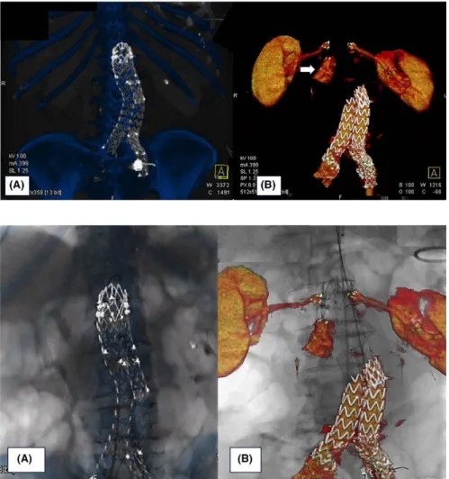
FIGURE 1 Bone and stent graft scaffolding volume rendering (VR) (A). VR of the type IA endoleak, renal arteries (B)
FIGURE 2 Moment of fusion with stent graft landmarks in AP projection (A). 3D‐IF guidance during the procedure: renal arteries and type IA endoleak VR on fluoroscopy (B)
|
531 TINELLI ETaL.atherosclerotic aortic disease. Graft migration and type IA EL could result from a compromised seal or fixation failure of the proximal aortic neck, particularly with necks that are short, angulated, large in diameter, conical, or those with sig-nificant mural thrombus or calcium.18
Endovascular redo of a previously treated aorta, typically for an abdominal aortic aneurysm, has become not uncommon procedure with challenging technical aspects.19 Considering
the technical evolvement of endovascular tools, most of these reinterventions were performed by endovascular means.
This case confirms that endoanchors placement serves as a viable treatment option for type IA EL following EVAR failure. Aortic extender cuffs were used in conjunction with endoanchors when a significant length of the aortic neck was present between the proximal margin of the endograft and the lowest renal artery.1,20
Accurate preoperative planning is a critical issue in endovas-cular procedures, especially in redo endovasendovas-cular aortic surgery.21
Correct endoanchors deployment represents the key point of technical success in creating a new sealing. In accordance with the instructions for use from the manufacturer, we pro-vided a circumferential anchoring (six anchors for an aortic neck diameter ≥30 mm; four for a diameter ≤29 mm) of the stent to the aortic wall. In the second step, we treat the type IA EL with a “suture line” along the leak channel. This tech-nical deployment is feasible with a correct and critical C‐arm positioning for proper spacing, visualization, and implanta-tion. However, leak channel identification is often not very accurate in 2D fluoroscopy.
3D‐IF support in endovascular reinterventions can en-able the safe alignment of anatomical structures shown in VR with 2D fluoroscopy, which can be accurately over-lapped with landmarks from the previously placed stent graft, as was demonstrated in this case. In redo endovas-cular procedures, the endograft scaffolding showed several landmarks to improve the volume alignment in the fusion technique, thus making it more accurate.
Precise EndoAnchor appositioning was achieved at the origin of the EL with a good result considering the rela-tive difficulty in orientation and navigation of the proxi-mal cuff device. Of course, EndoAnchor deployment along the EL channel could be placed in fluoroscopy with pre-cise degrees of angulation obtained in the planning study using preoperative CT; however, the situation may require further exposure to X‐ray radiography and the use of con-trast to confirm the EL. The major advantage of 3D‐IF is its capability of overcoming the known limitations of 2D angiographic roadmap registration.22 The fusion technique
provides an accurate visualization of compromised anat-omy in need of reintervention. This imaging technique has been shown in complex endovascular procedures to reduce procedure time and limit the use of contrast media and ra-diation exposure.16,22 Therefore, we favor the FI approach
in reinterventions because of its potential to significantly decrease radiation exposure while facilitating greater an-atomical and procedural visualization. Zero‐contrast rein-tervention procedures, such as zero‐contrast EVARs, have been reported in literature with FI 3D road mapping.23,24
This is an important development because patients who un-dergo endovascular procedures are very frequently exposed to follow‐up X‐ray examinations and secondary procedures, ultimately increasing their radiation and contrast exposure.
Redo endovascular aortic procedures are technically de-manding and carry increased risks. Improving experience and technology, such as fusion imaging, should mitigate some of this risk and are recommended.19,25
4
|
CONCLUSION
Redo endovascular aortic procedures are technically de-manding. Accurate planning and intraoperative imaging support are essential. The 3D‐IF technique represents a safe and effective imaging tool that is also capable of guiding FIGURE 3 Best intraoperative
projection of type IA endoleak channel (A). Positioning and deployment of the Heli‐FX endoanchors in the specific origin of the endoleak for the “endosuture line” (B)
532
|
TINELLI ETaL.precise EndoAnchor deployment with the goal of success-fully achieving strong fixation and a durable seal of the proximal aortic neck. The further combination of interesting and useful endovascular techniques is warranted to provide a tailored strategy for each patient.
CONFLICT OF INTEREST
Declaration of Conflicting Interests: nothing to declare.
AUTHOR CONTRIBUTION
GT, FM, FDN, and YT: involved in conception and design. GT, FM, and FDN: acquired the data. GT, FM, AS, and RF: analyzed and interpreted the data. GT, FM, FDN, RF, and YT: drafted the article. GT, AS, and YT: critically revised the manuscript. GT, FM, FDN, RF, AS, and YT: approved the manuscript.
ORCID
Giovanni Tinelli https://orcid.org/0000-0002-2212-3226
REFERENCES
1. Schlösser F, deVries J, Chaudhuri A. it Time to insert endoan-chors into routine EVAR [editorial]. Eur J Vasc Endovasc Surg. 2017;53(4):458‐459.
2. AbuRahma AF, Yacoub M, Mousa AY, et al. Aortic neck anatomic features and predictors of outcomes in endovascular repair of ab-dominal aortic aneurysms following vs not following instructions for use. J Am Coll Surg. 2016;222:579‐589.
3. Lederle FA, Freischlag JA, Kyriakides TC, et al. Long‐term com-parison of endovascular and open repair of abdominal aortic aneu-rysm. N Engl J Med. 2012;367:1988‐1997.
4. Zacharias N, Warner CJ, Taggert JB, et al. Anatomic charac-teristics of abdominal aortic aneurysms presenting with de-layed rupture after endovascular aneurysm repair. J Vasc Surg. 2016;64:1629‐1632.
5. Cox DE, Jacobs DL, Motaganahalli RL, Wittgen CM, Peterson GJ. Outcomes of endovascular AAA repair in patients with hostile neck anatomy using adjunctive balloon‐expandable stents. Vasc
Endovascular Surg. 2006;40:35‐40.
6. Farley SM, Rigberg D, Jimenez JC, Moore W, Quinones‐Baldrich W. A retrospective review of Palmaz stenting of the aortic neck for endovascular aneurysm repair. Ann Vasc Surg. 2011;25:735‐739. 7. Rajani RR, Arthurs ZM, Srivastava SD, Lyden SP, Clair DG,
Eagleton MJ. Repairing immediate proximal endoleaks during ab-dominal aortic aneurysm repair. J Vasc Surg. 2011;53:1174‐1177. 8. Kelso RL, Lyden SP, Butler B, Greenberg RK, Eagleton MJ,
Clair DG. Late conversion of aortic stent grafts. J Vasc Surg. 2009;49:589‐595.
9. Martin Z, Greenberg RK, Mastracci TM, Eagleton MJ, O’Callaghan A, Bena J. Late rescue of proximal endograft failure using fenes-trated and branched devices. J Vasc Surg. 2014;59:1479‐1487. 10. Coscas R, Kobeiter H, Desgranges P, Becquemin JP. Technical
as-pects, current indications, and results of chimney grafts for juxtare-nal aortic aneurysms. J Vasc Surg. 2011;53:1520‐1527.
11. De Vries JP, Van De Pavoordt HD, Jordan WD. Rationale of EndoAnchors in abdominal aortic aneurysms with short or angu-lated necks. J Cardiovasc Surg (Torino). 2014;55:103‐107. 12. de Vries JP, Ouriel K, Mehta M, et al. Analysis of EndoAnchors for
endovascular aneurysm repair by indications for use. J Vasc Surg. 2014;60:1460‐1467. e1.
13. Avci M, Vos JA, Kolvenbach RR, et al. The use of endoanchors in repair EVAR cases to improve proximal endograft fixation. J
Cardiovasc Surg (Torino). 2012;53:419‐426.
14. Perdikides T, Melas N, Lagios K, et al. Primary endoanchoring in the endovascular repair of abdominal aortic aneurysms with an unfavorable neck. J Endovasc Ther. 2012;19:707‐715.
15. de Ruiter QM, Reitsma JB, Moll FL, van Herwaarden JA. Meta‐ analysis of cumulative radiation duration and dose during EVAR using mobile, fixed, or fixed/3D fusion C‐arms. J Endovasc Ther. 2016;23:944‐956.
16. McNally MM, Scali ST, Feezor RJ, Neal D, Huber TS, Beck AW. Three‐dimensional fusion computed tomography decreases radiation exposure, procedure time, and contrast use during fenestrated endovascular aortic repair. J Vasc Surg. 2015;61: 309‐316.
17. Hertault A, Maurel B, Sobocinski J, et al. Impact of hybrid rooms with image fusion on radiation exposure during endovascular aor-tic repair. Eur J Vasc Endovasc Surg. 2014;48:382‐390.
18. Antoniou GA, Georgiadis GS, Antoniou SA, Kuhan G, Murray D. A meta‐analysis of outcomes of endovascular abdominal aortic an-eurysm repair in patients with hostile and friendly neck anatomy. J
Vasc Surg. 2013;57:527‐538.
19. Budtz‐Lilly J, Hongku K, Sonesson B, Dias N, Resch T. Endovascular redo aortic surgery. J Cardiovasc Surg (Torino). 2017;58(6):854-860.
20. Jordan WD, Mehta M, Varnagy D, et al. Results of the ANCHOR prospective, multicenter registry of EndoAnchors for type Ia en-doleaks and endograft migration in patients with challenging anat-omy. J Vasc Surg. 2014;60:885‐892. e2.
21. Tinelli G, Hertault A, Martin Gonzalez T, et al. Evaluation of a new imaging software for aortic endograft planning. Eur Rev Med
Pharmacol Sci. 2017;21:2717‐2724.
22. Tacher V, Lin M, Desgranges P, et al. Image guidance for endovas-cular repair of complex aortic aneurysms: comparison of two‐di-mensional and three‐ditwo‐di-mensional angiography and image fusion. J
Vasc Interv Radiol. 2013;24:1698‐1706.
23. Kobeiter H, Nahum J, Becquemin JP. Zero‐contrast thoracic endovascular aortic repair using image fusion. Circulation. 2011;124:e280‐e282.
24. Kaladji A, Dumenil A, Mahé G, et al. Safety and accuracy of endo-vascular aneurysm repair without pre‐operative and intra‐operative contrast agent. Eur J Vasc Endovasc Surg. 2015;49:255‐261. 25. Sveinsson M, Sobocinski J, Resch T, et al. Early versus late
ex-perience in fenestrated endovascular repair for abdominal aortic aneurysm. J Vasc Surg. 2015;61:895‐901.
How to cite this article: Tinelli G, De Nigris F,
Minelli F, Flore R, Santoliquido A, Tshomba Y. Endoanchors under 3D image fusion for a type IA endoleak after EVAR. Clin Case Rep. 2019;7:529– 532. https://doi.org/10.1002/ccr3.2033