Coexistence of Different Circulating Anti-Podocyte
Antibodies in Membranous Nephropathy
Corrado Murtas,* Maurizio Bruschi,†Giovanni Candiano,†Gabriella Moroni,‡Riccardo Magistroni,§Andrea Magnano,| Francesca Bruno,¶Antonella Radice,** Luciana Furci,§Lucia Argentiero,|Maria Luisa Carnevali,|Piergiorgio Messa,‡ Francesco Scolari,††Renato Alberto Sinico,** Loreto Gesualdo,¶Fernando C. Fervenza,‡‡Landino Allegri,|
Pietro Ravani,§§and Gian Marco Ghiggeri* Summary
Background and objectives The discovery of different podocyte autoantibodies in membranous nephropathy (MN) raises questions about their pathogenetic and clinical meaning. This study sought to define antibody isotypes and correlations; to compare levels in MN, other glomerulonephritides, and controls; and to determine their association with clinical outcomes.
Design, setting, participants, & measurements Serum IgG1, IgG3, and IgG4against aldose reductase (AR), SOD2,
anda-enolase (aENO) were measured at diagnosis in 186 consecutive MN patients, in 96 proteinuric controls (36 with FSGS, and 60 with IgA nephropathy), and in 92 healthy people recruited in four Italian nephrology units. Anti-phospholipase A2 receptor (PLA2r) and anti-neutral endopeptidase (NEP) IgG4were titrated in the same
specimens. Association with 1-year follow-up clinical parameters was studied in 120 patients.
Results IgG4was the most common isotype for all antibodies; IgG1and IgG3were nearly negligible. IgG4levels
were positive in a significant proportion of MN patients (AR, 34%; SOD2, 28%; aENO, 43%). Antibody titers were higher in MN than in healthy and pathologic controls (P,0.005). Anti-NEP IgG4did not differ from normal
controls (P=0.12). Anti-PLA2r IgG4was detected in 60% of patients and correlated with anti-AR, anti-SOD2, and
anti-aENO IgG4(P,0.001). In MN patients negative for the whole antibody panel (20%), 1-year proteinuria
was lower compared with patients with at least one antibody positivity (P,0.05).
Conclusions Our data suggest that IgG4is the prevalent isotype for antibodies against cytoplasmic antigens of
podocytes (AR, SOD2,aENO). Their levels were higher than in other proteinuric glomerulonephritides and in normal controls and were correlated with anti-PLA2r. Only baseline negativity for all known antibodies predicted lower 1-year proteinuria.
Clin J Am Soc Nephrol 7: ccc–ccc, 2012. doi: 10.2215/CJN.02170312
Introduction
Membranous nephropathy (MN) is a leading cause of nephrotic syndrome in adults (1). Glomerular damage is produced by the deposition of subepithelial im-mune deposits that consist mainly of IgG4and C5b-9
(2,3). The definition of causative autoantibodies and their renal targets is essential to understand the mech-anisms of disease development and progression.
Recentfindings indicate that more than one podocyte protein may act as an autoantigen in human MN (4). Identified antigens are membrane proteins, such as phos-pholipase A2 receptor (PLA2r) (5) and neutral endopep-tidase (NEP) (6), and components of the cytoplasm, such as aldose reductase (AR), SOD2, anda-enolase (aENO) (7,8). Circulating levels of the corresponding autoanti-body might be used as biomarker of disease activity.
With the exception of a report of maternal anti-NEP antibodies in antenatal MN (6,9), data on autoantibod-ies are limited to anti-PLA2r antibodautoantibod-ies. Anti-PLA2r IgG4 have been described as the largely predominant
circulating and glomerular isotype in MN patients (5). They seem specific (89%) for idiopathic MN (10–12) and can be utilized as support to exclude secondary MN (10). However, several MN patients are anti-PLA2r negative (30%–50% according to case series) whereas in others, anti-PLA2r positivity persists after response to therapy (11,13). Data on anti-PLA2r do not exclude the presence of other circulating autoan-tibodies such as anti-cytoplasmic antigens of podocytes, whose serum levels might be detected and might be useful in clinical practice.
To test this hypothesis, we measured the levels of antibodies against both membrane and cytoplasmic autoantigens in patients with and without MN. Our objectives were as follows: (1) to define the prevalent isotype for each anti-cytoplasmic antibody and the spec-trum of MN patients in respect to positivity or negativ-ity for the whole panel of currently known antibodies; (2) to compare their levels in participants with MN ver-sus patients suffering from other proteinuric glomerular
*Division of Nephrology, Dialysis, and Transplantation, and†Laboratory on Pathophysiology of Uremia, Istituto Giannina Gaslini, Genoa, Italy;‡Division of Nephrology and Dialysis, IRCCS Fondazione Ospedale Maggiore, Mangiagalli, Regina Elena, Milan, Italy; §Department of Nephrology, University of Modena, Modena, Italy;|Department of Clinical Medicine, Nephrology, and Health Sciences, University of Parma, Parma, Italy; ¶Division of Nephrology, University of Bari, Bari, Italy; **Division of Nephrology and Section of Clinical Immunology, San Carlo Hospital, Milan, Italy;††Division of Nephrology, University of Brescia and Montichiari Hospital, Brescia, Italy;‡‡Division of Nephrology and Hypertension, Mayo Clinic, Rochester, Minnesota; and §§Division of Nephrology, University of Calgary, Calgary, Alberta, Canada Correspondence: Dr. Gian Marco Ghiggeri, Division of Nephrology, Dialysis, and Transplantation, Laboratory on Pathophysiology of Uremia, Istituto Giannina Gaslini, Largo G. Gaslini 5, 16148 Genova, Italy, and Dr. Pietro Ravani, Division of Nephrology, University of Calgary, 29th St NW (r C210N), Calgary, AB, Canada. Email: labnefro@ ospedale-gaslini.ge.it or [email protected]
diseases or normal controls; and (3) to ascertain whether a relationship exists between the type and level of these antibodies and clinical outcome in individuals with MN.
Materials and Methods
Assembled Cohort
We conducted a retrospective study at four Italian nephrology centers in Parma, Modena, Milano, and Bari. We recruited patients with newly diagnosed MN or other proteinuric nephropathies and normal controls over 2 years (2008–2010). Ethical approval to test antibody levels was obtained from the Giannina Gaslini Institute Ethics Com-mittee. The study was registered in the EudraCT registry (EudraCT 2011–003942–41).
MN Patients. We recruited 186 consecutive patients with idiopathic MN, who gave informed consent to have the serum antibody levels titrated at the time of renal biopsy, before any therapy was started (Table 1). At the time enroll-ment was closed, 120 patients had completed a follow-up of 12 months or longer, during which they had received differ-ent therapy schemes, mainly the Ponticelli schedule (Supple-mental Table 1). Criteria for enrollment were as follows: histologically proven MN; negative tests for serum autoanti-bodies (antinuclear antibody, ANCA), cryoglobulins, and viral markers (hepatitis B surface antigen, HIV); absence of any clinical suspicion of secondary MN; or absence of any previous immunosuppressive treatment.
Proteinuric Controls. Ninety-two patients with different nephropathies were recruited at the same institutions: 32 patients with FSGS and 60 with IgA nephropathy (IgAN). Diagnosis was always based on histologic criteria. To pro-vide similar clinical conditions to MN patients, FSGS spec-imens were all collected during a relapse of nephrotic proteinuria. IgAN patients had to present with proteinuria .0.3 g/d and to be free of any immunosuppressive ther-apy at the time of serum collection (Table 1).
Normal Controls. Serum was obtained from 96 normal controls recruited at the same institutions. They consisted of normal blood donors who had at least one normal urinalysis and serum tests in the prior 6 months (Table 1). Assays for Autoantibodies
Anti-AR, Anti-SOD2, and Anti-aENO. Circulating IgG1,
IgG3, and IgG4levels against AR, SOD2, andaENO in sera
were determined with dot blot utilizing recombinant pro-teins fixed to nitrocellulose as antigens, as previously de-scribed (7,8). Details of the method and examples of variable positivity are given in the Supplemental Methods and Supplemental Figure 1. Antibody positivity was defined as a serum level exceeding the 95th percentile of levels titrated in normal controls.
Anti-PLA2r. Circulating anti-PLA2r IgG4 antibodies
were titrated by Western blot against podocyte protein extracts (kindly offered by Dr. Saleem, University of Bristol, Bristol, UK) previously separated in gradient monodimen-sional electrophoresis (14) and then incubated with serum. The technique is described in detail in the Supplemental Methods. Anti-PLA2r autoantibodies were also evaluated in a random subsample of MN patients (n=73) by indirect immunofluorescence with a commercially available test, according to the manufacturer’s instructions (Euroimmun, Lubeck, Germany) (15). Details of the method are given in the Supplemental Methods and Supplemental Table 2.
Anti-NEP. Circulating IgG4 antibodies against NEP
were assessed by dot blot utilizing recombinant NEP as fixed antigen. The assay is based on the same technique described for anti-AR, anti-SOD2, and anti-aENO, and is reported in the Supplemental Methods.
Statistical Analyses
Data were described using frequencies, means, and SDs, as appropriate. Natural log transformation was performed on non-normally distributed variables before using linear models. Due to the presence of several “zeros,” baseline correlations were performed using the Spearman rank cor-relation method for non-normally distributed variables; natural log transformation was performed after adding 0.01 to each variable value. The Mann–Whitney U test was used to compare non-normally distributed variables. Separate univariable logistic models were used with pres-ence of MN versus other nephropathies or controls as binary outcomes to estimate the area under the receiver operating characteristic (ROC) curves. Logistic regres-sion models of 1-year proteinuria were built on log2
-transformed antibody values adjusting for baseline levels of log2-proteinuria. Two models were built with the
dichot-omous outcome defined as complete or partial remission (proteinuria,0.3 or ,3.5 g/d). Linear regression models
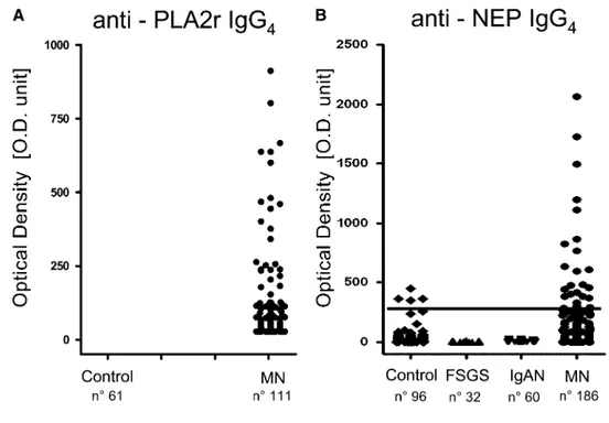
Table 1. Clinical characteristics of patients and controls enrolled in this study
MN (n=186) FSGS (n=32) IgAN (n=60) Normal (n=96) Male sex 121 (65) 19 (59) 38 (63) 56 (58) Age (yr) 59616 1863 4064 49610 Race Caucasian 184 (99) 32 (100) 56 (93) 96 (100) other 2 (1) 4 (7) Diabetes 24 (13) 0 (0) 4 (7) 0 (0) Serum creatinine (mg/dl) 1.1 (0.3–6) 0.6 (0.3–1) 0.9 (0.5–1.3) 0.9 (0.6–1.2) Proteinuria (g/d) 5.8 (0.3–28) 6.0 (3.6–12.4) 1.55 (0.9–3.4) 0
Data are presented asn (%) or mean 6 SD, or as median (range) for those with non-normal distribution. Clinical data were collected at the time of serum sample collection. Proteinuria in normal controls was tested by urine dipstick. MN, membranous nephropathy; IgAN, IgA nephropathy.
of log2-proteinuria adjusted for baseline proteinuria were
also built on each antibody level. Similarly, linear models were used to study the associations between levels of anti-bodies and serum albumin. Models with only one antibody at a time were built for reasons of colinearity. Model as-sumptions and goodness offit were verified looking at for-mal tests and graphical tests based on residuals. All statistical analyses were performed with STATA/MP 12.1 software (StataCorp, College Station, TX).
Results
Patients with MN were prevalently male (65%) and had a mean age of 59616 years. All were proteinuric at the time of enrollment and had variable creatinine levels (Table 1). Patients with FSGS (59% male) had proteinuria.3.5 g/d, had normal renal function in all cases, and were younger (age 1863 years). IgAN patients (63% male; 4064 years) had proteinuria 0.9–3.4 g/d and normal renal function. Normal controls were prevalently male (58%) with a mean age of 49610 years (Table 1).
Antibody Levels in MN and in Other Glomerulonephritides
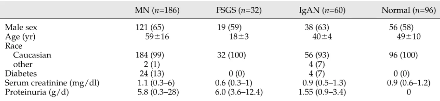
Anti-AR, Anti-SOD2, and Anti-aENO Antibody Isotypes/ Serum Levels. Circulating AR, SOD2, and anti-aENO isotypes (IgG1, IgG3, and IgG4) and levels were
de-termined by dot blot in the serum of 186 MN patients at diagnosis (Figure 1). Isotype characterization of each spec-ificity indicated that IgG4is the predominant IgG subclass.
Anti-AR and anti-SOD2 IgG1and IgG3were, in fact, only
sporadically positive (,3%). Anti-aENO IgG1–3 were
in-stead increased in a small but significant proportion of MN patients (13% IgG1and 7% IgG3) (Supplemental Figure 2).
In patients with other nephropathies, serum levels of each
IgG4antibody were found to be lower than in normal
con-trols; one IgAN patient presented an isolated anti-aENO positivity (Figure 1). The percentage of MN patients with IgG4positive levels was highly significant (AR, 34%; SOD,
26%;aENO, 43%; P,0.001 for all) (Supplemental Figure 3). Comparison of levels confirmed significantly higher circu-lating levels of anti-AR, anti-SOD2, and anti-aENO IgG4in
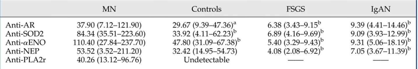
MN patients compared with either normal participants or other nephropathies (Table 2). The area under the ROC curves for each antibody was significantly greater in MN patients compared with patients with other nephropathies or normal participants, although to a lesser extent (Figure 2). Anti-PLA2r and Anti-NEP Antibodies. Anti-PLA2r IgG4 was detected by Western blot in 111 MN patients
(Figure 3), giving afinal estimate of 60% of patients being positive (Supplemental Figure 3), confirming previous studies (5,10). In a random portion of MN sera (73 pa-tients), anti-PLA2r total IgG as determined by indirect im-munofluorescence (12,15) confirmed results of Western blot titration (Spearman analysis, r=0.91; P,0.001) (Sup-plemental Table 2). Anti-NEP IgG4serum levels were
de-termined with dot blot analysis. The results shown in Figure 3 indicate a minor percentage (17%) of MN patients with anti-NEP positivity. NEP serum levels were not higher in MN patients than in normal serum (P=0.12) (Table 2).
Multiple Positivity. Many MN patients presented con-comitant high serum levels of more than one antibody. Anti-PLA2r serum levels correlated with all the other antibodies and anti-AR correlated with anti-SOD2 (Table 3) (P,0.001 in all cases). Simultaneous multiple positivity or negativity for the antibody panel was tested in the whole cohort of patients: 19 sera (10%) were completely positive and 37 (20%) were negative. Most patients (70%) presented intermediate positivity for one, two, or three antibodies
Figure 1. | Serum antibodies against cytoplasmic antigens of podocytes are increased in a significant portion of MN patients. Circulating IgG4 (A) anti-AR, (B) anti-SOD2, and (C) anti-aENO in MN and control populations are shown. In all cases, we utilized a technique based on dot blot analysis with recombinant protein linked to nitrocellulose as an antigen (Supplemental Figure 1). Results are given as chemi-luminescence OD arbitrary units that corresponds to one unit of signal intensity of chemichemi-luminescence detected by VersaDoc and computed with QuantyOne software (Bio-Rad). The horizontal line is set at the 95th percentile of levels titrated in normal controls. AR, aldose reductase; aENO, a-enolase; MN, membranous nephropathy.
(Table 4). Of the 75 anti-PLA2r–negative patients, 38 (51%) were positive for at least one other antibody (Supplemen-tal Figure 4).
Clinical Correlations
There was no relationship between antibody levels and proteinuria, renal function, and histologic stage at baseline. No histologic or clinical characteristics distinguished anti-PLA2r–positive and anti-PLA2r–negative patients. Antibody levels failed to predict the probability to reach complete (,0.3 g/d) or partial (, 3.5 g/d) remission after 1 year. When proteinuria was modeled as a continuous variable, only anti-AR IgG4levels predicted 1-year proteinuria.
Sim-ilarly, only anti-aENO IgG4significantly predicted serum
albumin levels at 12 months (Supplemental Table 3). Finally, 27 MN patients who were negative for all antibodies completed the 12-month follow-up (Table 4). Although treatment did not differ, they presented a mild tendency to have a better outcome. In fact, compared with MN patients who were positive for at least one antibody, completely negative patients had significantly lower 1-year proteinuria (Mann–Whitney test, P=0.03), at 0.33 g/d (inter-quartile range, 0.1–1.8) versus 1.50 g/d (inter(inter-quartile range,
0.2–4.2). A borderline significantly higher remission rate was also present in negative patients (48% versus 27%;P=0.07).
Discussion
The recent discovery that podocyte proteins are targets of circulating antibodies in human MN represented a break-through in the research on MN pathogenetic mechanisms (5,7,8). However, the implication of more than one antigen in the formation of subepithelial immune deposits raises questions about the clinical significance of each autoanti-body. Thus far, four podocyte antigens have been studied in primary MN: PLA2r was the first discovered (and deeply investigated) (5,11,13), and AR, SOD2, andaENO were subsequently identified (7,8). Our study was planned to bridge the gap of knowledge on antibodies against neo-expressed cytoplasm antigens (i.e., anti-AR, anti-SOD2, and anti-aENO). In fact, there is little information in the literature about their serum levels and their correlation with anti-PLA2r. Moreover, the evaluation of a potential as-sociation of serum antibody levels (including anti-PLA2r) with clinical outcomes requires studies in large cohorts of patients. Our strategy was therefore based on the concomi-tant determinations of serum levels of anti-cytoplasmic
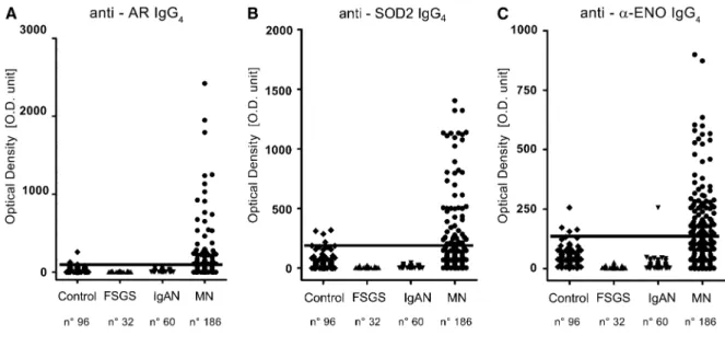
Table 2. Levels of circulating antibodies in MN patients, controls, and other nephropathies
MN Controls FSGS IgAN Anti-AR 37.90 (7.12–121.90) 29.67 (9.39–47.36)a 6.38 (3.43–9.15b 9.39 (4.41–14.46)b Anti-SOD2 84.34 (35.51–223.60) 33.92 (4.11–62.23)b 6.89 (4.16–9.69)b 9.09 (3.93–12.99)b Anti-aENO 110.40 (27.84–237.70) 47.80 (31.09–67.38)b 5.40 (3.29–9.43)b 9.31 (5.06–18.19)b Anti-NEP 53.52 (3.52–211.20) 32.42 (14.95–54.73) 4.08 (2.08–6.92)b 7.05 (3.67–11.39)b Anti-PLA2r 40.26 (13.12–96.76) Undetectable —— ——
Data are expressed as chemiluminescence OD arbitrary units and presented as median (interquartile range). Only data of 111 positive patients were reported for anti-PLA2r. MN, membranous nephropathy; IgAN, IgA nephropathy; AR, aldose reductase;aENO, a-enolase; NEP, neutral endopeptidase; PLA2r, phospholipase A2 receptor.
aA highly statistically significant difference with MN level (two tailed Mann–Whitney U test). P=0.004. bA highly statistically significant difference with MN level (two tailed Mann–Whitney U test). P,0.001.
Figure 2. | ROC curves. The area under the ROC curves for each antibody was significantly greater in MN patients compared with (A) patients with other nephropathies or (B) normal participants, although to a lesser extent. ROC, receiver operating characteristic; MN, membranous nephropathy.
antigen antibodies and comparisons with anti-PLA2r in the same patient population.
Our results showed that high levels of circulating anti-bodies against cytoplasm podocyte proteins (AR, SOD2, and aENO) are present in a significant number of MN patients. The predominant isotype was IgG4in all cases,
although anti-aENO IgG1–3 levels were not completely
negligible. On the other hand, circulating levels of IgG4
anti-PLA2r and other IgG4 antibodies correlated at the
time of diagnosis, suggesting a common formation mech-anism. Anti-NEP IgG4seemed to play a secondary role.
Some limitations of this study must be acknowledged. Follow-up sera were not available, and thus we could not perform a lengthwise evaluation of autoantibody levels and laboratory parameters. Further studies, now in prog-ress, will bridge this knowledge gap. Moreover, circulating
serum IgG subclass measurement was not available. A correlation of single specificities with the respective total IgG isotype level could not be verified. Truthfully, none of the previous seminal studies on anti-podocyte antibodies in MN reported such data (5,10–12); when planning brand new studies on MN, researchers will need to consider this recurring limitation. Lastly, a group of secondary MN was not available for comparison; a definitive evalu-ation of the specificity of cytoplasmic podocyte anti-gen antibodies is not possible with the data analyzed in our study.
Despite the considerations above, our study is thefirst to attempt correlating serum levels of different antibody specificity in the same population. Owing to the large number of patients recruited and the fact that they were all enrolled at the time of disease diagnosis (patients recruited in other studies had a variable disease duration from 4 to 144 months), our results add significant knowledge to the studies on MN that evaluated only anti-PLA2r antibodies. Moreover, our study adds relevant data on the definition of anti-PLA2r outliers (i.e., patients who are anti-PLA2r neg-ative despite an active disease). Indeed, we found that only 37 patients (20%) were negative for all antibodies, a small proportion of those negative for anti-PLA2r alone (40%). Interestingly, only negativity for the complete panel is associated with lower proteinuria after 1 year. Although no single antibody level has significant independent prog-nostic ability, negativity to all antibodies does. Our data cannot help understand whether this subgroup represents a different pathologic entity or a MN cluster with immunologic inactive disease that evolves toward spontaneous remission. However, recent advice to dose
Figure 3. | Serum levels of circulating anti-PLA2r and anti-NEP IgG4. (A) Anti-PLA2r was revealed utilizing a Western blot assay with podocyte
extracts as a fixed antigen. PLA2r was previously recognized by specific antibodies in the area of the gel between 116 and 220 kD where PLA2r was the unique spot (Supplemental Figure 1). Anti-PLA2r serum positivity was validated with a semiquantitative immunofluorescence test (Supplemental Table 2). (B) Anti-NEP IgG4was determined with dot blot analysis (Supplemental Figure 1). The horizontal line is set at the 95th
percentile of levels titrated in normal controls. Anti-PLA2r, anti-phospholipase A2 receptor; anti-NEP, anti-neutral endopeptidase.
Table 3. Spearman’s rank correlation coefficient between serum antibodies titers at the time of diagnosis in MN patients
SOD2 aENO PLA2r AR 0.385a 0.166 0.47a SOD2 0.097 0.36a
aENO 0.37a
Data are expressed asr for each correlation. MN, membranous nephropathy;aENO, a-enolase; PLA2r, phospholipase A2 re-ceptor; AR, aldose reductase.
aIndicates an highly statistically significant correlation
anti-PLA2r to define MN activity (16,17) and thus to drive the therapeutic approach is challenged by the results of this study. Further data from different MN cohorts are needed to confirm the advantage of the use of the complete panel of antibodies to individualize the therapeutic approach.
A few considerations on MN pathogenesis emerge from this study. Thefirst consideration is the temporal sequence of autoantibody production. Although this should be proven in an experimental model, we propose that the temporal appearance of detectable antibodies may follow a common mechanism. Although we cannot evaluate the timing of antibody production in the preclinical phase, it is possible that podocyte overexpression and de-localization of SOD2 and AR may represent an antioxidant response preceding the humoral immune response. In Heymann nephritis (18,19), podocyte-produced oxygen radicals in the presence of C5b-9 mediate glomerular damage (20,21). In this light, anti-SOD2 and anti-AR antibodies should follow afirst au-toimmune phase. However, autoantibody formation in MN may require a more complex and disease-specific mecha-nism than a generic podocyte injury, as confirmed by the minimal antibody serum levels in FSGS, the prototype of podocytopathy. In fact, we showed a lower circulating level of anti-AR, anti-SOD2, and anti-aENO in IgAN and FSGS compared with normal controls (as confirmed by the better performance of ROC curves comparing MN with other nephropathies). These data suggest an effect of pro-teinuria and/or minor total Ig serum levels in lowering anti-cytoplasmic podocyte antigen antibodies and support the concept that the production of such antibodies is a disease-specific mechanism in MN.
A second consideration concerns the role ofa2enolase. Circulating anti-aENO IgG has been reported in various autoimmune diseases (22–24). When characterized, the prevalent isotype was of the IgG1and IgG3subclasses also
in previous reports on MN patients (25), whereas anti-aENO IgG4seems to be more specific for MN (8). Because the shift
from IgG1to IgG4formation is a slow process that requires a
complex machinery involving T helper-2, B cell activation, and IL-3/13/10 (26), the prevalence of IgG4 in the serum
and within the glomeruli of MN patients suggests that the isotype switch may be a specific mechanism of the disease rather than a marker of inflammation. Because our data do not allow any conclusion on the role of anti-aENO IgG1and
IgG3in MN, we cannot exclude that they are also involved
in immune-deposit formation and podocyte damage.
In conclusion, our study demonstrates that all recently described serum anti-podocyte antibodies are increased in MN at diagnosis. Although no strong association with clinical outcome was found for any single autoantibody, follow-up proteinuria is lower in patients who are negative for all antibodies. A panel including all antibodies is therefore the most promising biomarker to be tested and utilized in prospective studies.
Coexistence of autoantibodies suggests a complex path-ogenetic pathway that involves different podocyte targets. New experimental models are needed to elucidate the ap-pearance time and the role of each anti-podocyte antibody in MN development and progression.
Acknowledgments
This study was investigator initiated and driven. The authors thank Dr. Saleem (University of Bristol, Bristol, UK) for supplying human conditionally immortalized podocyte cell lines, as well as Professor Gianni Cappelli for helpful discussion.
Istituto Giannina Gaslini providedfinancial and logistic support for this trial. This work was also supported by the Italian Ministry of Health“Ricerca Corrente,” the Renal Child Foundation, Fondazione Mara Wilma e Bianca Querci (“Ruolo dello stress reticolare nella progressione del danno renale e tumorale” project), and Fondazione La Nuova Speranza (“Progetto integrato per la definizione dei meccanismi implicati nella glomerulosclerosi focale”).
Part of these results were presented in abstract form at the Eighth International Congress on Autoimmunity, May 8–13, 2012, Granada, Spain.
Disclosures None.
References
1. Wasserstein AG: Membranous glomerulonephritis. J Am Soc Nephrol 8: 664–674, 1997
2. Glassock RJ: The pathogenesis of idiopathic membranous nephropathy: A 50-year odyssey. Am J Kidney Dis 56: 157–167, 2010
3. Makker SP, Tramontano A: Idiopathic membranous nephropathy: An autoimmune disease. Semin Nephrol 31: 333–340, 2011 4. Murtas C, Bruschi M, Carnevali ML, Petretto A, Corradini E,
Prunotto M, Candiano G, degl’Innocenti ML, Ghiggeri GM, Allegri L: In vivo characterization of renal auto-antigens involved in human auto-immune diseases: The case of membranous glomerulonephritis. Proteomics Clin Appl 5: 90–97, 2011 5. Beck LH Jr, Bonegio RG, Lambeau G, Beck DM, Powell DW,
Cummins TD, Klein JB, Salant DJ: M-type phospholipase A2 Table 4. Subgroup analysis of MN patients by number of baseline antibody positivity
Number of Positive Autoantibodies 4 3 2 1 0 In 186 patients 19 (10) 25 (13) 55 (30) 50 (27) 37 (20) In 120 patients 10 (8) 18 (15) 36 (30) 29 (24) 27 (23) Proteinuria T0 (g/d) 3.91 (2.8–6.5) 4.35 (2.8–8.6) 6.00 (4.3–9.2) 4.74 (2.7–7.3) 5.90 (3.6–7.8) Proteinuria T12 (g/d) 1.24 (0.2–2.2) 2.85 (0.5–6.8) 1.38 (0.1–4.4) 1.10 (0.4–3.2) 0.33 (0.1–1.8)
Complete remission 3 (30) 4 (22) 12 (33) 6 (21) 13 (48)
Data are presented asn (%) or median (interquartile range). Autoantibodies considered are against PLA2r, AR, SOD2, and aENO. Clinical data reported are referred to the group of patients who completed the 1-year clinical follow-up (120 patients). Complete re-mission is defined as proteinuria ,0.3 g/d. MN, membranous nephropathy; T0, proteinuria at diagnosis; T12, proteinuria after 1 year of follow-up; PLA2r, phospholipase A2 receptor; AR, aldose reductase;aENO, a-enolase.
receptor as target antigen in idiopathic membranous nephropa-thy. N Engl J Med 361: 11–21, 2009
6. Debiec H, Guigonis V, Mougenot B, Decobert F, Haymann JP, Bensman A, Descheˆnes G, Ronco PM: Antenatal membranous glomerulonephritis due to neutral endopeptidase anti-bodies. N Engl J Med 346: 2053–2060, 2002
7. Prunotto M, Carnevali ML, Candiano G, Murtas C, Bruschi M, Corradini E, Trivelli A, Magnasco A, Petretto A, Santucci L, Mattei S, Gatti R, Scolari F, Kador P, Allegri L, Ghiggeri GM: Autoim-munity in membranous nephropathy targets aldose reductase and SOD2. J Am Soc Nephrol 21: 507–519, 2010
8. Bruschi M, Carnevali ML, Murtas C, Candiano G, Petretto A, Prunotto M, Gatti R, Argentiero L, Magistroni R, Garibotto G, Scolari F, Ravani P, Gesualdo L, Allegri L, Ghiggeri GM: Direct characterization of target podocyte antigens and auto-antibodies in human membranous glomerulonephritis: Alfa-enolase and borderline antigens. J Proteomics 74: 2008–2017, 2011 9. Debiec H, Nauta J, Coulet F, van der Burg M, Guigonis V,
Schurmans T, de Heer E, Soubrier F, Janssen F, Ronco P: Role of truncating mutations in MME gene in fetomaternal
alloimmunisation and antenatal glomerulopathies. Lancet 364: 1252–1259, 2004
10. Qin W, Beck LH Jr, Zeng C, Chen Z, Li S, Zuo K, Salant DJ, Liu Z: Anti-phospholipase A2 receptor antibody in membranous nephropathy. J Am Soc Nephrol 22: 1137–1143, 2011 11. Beck LH Jr, Fervenza FC, Beck DM, Bonegio RG, Malik FA,
Erickson SB, Cosio FG, Cattran DC, Salant DJ: Rituximab-induced depletion of anti-PLA2R autoantibodies predicts response in membranous nephropathy. J Am Soc Nephrol 22: 1543–1550, 2011
12. Hoxha E, Harendza S, Zahner G, Panzer U, Steinmetz O, Fechner K, Helmchen U, Stahl RA: An immunofluorescence test for phospholipase-A₂-receptor antibodies and its clinical usefulness in patients with membranous glomerulonephritis. Nephrol Dial Transplant 26: 2526–2532, 2011
13. Murtas C, Ravani P, Ghiggeri GM: New insights into membranous glomerulonephritis: From bench to bedside. Nephrol Dial Transplant 26: 2428–2430, 2011
14. Laemmli UK: Cleavage of structural proteins during the assembly of the head of bacteriophage T4. Nature 227: 680–685, 1970
15. Debiec H, Ronco P: PLA2R autoantibodies and PLA2R glomer-ular deposits in membranous nephropathy. N Engl J Med 364: 689–690, 2011
16. Herrmann SM, Sethi S, Fervenza FC: Membranous nephropathy: The start of a paradigm shift. Curr Opin Nephrol Hypertens 21: 203–210, 2012
17. Couser WG: Basic and translational concepts of immune-mediated glomerular diseases. J Am Soc Nephrol 23: 381–399, 2012 18. Adler S, Huang H: Oxidant stress in kidneys of spontaneously
hypertensive rats involves both oxidase overexpression and loss of extracellular superoxide dismutase. Am J Physiol Renal Physiol 287: F907–F913, 2004
19. Heymann W, Lund HZ, Hackel DB: The nephrotic syndrome in rats; with special reference to the progression of the glomerular lesion and to the use of nephrotoxic sera obtained from ducks. J Lab Clin Med 39: 218–224, 1952
20. Adler S, Baker PJ, Pritzl P, Couser WG: Detection of terminal complement components in experimental immune glomerular injury. Kidney Int 26: 830–837, 1984
21. Haas M, Kerjaschki D, Mayer G: Lipid-lowering therapy in mem-branous nephropathy. Kidney Int Suppl 71: S110–S112, 1999 22. Migliorini P, Pratesi F, Bongiorni F, Moscato S, Scavuzzo M, Bombardieri S: The targets of nephritogenic antibodies in sys-temic autoimmune disorders. Autoimmun Rev 1: 168–173, 2002 23. Moodie FD, Leaker B, Cambridge G, Totty NF, Segal AW:
Alpha-enolase: A novel cytosolic autoantigen in ANCA posi-tive vasculitis. Kidney Int 43: 675–681, 1993
24. Terrier B, Degand N, Guilpain P, Servettaz A, Guillevin L, Mouthon L: Alpha-enolase: A target of antibodies in infectious and autoimmune diseases. Autoimmun Rev 6: 176–182, 2007 25. Wakui H, Imai H, Komatsuda A, Miura AB: Circulating
anti-bodies against alpha-enolase in patients with primary membra-nous nephropathy (MN). Clin Exp Immunol 118: 445–450, 1999 26. Aalberse RC, Stapel SO, Schuurman J, Rispens T:
Immunoglob-ulin G4: An odd antibody. Clin Exp Allergy 39: 469–477, 2009 Received: March 1, 2012 Accepted: June 5, 2012
C.M. and M.B. contributed equally to this work.
Published online ahead of print. Publication date available at www. cjasn.org.
This article contains supplemental material online at http://cjasn. asnjournals.org/lookup/suppl/doi:10.2215/CJN.02170312/-/ DCSupplemental.