Contents lists available atScienceDirect
Biomedicine & Pharmacotherapy
journal homepage:www.elsevier.com/locate/biophaReview
ADAM10 in Alzheimer's disease: Pharmacological modulation by natural
compounds and its role as a peripheral marker
Patricia Regina Manzine
a,b,⁎, Miren Ettcheto
b,c,d,e, Amanda Cano
c,f,g, Oriol Busquets
b,c,d,e,
Elena Marcello
h, Silvia Pelucchi
h,i, Monica Di Luca
h, Kristina Endres
j, Jordi Olloquequi
k,
Antoni Camins
b,c,d, Márcia Regina Cominetti
aaDepartment of Gerontology, Federal University of São Carlos, São Carlos, Brazil
bDepartament de Farmacologia, Toxicologia i Química Terapèutica, Facultat de Farmàcia i Ciències del’Alimentació, Universitat de Barcelona, Barcelona, Spain cBiomedical Research Networking Centre in Neurodegenerative Diseases (CIBERNED), 28031 Madrid, Spain
dInstitut de Neurociències, Universitat de Barcelona, Barcelona, Spain
eUnitats de Bioquímica i Farmacologia, Facultat de Medicina i Ciències de la Salut, Universitat Rovira i Virgili, Reus, Tarragona, Spain
fDepartment of Pharmacy, Pharmaceutical Technology and Physical Chemistry, Faculty of Pharmacy and Food Sciences, University of Barcelona, Barcelona, Spain gInstitute of Nanoscience and Nanotechnology (IN2UB), Barcelona, Spain
hDepartment of Pharmacological and Biomolecular Sciences, Università degli Studi di Milano, Milano, Italy iDepartment of Neurosciences, Psychology, Drug Research, and Child Health, University of Florence, Florence, Italy
jDepartment of Psychiatry and Psychotherapy, University Medical Center Johannes Gutenberg-University Mainz, Mainz, Germany kInstituto de Ciencias Biomédicas, Facultad de Ciencias de la Salud, Universidad Autónoma de Chile, Talca, Chile
A R T I C L E I N F O Keywords: Alzheimer’s disease ADAM10 Pharmaceutical Natural compounds α-Secretase A B S T R A C T
Alzheimer’s disease (AD) represents a global burden in the economics of healthcare systems. Amyloid-β (Aβ) peptides are formed by amyloid-β precursor protein (AβPP) cleavage, which can be processed by two pathways. The cleavage by theα-secretase A Disintegrin And Metalloprotease 10 (ADAM10) releases the soluble portion (sAβPPα) and prevents senile plaques. This pathway remains largely unknown and ignored, mainly regarding pharmacological approaches that may act via different signaling cascades and thus stimulate non-amyloidogenic cleavage through ADAM10. This review emphasizes the effects of natural compounds on ADAM10 modulation, which eventuates in a neuroprotective mechanism. Moreover, ADAM10 as an AD biomarker is revised. New treatments and preventive interventions targeting ADAM10 regulation for AD are necessary, considering the wide variety of ADAM10 substrates.
1. Introduction
The amyloid-β precursor protein (AβPP) is a transmembrane protein found in mostly all cell types. In pathological conditions, as Alzheimer’s disease (AD), AβPP is mainly processed by β- and γ-secretases, resulting in the production of amyloid-β (Aβ) peptides, the main players for the generation of senile plaques [1]. In physiological conditions, AβPP is mainly cleaved in the middle of the Aβ region by an α-secretase, identified as A Disintegrin And Metalloproteinase 10 (ADAM10), re-leasing a soluble fragment (sAβPPα) in a non-amyloidogenic and neu-roprotective pathway [2]. Therefore, reduction of Aβ formation and sAβPPα production by stimulating the non-amyloidogenic pathway seem promising strategies for the AD treatment [3]. In addition, the non-amyloidogenic cleavage of AβPP remains largely unknown, mainly regarding pharmacological approaches that may affect different
signaling pathways and improve the ADAM10 activity. This biblio-graphical review analyzes articles published in different levels of sci-entific evidence. Studies from Medline/PubMed databases (1990–2018) were included. These databases were chosen due to the great number of scientific articles with international availability focused on health follow-up, besides being provided with the most qualified forms of online search. For this, the search descriptors were defined according to the Medical Subject Headings (Mesh), using the vocabulary structured with the terms "ADAM10 protein", "Alzheimer disease", "alpha secre-tase" and "natural products". According to this specific search, about 70 studies were found within this theme, of which compounds of plant or animal origin were included for analysis.
This review emphasizes natural compounds that can directly or indirectly stimulate the neuroprotective mechanism achieved by ADAM10 induction, besides highlighting the challenges of this protein
https://doi.org/10.1016/j.biopha.2019.108661
Received 2 October 2018; Received in revised form 30 January 2019; Accepted 1 February 2019
⁎Corresponding author at: Department of Gerontology, Federal University of São Carlos, São Carlos, Brazil.
E-mail addresses:[email protected](P.R. Manzine),[email protected](J. Olloquequi).
Biomedicine & Pharmacotherapy 113 (2019) 108661
0753-3322/ © 2019 Elsevier Masson SAS. This is an open access article under the CC BY-NC-ND license (http://creativecommons.org/licenses/BY-NC-ND/4.0/).
as a biomarker for AD therapy.
2. ADAM10 and Alzheimer’s disease
ADAM10 belongs to the ADAMs family of proteins, which comprises transmembrane, as well as secreted metalloproteinases with functions in cell adhesion and proteolytic processing of diverse cell surface re-ceptors ectodomains and signaling molecules [4]. ADAM10 substrates comprise more than 40 molecules, including adhesion molecules (e.g., cadherins), chemokines (e.g., CX3CL1), growth factors (e.g., EGF and Tumor Necrosis Factor - α), signaling receptors, ligands (e.g., Notch receptor and Fas ligand) and AβPP [5–7].
ADAM10 is a multimodular transmembrane protein constitutively expressed in neurons, vascular cells, leukocytes and tumor cells [8]. It is synthesized in the endoplasmic reticulum (ER) as an inactive zymogen containing C-terminal, transmembrane, cysteine-rich, disintegrin and zinc-binding metalloprotease domains. There is also a pro-domain, which maintains the protease in a latent inactive form, via a cysteine switch mechanism, where a cysteine residue in the pro-domain co-ordinates the zinc ion in the catalytic site, preventing proteolytic ac-tivity [9]. During transport to the plasma membrane, ADAM10 is N-glycosylated at four positions and undergoes maturation through pro-domain removal by proprotein convertases [10]. Pro-ADAM10 has a molecular weight of∼90kDa and after pro-domain removal, the full-length active ADAM10 has∼65kDa [10,11].
Ectodomain shedding of ADAM10 leaves a ∼10kDa membrane-anchored C-terminal fragment and releases a ∼55kDa soluble ADAM10. Therefore, apart from its role in protein shedding and the initiation of regulated intramembrane proteolysis of several substrates, ADAM10 itself is subject to a similar proteolytic cascade by other ADAMs, such as ADAM9 and 15 and by γ-secretase. The γ-secretase presenilin releases ADAM10 intracellular domain, which then translo-cates to the nucleus and localizes to nuclear speckles, thought to be involved in gene regulation. This suggests that ADAM10, in addition to its important function as a membrane-tethered sheddase, also has the potential to be a signal transducing protein [12].
ADAM10 can be differentially regulated from transcriptional to translational and post-translational levels [7,13]. In neuronal cells, ADAM10 is localized at the synapses, at the presynaptic vesicles and at the postsynaptic side [14,15]. ADAM10 shedding activity at the synapse is under the control of trafficking mechanisms. SAP97 mediates ADAM10 trafficking from dendritic Golgi outposts to the synapse upon protein kinase C (PKC) activation [14,16], while AP2 interaction
triggers ADAM10 endocytosis [17]. Notably, ADAM10 is a relevant enzyme in the synapses because its activity participates in the re-modeling of the spines [18]. Indeed, its activity isfinely regulated by activity-dependent synaptic plasticity [17].
The results of ADAM10 regulation, as well as its action as a shedding molecule, are related to different physiological and pathological con-ditions, such as cancer and AD. For instance, the synaptic levels of ADAM10 are significantly reduced in AD patients hippocampi com-pared to age-matched healthy control subjects [19]. Since the seminal paper from Lammich and co-workers (1999), demonstrating that ADAM10 hasα-secretase activity and is responsible for the proteolytic processing of AβPP within the Aβ stretch, this metalloproteinase has been increasingly studied [20]. The argument that captivates and at-tracts interest from researchers is the strategy of increasing its activity in order to decrease Aβ production and hence, preventing or avoiding AD appearance or progression.
AβPP is a type I transmembrane glycoprotein constitutively ex-pressed in many types of mammalian cells, including neurons. It serves as the precursor of Aβ, whose sequence includes 28 amino acids of the extracellular and 12–15 residues of the membrane-spanning region of AβPP. The sequential AβPP cleavage by β and γ secretases is the basis of the amyloid cascade hypothesis,first proposed by Hardy and Higgins in 1992 [21]. Although it has been criticized over the years alternatively raised hypothesis were not able to fully explain the disease mechan-isms.
According to the amyloid hypothesis, AβPP ectodomain is detached from the neuronal membrane through sequential proteolytic cleavages that involveα, β and γ-secretases [22]. The mainβ-secretase involved in AβPP cleavage at β-site is the cleaving enzyme β-secretase (BACE-1) [23]. The action ofα-secretases, instead of β-, drives the pathway to a non-amyloidogenic cleavage, avoiding Aβ production. In the non-amyloidogenic pathway, AβPP is cleaved mainly by ADAM10, between Lys-16 and Leu-17 in the middle of the Aβ region, thus, releasing sAβPPα - a structure with neurotrophic and neuroprotective functions, retaining a 83-amino acid membrane-bound fraction (α-CTF or C83) residue in the membrane. The subsequent cleavage ofα-CTF byγ -se-cretases liberates P3, which is supposedly beneficial and not found in amyloid plaques [24] (Fig. 1).
Easily available, low invasive, cost-effective and early-stage disease detection biomarkers are urgently needed in AD clinical practice. Perhaps the massive failure faced so far in clinical trials for AD is due to the fact that the drugs were tested in patients with established dementia and a few in patients with mild cognitive impairment (MCI), but not
Fig. 1. Non-amyloidogenic and amyloidogenic pathways in AD through consecutive cleavages of AβPP protein in the membrane by ADAM10 and BACE-1, re-spectively.
early in the disease process - in prodromal or preclinical stages. Peripheral blood is a good source to investigate AD biomarkers, despite all difficulties regarding assay standardizations and replicability of re-sults [25]. Among the peripheral tissues, platelets present the highest AβPP expression levels [26]. They store and liberate neurotransmitters and carry appropriate transporters and receptors, normally expressed by neuronal cells [27]. In addition, platelets can produce all AβPP fragments found in neurons, thus indicating that they haveα, β and γ-secretases activities and that, as well as neurons, can process AβPP through the amyloidogenic and the non-amyloidogenic pathway. Finding the same products from the secretases in platelets and in neu-rons, it is acknowledged that the formers are easily accessible and po-tentially useful as a clinical tool to monitor the effects of new therapies based on β and γ-secretases inhibition and/or α-secretases activation [28]. In healthy individuals, the cleavage byα-secretase seems to be the dominant way used by platelets since the detected levels of sAβPPα are much higher than the levels of sAβPPβ [28].
Two studies have demonstrated that ADAM10 is the primordial α-secretase in neurons, which are the most affected cells in AD [29,30]. In addition, its beneficial role in alleviating the Aβ burden in AD has been demonstrated both in vivo and in vitro [31,32]. Furthermore, in addition to the four established AD susceptibility genes, AβPP, Presenilin-1, Presenilin-2 [33] and Apolipoprotein E [34], increasing evidence in-dicates that ADAM10 is a candidate for AD susceptibility gene [35,36]. As a consequence of the several roles of ADAM10 in the brain, it not only would act as a majorα-secretase, but also can affect tau pathology, synaptic functions, hippocampal neurogenesis, and gliogenesis by in-teracting with its substrates [2].
3. ADAM10 modulation by natural compounds
Based on the Aβ hypothesis, compounds that lower Aβ peptide brain levels, either by inhibiting its production or increasing its clearance and those that prevent Aβ aggregation represent disease-modifying ther-apeutic agents, which should alter its course. If we assume that the amyloidogenic hypothesis is important, one of the therapeutic strate-gies for AD prevention will be the inhibition of Aβ42formation, mis-folding and aggregation until substantial neurodegeneration is devel-oped and the cognitive decline appears as the main symptom of this progressing disease [37–39] .
The non-amyloidogenic pathway and the specific role of ADAM10 in AD neuropathology have been described above. The administration of drugs directed to increase ADAM10 activity is an interesting and future therapeutic option for AD treatment. The main feature of the α-secre-tase activation pathway is the increase in the generation of sAβPPα, which has neurotrophic, neuroprotective properties and is involved in the maintenance of dendritic integrity in the hippocampus. Hick and colleagues reported that acute application of exogenous recombinant sAβPPα to mouse models of AD improves synaptic strength and en-hance memory performance, while sAβPPβ was ineffective [40]. In addition, the α7-nAChRs has been recently identified as a crucial physiological receptor specific for sAβPPα, but not for sAβPPβ [41].
Therefore, this preclinical data reinforce the hypothesis that en-hancing brain sAβPPα levels is a potential strategy to improve AD-re-lated symptoms and attenuate synaptic deficits. It has been demon-strated that a virus-mediated intracranial expression of sAβPPα can mitigate the Aβ-related synaptic deficits of APP/PS1 mice in vivo [42]. sAβPPα has synaptotrophic effects in an Aβ-independent pathology [41].The functional synaptic plasticity was also modulated by sAβPPα in the hippocampus, including term potentiation (LTP) and long-term depression (LTD) of synaptic transmission, through the activation of the N-methyl-D-aspartate subtype of glutamate receptors (NMDAR)
[40]. Furthermore, the beneficial effects of sAβPPα could be explained by the inhibition of BACE-1 activity. Peters-Libeu and colleagues re-ported that sAβPPα can directly inhibit BACE-1, suggesting that it could be an endogenous ligand of this enzyme [43]. In addition, Tan and
colleagues reported that direct administration of sAβPPα into the hip-pocampus has a potential therapeutic impact by increasing the process of neurogenesis [44]. Therefore, the stimulation of the non-amyloido-genic pathway could significantly reduce Aβ release and sAβPPβ levels, being a suitable therapeutic strategy in AD. Furthermore, both en-zymes, ADAM10 and BACE-1 compete for the AβPP cleavage, therefore potentiating ADAM10 activity might inhibit the neurotoxic amyloid generation. Moreover, sAβPPα can prevent the activation of the JNK stress kinase-signaling pathway and has an anti-apoptotic effect [45]. Therefore, a drug able to promote ADAM10 activity in the brain can provide a multi-target therapy for AD to reduce Aβ generation and limit its toxic synaptic signaling, increasing Aβ clearance and favoring neu-rogenesis.
In this framework, in order to increase therapy effectiveness and to reduce the occurrence of potential side effects, the challenge is directly targeting the drug to the brain, attempting to tackle a brain and disease-specific mechanism able to regulate ADAM10 activity. Undesirable ef-fects obtained by non-specific ADAM10-targeting might be found in cancer proliferation [46], cell adhesion [47–49], promotion of T cell/ NK-cell precursor [50], inflammation [51,52] and others, since several in vitro studies have shown a wide variety of ADAM10 substrates [53]. A potential strategy could be the use of natural compounds for ther-apeutic approaches to treat AD, as in the recent years they have been shown to increase ADAM10 expression. Therefore, it seems more ap-propriate to promote the long-term intake of natural origin products that are being used for a long time in indigenous cultures, and mainly through the consumption of such compounds by diet. In this context, it recently emerged that some natural compounds such as the plant ex-tracts Ginkgo biloba and green tea–epigallocatechin-3-gallate can spe-cifically activate the α-secretase cleavage of AβPP, as will be shown below (Fig. 2).
In AD, ADAM10 activators could be disease-modifying therapies since they could increase the cognitive process and lead to slow neu-ronal loss and functional memory decline. Moreover, disease-modifying therapies should be introduced early in the course of a disease to achieve maximum benefit for patients. In addition to AD, ADAM10 is a suitable therapeutic target for other cognitive loss diseases such as Fragile X Syndrome (FXS) and autism spectrum disorder [54,55]. Fur-ther research is required to understand the molecular mechanisms of these agents involved in ADAM10 activation and their beneficial and adverse effects in patients with AD and other diseases.
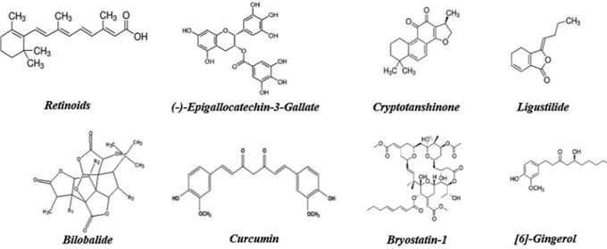
3.1. Naturalα-secretase inducers as potential disease modifiers
Three of membrane-anchored zinc-dependent metalloproteinases, ADAM10, ADAM17 and ADAM9 display alpha-secretase activity [56,57]. However, since the individual knock-out of these proteinases in neither case completely prevented alpha-secretase processing of AβPP, it is likely that different ADAMs are compensating mutually, and under different conditions may contribute to alpha-secretase cleavage of AβPP.
3.1.1. Retinoids
Several biological processes are regulated by Vitamin A (all-trans-retinol), like embryonic development, growth, differentiation and cel-lular apoptosis, as well as brain functions [58]. Derivate from pro-vi-tamin A carotenoids, these can be obtained from colorful fruits and vegetables or from animal sources such as dairy, liver and egg yolk [59] which are converted into retinal and subsequently to retinoids [60]. Retinoids play important roles in the regulation of adult brain functions such as neuronal differentiation, neurite growth, neurotransmitter re-lease and LTP [61].
Currently, the etiology of late-onset AD remains unclear, however, previous studies reported that retinoic acid (RA) signaling is essential for normal brain maintenance [62]. Retinoid signaling is mediated by two classes of receptors, namely, retinoic acid receptors (RARα, β, and
γ) and retinoid X receptors (RXRα, β, and γ). Both receptor types are widely distributed in the brain, being RARα and RXRα the main sub-types of receptors found in the hippocampus playing an important role in neuronal plasticity. Furthermore, Chiang and colleagues described that RARβ is also essential in hippocampal CA1 LTP, spatial learning and memory [63].
The research study of Corcoran and colleagues was the first monstrating that a loss of brain retinoid acid function leads to Aβ de-position in the brain of adult rats [64]. They suggest the decrease of cholinergic function and in acetylcholinesterase expression due to a lack of RA before Aβ deposition. In a preclinical study, Tippmann and colleagues evaluated the effects of acitretin, a synthetic retinoid effec-tive for the treatment of psoriasis. This drug stimulated ADAM10 pro-moter activity increasing mature ADAM10 as well asα-secretase ac-tivity. The authors suggested that acitretin activates the RAR/RXR-heterodimers bound to the ADAM10 promoter, through the major vi-tamin A metabolite all-trans retinoic acid, and thereby upregulates ADAM10 transcription [65]. Furthermore, acitretin regulates some genes linked to the AD neuropathology, such as choline acetyl-transferase (ChAT) [66].
In the preclinical Tg2576 mouse AD model, Jarvis and colleagues reported that the administration of RARα agonist significantly de-creased brain Aβ production and that these effects were receptor spe-cific because neither RARβ nor γ-agonists affected Aβ production [67]. Moreover, in neuronal cortical cultures, RARα agonists were demon-strated to be neuroprotective, as they prevented Aβ-induced neuronal cell death. Therefore, RARα agonists induce ADAM10 expression and have multiple effects apart of AβPP processing. In the non-amyloido-genic pathway, they decrease intracellular Aβ production and are neuroprotective, indicating that RARα agonists have therapeutic po-tential for the treatment of late-onset AD [67].
Using the same AD preclinical model, Gonçalves and coworkers evaluated the efficacy of several retinoid receptor agonists including AM 580, CD2019 and CD437, which are selective RARα, RARβ and RARγ agonists, respectively. They specifically demonstrated that acti-vation of RARα receptor signaling pathway favors Aβ clearance by upregulating the enzymes NEP and IDE and modulating the release of proinflammatory cytokine TNF-α by glial cells [68].
Likewise, in patients with mild to moderate AD, a key study per-formed by Endres and coworkers demonstrated that acitretin treatment
significantly increased CSF sAβPPα levels. Therefore, this research confirms preclinical data and was the first human evidence of an in-crease in ADAM10 activity by this drug in AD patients. In addition, this study gives support to the potential use of retinoid agonists as potential disease-modifying therapy drugs for AD treatment [69].
Similarly, Fukasawa and co-workers observed that Tamibarotene, a retinoid receptor agonist, lowered the insoluble Aβ40and Aβ42levels in AβPP23 transgenic mice by upregulating α-secretase expression [70]. Furthermore, in association with HX630, an RXR agonist, Tamibarotene significantly enhanced the cognitive process in the preclinical mouse model as observed by the Morris water maze test [71]. In addition, Tamibarotene has been also administered to 13-month-old SAMP8 mice and improved memory/learning. Likewise, this cognitive improvement could be due to RA activation and increased hippocampal ADAM10 mRNA expression and protein production, leading to the release of the soluble neuroprotective fragment sAβPPα [72].
In APP/PS1 transgenic mice, Ding and colleagues administrated all-trans retinoic acid (atRA) 20 mg/kg, intraperitoneally for 8 weeks [73]. This treatment improved spatial learning and memory compared with the vehicle-treated animals, assessed by the Morris water maze test. Furthermore, atRA inhibited Aβ formation, tau hyperphosphorylation, glial activation, and prevented loss of presynaptic terminals. Authors suggest that neuroprotective effects of atRA in AD could be also asso-ciated with an increase in the ChAT levels. However, potential adverse side effects of higher atRA concentrations could restrict its potential clinical applications in AD therapeutics.
Bexarotene is a highly specific RXR receptor agonist, which has a suitable safety profile in humans. In a clinical trial study, Cummings and colleagues (registered in ClinicalTrials.gov under the identifier NCT01782742) conducted a double-blind, randomized, placebo-con-trolled, parallel group study of a single dose (300 mg/day) of bexar-otene. This study also included groups of AD patients, carriers and non-carriers of APOE allele. Patients were treated for 4 weeks and an im-provement was observed in the APOE non-carriers treated group, showing a significant reduction in brain Aβ levels compared with the APOE4 carrier group [74].
In a recent clinical research, Ghosal and coworkers evaluated the effects of Bexarotene on the alteration of Aβ metabolism in cognitively healthy individuals (NCT02061878) and reported a problem with Bexarotene due to its poor CNS penetration in cognitively healthy
Fig. 2. Natural compounds can stimulate ADAM10 maturation and/or activation. The increased activity of ADAM10 in the membrane promotes the greater release of sAβPPα, a neuroprotective cleavage product of AβPP.
subjects [75].
It is important to emphasize that studies using natural products as a source of retinoids are needed and were not found in this literature search, so that only assays induced by retinoic acid, modulation of their receptors or of synthetic origin were analyzed.
3.1.2. (-)-Epigallocatechin-3-Gallate
(-)-Epigallocatechin-3-gallate (EGCG) is the major polyphenol of the Camellia sinensis leaf, from which green tea is produced. EGCG has antioxidant properties that might be useful since oxidative stress bears significance in AD development. It has been reported that this natural polyphenol shows neuroprotective properties in hippocampal neurons and neuronal-like cultures, inhibiting Aβ oligomer toxicity [76–78]. Apart of its potential antioxidant activity, neuroprotective EGCG properties could be explained through the modulation of several sig-naling pathways, among them the protein kinase C (PKC)/α-secretase/ sAβPPα pathway, leading to increasing the α-secretase activity.
A report by Obregon and coworkers was the first research sug-gesting that EGCG was able to induce ADAM10 maturation and in ad-dition to favoring AβPP non-amyloidogenic α-secretase processing in neuronal cells. They also describe the molecular mechanism involved in EGCG modulation of ADAM10 activity through estrogen receptor [79,80]. Additionally, preclinical research studies confirm that EGCG induced non-amyloidogenic sAβPPα release and inhibited the genera-tion of Aβ peptide via a PKC-dependent α-secretase activation. In AD preclinical models, such as APP/PS1 mice, the administration of EGCG induced cognitive improvement associated with the activation of brain insulin receptor [81]. In addition, there was Akt activation and in-hibition of GSK3β signaling with a significant decrease in the hippo-campal Aβ42levels, and an inhibition of JNK/TNFα pathway (anti-in-flammatory). In the SAMP8 mice model of AD and aging, EGCG significantly prevented Aβ42accumulation through the overexpression of NEP [82]. Likewise, in the same mouse model, Guo and coworkers reported that EGCG improved memory function evaluated by the Morris water maze through the decrease of Aβ42and BACE-1 levels. Likewise, the administration of EGCG (3 mg/kg for 1 week) in a mouse model of AD was able to prevent Aβ42-induced memory loss. The improvement in memory function was related to a decrease of Aβ42levels and a de-crease inβ-secretase levels [83].
Furthermore, additional targets involved in EGCG neuroprotection have been suggested, such as modulation of tyrosine-phosphorylation-regulated kinase 1 A (DYRK1 A) that could be important in Down’s syndrome neuropathology. Recent studies give support to the use of EGCG as a cognitive enhancer in Down’s syndrome patients. Torre and coworkers (NCT01699711) reported that patients with Down’s syn-drome treated with EGCG (9 mg/kg per day) during 12 months in parallel with cognitive training presented beneficial effects on memory compared to placebo-treated patients or the placebo plus cognitive training group in cognitive tests [84]. Despite the fact that this clinical trial has so far only been performed in Down’s syndrome patients evaluating the effects of EGCG on DYRK1 A and AβPP and its role im-proving cognitive performance, its action in AD should also be con-sidered.
In addition, a clinical trial completed in 2015 (NCT00951834) studied the effect of an EGCG formulation called Sunphenon in early-stage AD cases, however, the results about the potential disease-mod-ifying effects of the drug were not yet reported.
3.1.3. Cryptotanshinone
The dried root of Salvia miltiorrhiza BUNGE (Labiatae), called ‘Danshen’ in China, is a very popular traditional Chinese medicine. Preclinical studies have shown that this plant possesses multiple phar-macological activities and has been used for the treatment of various diseases such as cardiovascular, blood circulation disorders, angina pectoris, hyperlipidemia, insomnia, and inflammation [85]. Analysis of the chemical constituents of S. miltiorrhiza root revealed the presence of
two classes of secondary metabolites. Among them, a family of lipid-soluble, hydrophobic diterpene pigments known as tanshinones and water-soluble, hydrophilic, polyphenolic compounds mainly consisting of caffeic acid monomers, dimers, trimers or tetramers in the form of salvianolic acids.
Preclinical evidence suggests that cryptotanshinone has neuropro-tective effects and provides potential therapeutic benefit against neu-rodegenerative disorders such as AD. Interestingly, tanshinone deriva-tives cryptotanshinone and 15, 16-dihydrotanshinone I improve cognitive function and learning in scopolamine-treated rats through the inhibition of acetylcholinesterase activity in a dose-dependent manner [86]. Likewise, it was reported that cryptotanshinone shows anti-apoptotic effects in rat cortical cultures against glutamate-induced apoptosis through Bcl-2 activation, which is a downstream signaling target of PI3K/Akt pathway and mediated the neuroprotective effect [87]. In addition, the activation of this PI3K pathway in cortical neu-rons enhances α-secretase activity [88]. Cryptotanshinone prevents Aβ42-induced apoptosis and cytotoxicity in SH-SY5Y [89]. Moreover, in N2a mouse neuroblastoma cells it reduced Aβ production and fa-cilitated activation and translocation of ADAM10 and PKC-α to the cell membrane [90]. The increase of ADAM10 activity and secretion of sAβPPα could be attributed to a mechanism involving PKC activation. In the APP/PS1AD transgenic mouse model 4 months of daily oral cryptotanshinone administration beginning at 3 months of age, sig-nificantly improved learning and spatial navigation with a reduction of amyloid plaque deposition. Due to the ability to cross BBB, its periph-eral administration was able to stimulate ADAM10-mediated AβPP cleavage, increase sAβPPα production, and reduce the amounts of Aβ42 peptides in CNS [91].
3.1.4. Ligustilide
Ligustilide is a natural lipophilic compound present in the Umbelliferae family of medicinal plants. Some preclinical studies sug-gest that this natural compound might be a promising therapeutic candidate for the treatment of age-related neurodegenerative diseases, such as AD. A recent preclinical study in APP/PS1 mice demonstrated that intragastrically administered ligustilide (30 mg/kg) for 14 weeks, starting at 8.5 months of age, significantly improved the neurobeha-vioral deficits of transgenic compared with control wild-type mice. Authors suggested that ligustilide-evoked cognitive effects were medi-ated by an increase in non-amyloidogenic pathway, specifically by in-creasing ADAM10 expression and enzymatic activity [92]. Logically, this effect on ADAM10 decreased soluble and insoluble Aβ42levels and plaque deposition in the brains of APP/PS1 mice.
Kuang and colleagues reported that oral administration of 40 mg/kg ligustilide exerted a neuroprotective effect against Aβ neurotoxicity when administered intracerebroventricularly [93]. In addition, the compound improved the cognitive deficits and authors suggested that the effects could be explained by the inhibitory effect of glial cells and TNF-α-signaling pathway. Recently, in a 7-month-old APP/PS1 mice assay, Xu and co-authors [94] verified that 8 weeks of daily intragastric administration of ligustilide alleviate mitochondrial dysfunction and morphology issues, exert an antioxidation effect by reducing the levels of malondialdehyde (MDA) and reactive oxygen species (ROS), reduces Aβ levels, provided synaptic protection and ameliorates memory def-icit. Furthermore, it has been reported that the positive effect of li-gustilide on cognitive process can be inhibited by scopolamine - an inhibitor of acetylcholinesterase activity - and thus act via enhancing cholinergic function and memory processes [95].
Interestingly, chronic administration of ligustilide prevented the development of AD-like neuropathologies and memory impairment in SAMP8 mice. One potential neuroprotective mechanism proposed is through Klotho upregulation, which is an aging-suppressor gene that causes systemic anti-aging and increases longevity. The increase in brain Klotho levels inhibited the IGF-1 pathway and decreased oxida-tive stress in the brain [96].
3.1.5. Bilobalide - Ginkgo biloba extract
The seeds and leaves of Ginkgo biloba (Ginkgoaceae) have been used in traditional Chinese medicine. Although the extracts of the Ginkgo biloba leaves contain flavonoids, bilobalide and ginkgolides that can cross the BBB and also produce pharmacological effects in the CNS, a recent study published by Vellas and colleagues reported the Ginkgo biloba extract was ineffective in lowering the risk of AD [97]. It has been reported that the Ginkgo biloba extract presented an antioxidant effect on mitochondrial oxidative stress and prevented Aβ-induced neuronal apoptosis in neuronal cell cultures. However, although the total extract may not be effective in the prevention of AD, it has been reported that bilobalide, a sesquiterpenoid extracted from Ginkgo biloba leaves, pre-sented neuroprotective effects against Aβ42[98]. Furthermore, Shi and colleagues reported that bilobalide increased sAβPPα secretion in SH-SY5Y cells, while decreased Aβ production [99]. Moreover, bilobalide up-regulated the ADAM10 expression through an up-regulation of PI3K activity without increasing PKC activity [100]. Accordingly, Yin and coworkers reported that bilobalide prevented Aβ25–35induced cognitive loss in Morris water maze test. The authors suggested that bilobalide beneficial effects are mediated by the inhibition of oxidative stress and the prevention of neuronal apoptosis in the brain of treated rats [101].
3.1.6. Curcumin
Curcumin is the major constituent of the Asian spice, turmeric, isolated from the rhizome of Curcuma longa. This natural product, due to its size, can easily penetrate the BBB, and it was suggested as a promising AD therapy [102]. In an excellent review, Reddy and col-leagues described in detail the mechanisms involved in the neuropro-tective effects of curcumin in AD preclinical models [103]. Among them, curcumin demonstrated antioxidant, anti-inflammatory, autop-hagic effects in APP/PS1 mice, decreasing Aβ levels and BACE-1 ac-tivity, among other beneficial effects. Narasingapa and coworkers de-veloped curcumin amino acid conjugates that showed a potent α-secretase stimulatory activity [104]. The mechanisms underlying these effects are still unclear, however, it seems that curcumin can activate SIRT1 expression [105], transcriptionally increasing ADAM10 levels [106].
In addition, some clinical trials evaluated the efficacy of this drug in AD. The clinical trial NCT00164749 investigated the potential effec-tivity of curcumin and Ginkgo biloba on AD progression, but the results showed that the combination failed to reduce Aβ levels in the blood of AD patients or to improve their cognition.
The clinical trial NCT00099710 examined the safety and tolerability of curcumin and determined its effect on patients with mild to moderate AD. Ringman and colleagues published the results of this study,
demonstrating no beneficial effects of curcumin in decreasing CSF Aβ levels. They suggested that problems with bioavailability could be re-sponsible for the observed inefficacy of this drug [107].
3.1.7. Bryostatin-1
This drug is responsible for the stimulation ofα-secretase activity through activation of PKC and subsequent promotion of sAβPPα se-cretion [108]. Interestingly, this a typical compound of animal origin that is able, at nanomolar concentrations, to promote an increase in PKC activity, increase sAβPPα levels and decrease brain Aβ levels without inducing pro-tumor activity [109].
It was demonstrated that administration of Bryostatin-1 in the APP/ PS1 model of familial AD improved cognitive deficits compared to control mice [110]. Some clinical trials evaluated the efficacy of this drug in AD patients. The clinical trial NCT02221947 is a single center, randomized, double-blind, placebo-controlled, parallel group’s trial in AD patients. The patients received a single intravenous dose of placebo or 25μg/m2bryostatin-1 and safety, efficacy, pharmacokinetics, and pharmacodynamics of drug were investigated. The bryostatin was able to increase the Mini-Mental State Examination (MMSE) score, was well tolerated in AD patients and no drug-related adverse events were re-ported [108].
The clinical trial NCT02431468 evaluated the effects of bryostatin-1 (two doses with 20 and 40μg administered i.v.) in the treatment of moderately severe to severe AD. No clinical data from the study has been currently reported.
3.1.8. [6]-Gingerol
Gingerol (Zingiberaceae family) is a dietary compound that can be found in a number of plants. Its major phenolic component is the [6]-gingerol, which has antitumor, antimutagenic, antioxidant, anti-apop-totic, anti-inflammatory, cardio- and hepatoprotective properties [111]. [6]-Gingerol has neuroprotective effects by suppressing the GSK-3β activation and increasing Akt activity [112]. In addition [6],-gingerol was able to restore Aβ25-35-depleted endogenous antioxidant glu-tathione and affected Aβ25-35-induced intracellular ROS accumulation by upregulating heme oxygenase-1 (HO-1) andγ-glutamylcysteine li-gase (GCL) in SH-SY5Y cells mediated by NF-E2-related factor 2 (Nrf2). Therefore [6]-gingerol can be a potential drug for AD prevention and/ or treatment through its antioxidant capacity [113]Fig. 3.
3.2. Other compounds that activateα-secretase pathway
Besides to the natural compounds that can modulate the ADAM10 described earlier (Figs. 2 and 3), it was reported that Myricetin, a
quercetin isomer, induced α-secretase and decreased Aβ levels reg-ulating also BACE-1 activity [114]. Genistein is an isoflavonoid of the Leguminosae family that can activate PKC signaling and thus α-secre-tase activity increasing sAβPPα levels [115]. In addition, it can de-crease Aβ levels through the BACE-1 inhibition and improve cognition via inhibiting acetylcholinesterase activity. Interestingly, Avramovich and co-workers reported that non-steroidal drugs such as nimesulide, ibuprofen and indomethacin, can increaseα-secretase activity, thereby reducing the Aβ formation and thus, as we have proposed in previous studies, it is a suitable cofactor strategy for AD prevention [116].
Shuck and co-authors analyzed 313 extracts of medicinal plants indigenous to Korea for ADAM10 gene in SH-SY5Y cells. The extract of Caragana sinica (Buc'hoz) Rehder was identified as the best candidate for ADAM10 gene enhancers in peripheral tissue, without side effects. By fractionating Caragana sinica extract, alpha-viniferin was identified as one of the biologically active components that can achieve BBB pe-netrance and might be used as novel therapeutic options for treating AD by increasing ADAM10 gene expression [117].
Resveratrol (RSV) also is a natural polyphenolicflavonoid, which can be found in grapes and red wine, and exerts neuroprotective and antioxidant properties [118]. RSV decreased total cholesterol con-centration in hypercholesterolemic rats [119]. The direct and positive effects of RSV on AD pathology are related to the activation of nuclear retinoic acid receptors, which may activate ADAM10 gene transcription [65], as discussed earlier in this review. RSV treatment under experi-mental conditions in Chinese hamster ovary (CHO) cells expressing human AβPP695containing a Swedish mutation showed a significant increase in ADAM10 expression, especially its mature form and maybe the reason for the increase of the AβPP α-CTF fragment after RSV treatment [120].
Acetyl-L-carnitine (ALC) is a compound that helps to maintain mi-tochondrial bioenergetics and decreases the oxidative stress associated with aging [121]. ALC is present at high concentrations in the brain and contains portions of carnitine and acetyl, both with neurobiological properties. Carnitine is important in theβ-oxidation of fatty acids and the acetyl portion can be used to maintain acetyl-CoA levels. Other reported neurobiological effects of ALC include brain energetic mod-ulation and phospholipid metabolism, synaptic morphology and sy-naptic transmission via multiple neurotransmitters [122]. ALC is active in cholinergic neurons, where it is involved in acetylcholine production. ALC treatment has been shown to stimulateα-secretase activity and, consequently, to reduce theβ-secretase-mediated pathway [123]. It is known that ALC pre-treatment of cortical neurons in culture sig-nificantly reduced Aβ-induced cytotoxicity, protein oxidation and lipid peroxidation in a concentration-dependent manner [121]. In hippo-campal neurons, treatment with ALC caused an increase in ADAM10 levels in the post-synaptic compartment [124–126]. Another study showed that ALC can influence the non-amyloidogenic metabolism of AβPP, without affecting total AβPP and ADAM10 levels. The data suggest that ALC did not alter ADAM10 protein levels, but rather in-fluenced the delivery of ADAM10 to the post-synaptic compartment, and consequently positively modulated its enzymatic activity towards AβPP in neuronal cells [123].
4. Future perspective
4.1. Challenges of ADAM10 as a predictor of therapy outcome for Alzheimer’s disease
ADAM10 expression and/or activity dysregulation are involved in a number of human pathologies [127], ranging from those affecting the brain [32,128], liver [129], epithelium [130,131], immune system [132–134] to cancer [135–137]. In this way, ADAM10 activation or inhibition therapies will differ according to the pathology (see the re-view from Wetzel and co-authors) [127]. However, experimental trials verify better tolerance in its moderate upregulation than in its
inhibition [127]. ADAM10 overexpression in adult mice can cause changes in the expression of more than 300 genes, however, effects were mild and age-dependent as in the case of Notch signaling, one of ADAM10′s main substrates [138]. Another important point to consider is the great functional similarity of ADAM10 and ADAM17, so that they could be co-affected and, in some cases, develop undesirable situations, such as inflammation and impaired tissue regeneration [139].
New natural treatments and preventive interventions on ADAM10 regulation for AD patients are necessary, however, they have to be carefully investigated in animal models and later in clinical trials, considering the wide variety of ADAM10 substrates, with e.g. more than 90 just in the brain [140]. In addition, most studies have used in vitro approaches to identify substrates for ADAM10. Thus, besides the urgent need to confirm these candidates in vivo, also attention is re-quired for new substrates that may limit ADAM10 activation, due to toxic effects. Despite its possible benefit when activated for patients with AD, it can be highly detrimental in other situations, such as cancer, tumor progression, metastasis, FXS, inflammation and others [53,55]. In this way, for future perspectives, it will be necessary to develop fo-cused drugs on tissue-substrate-specific ADAM10 interaction as these approaches could reduce unwanted systemic effects. Moreover, since AD is currently seen as a multifactorial disease, multi-drug therapies should be considered that besides ADAM10 target other molecules or pathways, for example, neurogenesis.
Taken together, all the information about ADAM10 clearly indicates that more studies must be performed in order to definitely demonstrate its role in the underlying AD mechanisms. A deep investigation of ADAM10 functions would offer new understandings of its biological mechanisms of action, as well as provide novel possibilities for the development of AD treatments or predicting therapeutic outcomes. Finally, the possibility of measuring ADAM10 alterations in peripheral cells of AD patients can be exploited to monitor the response of the patients to therapeutic strategies targeting ADAM10 activity.
Funding acknowledgment
PRM and MRC are supported by grants 2015/26084-1,2017/13224-5 and 2012015/26084-1,2017/13224-5/24940-8, São Paulo Research Foundation (FAPESP,Brazil). This work was supported by the Spanish Ministry of Science and InnovationSAF2017-84283-R, PI2016/01, CB06/05/0024 (CIBERNED), the European Regional Development Funds and MAT 2014-59134-R project. SP by AIRAlzh Onlus-COOP Italia.
References
[1] J. Folch, D. Petrov, M. Ettcheto, S. Abad, E. Sanchez-Lopez, M.L. Garcia, J. Olloquequi, C. Beas-Zarate, C. Auladell, A. Camins, Current research therapeutic strategies for alzheimer’s disease treatment, Neural Plast. 2016 (2016) 8501693. [2] X.Z. Yuan, S. Sun, C.C. Tan, J.T. Yu, L. Tan, The role of ADAM10 in alzheimer’s
disease, J. Alzheimers Dis. 58 (2) (2017) 303–322.
[3] Y.Q. Wang, D.H. Qu, K. Wang, Therapeutic approaches to Alzheimer’s disease through stimulating of non-amyloidogenic processing of amyloid precursor pro-tein, Eur. Rev. Med. Pharmacol. Sci. 20 (11) (2016) 2389–2403.
[4] A.P.J. Huovila, A.J. Turner, M. Pelto-Huikko, L. Karkkainen, R.M. Ortiz, Shedding light on ADAM metalloproteinases, Trends Biochem. Sci. 30 (7) (2005) 413–422. [5] J. Pruessmeyer, A. Ludwig, The good, the bad and the ugly substrates for ADAM10 and ADAM17 in brain pathology, inflammation and cancer, Semin. Cell Dev. Biol. 20 (2) (2009) 164–174.
[6] S. Weber, P. Saftig, Ectodomain shedding and ADAMs in development, Development (Cambridge, England) 139 (20) (2012) 3693–3709.
[7] K. Endres, T. Deller, Regulation of alpha-secretase ADAM10 in vitro and in vivo: genetic, epigenetic, and protein-based mechanisms, Front. Mol. Neurosci. 10 (2017) 56.
[8] M.L. Moss, G. Powell, M.A. Miller, L. Edwards, B. Qi, Q.X. Sang, B. De Strooper, I. Tesseur, S.F. Lichtenthaler, M. Taverna, J.L. Zhong, C. Dingwall, T. Ferdous, U. Schlomann, P. Zhou, L.G. Griffith, D.A. Lauffenburger, R. Petrovich, J.W. Bartsch, ADAM9 inhibition increases membrane activity of ADAM10 and controls alpha-secretase processing of amyloid precursor protein, J. Biol. Chem. 286 (47) (2011) 40443–40451.
[9] H.E. Van Wart, H. Birkedal-Hansen, The cysteine switch: a principle of regulation of metalloproteinase activity with potential applicability to the entire matrix metalloproteinase gene family, Proc. Natl. Acad. Sci. U. S. A. 87 (14) (1990)
5578–5582.
[10] A. Anders, S. Gilbert, W. Garten, R. Postina, F. Fahrenholz, Regulation of the alpha-secretase ADAM10 by its prodomain and proprotein convertases, FASEB J. 15 (10) (2001) 1837–1839.
[11] L. Seipold, H. Altmeppen, T. Koudelka, A. Tholey, P. Kasparek, R. Sedlacek, M. Schweizer, J. Bar, M. Mikhaylova, M. Glatzel, P. Saftig, In Vivo Regulation of the a Disintegrin and Metalloproteinase 10 (ADAM10) by the Tetraspanin 15, Cellular and Molecular Life Sciences : CMLS, 2018.
[12] T. Tousseyn, A. Thathiah, E. Jorissen, T. Raemaekers, U. Konietzko, K. Reiss, E. Maes, A. Snellinx, L. Serneels, O. Nyabi, W. Annaert, P. Saftig, D. Hartmann, B. De Strooper, ADAM10, the rate-limiting protease of regulated intramembrane proteolysis of Notch and other proteins, is processed by ADAMS-9, ADAMS-15, and the gamma-secretase, J. Biol. Chem. 284 (17) (2009) 11738–11747. [13] R. Peron, I.P. Vatanabe, P.R. Manzine, A. Camins, M.R. Cominetti, Alpha-secretase
ADAM10 regulation: insights into alzheimer’s disease treatment, Pharmaceuticals 11 (1) (2018).
[14] E. Marcello, F. Gardoni, D. Mauceri, S. Romorini, A. Jeromin, R. Epis, B. Borroni, F. Cattabeni, C. Sala, A. Padovani, M. Di Luca, Synapse-associated protein-97 mediates alpha-secretase ADAM10 trafficking and promotes its activity, J. Neurosci. 27 (7) (2007) 1682–1691.
[15] J.L. Lundgren, S. Ahmed, S. Schedin-Weiss, G.K. Gouras, B. Winblad, L.O. Tjernberg, S. Frykman, ADAM10 and BACE1 are localized to synaptic ve-sicles, J. Neurochem. 135 (3) (2015) 606–615.
[16] C. Saraceno, E. Marcello, D. Di Marino, B. Borroni, S. Claeysen, J. Perroy, A. Padovani, A. Tramontano, F. Gardoni, M. Di Luca, SAP97-mediated ADAM10 trafficking from Golgi outposts depends on PKC phosphorylation, Cell Death Dis. 5 (2014) e1547.
[17] E. Marcello, C. Saraceno, S. Musardo, H. Vara, A.G. de la Fuente, S. Pelucchi, D. Di Marino, B. Borroni, A. Tramontano, xE, O. rez, xF, I. o, A. Padovani, M. Giustetto, F. Gardoni, M. Di Luca, Endocytosis of synaptic ADAM10 in neuronal plasticity and Alzheimer’s disease, J. Clin. Invest. 123 (6) (2013) 2523–2538.
[18] M. Malinverno, M. Carta, R. Epis, E. Marcello, C. Verpelli, F. Cattabeni, C. Sala, C. Mulle, M. Di Luca, F. Gardoni, Synaptic localization and activity of ADAM10 regulate excitatory synapses through N-Cadherin cleavage, J. Neurosci. 30 (48) (2010) 16343.
[19] E. Marcello, R. Epis, C. Saraceno, F. Gardoni, B. Borroni, F. Cattabeni, A. Padovani, M. Di Luca, SAP97-mediated local trafficking is altered in Alzheimer disease pa-tients’ hippocampus, Neurobiol. Aging 33 (2) (2012) 27.
[20] S. Lammich, E. Kojro, R. Postina, S. Gilbert, R. Pfeiffer, M. Jasionowski, C. Haass, F. Fahrenholz, Constitutive and regulated alpha-secretase cleavage of Alzheimer’s amyloid precursor protein by a disintegrin metalloprotease, Proc. Natl. Acad. Sci. U. S. A. 96 (7) (1999) 3922–3927.
[21] J.A. Hardy, G.A. Higgins, Alzheimer’s disease: the amyloid cascade hypothesis, Science (New York, N.Y.) 256 (5054) (1992) 184–185.
[22] L.M. Bekris, N.M. Galloway, S. Millard, D. Lockhart, G. Li, D.R. Galasko, M.R. Farlow, C.M. Clark, J.F. Quinn, J.A. Kaye, G.D. Schellenberg, J.B. Leverenz, P. Seubert, D.W. Tsuang, E.R. Peskind, C.E. Yu, Amyloid precursor protein (APP) processing genes and cerebrospinalfluid APP cleavage product levels in Alzheimer’s disease, Neurobiol. Aging 32 (3) (2011) 556.e13–556.e23. [23] S.L. Cole, R. Vassar, The Alzheimer’s disease beta-secretase enzyme, BACE1, Mol.
Neurodegener. 2 (1) (2007) 22.
[24] M. Morishima-Kawashima, Y. Ihara, Alzheimer’s disease: β-Amyloid protein and tau, J. Neurosci. Res. 70 (3) (2002) 392–401.
[25] L. Shi, A.L. Baird, S. Westwood, A. Hye, R. Dobson, M. Thambisetty, S. Lovestone, A decade of blood biomarkers for alzheimer’s disease research: an evolving field, improving study designs, and the challenge of replication, J. Alzheimers Dis. 62 (3) (2018) 1181–1198.
[26] B. Borroni, C. Agosti, E. Marcello, M. Di Luca, A. Padovani, Blood cell markers in Alzheimer Disease: amyloid Precursor Protein form ratio in platelets, Exp. Gerontol. 45 (1) (2010) 53–56.
[27] F. Colciaghi, B. Borroni, L. Pastorino, E. Marcello, M. Zimmermann, F. Cattabeni, A. Padovani, M. Di Luca, alpha-secretase ADAM10 as well as alpha APPs is re-duced in platelets and CSF of Alzheimer disease patients, Mol. Med. 8 (2) (2002) 67–74.
[28] G. Evin, A. Zhu, R.M.D. Holsinger, C.L. Masters, Q.-X. Li, Proteolytic processing of the Alzheimer’s disease amyloid precursor protein in brain and platelets, J. Neurosci. Res. 74 (3) (2003) 386–392.
[29] E. Jorissen, J. Prox, C. Bernreuther, S. Weber, R. Schwanbeck, L. Serneels, A. Snellinx, K. Craessaerts, A. Thathiah, I. Tesseur, U. Bartsch, G. Weskamp, C.P. Blobel, M. Glatzel, B. De Strooper, P. Saftig, The Disintegrin/
Metalloproteinase ADAM10 Is Essential for the Establishment of the Brain Cortex, J. Neurosci. 30 (14) (2010) 4833–4844.
[30] P.H. Kuhn, H. Wang, B. Dislich, A. Colombo, U. Zeitschel, J.W. Ellwart, E. Kremmer, S. Rossner, S.F. Lichtenthaler, ADAM10 is the physiologically re-levant, constitutive alpha-secretase of the amyloid precursor protein in primary neurons, EMBO J. 29 (17) (2010) 3020–3032.
[31] P.H. Kuhn, H. Wang, B. Dislich, A. Colombo, U. Zeitschel, J.W. Ellwart, E. Kremmer, S. Rossner, S.F. Lichtenthaler, ADAM10 is the physiologically re-levant, constitutive alpha-secretase of the amyloid precursor protein in primary neurons, EMBO J. 29 (17) (2010) 3020–3032.
[32] R. Postina, A. Schroeder, I. Dewachter, J. Bohl, U. Schmitt, E. Kojro, C. Prinzen, K. Endres, C. Hiemke, M. Blessing, P. Flamez, A. Dequenne, E. Godaux, F. van Leuven, F. Fahrenholz, A disintegrin-metalloproteinase prevents amyloid plaque formation and hippocampal defects in an Alzheimer disease mouse model, J. Clin. Invest. 113 (10) (2004) 1456–1464.
[33] R. Cacace, K. Sleegers, C. Van Broeckhoven, Molecular genetics of early-onset
Alzheimer’s disease revisited, Alzheimers Dement. 12 (6) (2016) 733–748. [34] J.T. Yu, L. Tan, J. Hardy, Apolipoprotein E in Alzheimer’s disease: an update,
Annu. Rev. Neurosci. 37 (2014) 79–100.
[35] J. Suh, S.H. Choi, D.M. Romano, M.A. Gannon, A.N. Lesinski, D.Y. Kim, R.E. Tanzi, ADAM10 missense mutations potentiate beta-amyloid accumulation by impairing prodomain chaperone function, Neuron 80 (2) (2013) 385–401.
[36] R. Vassar, ADAM10 prodomain mutations cause late-onset Alzheimer’s disease: not just the latest FAD, Neuron 80 (2) (2013) 250–253.
[37] R.J. Perrin, A.M. Fagan, D.M. Holtzman, Multimodal techniques for diagnosis and prognosis of Alzheimer’s disease, Nature 461 (7266) (2009) 916–922. [38] C.R. Jack Jr, H.J. Wiste, P. Vemuri, S.D. Weigand, M.L. Senjem, G. Zeng,
M.A. Bernstein, J.L. Gunter, V.S. Pankratz, P.S. Aisen, M.W. Weiner, R.C. Petersen, L.M. Shaw, J.Q. Trojanowski, D.S. Knopman, Brain beta-amyloid measures and magnetic resonance imaging atrophy both predict time-to-progression from mild cognitive impairment to Alzheimer’s disease, Brain 133 (11) (2010) 3336–3348. [39] C.R. Jack Jr., D.S. Knopman, W.J. Jagust, L.M. Shaw, P.S. Aisen, M.W. Weiner,
R.C. Petersen, J.Q. Trojanowski, Hypothetical model of dynamic biomarkers of the Alzheimer’s pathological cascade, Lancet Neurol. 9 (1) (2010) 119–128. [40] M. Hick, U. Herrmann, S.W. Weyer, J.P. Mallm, J.A. Tschape, M. Borgers,
M. Mercken, F.C. Roth, A. Draguhn, L. Slomianka, D.P. Wolfer, M. Korte, U.C. Muller, Acute function of secreted amyloid precursor protein fragment APPsalpha in synaptic plasticity, Acta Neuropathol. 129 (1) (2015) 21–37. [41] M.C. Richter, S. Ludewig, A. Winschel, T. Abel, C. Bold, L.R. Salzburger, S. Klein,
K. Han, S.W. Weyer, A.K. Fritz, B. Laube, D.P. Wolfer, C.J. Buchholz, M. Korte, U.C. Müller, Distinct in vivo roles of secreted APP ectodomain variants APPsα and APPsβ in regulation of spine density, synaptic plasticity, and cognition, EMBO J. 37 (11) (2018).
[42] R. Fol, J. Braudeau, S. Ludewig, T. Abel, S.W. Weyer, J.-P. Roederer, F. Brod, M. Audrain, A.-P. Bemelmans, C.J. Buchholz, M. Korte, N. Cartier, U.C. Müller, Viral gene transfer of APPsα rescues synaptic failure in an Alzheimer’s disease mouse model, Acta Neuropathol. 131 (2) (2016) 247–266.
[43] C. Peters-Libeu, J. Campagna, M. Mitsumori, K.S. Poksay, P. Spilman, A. Sabogal, D.E. Bredesen, V. John, sAbetaPPalpha is a potent endogenous inhibitor of BACE1, J. Alzheimers Dis. 47 (3) (2015) 545–555.
[44] V.T.Y. Tan, B.G. Mockett, S.M. Ohline, K.D. Parfitt, H.E. Wicky, K. Peppercorn, L. Schoderboeck, M.F.B. Yahaya, W.P. Tate, S.M. Hughes, W.C. Abraham, Lentivirus-mediated expression of human secreted amyloid precursor protein-alpha prevents development of memory and plasticity deficits in a mouse model of Alzheimer’s disease, Mol. Brain 11 (1) (2018) 7.
[45] J. Renziehausen, C. Hiebel, H. Nagel, A. Kundu, S. Kins, D. Kogel, C. Behl, P. Hajieva, The cleavage product of amyloid-beta protein precursor sAbetaPPalpha modulates BAG3-dependent aggresome formation and enhances cellular protea-somal activity, J. Alzheimers Dis. 44 (3) (2015) 879–896.
[46] U. Sahin, G. Weskamp, K. Kelly, H.M. Zhou, S. Higashiyama, J. Peschon, D. Hartmann, P. Saftig, C.P. Blobel, Distinct roles for ADAM10 and ADAM17 in ectodomain shedding of six EGFR ligands, J. Cell Biol. 164 (5) (2004) 769–779. [47] T. Maretzky, K. Reiss, A. Ludwig, J. Buchholz, F. Scholz, E. Proksch, B. de
Strooper, D. Hartmann, P. Saftig, ADAM10 mediates E-cadherin shedding and regulates epithelial cell-cell adhesion, migration, and beta-catenin translocation, Proc. Natl. Acad. Sci. U. S. A. 102 (26) (2005) 9182–9187.
[48] K. Reiss, T. Maretzky, A. Ludwig, T. Tousseyn, B. de Strooper, D. Hartmann, P. Saftig, ADAM10 cleavage of N-cadherin and regulation of cell-cell adhesion and beta-catenin nuclear signalling, EMBO J. 24 (4) (2005) 742–752.
[49] K. Uemura, T. Kihara, A. Kuzuya, K. Okawa, T. Nishimoto, H. Ninomiya, H. Sugimoto, A. Kinoshita, S. Shimohama, Characterization of sequential N-cad-herin cleavage by ADAM10 and PS1, Neurosci. Lett. 402 (3) (2006) 278–283. [50] E. Six, D. Ndiaye, Y. Laabi, C. Brou, N. Gupta-Rossi, A. Israel, F. Logeat, The Notch
ligand Delta1 is sequentially cleaved by an ADAM protease and gamma-secretase, Proc. Natl. Acad. Sci. U. S. A. 100 (13) (2003) 7638–7643.
[51] C. Arduise, T. Abache, L. Li, M. Billard, A. Chabanon, A. Ludwig, P. Mauduit, C. Boucheix, E. Rubinstein, F. Le Naour, Tetraspanins regulate ADAM10-mediated cleavage of TNF-alpha and epidermal growth factor, J. Immunol. (Baltimore, Md. : 1950) 181 (10) (2008) 7002–7013.
[52] Y. Hiraoka, K. Yoshida, M. Ohno, T. Matsuoka, T. Kita, E. Nishi, Ectodomain shedding of TNF-alpha is enhanced by nardilysin via activation of ADAM pro-teases, Biochem. Biophys. Res. Commun. 370 (1) (2008) 154–158. [53] H.C. Crawford, P.J. Dempsey, G. Brown, L. Adam, M.L. Moss, ADAM10 as a
therapeutic target for cancer and inflammation, Curr. Pharm. Des. 15 (20) (2009) 2288–2299.
[54] J. Chen, S. Yu, Y. Fu, X. Li, Synaptic proteins and receptors defects in autism spectrum disorders, Front. Cell. Neurosci. 8 (2014) 276.
[55] P. Saftig, S.F. Lichtenthaler, The alpha secretase ADAM10: a metalloprotease with multiple functions in the brain, Prog. Neurobiol. 135 (2015) 1–20.
[56] M. Asai, C. Hattori, B. Szabo, N. Sasagawa, K. Maruyama, S. Tanuma, S. Ishiura, Putative function of ADAM9, ADAM10, and ADAM17 as APP alpha-secretase, Biochem. Biophys. Res. Commun. 301 (1) (2003) 231–235.
[57] C. Tanabe, N. Hotoda, N. Sasagawa, A. Sehara-Fujisawa, K. Maruyama, S. Ishiura, ADAM19 is tightly associated with constitutive Alzheimer’s disease APP alpha-secretase in A172 cells, Biochem. Biophys. Res. Commun. 352 (1) (2007) 111–117. [58] J.S. Khillan, Vitamin A/retinol and maintenance of pluripotency of stem cells,
Nutrients 6 (3) (2014) 1209–1222.
[59] M. Chakrabarti, A.J. McDonald, J. Will Reed, M.A. Moss, B.C. Das, S.K. Ray, Molecular Signaling Mechanisms of Natural and Synthetic Retinoids for Inhibition of Pathogenesis in Alzheimer’s Disease, J. Alzheimers Dis. 50 (2) (2016) 335–352. [60] A.B. Barua, H.C. Furr, Properties of retinoids. Structure, handling, and
[61] R.K. Sodhi, N. Singh, Retinoids as potential targets for Alzheimer’s disease, Pharmacol. Biochem. Behav. 120 (2014) 117–123.
[62] M. Maden, Retinoic acid in the development, regeneration and maintenance of the nervous system, Nat. Rev. Neurosci. 8 (10) (2007) 755–765.
[63] M.Y. Chiang, D. Misner, G. Kempermann, T. Schikorski, V. Giguere, H.M. Sucov, F.H. Gage, C.F. Stevens, R.M. Evans, An essential role for retinoid receptors RARbeta and RXRgamma in long-term potentiation and depression, Neuron 21 (6) (1998) 1353–1361.
[64] J.P. Corcoran, P.L. So, M. Maden, Disruption of the retinoid signalling pathway causes a deposition of amyloid beta in the adult rat brain, Eur. J. Neurosci. 20 (4) (2004) 896–902.
[65] F. Tippmann, J. Hundt, A. Schneider, K. Endres, F. Fahrenholz, Up-regulation of the alpha-secretase ADAM10 by retinoic acid receptors and acitretin, FASEB J. 23 (6) (2009) 1643–1654.
[66] M. Kobayashi, I. Matsuoka, K. Kurihara, Cholinergic differentiation of cultured sympathetic neurons induced by retinoic acid. Induction of choline acetyl-transferase-mRNA and suppression of tyrosine hydroxylase-mRNA levels, FEBS Lett. 337 (3) (1994) 259–264.
[67] C.I. Jarvis, M.B. Goncalves, E. Clarke, M. Dogruel, S.B. Kalindjian, S.A. Thomas, M. Maden, J.P. Corcoran, Retinoic acid receptor-alpha signalling antagonizes both intracellular and extracellular amyloid-beta production and prevents neuronal cell death caused by amyloid-beta, Eur. J. Neurosci. 32 (8) (2010) 1246–1255. [68] M.B. Goncalves, E. Clarke, C. Hobbs, T. Malmqvist, R. Deacon, J. Jack,
J.P. Corcoran, Amyloid beta inhibits retinoic acid synthesis exacerbating Alzheimer disease pathology which can be attenuated by an retinoic acid receptor alpha agonist, Eur. J. Neurosci. 37 (7) (2013) 1182–1192.
[69] K. Endres, F. Fahrenholz, J. Lotz, C. Hiemke, S. Teipel, K. Lieb, O. Tuscher, A. Fellgiebel, Increased CSF APPs-alpha levels in patients with Alzheimer disease treated with acitretin, Neurology 83 (21) (2014) 1930–1935.
[70] H. Fukasawa, M. Nakagomi, N. Yamagata, H. Katsuki, K. Kawahara, K. Kitaoka, T. Miki, K. Shudo, Tamibarotene: a candidate retinoid drug for Alzheimer’s dis-ease, Biol. Pharm. Bull. 35 (8) (2012) 1206–1212.
[71] K. Kawahara, M. Suenobu, H. Ohtsuka, A. Kuniyasu, Y. Sugimoto, M. Nakagomi, H. Fukasawa, K. Shudo, H. Nakayama, Cooperative therapeutic action of retinoic acid receptor and retinoid x receptor agonists in a mouse model of Alzheimer’s disease, J. Alzheimers Dis. 42 (2) (2014) 587–605.
[72] K. Kitaoka, N. Shimizu, K. Ono, S. Chikahisa, M. Nakagomi, K. Shudo, K. Ishimura, H. Sei, K. Yoshizaki, The retinoic acid receptor agonist Am80 increases hippo-campal ADAM10 in aged SAMP8 mice, Neuropharmacology 72 (2013) 58–65. [73] Y. Ding, A. Qiao, Z. Wang, J.S. Goodwin, E.-S. Lee, M.L. Block, M. Allsbrook,
M.P. McDonald, G.-H. Fan, Retinoic Acid Attenuatesβ-Amyloid Deposition and Rescues Memory Deficits in an Alzheimer’s Disease Transgenic Mouse Model, J. Neurosci. 28 (45) (2008) 11622–11634.
[74] J.L. Cummings, K. Zhong, J.W. Kinney, C. Heaney, J. Moll-Tudla, A. Joshi, M. Pontecorvo, M. Devous, A. Tang, J. Bena, Double-blind, placebo-controlled, proof-of-concept trial of bexarotene Xin moderate Alzheimer’s disease, Alzheimers Res. Ther. 8 (2016) 4.
[75] K. Ghosal, M. Haag, P.B. Verghese, T. West, T. Veenstra, J.B. Braunstein, R.J. Bateman, D.M. Holtzman, G.E. Landreth, A randomized controlled study to evaluate the effect of bexarotene on amyloid-beta and apolipoprotein E metabo-lism in healthy subjects, Alzheimer’s Dement. (New York, N. Y.) 2 (2) (2016) 110–120.
[76] J. Bieschke, J. Russ, R.P. Friedrich, D.E. Ehrnhoefer, H. Wobst, K. Neugebauer, E.E. Wanker, EGCG remodels mature alpha-synuclein and amyloid-betafibrils and reduces cellular toxicity, Proc. Natl. Acad. Sci. U. S. A. 107 (17) (2010) 7710–7715.
[77] D.E. Ehrnhoefer, J. Bieschke, A. Boeddrich, M. Herbst, L. Masino, R. Lurz, S. Engemann, A. Pastore, E.E. Wanker, EGCG redirects amyloidogenic polypep-tides into unstructured, off-pathway oligomers, Nat. Struct. Mol. Biol. 15 (6) (2008) 558–566.
[78] S. Sinha, Z. Du, P. Maiti, F.G. Klarner, T. Schrader, C. Wang, G. Bitan, Comparison of three amyloid assembly inhibitors: the sugar scyllo-inositol, the polyphenol epigallocatechin gallate, and the molecular tweezer CLR01, ACS Chem. Neurosci. 3 (6) (2012) 451–458.
[79] J.W. Fernandez, K. Rezai-Zadeh, D. Obregon, J. Tan, EGCG functions through estrogen receptor-mediated activation of ADAM10 in the promotion of non-amy-loidogenic processing of APP, FEBS Lett. 584 (19) (2010) 4259–4267. [80] D.F. Obregon, K. Rezai-Zadeh, Y. Bai, N. Sun, H. Hou, J. Ehrhart, J. Zeng, T. Mori,
G.W. Arendash, D. Shytle, T. Town, J. Tan, ADAM10 activation is required for green tea (-)-epigallocatechin-3-gallate-induced alpha-secretase cleavage of amy-loid precursor protein, J. Biol. Chem. 281 (24) (2006) 16419–16427. [81] N. Jia, K. Han, J.J. Kong, X.M. Zhang, S. Sha, G.R. Ren, Y.P. Cao,
(-)-Epigallocatechin-3-gallate alleviates spatial memory impairment in APP/PS1 mice by restoring IRS-1 signaling defects in the hippocampus, Mol. Cell. Biochem. 380 (1-2) (2013) 211–218.
[82] X. Chang, C. Rong, Y. Chen, C. Yang, Q. Hu, Y. Mo, C. Zhang, X. Gu, L. Zhang, W. He, S. Cheng, X. Hou, R. Su, S. Liu, W. Dun, Q. Wang, S. Fang,
(-)-Epigallocatechin-3-gallate attenuates cognitive deterioration in Alzheimer’s disease model mice by upregulating neprilysin expression, Exp. Cell Res. 334 (1) (2015) 136–145.
[83] Y. Guo, Y. Zhao, Y. Nan, X. Wang, Y. Chen, S. Wang, (-)-Epigallocatechin-3-gallate ameliorates memory impairment and rescues the abnormal synaptic protein levels in the frontal cortex and hippocampus in a mouse model of Alzheimer’s disease, Neuroreport 28 (10) (2017) 590–597.
[84] R. de la Torre, S. de Sola, G. Hernandez, M. Farre, J. Pujol, J. Rodriguez, J.M. Espadaler, K. Langohr, A. Cuenca-Royo, A. Principe, L. Xicota, N. Janel,
S. Catuara-Solarz, G. Sanchez-Benavides, H. Blehaut, I. Duenas-Espin, L. Del Hoyo, B. Benejam, L. Blanco-Hinojo, S. Videla, M. Fito, J.M. Delabar, M. Dierssen, Safety and efficacy of cognitive training plus epigallocatechin-3-gallate in young adults with Down’s syndrome (TESDAD): a double-blind, randomised, placebo-con-trolled, phase 2 trial, Lancet Neurol. 15 (8) (2016) 801–810.
[85] X.Z. Zhang, S.S. Qian, Y.J. Zhang, R.Q. Wang, Salvia miltiorrhiza: A source for anti-Alzheimer’s disease drugs, Pharm. Biol. 54 (1) (2016) 18–24.
[86] K.K. Wong, M.T. Ho, H.Q. Lin, K.F. Lau, J.A. Rudd, R.C. Chung, K.P. Fung, P.C. Shaw, D.C. Wan, Cryptotanshinone, an acetylcholinesterase inhibitor from Salvia miltiorrhiza, ameliorates scopolamine-induced amnesia in Morris water maze task, Planta Med. 76 (3) (2010) 228–234.
[87] F. Zhang, W. Zheng, R. Pi, Z. Mei, Y. Bao, J. Gao, W. Tang, S. Chen, P. Liu, Cryptotanshinone protects primary rat cortical neurons from glutamate-induced neurotoxicity via the activation of the phosphatidylinositol 3-kinase/Akt signaling pathway, Exp. Brain Res. 193 (1) (2009) 109–118.
[88] Z. Mei, B. Situ, X. Tan, S. Zheng, F. Zhang, P. Yan, P. Liu, Cryptotanshinione up-regulates alpha-secretase by activation PI3K pathway in cortical neurons, Brain Res. 1348 (2010) 165–173.
[89] Z. Mei, P. Yan, B. Situ, Y. Mou, P. Liu, Cryptotanshinione inhibits beta-amyloid aggregation and protects damage from beta-amyloid in SH-SY5Y cells, Neurochem. Res. 37 (3) (2012) 622–628.
[90] S.S. Durairajan, L.F. Liu, J.H. Lu, I. Koo, K. Maruyama, S.K. Chung, J.D. Huang, M. Li, Stimulation of non-amyloidogenic processing of amyloid-beta protein pre-cursor by cryptotanshinone involves activation and translocation of ADAM10 and PKC-alpha, J. Alzheimers Dis. 25 (2) (2011) 245–262.
[91] Z. Mei, F. Zhang, L. Tao, W. Zheng, Y. Cao, Z. Wang, S. Tang, K. Le, S. Chen, R. Pi, P. Liu, Cryptotanshinone, a compound from Salvia miltiorrhiza modulates amyloid precursor protein metabolism and attenuates beta-amyloid deposition through upregulating alpha-secretase in vivo and in vitro, Neurosci. Lett. 452 (2) (2009) 90–95.
[92] X. Kuang, H.J. Zhou, A.H. Thorne, X.N. Chen, L.J. Li, J.R. Du, Neuroprotective Effect of Ligustilide through Induction of alpha-Secretase Processing of Both APP and Klotho in a Mouse Model of Alzheimer’s Disease, Front. Aging Neurosci. 9 (2017) 353.
[93] X. Kuang, J.R. Du, Y.S. Chen, J. Wang, Y.N. Wang, Protective effect of Z-ligustilide against amyloid beta-induced neurotoxicity is associated with decreased pro-in-flammatory markers in rat brains, Pharmacol. Biochem. Behav. 92 (4) (2009) 635–641.
[94] Y.J. Xu, Y. Mei, Z.L. Qu, S.J. Zhang, W. Zhao, J.S. Fang, J. Wu, C. Yang, S.J. Liu, Y.Q. Fang, Q. Wang, Y.B. Zhang, Ligustilide Ameliorates Memory Deficiency in APP/PS1 Transgenic Mice via Restoring Mitochondrial Dysfunction, Biomed Res. Int. 2018 (2018) 4606752.
[95] L.L. Cheng, X.N. Chen, Y. Wang, L. Yu, X. Kuang, L.L. Wang, W. Yang, J.R. Du, Z-ligustilide isolated from Radix Angelicae sinensis ameliorates the memory im-pairment induced by scopolamine in mice, Fitoterapia 82 (7) (2011) 1128–1132. [96] X. Kuang, Y.S. Chen, L.F. Wang, Y.J. Li, K. Liu, M.X. Zhang, L.J. Li, C. Chen, Q. He, Y. Wang, J.R. Du, Klotho upregulation contributes to the neuroprotection of li-gustilide in an Alzheimer’s disease mouse model, Neurobiol. Aging 35 (1) (2014) 169–178.
[97] B. Vellas, N. Coley, P.J. Ousset, G. Berrut, J.F. Dartigues, B. Dubois, H. Grandjean, F. Pasquier, F. Piette, P. Robert, J. Touchon, P. Garnier, H. Mathiex-Fortunet, S. Andrieu, Long-term use of standardised Ginkgo biloba extract for the prevention of Alzheimer’s disease (GuidAge): a randomised placebo-controlled trial, Lancet Neurol. 11 (10) (2012) 851–859.
[98] S. Bastianetto, W.H. Zheng, R. Quirion, The Ginkgo biloba extract (EGb 761) protects and rescues hippocampal cells against nitric oxide-induced toxicity: in-volvement of itsflavonoid constituents and protein kinase C, J. Neurochem. 74 (6) (2000) 2268–2277.
[99] C. Shi, D.D. Zheng, F.M. Wu, J. Liu, J. Xu, The phosphatidyl inositol 3 kinase-glycogen synthase kinase 3beta pathway mediates bilobalide-induced reduction in amyloid beta-peptide, Neurochem. Res. 37 (2) (2012) 298–306.
[100] C. Shi, F. Wu, J. Xu, J. Zou, Bilobalide regulates soluble amyloid precursor protein release via phosphatidyl inositol 3 kinase-dependent pathway, Neurochem. Int. 59 (1) (2011) 59–64.
[101] Y. Yin, Y. Ren, W. Wu, Y. Wang, M. Cao, Z. Zhu, M. Wang, W. Li, Protective effects of bilobalide on Abeta(25-35) induced learning and memory impairments in male rats, Pharmacol. Biochem. Behav. 106 (2013) 77–84.
[102] F. Ullah, A. Liang, A. Rangel, E. Gyengesi, G. Niedermayer, G. Munch, High bioavailability curcumin: an anti-inflammatory and neurosupportive bioactive nutrient for neurodegenerative diseases characterized by chronic neuroin-flammation, Arch. Toxicol. 91 (4) (2017) 1623–1634.
[103] P.H. Reddy, M. Manczak, X. Yin, M.C. Grady, A. Mitchell, S. Tonk, C.S. Kuruva, J.S. Bhatti, R. Kandimalla, M. Vijayan, S. Kumar, R. Wang, J.A. Pradeepkiran, G. Ogunmokun, K. Thamarai, K. Quesada, A. Boles, A.P. Reddy, Protective effects of indian spice curcumin against amyloid-beta in alzheimer’s disease, J. Alzheimers Dis. 61 (3) (2018) 843–866.
[104] R.B. Narasingappa, M.R. Javagal, S. Pullabhatla, H.H. Htoo, J.K. Rao, J.F. Hernandez, P. Govitrapong, B. Vincent, Activation of alpha-secretase by cur-cumin-aminoacid conjugates, Biochem. Biophys. Res. Commun. 424 (4) (2012) 691–696.
[105] Q. Sun, N. Jia, W. Wang, H. Jin, J. Xu, H. Hu, Activation of SIRT1 by curcumin blocks the neurotoxicity of amyloid-beta25-35 in rat cortical neurons, Biochem. Biophys. Res. Commun. 448 (1) (2014) 89–94.
[106] H.R. Lee, H.K. Shin, S.Y. Park, H.Y. Kim, W.S. Lee, B.Y. Rhim, K.W. Hong, C.D. Kim, Cilostazol suppresses beta-amyloid production by activating a disin-tegrin and metalloproteinase 10 via the upregulation of SIRT1-coupled retinoic