Documento de Consenso sobre la prevención,
diagnóstico y tratamiento de las infecciones
por catéter venoso periférico en adultos
RESUMEN
El uso de catéteres vasculares es una práctica muy
utiliza-da en los hospitales. El uso de catéteres venosos periféricos de
corta duración se ha asociado con un elevado riesgo de
bacte-riemia nosocomial, lo que comporta una no despreciable
mor-bilidad y mortalidad. La etiología de estas infecciones suele ser
frecuentemente por Staphylococcus aureus, lo que explica su
gravedad. En este documento de consenso, elaborado por un
panel de expertos de La Sociedad Española de Infecciones
Car-diovasculares con la colaboración de expertos de la Sociedad
Española de Medicina Interna, La Sociedad Española de
Qui-mioterapia y la Sociedad Española de Cirugía Torácica y
Car-diovascular, pretende establecer unes normes para un mejor
uso de los catéteres venosos periféricos de corta duración. El
Documento revisa las indicaciones para su inserción,
manten-imiento, registro, diagnóstico y tratamiento de las infecciones
derivadas y las indicaciones para su retirada; haciendo énfasis
en la formación continuada del personal sanitario para lograr
una mayor calidad asistencial. Seguir las recomendaciones del
consenso permitirá utilizar de una manera más homogénea los
catéteres venosos periféricos minimizando el riesgo de
infec-ción y sus complicaciones.
Palabras Clave: Infección de catéter; catéter venoso periférico; bacter-iemia, prevención de la infección de catéter; diagnóstico de la infección de catéter; tratamiento de la infección de catéter, infección nosocomial
ABSTRACT
The use of endovascular catheters is a routine practice in
secondary and tertiary care level hospitals. Short peripheral
catheters have been found to be associated with the risk of
nosocomial bacteremia resulting in morbidity and mortality.
Staphyloccus aureus is mostly associated with peripheral
cath-eter insertion. This Consensus Document has been elaborated
by a panel of experts of the Spanish Society of Cardiovascular
Infections in cooperation with experts from the Spanish
So-ciety of Internal Medicine, Spanish SoSo-ciety of Chemotherapy
and Spanish Society of Thoracic-Cardiovascular Surgery and
aims at define and establish the norm for management of
short duration peripheral vascular catheters. The document
addresses the indications for insertion, catheter maintenance
and registry, diagnosis and treatment of infection, indications
for removal and stresses on continuous education as a driver
for quality. Implementation of this norm will allow uniformity
in usage thus minimizing the risk of infection and its
compli-cations.
Key words: catheter infection, peripheral venous catheter, bacteremia, prevention of catheter infection, diagnosis of catheter infection, treatment of catheter infection, nosocomial infection
2016 Expert consensus document on prevention,
diagnosis and treatment of short-term peripheral
venous catheter-related infections in adult
1Servicio de Medicina Interna. Consorcio Sanitario de Mataró, Mataró, Barcelona.
2Servicio de Microbiología Clínica y Enfermedades Infecciosas, Hospital General Universitario Gregorio Marañón, Madrid 3Servicio de Medicina Interna. Hospital Universitario MontePrincipe, Madrid
4Servicio de Enfermedades Infecciosas. Hospital Universitario Virgen del Rocío, Sevilla.
5Servicio de Enfermedades Infecciosas Hospital Universitario Marqués de Valdecilla, Universidad de Cantabria. Santander. 6Servicio de Enfermedades Infecciosas y Microbiología Clínica. Hospital Universitario Virgen Macarena. Sevilla
7Servicio de Enfermedades Infecciosas. Hospital Universitario Donosti. San Sebastián. 8Servicio de Cirugía Cardiovascular. Hospital Universitario Marqués de Valdecilla, Santander. 9Servicio de Enfermedades Infecciosas, Complejo Hospitalario Universitario A Coruña, A Coruña. 10Servicio de Enfermedades Infecciosas, Hospital Clinic-IDIBAPS. Universidad de Barcelona, Barcelona. 11Unidad de Enfermedades Infecciosas. Hospital Universitario de Cruces, Bilbao. Universidad del País Vasco 12Servicio de Cirugía Cardiaca. Hospital Universitario de A Coruña. A Coruña
13Cardiothoracic and Vascular Surgery. Heart & Vascular Institute, Cleveland Clinic Abu Dhabi, Abu Dhabi, United Arab Emirates
Representing the a) Spanish Society of Cardiovascular Infections (SEICAV), b) Spanish Society of Internal Medicine (SEMI), c) Spanish Society of Chemotherapy (SEQ) and d) Spanish Society of Thoracic and Cardivacular Surgery (SECTCV)
Josep A. Capdevila
1,a,bMaría Guembe
2José Barberán
3,b,cArístides de Alarcón
4,aEmilio Bouza
2,aM. Carmen Fariñas
5,aJuan Gálvez
6,aMiguel Ángel Goenaga
8,aFrancisco Gutiérrez
8,aMartha Kestler
2,aPedro Llinares
9,aJosé M. Miró
10,aMiguel Montejo
11,aPatricia Muñoz
2,aMarta
Rodríguez-Creixems
2,aDolores Sousa
9,aJosé Cuenca
12,dCarlos-A. Mestres
10,13,a,don behalf the SEICAV, SEMI,
SEQ and SECTCV Societies
Correspondence: Josep A. Capdevila Internal Medicine
Consorcio Hospitalario de Mataró Mataró (Barcelona)
E-mail: [email protected]
*Este Documento también se ha publicado en: Cirugía Cardiovascular:
PARTICIPATING ORGANIZATIONS
This Consensus Document has been elaborated by a panel
of experts of the Spanish Society of Cardiovascular Infections
(SEICAV) in cooperation with experts from the following
sci-entific societies: Spanish Society of Internal Medicine (SEMI),
Spanish Society of Chemotherapy (SEQ) and Spanish Society of
Thoracic-Cardiovascular Surgery (SECTCV).
METHODS
The recommendations for insertion, handling and removal
of PVCs and also what to do when suspecting infection
(di-agnosis) and its treatment are issued based on the best
sci-entific available evidence or, when not available, on expert
opinion. Therefore, PubMed (www.PubMed.org) literature
search between 1986 and 2015 has been performed. This is a
well-known free access resource established and maintained
by the National Center for Biotechnology Information (NCBI)
of the National Library of Medicine (NLM) of the USA, which
provides free access to MEDLINE, the database of citations and
abstracts of the NLM. It currently stores over 24 million
cita-tions from over 5,600 biomedical journals.
In our PubMed search using the Medical Subject
Head-ings (MeSH) terms “management of peripheral venous catheter”
(N=363) and “peripheral catheter-related bacteremia” (N=260),
studies related to newborns or pediatric patients and studies on
peripherally inserted central catheters (PICC) were discarded. MeSH
terms is the NLM controlled vocabulary thesaurus used for indexing
articles for PubMed (www.pubmed.org). Guidelines on prevention,
diagnosis and treatment of catheter infection were reviewed
7-10.
The levels of evidence and strength of recommendations
according to the below definitions will be shown in bold within
brackets when a recommendation is made in the text.
BACKGROUND
The use of endovascular catheters is generalized practice
in the hospital setting
1. A recent prevalence study showed that
81.9% of patients admitted to Internal Medicine services are
inserted with one or more catheters, out of which 95.4% are
short duration peripheral lines
2. It has also recently been
doc-umented the increasing influence of peripheral catheters as a
driver for nosocomial bacteremia with high associated
morbid-ity and mortalmorbid-ity
3-5. Several studies have shown that the risk
of bacteremia related to a peripheral venous catheter (PVC) is
similar to that of central venous lines
6with an estimate of 0
to 5 bacteremia episodes per 1000 catheter-days in admitted
adult patients
4,6. Furthermore, the vast majority of cases of
PVC-related bacteremia are S. aureus bacteremia; this is
dif-ferent from central venous lines, being S. epidermidis the most
frequent isolated pathogen in the latter setting
3,4. This yields
a higher complication rate including nosocomial endocarditis
thus making treatment difficult. There are several guidelines
and consensus documents on prevention, diagnosis and
treat-ment of central venous catheter-related infections
7-10that
have greatly contributed to reduce the infection rate and
facil-itate its management, especially in Intensive Care Units (ICU).
However, there is scanty literature focusing on short duration
peripheral catheters which are those mostly used out of the
ICU setting
1,11. Several observational studies have shown that
there is lack of knowledge on how to use PVC by the attending
staff
12and on the opportunities to improve its handling
1,12-14.
OBJECTIVE
The objective of this Consensus Document is to review
ev-idence and make recommendations for management of short
duration PVC in adults. This will allow uniformity in usage thus
minimizing the risk of infection and its complications.
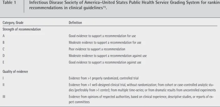
Table 1
Infectious Disease Society of America–United States Public Health Service Grading System for ranking
recommendations in clinical guidelines
15.
Category, Grade Definition Strength of recommendation
A Good evidence to support a recommendation for use
B Moderate evidence to support a recommendation for use
C Poor evidence to support a recommendation
D Moderate evidence to support a recommendation against use
E Good evidence to support a recommendation against use
Quality of evidence
I Evidence from >1 properly randomized, controlled trial
II Evidence from >1 well-designed clinical trial, without randomization; from cohort or case-controlled analytic stu-dies (preferably from >1 center); from multiple time-series; or from dramatic results from uncontrolled experiments III Evidence from opinions of respected authorities, based on clinical experience, descriptive studies, or reports of
hanced asepsis is not required if the endovenous segment of the
PVC is not manipulated
9(III-B). As it is the case when inserting
central venous lines, the use of additional protection measures
like facemask is not recommended. However, this is a topic for
consideration and analysis if in a given institution higher than
expected rates of PVC-related bacteremia are observed.
Sterile gauze dressing or semi permeable transparent sterile
dressing to cover the insertion site will be used
23,24(II-A).
Ster-ile gauze dressing will be inspected and replaced every other day
and transparent dressing should not stay in place over 7 days
9. If
there is humidity, sweating or blood it is more appropriate to use
non-occlusive gauze dressing
24,25(III-B). Revision or replacement
of dressing must be performed with single-use clean gloves
9.
PVCs placed on urgent basis or without considering
min-imal hygiene rules must be removed and replaced before 48
hours to avoid the risk of infection
17,26,27(II-A).
The use of techniques facilitating identification of veins as
laser or ultrasound
28,29in patients with poor venous flow are
also recommended for insertion. However, these techniques do
not reduce the risk of infection. A meta-analysis on this topic
showed that its routine use is not justified
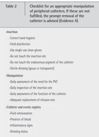
30(I-A).
4. Checklist
The adhesion to recommendations in the form of
check-list is associated to better results in prevention of
post-inser-tion complicapost-inser-tions after inserpost-inser-tion of central venous lines and
PVCs
10,31(I-A). This is reflected in table 2.
DEFINITIONS
Table 1 describes the levels of evidence and the strength
of recommendations according to the criteria of the Infectious
Disease Society of America (ISDA)
15.
PVC is a catheter shorter than 7.62 cm (3 inches).
Sepsis is a systemic inflammatory response syndrome
sec-ondary to an infection
16. The term phlebitis is used if one of
the following criteria was fulfilled: swelling and erythema > 4
mm, tenderness, palpable venous cord, pain or fever with local
symptoms. Isolated swelling is not defined as phlebitis.
INSERTION
1. When?
PVC will be inserted when the duration of a given
endo-venous therapy is expected to be shorter than 6 days and the
PVC will not be used for major procedures as hemodialysis,
plas-mapheresis, chemotherapy, parenteral nutrition, monitoring or
administration of fluid large volumes. When any of these
cir-cumstances is to be expected, it is preferable to insert a single-,
double- or triple-lumen central venous line (peripherally
insert-ed or not) as the risk of chemical phlebitis, the neinsert-ed for
high-speed volume infusion or frequent manipulations do not
sup-port a short catheter
(I-A)
17,18. An isolated transfusion does not
need a central venous line insertion. Before placing any venous
line, even peripheral, it is mandatory the evaluation of the actual
need. Venous lines are often placed as routine; this meant to be
an act reflecting the provision of care. It is also frequently shown
that to treat the patient a “prophylactic” line was not
mandato-ry. A study showed that up to 35% of peripheral venous lines
place in the emergency department are unnecessary
19.
2. Where?
A PVC can be inserted in every accessible vein. However,
upper extremity veins are preferable for patient comfort and
lesser risk of contamination. Some studies reported a higher
risk of phlebitis after lines were placed at the cubital crease,
thus becoming preferable avoiding this site in benefit of arm,
forearm or dorsal aspect of the hand/wrist
20,21(II-A).
Furthermore, other patient-related factors like
accessibil-ity to the venous system or comfort after insertion have to be
taken into account. It does not make much sense to insert a
PVC onto a central vein
(III-A).
3. How?
The insertion of PVC must be performed under maximal
aseptic techniques. It is not necessary to prep a surgical field
as it is the when inserting a central venous line. The skin must
be disinfected with 2% alcoholic chlorhexidine solution or, if
not available, with a 70% iodine or alcohol solution
9,22,23(I-A).
The insertion site should not be touched after disinfection.
The catheter must be handled from its proximal end when
in-serted. The caregiver inserting the PVC must previously perform
hand hygiene with water and soap and/or wash hands with
alcohol solution. Single-use clean gloves must be used. An
en-Table 2
Checklist for an appropriate manipulation
of peripheral catheters. If these are not
fulfilled, the prompt removal of the
catheter is advised (Evidence A)
-Insertion
-Correct hand hygiene -Field disinfection -Use single-use clean gloves -Do not touch the insertion site
-Do not touch the endovenous segment of the catheter -Sterile dressing (gauze or transparent)
-Manipulation
-Daily assessment of the need for the PVC -Daily inspection of the insertion site -Daily assessment of the function of the catheter -Adequate replacement of infusion sets
-Catheter and events registry
-Fluid extravasation -Presence of blood -Inflammatory signs -Dressing status
MAINTENANCE
The catheter and the need for usage have to be assessed
daily. It is advisable to remove the PVC if it is not necessary
as the risk of infection or phlebitis gradually increases as PVC
days go by
18,32,33(II-A). It is advisable to insert new PVC, if
re-quired, than keeping in place an inactive line that might be
useful at later stage.
The status of the insertion site must also be assessed daily,
seeking for eventual discomfort/symptoms at the endovascular
segment suggesting early stages of phlebitis and checking its
functional status. Phlebitis should be suspected if any of the
fol-lowing signs develop: warmth, tenderness, erythema or palpable
cord. In an abnormality at the insertion site is detected, dressing
must be removed and the site inspected
34,35(III-A). The catheter
must then be removed and its tip sent for Microbiology
accord-ing to the criterion of the attendaccord-ing physician
17(III-A).
No antiseptic cream shall be used at the insertion point
36(III-C).
Every manipulation of the catheter must be performed
with single-use clean gloves. There is no consensus on the type
of connectors to be used. It is preferable a three-way stopcock
than caps requiring connection-disconnection after every use.
Closed connectors for catheter access can be used as long as
they are disinfected with alcohol-impregnated wipes at every
attempt to access the catheter
37(
II-A).
A meta-analysis revealed that there are no advantages of
replacing the infusion system earlier than 96 hours
38,39(I-A)
other when they are used for blood transfusion or infusion of
lipid emulsions (should this be the case, they have to be
re-placed every time). There is no evidence that neither antibiotic
prophylaxis at insertion nor the antibiotic-lock are
cost-effi-cient to keep PVC free from infection.
REGISTRY
It is mandatory to keep daily record of characteristics and
conditions of the catheter. In this registry the type of catheter,
insertion date, anatomic location, daily inspection of dressing,
removal date and cause of removal (malfunction, infection,
not required,…) must be recorded
(III-A). The lack of a registry
is synonymous of lack of knowledge on how to use catheters,
their complications and the inability to establish corrective
measurements should an event occur
40. These registries should
ideally be electronically supported to facilitated data collection
and analysis.
REMOVAL
1. When?
As there is a causal relationship between the duration
of PVC and the risk of phlebitis, the need for systematic
re-placement of PVC at a given time interval to avoid local and
systemic complications has been proposed
18,41,42. However,
this strategy may render expensive the provision of care by
increasing in over 25% the cost and number of catheters to
use and make the catheter resite more difficult
42,43. This, on the
other side, has not avoided the complications of the use of the
new catheter regardless of the inconveniences of replacing a
line for the patient and caregiver.
More recently, prospective and randomized studies
com-paring systematic replacement at 72 hours versus clinically
indicated replacement of PVC did not found statistically
signif-icant differences in the incidence of phlebitis/local infection/
bacteremia and the number of malfunctioning catheters both
in hospitalized patients and in patients on home therapy
18,41-51.
These observations support the replacement of PVC only when
indicated
(I-A).
Systematic removal of PVC after 3-4 days is not
support-ed, although it is not advised to keep PVC in place beyond 5
days
(III-B).
Although keeping in place an unused catheter increases
the risk of phlebitis
51, it is not clear if they must be rinsed with
normal saline or heparin. It seems that the risk of phlebitis is
reduced with heparin but it continues to be at 45%
52, thus
be-ing removal advisable if unused. Therefore, unused catheters
should not be kept in place as the risk of inflammation and
infection increases
10,53-55(I-A).
PVC must be removed if the following circumstances
ap-ply: end of therapy, signs of chemical phlebitis, malfunction,
suspicion of infection or suspicion of inappropriate insertion
or manipulation as in cases of vital emergency
56,57(II-A).
2. How?
Simple removal will be performed with single-use clean
gloves and gauze dressing applied thereafter. Removal for
sus-pected infection implies sending the tip of the catheter (2-3
mm of distal end) in a sterile container for Microbiology. In the
latter case, single-use sterile gloves and sterile instrument to
cut the tip of the PVC must be used. Only catheters with
sus-pected infection must be sent for Microbiology
(III-A). There
will be suspected infection if fever or signs of sepsis without
evident focus and/or suppurated phlebitis appeared. Chemical
phlebitis alone is not enough to submit the catheter for
Mi-crobiology. It has to be reminded that catheter-related
bac-teremia may develop without any suspicion that the catheter
may be the cause
8,58.
DIAGNOSIS
PVC infection shall be suspected when a patient with one
or more PVC develops fever and/or signs of sepsis without
ad-ditional clinical focus. Under this circumstance, past history of
inappropriate manipulation and prolonged duration support a
PVC-suspected origin of infection. Septic phlebitis or
suppura-tion at the insersuppura-tion site support this hypothesis
58,59; however
simple chemical phlebitis may cause low-grade fever.
If infection is suspected, 2-3 samples for blood culture
must be collected. Sampling from PVC must be performed
un-der aseptic conditions. A cotton swab should be used to take
samples from purulent exudate if present. As PVCs should be
of short duration and of easy replacement it is not justified to
keep a catheter in situ while awaiting results from
Microbiol-ogy if infection is suspected
(III-B). We then believe that
con-servative diagnostic techniques for diagnosis of infection are
not applicable
60,61(III-A). Gram stain of a PVC segment may
quickly draw the attention on the possibility of infection
62.
TREATMENT
In the treatment of PVC infection, the first step is
re-moval of the PVC as it has been mentioned above. Once the
PVC is removed and blood samples taken for culture, the
need for empirical antibiotic treatment will be related to
the clinical condition of the patient (including fever and
el-evation of biomarkers). Treatment should be directed to PVC
bacteremia. Isolated positive tips cultures don´t need
antibi-otic treatment.
If empirical antibiotic treatment is initiated, Gram-positive
cocci (including methicillin-resistant S. aureus) and
Gram-neg-ative bacilli (including P. aeruginosa) must be addressed
ac-cording to individual patient risk factors and the institutional
flora. Other possible etiologies, albeit infrequent, have to be
considered in special subsets of patients as those previously
treated with antibiotics, with multiple comorbidities, immune
depressed or hospitalized for long periods of time
63. S. aureus
has become an increasingly impactful etiologic pathogen for
bacteremia as it has been shown in several studies
3,4,64-66. For
bacteremia related to central venous catheters, the etiology is
well diversified.
A reasonable empirical regimen is a combination of
dap-tomycin and a ß-lactam active against P. aeruginosa. In
pa-tients with ß-lactam allergies, aztreonam, an aminoglycoside
or a quinolone could be an alternative. In any case, treatment
should follow sensitivity patterns at 24-72 hours after cultures
are taken
67,68(I-A).
The duration of antibiotic treatment will be related to the
isolated pathogen. S. epidermidis can be treated with removal
of PVC if no other inert material that can be colonized and/
or infected exists; duration of treatment should not be longer
than 7 days. If no antibiotic treatment is given, the patient
must be symptom-free and cultures must be negative upon
removal of PVC.
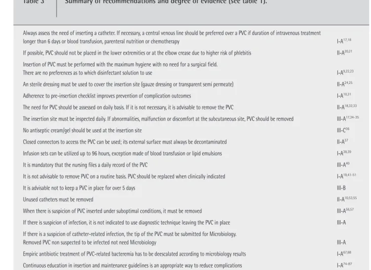
Table 3
Summary of recommendations and degree of evidence (see table 1).
Always assess the need of inserting a catheter. If necessary, a central venous line should be preferred over a PVC if duration of intravenous treatment
longer than 6 days or blood transfusion, parenteral nutrition or chemotherapy I-A17,18
If possible, PVC should not be placed in the lower extremities or at the elbow crease due to higher risk of phlebitis II-A20,21
Insertion of PVC must be performed with the maximum hygiene with no need for a surgical field.
There are no preferences as to which disinfectant solution to use I-A9,22,23
An sterile dressing must be used to cover the insertion site (gauze dressing or transparent semi permeate) II-A24,25
Adherence to pre-insertion checklist improves prevention of complication outcomes I-A10,31
The need for PVC should be assessed on daily basis. If it is not necessary, it is advisable to remove the PVC II-A18,32,33
The insertion site must be inspected daily. If abnormalities, malfunction or discomfort at the subcutaneous site, PVC should be removed III-A17,34-35
No antiseptic cream/gel should be used at the insertion site III-C36
Closed connectors to access the PVC can be used; its external surface must always be decontaminated II-A37
Infusion sets can be utilized up to 96 hours, exception made of blood transfusion or lipid emulsions I-A38,39
It is mandatory that the nursing files a daily record of the PVC III-A40
It is not advisable to remove PVC on a routine basis. PVC should be replaced when clinically indicated I-A18,41-51
It is advisable not to keep a PVC in place for over 5 days III-B
Unused catheters must be removed II-A10,53,55
When there is suspicion of PVC inserted under suboptimal conditions, it must be removed III-A56,57
If there is suspicion of infection, it is not indicated to use diagnostic technique leaving the PVC in place III-A If there is a suspicion of catheter-related infection, the tip of the PVC must be submitted for Microbiology.
Removed PVC non suspected to be infected not need Microbiology III-A
Empiric antibiotic treatment of PVC-related bacteremia has to be deescalated according to microbiology results I-A67,68
A different situation is S. aureus or C. albicans infection
as those require a minimum of 14 days of treatment
69and
fol-low-up cultures at 72 hours. Secondary infectious foci like
en-docarditis and/or osteomyelitis must be ruled out
70. This is even
more important if bacteremia persists after removal of the PVC
thus indicating a more prolonged presence of bacteria in the
blood stream
70-73. This Consensus Document does not pretend
reviewing the treatment of S. aureus or other bacteremias and
the reader is referred to specific guidelines
70,71. Gram-negative
bacilli infections usually need 7 to 14 days of treatment after
removal of PVC and after the first negative blood culture is
confirmed
7.
CONTINUOUS EDUCATION
Continuous education of healthcare caregivers on the
indications for PVC insertion and the convenience of having
PVC inserted is necessary. It is necessary to periodically
re-mind the nursing staff inserting PVC the guidelines for
in-sertion and maintenance
74-81(I-A). Table 3 summarizes the
recommendations and degree of evidence and references as
produced in this document.
The lack of a continuous
edu-cation programme leads to relaxation of the norm,
aban-donment of good clinical practices and increase in infection
and complication rates. On the contrary, specific educational
programmes help in reducing infection rates
82-87. There are
different ways to provide education. Education among peers
has shown the best benefits in guideline follow-up as the
staff is engaged in education.
It is advisable that the infection and complication rates
are periodically disclosed to the staff in charge of inserting
PVCs. This is positive reinforcement on guideline/protocol
fol-low-up and a warning if deviations occur. Furthermore, the
adherence to the checklist can be monitored (table 2).
CONFLICT OF INTEREST
None declared
ABBREVIATIONS AND ACRONYMS
SEICAV = Sociedad Española de Infecciones Cardiovasculares
SEMI = Sociedad Española de Medicina Interna
SEQ = Sociedad Española de Quimioterapia
SECTCV = Sociedad Española de Cirugia Torácica-Cardiovascular
PVC = Peripheral venous catheter
ICU = Intensive Care Unit
NCBI = National Center for Biotechnology Information
NLM = National Library of Medicine
PICC = Peripherally-inserted central catheters
ISDA = Infectious Disease Society of America
REFERENCES
1. Pérez-Granda MJ, Guembe MR, Rincón C, Muñoz P, Bouza E. A prevalence survey of intravascular catéter use in a general hospital. J Vasc Access 2014: 25:524-8.
2. Guembe M, Pérez-Granda MJ, Capdevila JA, Barberan J, Pinilla B, Martín-Rabadán, Bouza E. on behalf of the NUVE Study. Nation-wide study of the use of intravascular catheters in internal medi-cine departments. J Hosp Infect 2015; 90(2):135-41.
3. Pujol M, Hornero A, Saballs M, Argerich MJ, Verdaguer R, Cisnal M et al. Clinical epidemiology and outcomes of peripheral venous catheter-related bloodstream infections at a university-affiliated hospital. J Hosp Infect 2007; 67:22-9.
4. Delgado M,Gabillo A, Elias L, Yebenes JC, Sauca G, Capdevila JA. Caracteristicas de la bacteriemia relacionada con catéter venoso periférico en un hospital general. Rev Esp Quimioter 2012; 25:129-33.
5. Almirante B, Limón E, Freixas N, Gudiol F. Vigilancia de bacteriemi-as relacionadbacteriemi-as con el uso de catéteres venosos en los hospitales de Catalunya. Resultados del Programa VINCAT (2007-2010). Enf. Infecc Microbiol Clin 2012; 30 (supl 3):13-9.
6. Chopra V, Anand S, Krein SL, Chenoweth C, Saint S. Bloodstream infection, venous thrombosis, and peripherally inserted central catheters: reappraising the evidence. Am J Med 2012; 125:733-41 7. León C, Ariza X. Guías para el tratamiento de las infecciones
rel-acionadas con catéteres intravasculares de corta permanencia en adultos: conferencia de consenso SEIMC-SEMICYUC”. Enf Infecc Microbiol Clin 2004; 22:99-101.
8. Mermel LA, Allon M, Bouza E, Craven DE, Flynn P, O’Grady NP et al. Clinical practice guidelines for the diagnosis and management of intravascular catheter-related infection:2009. Update by the Infec-tious Diseases Society of America. Clin Infect Dis 2009; 49:1-45. 9. O´Grady N, Alexander M, Burns LA, Dellinger EP, Garland J, Heard
SO, et al. Guideline for the prevention of intravascular Cathe-ter-related infections. Clin Infect Dis 2011; 52:162-93.
10. Pronovost P, Needham D, Berenholtz S, Sinopoli D, Chu H, Cos-grove S, et al. An intervention to decrease catheter-related blood-stream infections in the ICU. N Engl J Med 2006; 355:2725-32. 11. Capdevila JA. El catéter periférico: el gran olvidado de la infección
nosocomial. Rev Esp Quimioter 2013; 26:1-5.
12. Cicolini G, Simonetti V, Comparcini D, Labeau S, Blot S, Pelusi G, et al. Nurse’s knowledge of evidence-based guidelines on the preven-tion of peripheral venous catheter-related infecpreven-tions: a multicen-tre suevey. J Clin Nurs 2014; 17-18:2578-88.
13. Ahlqvist M, Beerglund B, Wiren M, Klang B, Johansson E. Accuracy in documentation-a study of peripheral venous catheters. J Clin Nurs 2009; 13:1945-52.
14. Véliz E, Vergara T, Fica A. Evaluación de las condiciones de manejo de catéteres vasculares periféricos en pacientes adultos. Rev Chile-na Infectol 2014; 31: 666-9.
15. Kish MA. Guide to development of practice guidelines. Clin Infect Dis 2001; 32:851-4
16. American College of Chest Physicians/ Society of Critical Care Medicine. Consensus Conference: definitions for sepsis and organ failure and guidelines for the use of innovative therapies in sepsis. Crit Care Med 1992; 20:864-74
17. Maki D, Ringer M. Risk Factors for infusion-related phlebitis with small peripheral venous catheters. A randomized controlled trial. Ann Intern Med 1991; 114:845-54.
18. Mestre G, Berbel C, Tortajada P, Gallemí G, Aguilar M, Caylà J, et al. Assessing the influence of risk factors on rates and dynamics of peripheral vein phlebitis: an observational cohort study. Med Clin 2011; 139:185-91.
19. Göransson KE, Johansson E. Indication and usage of peripheral ve-nous catheters inserted in adult patients during emergency care. J Vasc Access 2011; 3:193-9.
20. Dunda S, Demir E, Mefful O, Grieb G, Bozkurt A, Pallua N. Manage-ment, clinical outcomes, and complications of acute cannula-re-lated peripheral vein phlebitis of the upper extremity: A retrospec-tive study. Phlebology 2014; 30:381-8.
21. Uslusoy E, Mete S. Predisposing factors to phlebitis in patients with peripheral intravenous catheters: a descriptive study. J Am Acad Nurse Pract 2008; 20: 172-80.
22. Maki DG, Ringer M, Alvarado CJ. Prospective randomized trial of povidone-iodine, alcohol, and chlorhexidine for prevention of in-fection associated with central venous and arterial catheters. Lan-cet 1991; 338:339–43.
23. Canadian Agency for drugs and technology in health. Use of clor-hexidine gluconate with alcohol for the prevention of peripheral intravenous device infections: A review of clinical and cost effec-tiveness and Guidelines. 2014, April 3.
24. Maki DG, Stolz SS, Wheeler S, Mermel LA. A prospective, rand-omized trial of gauze and two polyurethane dressings for site care of pulmonary artery catheters: implications for catheter manage-ment. Crit Care Med 1994; 22:1729–37.
25. Bijma R, Girbes AR, Kleijer DJ, Zwaveling JH. Preventing central ve-nous catheter-related infection in a surgical intensive-care unit. Infect Control Hosp Epidemiol 1999; 20:618–20.
26. Göransson KE, Johansson E. Prehospital peripheral venous cathe-ters: a prospective study of patients complications. J Vasc Access 2012; 13:16-21.
27. Forni C, Loro L, Tremosini M, Trofa C, D’Alessandro F, Sabattini T, et al. Cohort study of peripheral catheter-related complications and identification of predictive factors in a population of orthopedic patients. Assist Inferm Ric 2010; 29:166-77.
28. Aulagnier J, Hoc C, Mathieu E, Dreyfus JF, Fischler M, Le Guen M. Efficacy of accuvein to facilitate peripheral intravenous placement in adults presenting to an emergency department: a randomized clinical trial. Acad Emerg Med 2014; 21:858-63.
29. Au AK, Rotte MJ, Grzybowski RJ, Ku BS, Fields JM. Decrease in central venous catéter placement due to use of ultrasound guid-ance for peripheral intravenous catheters. Am J Emerg Med 2012; 30:1950-4.
30. Liu YT, Alsaawi A, Bjornsson HM. Ultrasound (US) guidance for the placement of peripheral venous access: a systematic review of
ran-domized-controlled trials. Eur J Emerg Med 2014; 21:18-23 31. Chiu PC, Lee YH, Hsu HT, Feng YT, Lu IC, Chiu SL, Cheng KI.
Estab-lish a perioperative check forum for peripheral intravenous acces to prevent the occurrence of phlebitis. Kaohsiung J Med Sci 2015; 31:215-21.
32. Targer IB, Ginsberg MB, Ellis SE, Walsh NE, Dupont I, Simchen EA et al. An Epidemiological study of the risks associated with peripher-als intravenous catheters. Am J Epidemiol 1983; 118:839-51. 33. O´Grady NP, Alexander M, Dellinger EP, Gerberding JL, Heard SO,
Maki DG, et al. Guidelines for the prevention of intravascular cath-eter-related infections. Infect Control Hospital Epidemiol 2002; 23:759-69.
34. White MC. Infections and infection risks in home care settings. In-fect Control Hosp Epidemiol 1992; 13:535–9.
35. White MC, Ragland KE. Surveillance of intravenous catheter-relat-ed infections among home care clients. Am J Infect Control 1994; 22:231–5.
36. Zakrzewska-Bode A, Muytjens HL, Liem KD, Hoogkamp-Korstanje JA. Mupirocin resistance in coagulase-negative staphylococci, after topical prophylaxis for the reduction of colonization of central ve-nous catheters. J Hosp Infect 1995; 31:189–93.
37. Yébenes JC, Delgado M, Sauca G, Serra-Prat M, Solsona M, Almirall J, Capdevila JA, Balanzó X. Efficacy of three different valve systems of needle-free closed connectors in avoiding access of microor-ganisms to endovascular catheters after incorrect handling. Crit Care Med 2008; 36:2558-61.
38. Ullman AJ, Cooke ML, Gillies ED, Marsh NM, Daud A, McGrail MR, et al. Optimal timing for intravascular administration set replace-ment. Cochrane Database Syst Rev 2013; 9:CD003588.
39. Lai KK. Safety of prolonging peripheral cannula and IV tubing use from 72 hours to 96 hours. Am J Infect Control 1998; 26:66-70. 40. Ahlqvist M, Berglund B, Wirén M, Klang B, Johansson E. Accuracy
in documentation-a study of peripheral venous catheters. J Clin Nurs 2009; 18:1945-52.
41. Bregenzer T. Conen D, Sackmann P, Widmer A. Is routine replace-ment of peripheral intravenous catheters necessary?. Arch Intern Med 1998; 158: 151-6.
42. Van Donk P, Rickard CM, McGrail MR, Doolan G. Routine replace-ment versus clinical monitoring of peripheral intravenous cath-eters in a regional hospital in the home program: A randomized controlled trial. Infect Control Hosp Epidemiol 2009; 30:915-7. 43. Webster J. Clarke S, Paterson D, Hutton A, van Dyk S, Gale C,
Hop-kins T. Routine care of peripheral intravenous catheters versus clinically indicated replacement; randomized controlled trial. BMJ 2008; 337:a339.
44. Webster J, Osborne S, Rickard C, Hall J. Clinically-indicated replace-ment versus routine replacereplace-ment of peripheral venous catheters. Cochrane Database Syst Rev 2010; (3):CD007798.
45. Mestre G, Berbel C, Tortajada P, Alarcia M, Coca R, Fernández M et al. Successful multifaceted intervention aimed to reduce short peripheral venous catheter-related adverse events; A quasi experi-mental cohort study. Am J Infect Control 2013; 41(6):520-6.
46. Grüne F, Schrappe M, Basten J, Wenchel HM, Tual E, Stützer H. Phlebitis rate and time kinetics of short peripheral intravenous catheters. Infection 2004; 32:30-2.
47. Lee WL, Chen HL, Tsai TY, Lai IC, Chang WC, Huang CH, et al. Risk factors for peripheral intravenous catheter infection in hospital-ized patients: A prospective study of 3165 patients. Am J Infect Control 2009; 37:683-6.
48. Giménez Pérez M. Systematic withdrawal of peripheral vein cath-eters: does it salvage lives or increase costs?. Med Clin 2012; 139:203-5.
49. Hasselberg D, Ivarsson B, Andersson R, Tingstedt B. The handling of peripheral venous catheters--from non-compliance to evi-dence-based needs. J Clin Nurs 2010; 19:3358-563.
50. Juvé ME, Carbonell MD, Soldevila RM, Campa I, Juarez M. Manten-imiento de catéteres venosos periféricos durante más de 4 días. En busca de la mejor evidencia. Enf Clin 2003; 13:208-16.
51. Do Rego Furtado LC. Maintenance of peripheral venous access and its impact on the development of phlebitis: a survey of 186 catheters in a general surgery department in Portugal. J Infus Nurs 2011; 34:382-90.
52. Bertolino G, Pitassi A, Tinelli C, Staniscia A, Guglielmana B, Scu-deller L, et al. Intermittent flushing with heparin versus saline for maintenance of peripheral intravenous catheters in a medical department: a pragmatic cluster-randomized controlled study. Worldviews Evid Based Nurs 2012; 9:221-6.
53. Berenholtz SM, Pronovost PJ, Lipsett PA, et al. Eliminating cathe-ter-related boodstream infections in the intensive care unit. Crit Care Med 2004; 32:2014–2020
54. Lederle FA, Parenti CM, Berskow LC, Ellingson KJ. The idle intrave-nous catheter. Ann Intern Med 1992; 116:737–8.
55. Parenti CM, Lederle FA, Impola CL, Peterson LR. Reduction of un-necessary intravenous catheter use. Internal medicine house staff participate in a successful quality improvement project. Arch In-tern Med 1994; 154:1829–32.
56. Raad II, Hohn DC, Gilbreath BJ, et al. Prevention of central ve-nous catheter-related infections by using maximal sterile barrier precautions during insertion. Infect Control Hosp Epidemiol 1994; 15:231–8.
57. Boyce JM, Pittet D. Guideline for hand hygiene in health-care set-tings: recommendations of the Healthcare Infection Control Prac-tices Advisory Committee and the HICPAC/SHEA/APIC/IDSA Hand Hygiene Task Force. Infect Control Hosp Epidemiol 2002; 23:S3–40. 58. Yébenes JC, Capdevila JA. Infección relacionada con catéteres
in-travasculares. Med Clin 2003; 121:238.
59. Bouza E, Burillo A, Muñoz P. Catheter-related infections: diagnosis and intravascular treatment. Clin Microb Infect 2002; 8:265-74. 60. Capdevila JA, Planes AM, Palomar M, Gasser I, Pahissa A, Crespo E,
Almirante B, Martínez Vázquez JM. Value of differential quantita-tive blood cultures in diagnosis of catheter related sepsis. Eur J Clin Microbiol 1992; 11:403-7.
61. Blot F, Schmidt E, Nitemberg G, Tancredo C, Leclercq B, Laplanche A, et al. Earlier positivity of Central-venous-versus
perioher-al-blood cultures is highly predictive of catéter-related sepsis. J Clin Microbiol 1998; 36:105-9.
62. Aygun G, Yasar H, Yilmaz M, Karasahin K, Dikmen Y, Polat E, et al. The value of gram staining of catheter segments for rapid de-tection of peripheral venous catheter infections. Diagn Microbiol Infect Dis 2006; 54:165-7.
63. Reigades E, Rodriguez-Créixems M, Sánchez-Carrillo C, Martín-Ra-badán P, Bouza E. Uncommon aetiological agents of catheter-re-lated bloodstream infections. Epidemiol Infect 2015; 143:741-4. 64. Stuart RL, Cameron DR, Scout C, Kotsanas D, Grayson ML, Korman
TM, et al. Peripheral intravenous catheter-associated
Staphylo-coccus aureus bacteremia: more than 5 years of prospective data
from two tertiary health services. Med J Aus 2013; 198:551-3. 65. Trinh TT, Chan PA, Edwards O, Hollenbeck B, Huang B, Burdick N, et
al. Peripheral venous catheter-related Staphylococcus aureus bac-teremia. Infect Control Hosp Epidemiol 2011; 32:579-83. 66. Almirante B, Limón E, Freixas N, Gudiol F; Vincat Program.
Labo-ratory-based surveillance of hospital-acquired catheter-related bloodstream infections in Catalonia. Results of the Vincat Program (2007-2010). Enf Infecc Microbiol Clin 2012; Suppl 3:13-9. 67. Dellit TH, Owens RC, McGowan JE, et al. Infectious Diseases Society
of America and the Society for Healthcare Epidemiology of Ameri-ca guidelines for developing and institutional program to enhance antimicrobial stewardship. Clin Infect Dis 2007; 44:159–77. 68. Rodríguez-Baño J, Paño-Pardo JR, Alvarez-Rocha L, Asensio A,
Calbo E, Cercenado E, et al. Programas de optimización de uso de antimicrobianos (PROA en hospitales españoles: documento de consenso GEIH-SEIMC, SEFH y SEMPSPH. Enferm Infecc Microbiol Clin 2012; 30:22.e1-22.e23.
69. Jernigan JA, Farr BM. Short-course therapy of catheter-related
Staphylococcus aureus bacteremia: a meta-analysis. Ann Intern
Med 1993; 119:304-11.
70. Gudiol F, Aguado JM, Almirante B, Bouza B, Cercenado E Domínguez MA, et al. Diagnosis and treatment of bacteremia and endocarditis due to Staphylococcus aureus. A clinical guideline from the Span-ish Society of Clinical Microbiology and Infectious Diseases. Enferm Infecc Microbiol Clin 2015; 33(9):625.e1-625.e23.
71. Cisneros-Herrerosa JM, Cobo-Reinoso J, Pujol-Rojo M, Rodríguez-Baño J, Salavert-Lletíe M. Guía para el diagnóstico y tratamiento del paciente con bacteriemia. Guías de la Sociedad Española de Enfermedades Infecciosas y Microbiología Clínica (SEIMC). Enferm Infecc Microbiol Clin 2007; 25:111-30.
72. Fowler VC, Justice A, Moore C, Benjamin DK, Woods CW, Campbell S, et al. Risk factors for haematogenous complications of intra-vascular catheters-associated S. aureus bacteremia. Clin Infect Dis 2005; 40:95-103.
73. Sanchez KT, Obeid KM, Szpunar S, Fakih MG, Khatib R. Delayed peripheral venous catheter-related Staphylococcus aureus bacte-remia: onset ≥ 24 hours after catheter removal. Scand J Infect Dis 2012; 44:551-4.
74. Yoo S, Ha M, Choi D, Pai H. Effectiveness of surveillance of central catheter-related bloodstream infection in an ICU in Korea. Infect Control Hosp Epidemiol 2001; 22:433–6.
75. Warren DK, Zack JE, Cox MJ, Cohen MM, Fraser VJ. An educational intervention to prevent catheter-associated bloodstream infec-tions in a non-teaching community medical center. Crit Care Med 2003; 31:1959–63.
76. Warren DK, Zack JE, Mayfield JL, et al. The effect of an education program on the incidence of central venous catheter-associated bloodstream infection in a medical ICU. Chest 2004; 126:1612–8. 77. Warren DK, Cosgrove SE, Diekema DJ, et al. A multicenter
interven-tion to prevent catheter-associated bloodstream infecinterven-tions. Infect Control Hosp Epidemiol 2006; 27:662–9.
78. Higuera F, Rosenthal VD, Duarte P, Ruiz J, Franco G, Safdar N. The effect of process control on the incidence of central venous cath-eter-associated bloodstream infections and mortality in intensive care units in Mexico. Crit Care Med 2005; 33:2022–7.
79. Coopersmith CM, Rebmann TL, Zack JE, et al. Effect of an education program on decreasing catheter-related bloodstream infections in the surgical intensive care unit. Crit Care Med 2002; 30:59–64. 80. Coopersmith CM, Zack JE, Ward MR, et al. The impact of bedside
behavior on catheter-related bacteremia in the intensive care unit. Arch Surg 2004; 139:131–6.
81. Sherertz RJ, Ely EW, Westbrook DM, et al. Education of physicians in-training can decrease the risk for vascular catheter infection. Ann Intern Med 2000; 132:641–8.
82. Eggimann P, Harbarth S, Constantin MN, Touveneau S, Chevrolet JC, Pittet D. Impact of a prevention strategy targeted at vascu-lar-access care on incidence of infections acquired in intensive care. Lancet 2000; 355:1864–68.
83. Lolom I, Deblangy C, Capelle A, Guerinot W, Bouvet E, Barry B, et al. Effect of a long-term quality improvement program on the risk of infection related peripheral venous catheters. Presse Med 2009; 38:34-42.
84. Boy S, Aggarwal I, Davey P, Logan M, Nathwani D. Peripheral in-travenous catheters: the road to quality improvement and safer patient care. J Hosp. Infect 2011; 77:37-41.
85. Frigerio S, Di Giulio P, Gregori D, Gavetti D, Ballali S, Bagnato S, et al. Managing peripheral venous catheters: an investigation on the efficacy of a strategy for the implementation of evidence-based guidelines. J Eval Clin Pract 2012; 18:414-9.
86. Soifer NE, Borzak S, Edin BR, Weinstein RA. Prevention of periph-eral venous catheter complications with an intravenous ther-apy team: a randomized controlled trial. Arch Intern Med 1998, 158:473-7.
87. Vidal E, Capdevila JA., Sauca G, Force L, Floriach N, Usas M, et al. Reducción de la tasa de bacteriemia asociada a catéter venoso periférico después de aplicar un programa de prevención. Abstract presentado en el XV Congreso SEIMC. Bilbao 2012