Scuola Dottorale in Biologia
Dottorato di Ricerca in BIOLOGIA APPLICATA ALLA SALUTE DELL'UOMO
XXVII Ciclo
INTERAZIONI TRA LE ATTIVITA' DEL RECETTORE PER GLI ESTROGENI ALFA E LA
RETE DI SEGNALI BASATI SULL'UBIQUITINA INTERACTIONS BETWEEN ESTROGEN RECEPTOR ALPHA ACTIVITIES AND THE UBIQUITIN-BASED SIGNALLING NETWORK
Dottorando: VALERIA PESIRI ______________ Tutor: Dott. FILIPPO ACCONCIA ______________
Alla mia numerosa famiglia
INDEX
RIASSUNTO...Pag. I SUMMARY...Pag. V 1. BACKGROUND...Pag. 1 1.1 Estrogens...Pag. 1 1.2 Estrogen Receptor α (ERα)...Pag. 2
1.2.1 ERα structure.
1.2.2 ERα nuclear signalling. 1.2.3 ERα extra-nuclear signalling.
1.2.4 Integration of E2:ERα signalling to physiological processes.
1.2.5 Modulation of ERα activities by post-translational modifications.
1.3Ubiquitin (Ub)-based signalling
network...Pag. 11
1.3.1 Proteolytic and non-proteolytic ubiquitination. 1.3.2 The Ub-Binding Domains.
1.4Interactions between the ERα and Ub
signalling...Pag. 17
1.4.1 Ub-dependent proteolytic control of ERα expression.
1.4.2 Ub-dependent non-proteolytic control of ERα signalling.
2. AIM...Pag. 21 3. IDENTIFICATION OF NON-COVALENT Ub-BINDING SURFACE (UBS) ON ERα...Pag. 23 3.1 Introduction.
3.2 Results.
3.2.1 Indentification of two UBSs on ERα. 3.2.2 Characterization of the full length ERα-UBS mutant in cells.
4. ROLE OF THE ERα-UBS IN E2-INDUCED CELL PROLIFERATION...Pag. 32 4.1 Introduction.
4.2 Results.
4.2.1 The ERα-UBS is required for E2-induced cell proliferation.
4.2.2 The ERα-UBS functions are necessary for the E2-induced cholesterol sourcing.
4.2.3 The ERα-UBS controls the receptor full transcriptional activation.
5. ROLE OF THE ERα-UBS IN E2-INDUCED GENE EXPRESSION...Pag. 41 5.1 Introduction.
5.2 Results.
5.2.1 DNA Micro Arrays: the ERα-UBS is necessary for E2-induced gene expression.
5.2.2 Ingenuity Pathways Analysis: CREB1
activation by E2 relies on a functional UBS on ERα. 5.2.3 Dissection of the E2-triggered ERα-UBS dependent CREB1 pathway.
6. DISCUSSION AND CONCLUSIONS...Pag. 54 7. REFERENCES...Pag. 66 ACKNOWLEDGEMENTS...Pag. 76 APPENDIX A.
MATHERIALS AND METHODS. APPENDIX B.
SUPPLEMENTAL FIGURES. APPENDIX C.
RIASSUNTO.
Il 17β-estradiolo (E2) è l'estrogeno più efficace negli esseri umani e svolge un ruolo critico nel controllo di numerosi processi cellulari che influenzano fortemente diversi aspetti della fisiologia femminile e maschile. Gli effetti cellulari di E2 (ad es. proliferazione, apoptosi, differenziamento) dipendono dalla fine regolazione dell'attività dei suoi recettori, il ERα ed il ERβ, che in seguito al legame dell'ormone mediano l'attivazione di vie extra-nucleari (ad es. attivazione di protein chinasi) e nucleari (ad es. trascrizione genica). La deregolazione degli eventi indotti dal E2 è considerato un fattore di rischio per l'insorgenza e la progressione di diverse tipologie di cancro, per cui la comprensione del meccanismo d'azione dei suoi recettori riveste una notevole importanza per l'identificazione di possibili bersagli farmacologici. In particolare, molta attenzione è rivolta al ERα, che media gli effetti proliferativi del E2 in cellule di cancro alla mammella. Gli attuali approcci terapeutici al cancro alla mammella ERα-positivo dipendono anche dall'utilizzo di farmaci che legano il ERα e modificano i suoi livelli cellulari (ad es. Tamoxifene, Faslodex); tuttavia, i possibili effetti collaterali di questi farmaci [es. resistenza, cancro in altri tessuti (es. endometriale)] spingono alla continua ricerca di nuovi bersagli farmacologici.
Tra le terapie contro il cancro, il sistema dell'ubiquitina (Ub) è oggetto di interesse in quanto è alla base di una complessa rete di interazioni proteiche che è critica per la trasduzione del segnale responsabile di molti processi cellulari. Le interazioni molecolari dipendenti dall'Ub si basano su associazioni non-covalenti tra proteine modificate con l’Ub (ubiquitinazione) e proteine (chiamate recettori per l'Ub) che possiedono un dominio di legame all'Ub (UBD). La deregolazione di questo intricato sistema è stata associata a diverse condizioni patologiche, incluse diverse tipologie di cancro. Il sistema dell'Ub controlla la segnalazione del complesso E2:ERα. Infatti, mentre la modificazione con catene di Ub (poliubiquitinazione)
controlla la degradazione del recettore e la sua attività trascrizionale, la modificazione del ERα con una singola molecola di Ub (monoubiquitinazione) è richiesta per l'attivazione dipendente dal E2 di segnali rapidi che portano alla proliferazione cellulare. Anche se non è stato chiarito il meccanismo molecolare attraverso il quale la modificazione con l’Ub modula le attività del ERα, una possibilità è che il ERα possa comportarsi come un recettore per l'Ub che lega la monoubiquitina (monoUb) su se stesso o su proteine interagenti e trasduce il segnale che porta alla proliferazione cellulare.
L'obiettivo di questo progetto di dottorato è stato valutare la capacità del ERα di legare non-covalentemente l'Ub e l'eventuale ruolo regolatorio di questo legame nei processi cellulari dipendenti dal E2.
Inizialmente, sono stati condotti esperimenti in vitro per comprendere l'abilità di diversi domini del ERα (A/B, C, E) di legare l'Ub derivante da lisati cellulari o ricombinante. Questi esperimenti hanno rivelato che il ERα possiede due domini che legano l'Ub (A/B ed E) contattandola direttamente. In seguito, è stata posta maggiore attenzione sul dominio E (del quale è nota la struttura) ed è stato osservato che la sua porzione N-terminale (aminoacidi 301-439) costituisce la minima superficie di legame all'Ub (UBS). All'interno dell'UBS sono stati poi identificati i residui critici per l'associazione non-covalente all'Ub (L429, A430).
L'introduzione della doppia mutazione L429,A430 (LAAG), nel dominio E in vitro e nell'intero (wt) ERα trasfettato nelle cellule, riduceva significativamente il legame all'Ub. Successivamente è stato valutato il ruolo della ERα-UBS negli effetti cellulari indotti dal E2. Per fare questo, sono state selezionate le cellule HEK293 che dopo la trasfezione esprimessero stabilmente il ERα wt e LAAG. In queste cellule è stata analizzata sia la proliferazione cellulare che l'approvigionamento di colesterolo indotte dal E2 ed è stato trovato che la mutazione dell'UBS sul ERα le previene. In seguito, poichè l'induzione della proliferazione da parte del E2 dipende fortemente dall'attivazione
extra-nucleare di chinasi di segnale, è stata valutata la fosforilazione attivatoria di alcune vie di segnale attivate del E2 in maniera conservata in diversi tipi cellulari. I risultati ottenuti indicano che la mutazione della UBS impedisce l'attivazione mediata dal E2 della via PI3K/AKT ma non della via ERK/MAPK. Questi dati indicano che la UBS sul dominio E del ERα gioca un ruolo critico per gli effetti mitogenici del E2. In particolare, l'incapacità del ERα LAAG di attivare la via PI3K/AKT sembra essere il principale responsabile di questo effetto. L'attivazione della via PI3K/AKT dipende dal fatto che il E2 induce una maggiore associazione del ERα con recettori per i fattori di crescita (ad es. IGF1-R) e richiama proteine di segnale alla membrana. Poichè i dati ottenuti mostrano anche una deregolata associazione del ERα LAAG con il IGF1-R è possibile che questo possa contribuire a prevenire la corretta attivazione della via di segnale a valle. Inoltre, la via PI3K/AKT controlla le attività del ERα anche fosforilando il recettore in risposta al E2. In particolare, la fosforilazione del recettore sul residuo di Ser118 dipende dalla via PI3K/AKT e gioca un ruolo critico per l'attività trascrizionale del E2:ERα. In accordo con i dati precedenti, il ERα LAAG non risultava fosforilato in Ser118 ne’ era in grado di mediare l'incremento dell'mRNA dei livelli della Ciclina D1 dopo il trattamento con E2.
In seguito, poichè i dati ottenuti suggerivano una ridotta abilità del ERα LAAG nel mediare l'espressione genica dei geni bersaglio del E2, sono stati effettuati esperimenti di DNA Micro Arrays che lo hanno definitivamente dimostrato. Inoltre, l'Ingenuity Pathway Analysis applicata ai dati ottenuti ha permesso di identificare CREB1 come altro fattore di trascrizione attivato dal E2 attraverso il wt ma non LAAG ERα. CREB1 controlla direttamente la trascrizione di geni coinvolti nella proliferazione cellulare (ad es. Ciclina D1), sopravvivenza cellulare (ad es. Bcl-2) e metastasi (ad es. VEGF), la cui deregolazione favorisce la progressione tumorale. I dati ottenuti mostrano che il E2 non è in grado di indurre l'attivazione di CREB1 ne’ la conseguente trascrizione di
geni target (ad es. Bcl-2, Cyclin D1) in cellule esprimenti ERα LAAG. Quindi l'UBS sul ERα è importante per l'attivazione trascrizionale di CREB1 indotta dal E2. Per comprendere se questo effetto fosse dipeso dall'incapacità del ERα LAAG di attivare la via PI3K/AKT è stata inibita questa via di segnale. I dati ottenuti indicano che l'attivazione della PI3K/AKT contribuiva alla fosforilazione di CREB1 indotta dal E2 in cellule HEK293 esprimenti il ERα wt e in cellule MCF-7. Infine, bloccando, attraverso un approccio farmacologico, la fosforilazione di CREB1 a monte o la sua attività trascrizionale a valle, è stato trovato che queste linee cellulari non sono in grado di proliferare in seguito alla stimolazione con il E2. Questi dati permettono di ipotizzare che un complesso di membrana dipendente dall'UBS sul ERα, e responsabile dell'attivazione della via di segnale PI3K/AKT indotta dal E2, controlli la proliferazione cellulare attraverso la modulazione dell'attivazione del ERα e di CREB1, richiesti per la trascrizione genica. Nel complesso, i dati riportati in questo progetto di dottorato indicano che il ERα possiede una UBS sul suo dominio E che gioca un ruolo chiave per garantire le attività nucleari ed extra-nucleari indotte dal E2 necessarie per la proliferazione cellulare.
Concludendo, le nostre scoperte indicano che il ruolo regolatorio del legame non-covalente all'Ub sul ERα deve essere aggiunto ai meccanismi molecolari noti ed attivati dal E2 per l'induzione della proliferazione cellulare. Poichè il ERα riveste un ruolo chiave nella progressione del cancro alla mammella, questo progetto mette in luce nuovi possibili bersagli farmacologici. A tal proposito, l'interferenza nel legame UBD:Ub tramite specifiche piccole molecole è stato già proposto come futuro bersaglio farmacologico contro alcuni tipi di cancro.
SUMMARY.
The E2:ERα signalling controls a plethora of physiological processes but plays also a critical role in breast cancer progression, thus the deep understanding of the mechanisms that control E2-induced cell proliferation would help to identify new putative druggable targets for the treatment of breast cancer. The ubiquitin (Ub)-system is gaining much attention for cancer therapies because it allows to build complex interactions network that is critical for signal transduction to many cellular processes. The Ub-based network depends on non-covalent binding between ubiquitinated proteins and proteins that possess an ubiquitin binding domain (UBD), called Ub-receptors. The deregulation of this intricate system has been associated with several pathological conditions including several types of cancers. Recent papers reported that the Ub-system deeply impacts the E2:ERα signalling; indeed, while ERα modification with polyUb chains controls the receptor turnover and transcriptional activity, ERα modification with single Ub molecule (i.e., monoubiquitination) is required for the E2-dependent activation of rapid signalling to cell proliferation. Even if it has not been clarified how the Ub modification on ERα modulates the receptor activities, one possibility, which has never been considered is that ERα could recognize and transduce the Ub modification on itself or on interacting proteins through an UBD.
The main goal of the present PhD project was to understand the non-covalent Ub-binding abilities of ERα and their regulatory role in E2-dependent cellular processes.
To this purpose, initial experiments were performed in vitro by using purified ERα domains and recombinant or cell lysates-derived Ub molecules. We found that ERα has two different Ub-binding surfaces (UBSs) (in A/B and E domains) that have different binding abilities towards specific based chains. By focusing on the E domain Ub-binding ability we mapped an UBS on its N-terminal portion (i.e., 301-439) and identified the structural determinants
required for ERα to non-covalently associate to Ub (i.e., L429, A430). Remarkably, the introduction of L429,A430 double mutations (i.e., LAAG) in the context of the full lenght ERα reduced Ub-binding also in cells. However, the ERα-UBS mutant basal intracellular localization as well as E2 binding affinity was not affected by the introduction of LAAG mutations.
Next, we found that the LAAG mutation blocks the E2-induced cell proliferation. Since proliferating cells have an increased cholesterol requirement, E2-dependent modulation of the cellular levels of the master regulators [i.e., the 3-hydroxy-3-methylglutaryl coenzyme A reductase (HMGR) and the low-density lipoprotein receptor (LDLr)] of cholesterol sourcing was next evaluated. We found that, differently from the wt, the LAAG ERα was not able to increase the cellular levels of the HMGR as well as LDLr after E2 treatment. Because the E2-dependent cell proliferation depends on the activation of extra-nuclear signalling kinases we evaluated the activating phosphorylation of some signalling pathways activated by E2 in several cell lines. Our results indicate that the ERα-UBS mutation impairs the E2-induced activation of the PI3K/AKT but not ERK/MAPK signalling. Moreover, the PI3K inhibitor blocked the E2-dependent increase of the HMGR and LDLr, as well as of cell proliferation also in breast cancer MCF-7 cells, endogenously expressing ERα. These data indicate that the UBS on ERα E domain plays a critical role for E2 mitogenic effects. In particular, the LAAG ERα inability to activate the PI3K/AKT pathway seems to be the principal responsible for this effect in MCF-7 cells as well as in HEK293 transfected cells because this mutant is unable to direct the physiological association of the ERα with the IGF1-R. The PI3K/AKT pathway also controls the ERα activities by phosphorylating the receptor in response to E2. In particular, the receptor phosphorylation on Ser118 residue depends on the PI3K/AKT pathway and plays a critical role in E2:ERα transcriptional activity. In accordance with previous data, we found no phosphorylation on this residue as well as no
induction of the Cyclin D1 mRNA levels after E2 treatment in LAAG ERα cells. These data suggest an impairment of the ERα-UBS mutant ability to mediate E2 target genes expression.
DNA Micro Arrays experiments definitively demonstrated that the LAAG mutant ERα was less transcriptionally active than the wt receptor. Interestingly, the Ingenuity Pathway Analysis on the obtained data helped us identifying CREB1 as another transcription factor activated by E2 through wt but not LAAG ERα. CREB1 directly controls the transcription of genes involved in cell proliferation (i.e., Cyclin D1), cell survival (i.e., Bcl-2) and metastasis (i.e., VEGF), whose deregulation promotes the tumoral progression. Interestingly, we found that CREB1 activating phosphorylation on Ser133 residue as well as target genes expression (e.g., Bcl-2, Cyclin D1) were not induced by E2 in LAAG ERα HEK293 cells. Thus, the ERα-UBS is important for the E2-triggered CREB1 transcriptional activation. To understand if the lack of CREB1 activation after E2 treatment could be ascribed to the LAAG ERα inability to mediate the activation of the PI3K/AKT, we inhibited this pathway. The obtained data indicate that the PI3K/AKT contributed to E2-induced CREB1 phosphorylation in wt ERα HEK293 as well as MCF-7 cells. Finally, by using specific inhibitors we blocked E2-activated CREB1 activities upstream (i.e., block of the activating phosphorylation) or downstream (i.e., block of the transcriptional activity) and found an inability of these cell lines to proliferate. It is tempting to speculate that an ERα-UBS-dependent membrane complex, responsible for the E2-triggered PI3K/AKT signalling activation, controls cell proliferation through the regulation of ERα and CREB1 activation required for gene transcription.
Overall, the data reported in this PhD project indicate that the ERα possesses an UBS on its E domain that plays a critical role for E2-induced nuclear and extra-nuclear signalling to cell proliferation.
In conclusion, our findings open new avenues in the field of E2-activated molecular mechanisms to physiological effects that now have to include also the non-covalent Ub-binding abilities of ERα. Given the key role played by ERα in breast cancer progression, the comprehension of the regulatory role of the ERα-UBS on E2 mitogenic effects reveals new putative druggable targets. In this respect, the interference of the UBD:Ub interaction by using specific small molecules has just been proposed as a future pharmacological target against cancer.
1. BACKGROUND. 1.1 Estrogens.
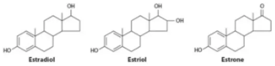
Estrogens are steroid hormones synthesized from cholesterol in a series of enzymatic reactions in which the last step is catalyzed by the aromatase enzyme. The three physiologically occurring estrogens are estrone (E1), 17β-estradiol (E2) and estriol (E3), that share a common four-ring structure (Fig. 1). E2 is the most active and the major estrogen in pre-menopausal women; its production is under the control of the hypothalamic-pituitary-ovarian (HPO) axis and predominantly depends on ovaries. Smaller amount of E2 is synthesized by peripheral tissues (e.g., adipose tissue, adrenal gland). Estrogens are transported in the blood bound to the sex hormone binding globulin and diffuse in target tissues (e.g., breast, endometrium, bone, brain, liver) where they control a plethora of physiological processes including reproduction, bone density, brain functions and cholesterol metabolism. Despite the physiological actions of endogenous E2, abnormally high levels of E2 or deregulated E2 signalling are associated with the increased incidence of certain types of cancer (e.g., breast and endometrial) [1-3]. The biological actions of E2 are mediated by the estrogen receptors (ERs), the ERα and ERβ that exert opposite effects on cellular processes including proliferation and apoptosis. ERs possess a subtype-specific expression and differentially influence the development and progression of E2-dependent cancer. Indeed, the oncogenic effects of E2 are mainly dependent on the ERα-mediated activities, which promote an increase of the cell proliferation and a reduction of the apoptosis [4, 5]. Indeed, the therapeutic protocol for ERα-positive breast cancer relies, among others, on drugs that bind ERα and change the breast cancer cell intracellular content (e.g., 4-OH-tamoxifen- TAM; ICI 182,780- Faslodex); however, these drugs display serious side effects (e.g., endometrial cancer for TAM) and determine tumor resistance. Moreover, when a breast cancer sample needs to be classified to select a therapy, only the nuclear pool of
ERα is considered while it has been reported that the membrane pool of the receptor plays a critical role for E2-induced cell proliferation [4].
Thus, it is required to find alternative approaches to fight breast cancer; in this respect, a deep understanding of the mechanisms of ERα-mediated E2-induced cell proliferation would help to identify new putative pharmacological targets.
Figure 1. Chemical structures of estrogens. 17β-Estradiol, Estriol and
Estrone share the typical steroid structure: three ciclohexane rings and one ciclopentane ring. [2]
1.2 Estrogen Receptor α (ERα).
E2 is a pleiotropic hormone that regulates human physiology far behind the control of reproductive tissues by binding to its cognate ERs. ERs are homologous members of the nuclear receptors super-family that act as a ligand-activated transcription factors; they are encoded by different genes, are expressed in many different tissues and regulate the expression of different target genes, thus transducing the E2 signalling in opposite ways (i.e., mitogenic for ERα and pro-apoptotic for ERβ) [4].
The ERα signalling is strictly dependent on the receptor structural characteristics, which allow it to localize to the plasma membrane as well as in the nucleus. These biochemical features permit the receptor to activate both E2-induced rapid (i.e., activation of signalling kinases) and delayed (i.e., gene transcription) effects. The ERα-mediated E2-triggered signals starting from plasma membrane, cytosolic, and nuclear compartments integrate to control the E2-dependent physiological effects [6].
1.2.1 ERα structure.
The pleiotropic action of E2 depends also on the highly allosteric plasticity of the ERs, which is due to their six modular domains biochemical architecture (Fig. 2). The N-terminal A/B domains of ERα is flexible, un-structured and allows intra- and inter-molecular protein interactions necessary for gene transcription activation; within this protein portion, a ligand-independent transcriptional activation function (AF-1) is present and can be activated by growth factor-evoked signalling cascades [6]. The C and D domains allow ERα docking to DNA and/or trafficking. In particular, the C domain (i.e., DNA binding domain- DBD) consists in the repetition of two zinc-finger motifs, which bind estrogen response elements (EREs) located in the DNA sequence of the target gene promoters [6]. The C domain also contributes to ERα dimerization. The flexible hinge, or D domain, contains the nuclear localization signals (NLS) required for the receptor trafficking to this compartment. Although the D domain structure-function relationship is not very well understood, the length of the D domain has been found to affect, together with the AF-1 and AF-2 regions, the E2-driven ERα transactivation [7]. Moreover, the D domain is a target of extensive post-translational modifications that affect the stability and/or activity of the receptor. The E domain contains the cavity where E2 binds (i.e., ligand binding domain- LBD). This protein segment has a hydrophobic structure based on α-helices and is also able to lodge agonists and antagonists [6].
A ligand-dependent activation function (AF-2) is present also within the C-terminal part of this domain. The AF-2 region, together with the AF-1, guarantees receptors association with activators and repressors (i.e., co-factors) that bridge the activated ERs with basal transcriptional apparatus. Remarkably, specific receptor:co-factors association selectively occur in different tissues thus contributing to the diversification of the E2-dependent effects. Finally the F domain is located at the very end of the C-terminus and its functions are at the present poorly
understood. Nonetheless, a role for this domain in the E2-induced ERα proteasomal degradation has been reported [8].
Figure 2. Estrogen Receptors (ERs) structure. The ERs possess six
functional domains (A-F): the N-terminal A/B domain, the C domain DNA binding domain (DBD), the D domain hinge region, the E domain ligand binding domain (LBD) and the C-terminal F region. Regions with transcriptional activation functions, AF-1 and AF-2, are located on A/B and E domain.
1.2.2 ERα nuclear signalling.
The E2-dependent ERα activities are usually classified in nuclear, when the nuclear receptor pool is involved, or extra-nuclear, when the plasma membrane pool of ERα is involved. The ERα nuclear activities have been extensively studied and the nuclear one is the unique pool of ERα considered when a breast cancer sample needs to be categorized to select a pharmacological treatment [4]. Into the nucleus, the E2-activated dimeric ERα directly contacts ERE sequences in the promoter of related genes (e.g., presenilin2- pS2/TIFF); remarkably, ERα can also indirectly regulate gene transcription (e.g., Cyclin D1) through transcription factors such as the stimulating protein 1 (Sp-1), the activator protein 1 (AP-1) and the cAMP response element-binding protein (CREB) [6] (Fig. 3). Both in direct and in indirect transcriptional mechanism, ERα physically associates with co-regulators, like chromatin-remodeling complexes (e.g., the SNF complex), histone acetyl transferases (HATs) [e.g., CREB binding protein (CBP)/p300, pCAF, and steroid receptor co-activators (SRCs)], methyltransferases (e.g., CARM1) and ubiquitin (Ub) ligases (e.g., E6-AP and Rsp-5) [6]. These proteins allow the RNA polimerase II recruitment and facilitate ERα transcriptional activity. Many co-activators contain the ER-interaction domain consisting in the
conserved LXXLL (L= leucine, X= any amino acid) sequence, also called NR-box, which is primarily required for AF-2 association. Moreover, also AF-1 works as a docking site for co-activators (e.g., SRC, CBP/p300).
ERα binding to estrogen responsive promoters occur cyclically both in the presence and in absence of E2. However, the E2-treatment increases the time of ERα:promoters association to guarantee the recruitment and the assembly of the transcription machinery. In each cycle, 26S proteasome-dependent degradation of ERα occurrs to allow some steps of the transcription process as well as to permit the association of newly synthesized receptors for further cycles. These events, in addition to allowing a rapid and limited response to E2, strongly link the ERα transcription and degradation mechanisms. Indeed, transcription inhibition blocks the receptor degradation and 26S proteasome inhibitors abolish ERα transcriptional activity [9, 10]. Remarkably, many signalling pathways control ERα-mediated gene expression by post-translationally modifying the receptor itself or its regulators, thus influencing the ERα folding or co-regulators recruitment.
Figure 3. Nuclear and extra-nuclear signalling of the Estrogen receptor α (ERα). 17β-Estradiol (E2) binding to ERα activates different
pathways that integrate to control several cellular processes (e.g., cell proliferation an survival). (a) E2:ERα complex activate gene expression by directly binding the estrogen response elements (EREs) sequences on DNA or indirectly contacting other transcription factors [e.g., the activator protein 1 (AP-1), the stimulating protein 1 (Sp-1)] that bind gene target promoters. Co-activators (CoA) and histone acetyl transferases (HATs) also associate to these protein complexes to activate gene transcription.
(b) ERα can also be transcriptionally activated by growth factors that
trigger the receptor phosphorylation. In addition, in the presence of E2, ERα increase its association with growth factors receptors (e.g., IGF1-R) and activate rapid signalling pathways (e.g., Shc/Src/Ras/ERK or PI3K/AKT) that converge to the nucleus to modulate gene expression. (c) E2 also triggers the ERα association with membrane signalling proteins that activate kinase cascades [e.g., (d) methylated ERα (MERα)/PI3K/Src/focal adhesion kinase (FAK) for AKT activation; (e) ERα/PELP1/Src for ERK activation] resulting in the activation of transcription factors (TFs). [15]
1.2.3 ERα extra-nuclear signalling.
The E2:ERα nuclear effects occur at least 2 hours after hormone treatment [9, 10] and can explain only some E2 functions; indeed, rapid (i.e., in seconds to minutes) E2-triggered effects have been reported that cannot be ascribed to the ERα nuclear activities [11]. These E2 rapid effects are independent on ERα transcriptional activity and are activated by membrane-impermeable E2-conjugates (e.g., E2:BSA) [6]. The E2-triggered extra-nuclear signalling involve the activation of several signalling kinase cascades [e.g., phospholipase C (PLC)/protein kinase C (PKC), avian Sarcoma virus (Src)/extracellular activated kinase, phosphatidyl-inositol 3 kinase (PI3K)/protein kinase B (PKB or AKT), p38/mitogen-activated protein kinase (MAPK)] that are selective for cell type as well as for ER sub-type. Remarkably, the PI3K/AKT and the ERK/MAPK pathways were found to be conserved in E2:ERα rapid signalling both in normal and transformed cell lines (e.g., epithelial cells, breast cancer cells) [4].
These signalling pathways are activated by E2 after binding with a small portion of the total ERα, originating from the same gene as nuclear ERα, that localizes to the plasma membrane caveolae and lipid rafts. Since ERα is not a trans-membrane receptor, its ability to localize to the plasma membrane must be ascribed to membrane proteins association and/or lipidation. In fact, our group demonstrated that ERα undergoes to palmitoylation on the E domain. This post-translational modification is required for receptor plasma membrane association. Indeed, ERα palmitoylation site cysteine (Cys- C) 447 mutation [i.e., C447 to alanine (A)] abrogates the receptor plasma membrane localization and the E2 ability to activate signal transduction pathways [12]. Moreover, S522 of ERα is necessary for receptor association with the caveolar protein caveolin-1, which facilitates ERα transport to the plasma membrane [13]. However, an unpalmitoylable ERα mutant is not able to interact with caveolin-1 [12].
Interestingly, in breast cancer cells E2 treatment induces a reduction of palmitate incorporation for ERα and
a parallel increase of its interaction with transmembrane growth factor receptors (e.g., IGF1-R) as well as signalling proteins. In particular, the PI3K/AKT and the ERK/MAPK pathways require different membrane complexes for their activation. Indeed, ERK/MAPK pathway has been shown to be activated after ERα/PELP1/Src as well as ERα/Shc/Src/Ras complexes formation while the PI3K/AKT pathway requires for example methylated ERα/p85 subunit of PI3K p85/Src/focal adhesion kinase (FAK) or ERα/PELP1/Src/p85 complexes [4, 14, 15, 16] (Fig. 3).
As anticipated, the ERα association with IGF1-R plays a critical role for the assembly of some of these complexes and then for signalling activation. For example, the adaptor protein Shc has been reported to translocate ERα to Shc-binding sites of IGF1-R on plasma membrane where the ERα/Shc/Src/Ras complex is assembled and rapid signalling is activated [17]. Moreover, both ERα and IGF1-R recruit the p85 subunit and also the PI3K pathway can be activated after ERα:IGF1-R interaction without Shc requirement [17].
Although the occurrence of this membrane ERα dependent extra-nuclear signalling in vivo has long been questioned, the recent characterization of many phenotypes of knock-in mouse models, in which the ability of the ERα to localize to the plasma membrane was impaired by mutation of the palmitoylation site, strongly demonstrates the physiological relevance of such E2-triggered effects in vivo (e.g., female and male fertility) [18, 19].
1.2.4 Integration of E2:ERα signalling to physiological processes.
In cells, the E2:ERα complex-dependent rapid and delayed signals integrate to control several physiological processes. For example, the activation of Src and PI3K leads to the Cyclin D1 expression, that promotes the G1-S phase transition [20] and to an increase of the pro-survival factor B-cell leukemia-2 (Bcl-2) [21]. In parallel, the E2-activated PI3K/AKT pathway induces the inhibitory
phosphorylation of the pro-apoptotic protein BAD, thus leading to an abrogation of the apoptotic processes [22]. The coordination of these events, induced by E2 and dependent on nuclear as well as extra-nuclear signalling activation results in cell survival and proliferation. The proliferative response of E2 target cells also depend on an adequate cholesterol sourcing (i.e., synthesis and uptake of cholesterol), which represents an absolute requirement for the cell membrane fluidity and stability of the newly divided cells [23, 24, 25]. In particular, cholesterol homeostasis maintenance depends on the 3-hydroxy-3-methylglutaryl coenzyme A reductase (HMGR), which is a key regulator for cholesterol biosynthesis, and the low-density lipoprotein receptor (LDLr), which controls cholesterol uptake [26]. E2 stimulation induces the direct transcription of the HMGR, which is an ERE-like containing gene [23] and the indirect, Sp1-mediated, transcription of the LDLr, which is a non-ERE containing gene [24]. Thus, the E2 cellular effects that lead cells to proliferate depend on the activation of several different intracellular pathways that require both nuclear and extra-nuclear ERα activities.
Recently, the generation of mutant mouse lacking the ERα membrane localization (i.e., ERα-C451A mouse) or the activation function AF-2 (i.e., ERα-AF2°) provided selective loss-of-function of the ERα extra-nuclear and nuclear activities, respectively [18]. The obtained data showed that the ERα membrane localization is critical for ovarian functions and thereby for fertility. On the contrary, while in ERα-C451A mouse E2-dependent uterine gene expression was preserved, it was abrogated in ERα-AF2° mouse, indicating that the nuclear and extra-nuclear cross-talk in vivo is modest in the uterus [18]. On the contrary, another group reported that nuclear and extra-nuclear signalling cross-talks in vivo in all the tested organs, including the uterus [19]. Thus, notwithstanding the discrepancies in the mice models, it is now very much accepted that nuclear and extra-nuclear activities of ERα play critical tissue-specific roles in vivo and are required
for organ development and function [19].
1.2.5 Modulation of ERα activities by post-translational modifications.
Membrane-originated E2 signals cross-talk to the nucleus by controlling ERα post-translational modifications that affect the receptor activities at different levels (e.g., signalling cascades activation, subcellular localization, transcriptional activity, turnover). The best documented ERα post-translational modification is phosphorylation, which occurs in response to E2 predominantly at serine (Ser- S) 118 and to a lesser extent on Ser104 and Ser106, all located in the ERα A/B domain, by different pathway [6]. These modifications principally affect the nuclear signalling of ERα. For example, Ser118 phosphorylation causes ERα dissociation from co-repressors and allows ERα interaction with specific co-activators (e.g., CBP/p300 and SRC1); then, liganddependent as well as -independent activation of the receptor occurs [6]. Ser118 phosphorylation has been reported to be mediated by E2-induced PI3K/AKT signalling in MCF-7 cells [27]. Other phosphorylation residues include Ser167, regulating ERα transcriptional activity, Ser236, required for ERα dimerization, and Ser305, important for gene transcription (e.g., Cyclin D1) [16]. Acetylation of ERα also occur at different lysine (Lys- K) residues by the ERα HAT co-activator p300 but while in some residues (e.g., K299, K302, K303) it has an inhibitory effect on the receptor transcriptional activity in other (e.g., K266, K268) is stimulatory [16].
In addition, ERα undergoes to other post-translational modifications that affect extra-nuclear signalling; among these, the previously described palmitoylation, which is required for ERα membrane localization and rapid signalling activation [12]. In addition, ERα arginine (Arg- R) 260 methylation by the protein arginine methyltransferase 1 (PRMT1) is necessary for the formation of the ERα complex with p85/Src/FAK required for the PI3K/AKT pathway activation [16, 28].
Finally, ERα is also ubiquitinated both for proteolytic (i.e., polyubiquitination) and non-proteolytic (i.e., monoubiquitination) functions. Of note, ERα modification with Ub affects both nuclear and extra-nuclear signalling. In particular, even if the exact site of ERα polyubiquitination is uncertain, it is well accepted that this modification regulates the receptor turnover and transcriptional activity [9, 10, 29]. On the contrary, ERα monoubiquitination on K302, K303 residues is mediated by the Ub ligase BRCA1/BARD1 [30] that could also be responsible for an impairment of p300-mediated ERα acetylation on the same residues [16, 31]. Our group found that ERα monoubiquitination is negatively modulated by E2 and control the E2-triggered PI3K/AKT pathway activation, transcriptional activity and cell proliferation [32-34].
Since this modification is the object of the present PhD project, it will be extensively described in the following paragraphs. Thus, in the absence and in the presence of E2, several post-translational modifications of ERα occur and control the receptor activities through the coordination of nuclear and extra-nuclear mechanisms [16, 27].
1.3 Ubiquitin (Ub)-based signalling network.
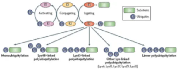
Ubiquitination is a highly controlled process that covalently labels proteins with the attachment of Ub to the target lysine (Lys- K) residues. Conjugation of Ub occurs through a cascade of enzymatic reactions mediated by the activating (E1), the conjugating (E2), and the Ub-ligases (E3) enzymes. These reactions create an isopeptide bond between the Ub C-terminal glycine (G) residue and the K residue on the target protein [35] (Fig. 4). Three different kind of E3 ligase exist: namely HECT (homologous with E6-associated protein C-terminus), which works as a single enzyme, U-box, and RING (really interesting new gene) ligases, which bind both the substrate and the Ub-loaded E2 thus facilitating the formation of the
covalent binding between Ub and substrate. Accordingly, the action of the E3 alone or in association with its E2 guarantees the specificity of the ubiquitination cascade. Because Ub itself contains seven K residues (K6, K11, K27, K29, K33, K48 and K63) that can serve as acceptor sites for chain elongation (Fig. 4), chains that contain multiple Ub moieties can be formed by either homotypic or heterotypic linkages. In addition, Ub chains assembled head-to-tail exist. Linear polyUb chains have been found in vivo as the resulting action on monoUb of the E3 ligase called LUBAC (linear Ub chain assembly complex). The difference between linear and K-linked chains has the physiological consequence to address selective recognition for specific interacting partners [36].
The ubiquitination pathway is further complicated by the possibility of the substrate to be differentially modified with Ub: 1) monoubiquitination, the attachment of a single Ub moiety, 2) multimonoubiquitination, the attachment of multiple Ub moiety to several K residues within the target protein and 3) polyubiquitination, a modification with a Ub chain [37] (Fig. 4). In vivo, all K residues are used for chain formation, but K48- and K63-based polyUb chains appear to be the most represented ones. Functionally, the modification of the target protein with a specific K-based modification has a specific signalling meaning and thus results in a particular Ub-dependent modulation of the physiological process, which is regulated by the ubiquitinated protein. Remarkably, all the Ub modifications are reversible and de-ubiquitination is achieved through the activity of specific proteases known as de-ubiquitinating enzymes (DUBs) [32]. This process reversibility renders ubiquitination a versatile post-translational modification.
Figure 4. The ubiquitin (Ub)-system complexity. Protein modification
with Ub occurs through a cascade of enzymatic reactions mediated by Ub-activating (E1), Ub-conjugating (E2) and Ub-ligating (E3) enzymes. These reactions covalently attach Ub to target lysine (Lys) residues of the substrate. Diverse modifications with Ub can occur: monoubiquitination is the attachment of a single Ub to a Lys of the target protein while polyubiquitination is the modification of Lys residues on target proteins with a chain of Ub. Since Ub possesses seven Lys that can be target of Ub modification for chain formation, several chains with different functions can be built on proteins (Lys6-, Lys11-, Lys27-, Lys29-, Lys33-, Lys48-, Lys63-linked). Alternatively, ubiquitin molecules can be linked head to tail to form linear chains. [37]
1.3.1 Proteolytic and non-proteolytic ubiquitination. It is known that each K-based polyUb chain assumes a particular three-dimensional topology, which in turn serves physiological processes. However, atypical Ub chains (i.e., based on linkages other than K48 or K63) exist but possess roles which are still elusive and difficult to study [36].
It is well known that the attachment of a K48-based polyUb chain labels proteins for 26S proteasome-mediated degradation. In addition, it has recently been reported that also K11-based polyUb chains serve as proteasomal degradation signal in yeast [36]. Thus, ubiquitination regulates protein half-life and turnover. In recent years it has become increasingly evident that the modification of proteins with a Ub chain based on K63 linkage or with the addition of a single Ub moiety (i.e., monoubiquitination) fulfills non-degradative functions (e.g., endocytosis, intracellular trafficking, DNA-damage response). For example, polyUb chains based on K6 linkages can also play a role in DNA repair. Indeed, the E3 ligase breast
cancer-susceptibility protein (BRCA1) and BRCA1-associated ring domain 1 (BARD1) localizes at DNA lesions with a mechanism involving the binding to K6- (and K63-) linked Ub chains. At the present K27-linked Ub polymers remains without a defined function while linear Ub chains, which represent the source of cellular monoUb, play a physiological role in the nuclear factor-κB (NF-κB)
pathway. Finally, some protein kinase [e.g., members of the AMP-activated protein kinase-related family (AMPK)] activity has been found to be blocked after modification with K29- and K33-linkages [36].
1.3.2 The Ub-Binding Domains.
The recognition of the ubiquitination diversity depends on an array of Ub-binding domains (UBDs) that bind non-covalently to Ub and which are found in a plethora of effector proteins, called Ub-receptors, with different functions (e.g., endocytosis, DNA repair). Ub-receptors generally have small (20-150 amino acids) UBDs, which do not possess any specific sequence conservation. However, the most common structural motifs in which an UBD folds are based on single or multiple α-helices [e.g., UBA (Ub-associated), UIM (Ub-interacting motif), MIU (motif interacting with Ub), UMI (UIM- and MIU-related UBD) domains], zinc-fingers [e.g., NZF (nuclear protein localization 4 zinc finger) domain], pleckstrin-homology (PH) fold [e.g., pleckstrin-like receptor for Ub (PRU)] and Ub-conjugating-like (e.g., UBC) structures [45]. The majority of the identified UBDs contact Ub on an hydrophobic patch around the isoleucine (I) 44 residue but also other Ub-surfaces contribute to binding specificity [38].
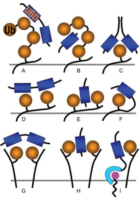
Interestingly, UBDs weakly bind to Ub (Kd=10-500 µM) thus avidity-based associations to ubiquitinated substrates are the mechanism by which high affinity interactions are obtained for efficient regulation of physiological processes. Indeed, protein oligomerization together with multiple UBDs on a single protein are often found in transient Ub-binding complexes [38] (Fig. 5). It has been reported that
many Ub-receptors possess UBDs with high binding selectivity toward polyUb chains rather than to monoUb in vitro. However, Ub-chains preference of an isolated UBD could differ from that of the full length Ub-receptor due to its subcellular location [38]. Moreover, selective binding of UBDs to specifically linked polyUb-chains has been reported and can depend on the topology of different polyUb. For example, K48-linked chains alternate in solution between a closed and an opened conformation, while K63 ones adopt an extended conformation [37]. Moreover, also UBDs organization can define Ub-binding specificity because linker regions of in tandem UBD repetitions arrange them in a way in which interactions are favored by one Ub chain type but not by others [37].
Interestingly, many Ub-receptors are ubiquitinated and this modification requires their UBDs. In this process, called coupled monoubiquitination, UBDs function as signals for ubiquitination because they recruit the ubiquitination machinery (i.e., E2-E3 complex) [38, 40]. Most of the Ub-receptors are monoubiquitinated rather than polyubiquitinated maybe because the UBD:Ub-binding mask the I44 residue required for Ub chain formation or because of steric hindrance of the E2-E3 complex with polyUb chains. The ubiquitination of Ub-receptors might have a regulatory function: for example, to mantain the Ub-receptor in an autoinhibitory state by inducing intra-molecular interaction among the monoUb and UBDs (Fig. 6). In this respect, it has been reported that the monoubiquitination of the UIM-containing protein EPSIN, important for the regulation of endocytosis, negatively influences the in vitro binding to some binding partners, but not others. Thus, the regulation of Ub-receptor monoubiquitination deeply impacts on signal transduction. Moreover, coupled ubiquitination plays a crucial role also for the formation of a signal relay network in which the monoUb leads to interactions with other downstream Ub-receptors, thus amplifying the Ub-based signalling [38] (Fig. 6). Additional complexity is given by the fact that several post-translational modifications (e.g.,
phosphorylation) regulate the Ub:UBDs interactions thus controlling the Ub-receptor functions [38].
Figure 5. Mechanisms for ubiquitin (Ub):Ub-binding domains (UBDs) interaction. (A) A polyUb chain can be bound by tandem UBDs each
contacting monoUb; alternatively, (B) a poly Ub chain can bind a single UBD by contacting it on two different surfaces. (C) MonoUb can be bound with high affinity from dimeric proteins that contact Ub on different surfaces through different faces of the UBDs. Protein modified with Ub in different Lys residues (i.e., multi-monoubiquitination) can contact (E) one or (D) more UBDs. (F) Monoubiquitination can direct intra-molecular monoUb:UBD interactions that mantain the protein in an auto-inhibitory status. (G) MonoUb on clustered membrane proteins can be bound by tandem UBDs or (H) can contact a single UBD on different surfaces. (I) MonoUb on membrane protein can be bound by a lipid-modified membrane protein containing an UBD. [39]
Figure 6. Models for coupled-monoubiquitination functions. (a)
Ubiquitin (Ub)-receptors monoubiquitination can mantain the receptor in an auto-inhibitory state that prevent its association to free Ub or ubiquitinated partners. Monoubiquitination can also induce a conformational change on the Ub-receptor that (b) activate adjacent enzymatic reactions or that (c) expose previously masked binding sites.
(d) Monoubiquitination of Ub-receptors can initiate a signal cascade in
which the modification with monoUb could induce a conformational change that expose the UBD to the interaction with ubiquitinated partners or with other Ub-receptors. [38]
1.4 Interactions between the ERα and Ub signalling.
In recent years, several papers reported a strict correlation between the ERα and the proteolytic- as well as non-proteolytic Ub-based pathways. Moreover, mounting evidence indicates how targeting the Ub-system could be a pharmacological option for cancer therapy [41]. Several point of intervention can be considered; for example, at the levels of the enzymes of the ubiquitination cascade. In this respect, our group found a strong effect of 4[4-(5-nitro-furan-2-ylmethylene)-3,5-dioxo-pyrazolidin-1-yl]-benzoic
acid ethyl ester (Pyr-41), the first cell permeable inhibitor of the key initiating enzyme E1 [42] on E2:ERα signalling and cellular processes (i.e., cell proliferation, cell migration) in MCF-7 cells [43], thus supporting the notion of the intertwined nature between the Ub-based system and ERα signalling. Other known drugs target the E3 ligase activity (i.e., HLI98 to inhibit Hdm2), substrate recognition (i.e., small compounds named RITA to block p53) or proteasomal activity (i.e., bortezomib), while new putative drugs might be designed to specifically occupy the UBDs of specific effector proteins [37, 41].
1.4.1 Ub-dependent proteolytic control of ERα expression. Ligand-induced ubiquitination and subsequent degradation appears to be a conserved pattern for limiting the cellular response to a given hormone. Indeed also in the case of E2, all the hormone effects occur in parallel while the ERα intracellular levels are regulated through a dynamic balance between ERα synthesis and ERα degradation. Proteolysis of ERα is principally under the control of the 26S proteasome since E2 binding induces the ERα polyubiquitination and subsequent degradation [29]. Furthermore, the un-liganded receptor is polyubiquitinated by the E3 Ub ligase MDM2 (mouse double minute-2) and degraded by the 26S proteasome. Interestingly, while polyubiquitination of the apo-ERα has the function to address any mis-folded ERα molecule for 26S proteasome-based removal, the E2-induced ERα polyubiquitination and degradation are necessary for receptor transcriptional functions [9, 10]. In particular, in the nucleus, both in the absence and in the presence of E2, ERα undergoes to a cyclic recruitment to the ERE containing promoters where, through sequential and ordered 26S proteasome-dependent events, it produces the transcriptional response to E2. In this way, the receptor proteolytic breakdown guarantees the time- and space-dependent synchronization of the cellular responses with the variable E2 extra-cellular concentrations [9, 10]. Accordingly, as transcription progresses ERα is degraded as consequence of the recruitment of Ub ligases
(e.g., E6-AP, MDM2, EFP) as well as coactivators (e.g., SRC-1 and SRC-3) [9, 10, 16]. Thus, the 26S proteasome plays a pivotal function of in the molecular steps leading to the expression of estrogen-responsive genes [44].
Our group also reported a critical role for ERα plasma membrane localization and signalling in the regulation of ERα degradation in breast cancer cells [27]. Indeed, mutation of ERα in the palmitoylation site accelerates ERα degradation in response to E2 because the PI3K pathway is not activated. In parallel, irrespective of E2 treatment, inhibition of ERα palmitoylation constitutively addresses ERα to the nuclear matrix and induces the basal degradation of the neo-synthesized ERα, suggesting that the native ERα pool requires palmitoylation for stabilization [27].
Signalling modulation of the ERα proteasome-dependent pathway has been reported to occur possibly through ERα phosphorylation in the Ser118 residue [29]. Indeed, our group found that the lack of palmitoylation as well as the inhibition of the PI3K/AKT pathway prevents the E2-dependent ERα Ser118 phosphorylation, ERα association with ERE-containing promoter and ERα transcriptional activity [27]. Moreover, our group found that the receptor pool that is addressed to the nuclear matrix for degradation is not phosphorylated on Ser118 [27]. However, if Ser118 phosphorylation is an important regulator of E2-induced proteolytic ERα ubiquitination remains to be established.
Thus, the regulation of ERα intracellular levels depend on the plasma membrane-starting signals as well as on transcriptional activity.
1.4.2 Ub-dependent non-proteolytic control of ERα signalling.
Ub proteolytic functions in the E2-induced 26S proteasome-dependent control of ERα cellular levels exist but at the present the identity of the E3 Ub ligase that directly polyubiquitinates ERα is unknown. Recent data have however defined that ERα is an actual substrate for the
E3 Ub ligase BRCA1 [30]. Interestingly, BRCA1, which is often mutated and amplified in several kinds of breast cancers, catalyzes the monoubiquitination of the ERα [30]. In particular, BRCA1 monoubiquitination has been shown to occur in in vitro assays on the K302 and K303 of the ERα E domain. Moreover, BRCA1 mutations frequently associated with cancer development prevent ERα monoubiquitination [30]. According to this initial evidence, our group found that ERα monoubiquitination also occurs in cell lines [32-34] and found that it is required for the activation of the E2-induced ERα rapid signalling (i.e., AKT activation) that controls the key steps for E2-induced cell proliferation (i.e., Cyclin D1 transcription, G1-to-S phase transition, cell cycle progression). Furthermore, we observed that the lack of ERα monoubiquitination impairs the E2-dependent increase of ERα:IGF1-R association, blocks ERα Ser118 phosphorylation and the ERα ability to regulate gene expression. Thus, endogenous ERα monoubiquitination regulates E2-dependent nuclear and extra-nuclear ERα activities and monoubiquitination seems to work as a negative feedback signal for the ERα-mediated E2 effects (e.g., cell proliferation) activation [32-34]. This evidence strongly suggests non-proteolytic functions (i.e., monoubiquitination-dependent) of the Ub-system in the regulation of E2:ERα signalling. However, the exact mechanism by which monoUb modulates ERα has not been defined.
For example, it cannot be excluded that the ERα monoubiquitination could depend on an UBD on the receptor itself. In that case the UBD:monoUb interaction could maintain ERα in an autoinhibitory state until the E2 binding allows deubiquitination and activation of several pathways. However, no information are available on the presence of UBDs on ERα. Another possibility is that monoUb creates new binding surfaces on ERα that guarantee the receptor association with specific interactors. The Ub-mediated interactions should then occur between ERα and the UBD of an Ub-receptor; however, the putative ERα Ub-receptor partner(s) has not been identified yet.
2. AIM.
Non-covalent binding between ubiquitinated proteins and Ub-receptors generates a complex network of interactions that is critical for signal transduction and controls many cellular processes. Indeed, the deregulation of this intricate system has been associated with several pathological conditions including several types of cancer [41].
Recent papers reported that the Ub system deeply impacts the E2:ERα signalling; indeed, while ERα polyubiquitination controls the receptor turnover and transcriptional activity [9, 10, 29], ERα monoubiquitination is required for the E2-dependent activation of rapid signalling to cell proliferation [34]. Even if it has not been clarified how the Ub modification on ERα modulates the receptor activities, it is possible that it creates new surfaces for the interaction with other molecules that possess an UBD and transfer the signal to the final outcome (e.g., gene expression, cell proliferation). On the other hand, it is also possible that ERα could recognize and transduce the Ub modification on itself or on interacting proteins through an UBD; however, no information are available on this issue.
Interestingly, monoubiquitinated proteins often possess and require an UBD to correctly function (e.g., to be monoubiquitinated, to interact with other proteins) [32]; thus, ERα monoubiquitination could depend on the presence of a UBD within the receptor structure. Moreover, among the six domains of ERα structure only the C and the E ones display a folded structure: two zinc-finger motifs for C domain and 12 α-helices for E domain [6]. Remarkably, the structure of most of the UBDs identified so far is based either on zinc-finger or α-helices [38]. These evidence suggests that ERα could behave as an Ub-receptor that decodes the Ub signal on other proteins and is itself modulated by it.
Thus, the main goal of the present PhD project was to understand the non-covalent Ub-binding abilities of ERα
and their regulatory role in E2-dependent cellular processes.
To this purpose, in vitro experiments were performed by using purified ERα domains and recombinant or cell lysates-derived ubiquitinated molecules. Next, the Human Embrionic Kidney (HEK293) and the cervix adenocarcinoma HeLa cell lines, which we endowed with the wt or mutant ERα, were used to analyze the impact of the ERα Ub-binding on E2:ERα signalling. We also used human breast cancer MCF-7 cells and treated them with specific inhibitors to block pathways that we found to be required for the ERα Ub-binding functions and to verify that they were conserved also in breast cancer cells.
3. IDENTIFICATION OF NON-COVALENT Ub-BINDING SURFACE (UBS) ON ERα.
3.1 Introduction.
Monoubiquitinated proteins are often Ub-receptors that possess at least one UBD [40]. Although many different UBDs exist, no specific conservation in terms of UBDs 3D-structure has been recognized. In fact, structural folds that have a regular secondary structure (e.g., α-helix and/or zinc-finger) are thought to be the only UBDs common features [37, 40, 45]. Interestingly, ERα biochemical anatomy consists in six modular domains (Fig. 2): while the A/B, D and F domains do not display a folded structure, the C domain consists in two zinc-finger motifs and the E domain is composed of 12 α-helices [6].
On this basis, we speculated that an UBD could be present in ERα. To this purpose, pull-down assays have been performed by using all ERα domains against recombinant or cell lysate-derived ubiquitinated species.
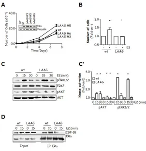
3.2 Results.
3.2.1 Indentification of two UBSs on ERα.
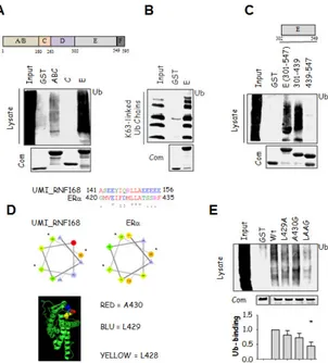
Initial in vitro experiments were performed to understand if ERα could possess an UBD. The ERα A/BC, C and E domains were cloned, expressed and purified as GST-fusion proteins as described in [46] except that all ERα-E domain encoding constructs were prepared in the presence of 20 µM E2. Next, GST-fusion proteins were used in pull-down assays by incubating them with HeLa cell lysates as source of ubiquitinated species. Subsequent anti-Ub immunoblot revealed that the A/B and the E domains but not the C domain were able to pull-down ubiquitinated species from total cellular lysates (Fig. 7A). Interestingly, the A/B and E domains showed different abilities to pull-down ubiquitinated species. The comassie (Com.) normalization, corresponding to 1/10 of the pull-down, indicate no differences between A/B and E protein
levels used for the experiment; thus, the observed variation in Ub-binding was not an artifact. Next, to exclude the possibility that the ubiquitinated species were pulled down through surface different than Ub, we performed pull-down experiments by using only the A/B or E domains against recombinant polyUb chains linked through K63. We found that either these domains were able to pull-down recombinant K63-linked polyUb chains (Supplemental Fig. 1 and Fig. 7B), thus demonstrating that the ERα A/B and E domains non-covalently contact Ub on the Ub-modified proteins. Next, to understand the reason for the observed differences between the A/B and E domain Ub-binding, we analyzed their ability to pull-down all the other known recombinant Ub chains (i.e., K6-, K11-, K27-, K29-, K33-, K48-linked). Interestingly, we found that while the E domain bound all (Fig. 2) but recombinant K33-linked Ub chains (data not shown), the A/B domain was able to pull-down only K6-linked Ub chains (Supplemental Fig. 1). Thus, the ERα Ub-binding A/B and E domains display preferential Ub-binding ability.
Because the A/B domain is non-structured and displays weak association to ubiquitinated species when compared to the 12 α helices-containing E domain, we focused on the E domain Ub-binding ability. Thus, we investigated which portion(s) of the ERα E domain is necessary for Ub-binding. We found that the N-terminal part of the E domain (i.e., amino acids 301-439) but not the E domain C-terminus (i.e., amino acids 439-547) was able to pull-down ubiquitinated species from total cellular lysates (Fig. 7C). This evidence indicates that the region of the ERα E domain that non-covalently associates in vitro with Ub is located within the protein region encompassing the amino acids 301-439. Unfortunately, we could not narrow down a smaller section of the E domain that associated to ubiquitinated species by using rational deletions of the N-terminal protein portion (i.e., 301-439) (data not shown). It is most likely that the ERα E domain Ub-binding portion requires an intact E domain 3D-folding. Thus, we considered the 301-439 region of the E domain as
the minimal binding region to Ub and called it ERα-Ub-binding surface (ERα-UBS).
In turn, in silico molecular modelling experiments would help rationalize the biochemical structural requirements for ERα E domain:Ub interaction. For these reasons, we decided to introduce specific point mutations within the minimal Ub-binding region (i.e., 301-439) in the context of the full length E domain in order to find the critical residues required for Ub-binding.
Interestingly, bioinformatic analysis identified L428, L429 and A430 in the E domain to have a spatial distribution reminiscent of the UBD called UMI [47], with L428 buried in the E domain core protein matrix and L429 and A430 at least partially exposed to solvent (Fig. 7D). Consistently, while single substitution of either L429 or A430 barely affected the Ub-binding abilities of the E domain in pull-down assays, double mutation of L429 and A430 (L429A, A430 to glycine (G)- LAAG) strongly prevented the E domain Ub-binding (Fig. 7E). To understand if the observed reduction in LAAG E domain Ub-binding was due to an impaired association to specific Ub chains, we performed pull-down experiments by using the wt and mutated E domain against all but K33-linked recombinant Ub chains. Remarkably, we found that the introduction of the LAAG mutations reduced the E domain ability to bind all but K48-linked Ub chains (Fig. 8). Thus, the observed reduction in Ub-binding ability of the mutant UBS depends on an overall lower ability to bind Ub chains. Next, we wondered if the reduced Ub-binding of the LAAG E domain could be due to an impairment of the E domain structure caused by the introduction of the mutations. To solve this problem, we performed circular dichroism (CD) experiments on the E-domain GST-fusion proteins, which showed that the introduction of the LAAG mutations in ERα did not significantly affect proper receptor folding (Supplemental figure 2).
Figure 7. Ub-binding ability of ERα. (A, B, C, E) Schematic of ERα and
E domain structure, in which the aminoacid positions are depicted. In vitro pull-down assays were performed using the indicated GST-tagged ERα constructs. GST-fusion proteins were incubated with (A, C, E) total cellular lysates extracted by growing HeLa cells or (B) with synthetic polyUb2-7 linked by Lys (K)63 and analyzed by immunoblot as indicated. *indicates significant differences with respect to the relative wild-type sample. (D) Top: ClustalW (http://www.ebi.ac.uk/Tools/msa/clustalw2/) alignment of ERα E domain with RNF168 UMI domain [47]. Middle: Projected helices using the Helical Wheel Projections software tool (http://rzlab.ucr.edu/scripts/wheel/wheel.cgi). Bottom: Three-dimensional structure of the human ERα LBD-E2 complex (1ERE) [48]; E2 has been removed. The structure is drawn in green; conserved amino acids between the ERα E domain and RNF168 UMI domain are space-filled and stained as indicated.
Figure 8. Ub-binding ability of the wt and L429A, A430G (LAAG) ERα E domain. In vitro pull-down assays using the GST-tagged ERα E domain constructs. GST-fusion proteins were incubated with synthetic polyUb2-7 K48- or K63-linked chains, or K6-, K11-, K27-, K29-linked di-Ub and analyzed in immunoblot as indicated.
3.2.2 Characterization of the full lenght ERα-UBS mutant in cells.
The pull-down experiments helped us identifying the in vitro key residues of the ERα E domain UBS. However, to understand if these residues are required also for the full length ERα binding to ubiquitinated species we introduced the LAAG mutations in wt ERα and transiently transfected it in HeLa or HEK293 cells.
Next, the wt and LAAG mutant receptor expressing cell lysates were treated with SDS in order to reduce the amount of receptor interactors that could associate to the receptor through a protein surface different from UBS [40]. Then, immunoprecipitation analysis was performed and the obtained results revealed that the introduction of the double LAAG mutation in ERα strongly prevents the receptor ability to associate with ubiquitinated proteins (Fig. 9A). These data demonstrate that the ERα-UBS directs Ub-binding also in cells.
Next, we sought to determine the role of ERα non-covalent Ub-binding on receptor activities. Since the ERα-based signalling is a function of receptor intracellular localization (i.e., nuclear and extra-nuclear) [6, 27], we tested the effect of the LAAG mutation on ERα sub-cellular distribution. Immunofluorescence analysis showed that both GFP-tagged wt and LAAG mutant ERα have the same nuclear, cytoplasmic and membrane tethered localization (Fig. 9B). Thus we conclude that mutation of L429 and A430 residues does not influence basal ERα sub-cellular localization.
Because the LAAG mutation is within the LBD, we also evaluated E2 binding affinity to the ERα-UBS mutant. Importantly, we found that the wt ERα E2 binding affinity was not impaired by the introduction of the LAAG mutations (Table 1).
In cells, ERα dimerizes and binds DNA to ERE sequences and induces gene transcription after E2-treatment [6]. To evaluate if the LAAG mutation could impair the receptor ability to dimerize under basal conditions, we transiently co-transfected flag-ERα wt and