UNIVERSITA’ DEGLI STUDI DI PISA
FACOLTA’ DI MEDICINA E CHIRURGIA Scuola di Specializzazione in Radiodiagnostica
TESI DI SPECIALIZZAZIONE
ROLE OF TRANSARTERIAL CHEMOEMBOLIZATION AS NEOADJUVANT TREATMENT IN T2 STAGE PATIENTS WAITING FOR LIVER
TRANSPLANTATION
Relatore
Chiar.mo Prof. Carlo Bartolozzi
Candidato Matteo Barattini
SUMMARY
Aim: To provide a retrospective assessment of long-term results in cirrhotic patients with stage T2 hepatocellular carcinoma (HCC) treated by orthotopic liver transplantation (LT).
Materials e Methods: The study included 168 cirrhotic patients (147 males, mean age 55.8 years) transplanted for stage T2 HCC from 1996 to 2010. In patients submitted to pre-LT transarterial chemoembolization (TACE), the CT study carried out prior to LT was reviewed to estimate tumour response to treatment in accordance with the modified RECIST criteria. The patients were subdivided into 3 groups: A) no pre-LT treatment; B) complete response (CR) after TACE; C) partial response, stable or progressive disease after TACE. Overall survival (OS) and recurrence-free (RFS) and disease-free (DFS) survival were calculated according to the Kaplan-Meyer analysis and compared using log-rank tests.
Results: Group A was made up of 56 patients, group B of 60 and Group C of 52. The clinical data were comparable for the three groups. At 5 years, OS, RFS and DFS were 75.2%, 92.7% and 73.2%, respectively. Survival rates were significantly higher in Group B than in Group C, and comparable in Groups A and B. In patients with total tumour diameter of >3cm, RFS and DFS censored in peri-procedural deaths (n=7) were significantly higher in Group B than in Group A.
Conclusions: In patients with T2-stage HCC submitted to LT, CR after pre-operative TACE is a favourable prognostic factor for survival and absence of tumour recurrence. Pre-OLT TACE may be clinically beneficial for T2-stage HCC patients with tumour size >3cm.
INTRODUCTION
Orthotopic liver transplantation (LT) is the preferred therapeutic option in selected patients with HCC, with over 70% survival rates at 5 years [1, 2, 3]. Currently the internationally validated criteria for inclusion in transplantation are the so-called Milan criteria, i.e. the presence of a single nodule <5cm, or of fewer than 3 nodules each one less than 3 cm [3], but a wide international debate is in progress as to the possibility of extending these criteria.
Besides these selection criteria, a major issue in potential candidates for LT is represented by the waiting time, with possible tumour progression and subsequently exclusion from transplantation (so-called “drop-out”). To reduce the incidence of this phenomenon, the use of percutaneous or trans-arterial loco-regional therapy has been proposed for patients on the waiting list for a transplant.
Although pre-transplant loco-regional treatments have entered the daily clinical practice of many centres, there is to date no demonstration that they can lead to any real benefit in terms of drop-out reduction and post-transplant survival, and debate on this point is still heated.
The aim of this single-institution retrospective study has been to assess the long-term clinical results of patients submitted to LT for stage T2 HCC, and to identify significant prognostic factors for survival and tumour recurrence, with particular reference to the radiological tumour response to the TACE.
MATERIALS AND METHODS
A single-institution retrospective study was carried out on cirrhotic patients with HCC at stage T2 of the modified TNM [4] who had undergone orthotopic liver transplant (LT) from January 1996 to December 2010.
Patients who had undergone surgical resections or percutaneous treatments prior to transplantation were excluded from the study, while patients submitted to pre-transplant TACE with at least one CT scan carried out between TACE and LT were admitted.
The final population consisted of 168 patients (147 males, mean age 55.8 years). Of these patients, 92 (54.8%) presented a single nodule, while the remaining 76 (45.2%) presented multiple nodules. Overall tumour diameter calculated according to RECIST criteria was ≤30 mm in 84 (50%) patients.
HCC diagnosis and pre-operative assessment
The following demographic data were collected for all patients: age, sex, cause of cirrhosis and MELD (Model for End-stage Liver Disease) score [5]. The latter was calculated retrospectively for patients operated on before 2002, on the basis of the clinical/laboratory data at the time of transplant.
Diagnosis of HCC in the absence of thrombosis was obtained through dynamic triphasic CT or, more rarely, MR examination.
The CT examination (Hi Speed CT/i or Light Speed Plus, GE Medical Systems, Milwaukee, USA) consisted of pre-contrast acquisition followed by intravenous administration of a bolus of iodinated contrast material (120 mL, flow rate 3-4 mL/
sec), with subsequent acquisitions in the arterial (30 seconds’ delay), the portal (70-80 seconds’ delay) and the equilibrium (180 seconds’ delay) phases. The acquisition protocol was chosen on the basis of the CT equipment available at the time, with slice thickness variability of 5 to 7 mm and reconstruction algorithm variability of 2.5 to 5mm.
The MR study (Signa HDX; GE Healthcare, Milwaukee, WI) was carried out only in cases which were considered dubious at CT. The dynamic study consisted of T1-weighted 3D-Fast Spoiled Gradient Echo sequences (TR 3.5msec, TE 1.7 msec, flip angle 12°, NEX 0,75 and slice thickness 4-5mm) after intravenous injection of contrast medium (doses: 0.1-0.2ml/kg body weight; flow 2ml/sec).
Diagnosis of HCC was made on the basis of the criteria established by the international guidelines [6]. The overall axial diameter of the tumours was obtained according to the RECIST (Response Evaluation Criteria in Solid Tumours) guidelines [7], adding together the maximum axial diameters of the target lesions. The absence of metastases was assessed by thoraco-abdominal CT and bone scintigraphy.
TACE procedure
At the moment of TACE, a digital-subtraction angiography (Multistar, Siemens, Erlangen, Germania) was carried out of the celiac trunk, superior mesenteric and hepatic arteries to evaluate the vascular anatomy of the liver and the presence of arteriovenous shunts, and to identify the arterial feeders of the tumours. All procedures were carried out under local anaesthaesia and using antiemetic and antibiotic prophylaxis, after obtaining the informed consent from all the patients.
TACE was performed with selective catheterisation of the segmental or sub-segmental hepatic arteries supplying the lesions, using 5-F catheters or 3-F coaxial microcatheters (TurboTracker 18; Boston Scientific, Cork, Ireland).
Lipiodol-TACE was performed injecting a mixture iodised oil (Lipiodol UltraFluid; Laboratori Guerbet, Aulnay-sous- Bois, France) and doxorubicin hydrochloride (Adriblastina®; Pfizer Italia srl, Borgo San Michele, Italy), followed by selective arterial embolisation with an emulsion of grated gelatin sponge particles (Spongostan standard®; Johnson & Johnson Medical Limited, Gargnano, Skipton, UK). The amount of iodised oil and doxorubicin to be administered was decided on the basis of the number and diameter of the lesions.
Drug Eluting Beads (DEB) TACE protocol was performed by superselective injection of 2-4 ml of 100-300 micron DC Bead® microspheres (Biocompatibles UK, Farnham, Surrey, UK) loaded with doxorubicin (50 mg per vial). Prior to delivery, the beads were mixed with a volume of non-ionic contrast medium, in a ratio of 1:3.
CT procedure and assessment of the response after TACE
All patients were submitted to clinical/laboratory tests and abdominal CT or MR examinations one month after TACE and every 3 months up to LT.
The CT examination included unenhanced scans, followed by intravenous administration of iodinated contrast agent (120 mL) at a flow rate of 3-4 ml/sec, obtaining acquisitions in the arterial, portal and equilibrium phases. As for the pre-procedural examination, the acquisition protocol varied according to the CT
equipment available each time, with variability of slice thicknesses from 5mm to 7mm and a variability of the reconstruction algorithm from 2.5 to 5mm.
The MR study (Sigma HDX; GE Healthcare, Milwaukee, WI) was carried out only in cases which were considered dubious at the CT test. The dynamic study consisted of T1-weighted 3D-Fast Spoiled Gradient Echo sequences (TR 3,5msec, TE 1,7 msec, flip angle 12°, NEX 0,75 and layer thickness 4-5mm), after intravenous injection of a contrast medium (doses: 0,1-0,2ml/kg bodyweight; flow 2ml/sec).
The CT examinations performed after TACE were retrospectively collected; if patients had undergone multiple CT tests in the interval between TACE and LT, the latest exam before LT was taken into consideration. Two expert radiologists blinded to the anatomo-pathological data, reviewed the pre- and post-TACE images in order to define tumour response to TACE according to the modified RECIST criteria [8]: - Complete response (CR): disappearance of any intratumoral arterial enhancement in all the target lesions;
-Partial response (PR): at least 30% reduction of the sum of the diameters of the viable target lesions (corresponding to the contrast uptake areas in the arterial phase), using as reference the sum of the pre-TACE diameters;
-Progressive disease (PD): at least 20% increase in the sum of the diameters of the viable target lesions (corresponding to the contrast uptake areas in the arterial phase), using as reference the minimum value of the sum of the diameters of the viable tissue of the target lesions obtained at the beginning of treatment. Appearance of new lesions of at least 1 cm diameter, with the contrast behaviour typical of HCC, is also considered as disease progression;
-Stable disease (SD): any condition not corresponding to CR, PR or PD.
The tumour was considered viable if contrast uptake was present in the arterial phase and washout in the portal or late phases. After Lipiodol-TACE, comparison between the unenhanced images and the arterial phase images was necessary in order to distinguish contrast uptake from the retention of iodinated oil.
In dubious cases, quantitative measurements were obtained by positioning a Region of Interest (ROI) in specific areas in all the post-contrast phases, following the indications proposed by Kim et al. [9]. A difference of at least 20 HU between baseline acquisition and at least one of the post-contrast acquisitions was considered to be indicative of a viable tumour.
Anatomo-pathological analysis
The explanted livers were assessed by the same Pathological Anatomy team. Macroscopic assessment was effected after cutting the liver into slices 0.5-1 cm thick along an axis parallel to the plane of the round ligament.
Slides were then prepared with haematoxylin and eosin dye and the microscopic assessment of each hepatic nodule was carried out, recording their sites and dimensions. The treated lesions were examined to assess the presence of vital tissue and the percentage of tumour necrosis.
The percentage of necrosis was classified as follows: 100% (absence of any vital residue); >50% (significant necrosis); <50% (minimal necrosis). In the presence of multiple nodules, the anatomo-pathologists gave an estimate (100%, >50% or <50%) of the percentage of necrosis as a ratio of the overall tumour tissue.
For the purposes of this retrospective analysis, the presence of microvascular invasion and the grade of the residual tumour were also recorded.
Post-LT follow-up
All patients were submitted to clinical controls, alpha-fetoprotein assay and abdominal ultrasound every 3 months, and to thorax X-ray examinations every 6 months. Where tumour recurrence was suspected, CT or MR examinations were carried out with the protocols described above. None of the patients included in the study was submitted to post-LT adjuvant chemotherapy.
For each patient, data relative to mortality (date and cause) and tumour recurrence (date and site) were recorded if present, and data collection was concluded on 31st
December 2011.
Statistical analysis
The statistical analysis was carried out with SAS software (SAS Institute, Cary, NC), considering P<0.05 as statistically significant.
The data were analysed with descriptive statistics (mean and standard deviation, SD), and compared using the Chi-square test or Fisher’s test for categorical data and Student’s t-test for paired data.
Overall survival (OS) was calculated from the date of transplant to the date of death or of the latest follow-up; Recurrence-free survival (RFS) from the date of the transplant to the date of tumour recurrence, and the patients who died of causes unrelated to tumour recurrence were counted at the moment of death; Disease-free
survival (DFS) was calculated from the date of transplant to the date of diagnosis of tumour recurrence, or death in the case of lack of recurrence.
Survival curves were obtained using the Kaplan-Meyer method and inter-group differences were assessed using the log-rank test.
The variables which were found to be statistically correlated to the survival data at univariate analysis were tested with multivariate analysis using the Cox model.
RESULTS
Of the 168 patients included in the study, 56 (33.3%) had not undergone any loco-regional treatment prior to LT, while the remaining 112 (66.7%) patients had undergone TACE at a mean of 138±100 days before transplantation (mean 127, interval 23±515 days). Ninety-five patients (84.8%) underwent Lipidiol-TACE, while the remaining 17 (15.2%) received DEB-TACE. The duration of post-LT follow-up was 53±42 months (interval 0-178, mean 38 months).
At latest follow-up, 10 (6%) patients had developed HCC recurrence, at the intrahepatic site (n=3), extrahepatic site (n=1) or intra and exptrahepatic sites (n=6). Forty-nine (29.2%) deaths were recorded, of which 7 were periprocedural (within 30 days of the operation). The remaining causes of death were: recurrence of hepatitis (n=18), tumour recurrence (n=6), infections (n=4), multi-organ failure (n=5), other tumours (n=5), other causes (n=4).
Tumour response to TACE
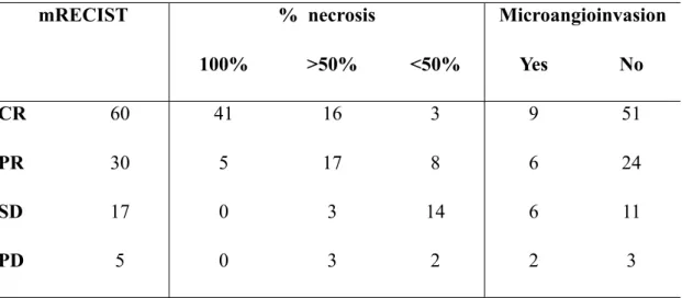
In patients submitted to TACE, at the latest CT scan prior to transplantation CR was observed in 60 patients (53.6%), PR in 30 patients (26.8%), SD in 17 patients (15.2%) and PD in the remaining 5 patients (4.4%).
A statistically significant correlation (p<0.0001) was observed between the tumour response according to mRECIST and the percentage of necrosis assessed on the explanted liver (Table 1).
Tumour response was found not to be significantly influenced by any clinical/ laboratory variable included in the analysis or by tumour extension (in terms of number and dimension of the lesions).
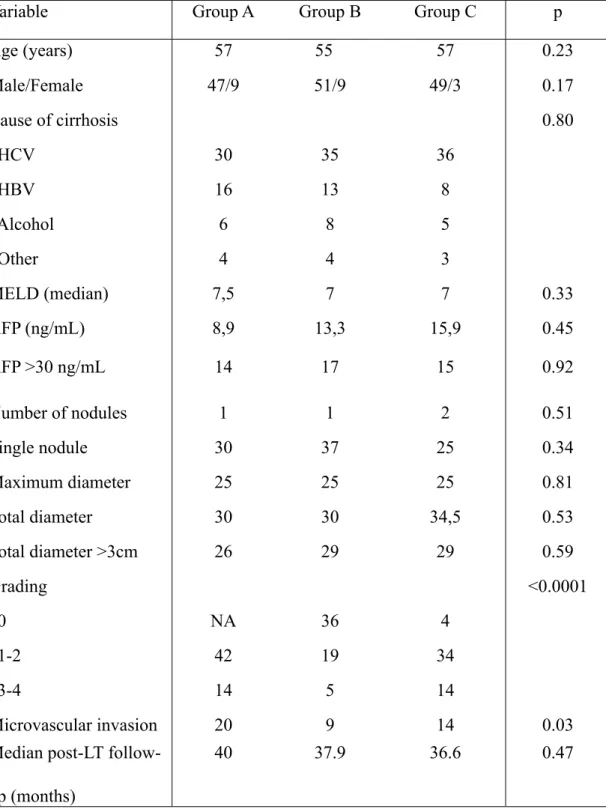
In the present study, the patients were thus divided into three groups: 1) Group A: patients not submitted to any pre-LT treatment (n=56); 2) Group B: patients with CR after TACE (n=60); 3) Group C: patients with PR, SD or PD after TACE (n=52). The clinical/demographic variables of these groups were found to be comparable (Table 2), while the anatomo-pathological data differed in terms of tumour grading and presence of micro-angioinvasion, due to the presence of 100% post-TACE necrosis in 41 patients of Group B and 5 patients of Group C (Tables 1 and 2):
Overall survival
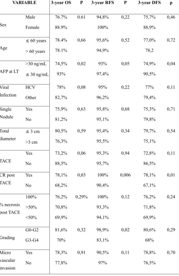
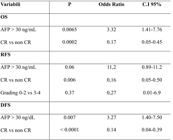
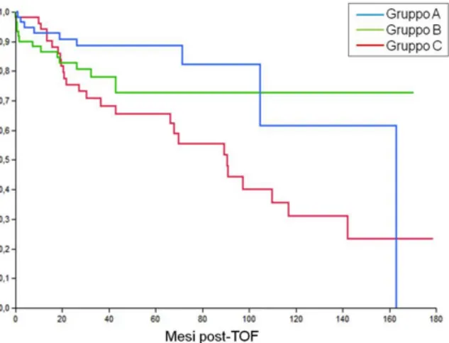
OS at 3 and 5 years was 78.3% and 75.2%, respectively, with median survival of 142 months. At the univariate analysis, the only factors correlated to mortality were the tumour response according to the mRECIST criteria (p=0.03) and the pre-LT AFP values (p=0.02) (Table 3). Both variables were confirmed at the multivariate analysis as independent factors of OS (Table 4). OS was found to be significantly higher (p=0.01) in groups A and B than in group C (Figure 1).
Recurrence-free survival (RFS)
RFS at 3 and 5 years was 95.5% and 92.7%, respectively. Of the 10 cases of tumour recurrence, 3 cases occurred in group A and 7 in group C. At the univariate analysis, RFS was found to be statistically correlated to tumour grading (p>0.0001), to AFP
values at transplantation (p=0.05), and to CR according to mRECIST (p=0.006) (Table 3). At the multivariate analysis, CR (p=0.006) and pre-LT AFP values (p=0.06) were found to be independent RFS variables (Table 4).
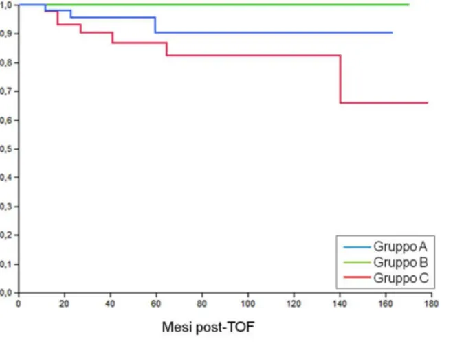
RFS was found to be higher in Group B than in Group A (p=0.07) and Group C (p=0.006) (Figure 2).
Disease-free survival (DFS)
DFS at 3 and 5 years was 77.3% and 73.2%, respectively, with a median disease-free survival of 140 months. As also seen for OS, DFS was statistically correlated to pre-LT AFP values (p=0.04) and CR (p=0.01) (Table 3), both factors being confirmed at the multivariate analysis (Table 4).
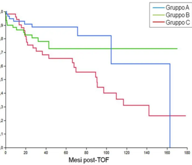
DFS was found to be significantly higher in Groups A and B than in Group C, with comparable results in Groups A and B (Figure 3).
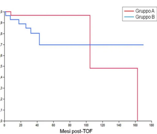
Censoring the peri-procedural deaths, DFS was found to be comparable in Groups A and B in the presence of a total tumour diameter ≤30 mm (P=0.23) (Figure 4); on the other hand, in patients with tumour diameter >30mm, DFS was found to be statistically higher in Group B than in Group A (p=0.05) (Figure 5).
DISCUSSION
LT is the preferred therapeutic option in selected patients with HCC, with over 70% survival rates at 5 years [1, 2, 3]. Currently the criteria most used and validated at international level for inclusion in transplantation are the so-called Milan criteria, i.e. the presence of a single nodule <5cm, or of fewer than 3 nodules each one less than 3 cm [3], but a wide international debate is in progress as to the possibility of extending these criteria. Other systems of selection adopted are the UCSF criteria and the “up-to-seven” criteria [10, 11, 12, 13].
Besides the selection criteria, another key factor to be borne in mind is the long waiting lists with possible consequent tumour progression and exclusion from transplantation because the above criteria of inclusion have been exceeded (so-called “drop-out”). To reduce the incidence of this phenomenon, the use of percutaneous or trans-arterial loco-regional therapies has been proposed for patients on the waiting list for LT.
Neo-adjuvant loco-regional therapies could moreover be used for reducing tumour extension in patients falling outside the Milan criteria, in order to allow them to be admitted to the waiting list and hence to transplant (so-called “downstaging”). More recent studies have moreover shown that response to loco-regional therapies may be a favourable prognostic factor in post-LT follow-up, and as such might constitute a further criterion for inclusion. Milonig et al. [14] have shown that patients falling within the Milan criteria with post-TACE CR or PR have a better rate of survival at 1, 2 and 5 years (100%, 93.2%, 85.7% and 93.8%, 83.6%, 66.2%) as compared with patients with PD or SD (82.4%, 50.7% and 19.3%, respectively).
Although pre-transplant loco-regional treatments have entered the daily clinical practice of many centres, there is to date no demonstration that they can lead to any real benefit in terms of drop-out reduction and post-transplant survival, and debate on this point is still heated.
According to a recent international consensus (International Consensus Conference on Liver Transplantation for Hepatocellular Carcinoma, Zurich 2010), bridge treatments are not substantially indicated in patients with T1-stage HCC; studies on large cohorts have in fact demonstrated that in these patients no significant progression of the disease is seen over a 12-month period, and the decision to undertake treatment must be taken on the basis of the risk of periprocedural complications and on the basis of the waiting time established by the waiting list [15-16].
In patients with stage T2 HCC, instead, the same consensus recommends neo-adjuvant loco-regional treatment if the time of waiting is over 6 months, above all for tumours at the upper limit of 5 cm and/or in the presence of high levels of AFP. For waiting times of less than 4 months or more than 9 months, no difference in terms of probable progression of the disease was found either in the treated or the non-treated groups of patients [17, 18].
The aim of this single-institution retrospective study was to assess the long-term clinical results of patients submitted to LT for stage T2 HCC, and to identify significant prognostic factors for survival and tumour recurrence, with particular reference to pre-operative TACE performance and response to TACE. The overall
survival rate and recurrence-free survival rate at 5 years were found to be favourable and substantially in line with literature data for similar patient populations [19].
In this relatively limited series of patients both overall survival and tumour recurrences were found not to be significantly influenced by clinical factors such as age or cause of cirrhosis, or by factors strictly correlated to tumour extension, particularly the presence of microangioinvasion. On the other hand, a factor closely correlated to mortality and tumour recurrence was found to be the response to the TACE as identified at the pre-operative CT scan on the basis of mRECIST criteria. Some recent studies have shown that the response to loco-regional treatment in non-surgical patients correlates with survival [14]. In one of our previous studies [20], carried out on a restricted number of patients falling outside the Milan criteria, complete tumour response assessed at post-TACE CT was found to be a possible criterion for selection for transplant in patients originally outside the Milan criteria, and a possible independent marker of survival and recurrence. Complete post-TACE response may thus be interpreted as an expression of favourable biological tumoral behaviour, and as a marker of absent vascular invasion and of low risk of post-transplant recurrence.
Other authors, on the other hand, have proposed disease progression after a valid tumour response as a criterion for exclusion from transplant [21].
According to our present experience, the use of mRECIST criteria as a further prognostic factor seems to be applicable also to patients originally falling within Milan criteria at stage T2. CR correlates positively both with survival (OS of 78.1% at 3 years) and with the absence of tumour recurrence (RFS of 100% at 3 years), with
markedly different curves from those obtained in patients with PR, SD or PD, regardless of other factors such as tumour extension or presence of microvascular invasion.
The survival curves of patients with CR were seen to be substantially comparable to those of patients not submitted to any pre-operative treatment, with a minimum margin in favour of CR as regards tumour recurrence, and a certain margin in favour of non-treatment for OS and DFS. This finding may indicate the lack of need for any pre-transplant treatment in patients with stage T2 HCC, above all in centres like ours in which the mean waiting time is less than 6 months. However, this result needs careful evaluation, particularly if we consider the relatively low number of patients included in each group.
It is interesting to note that if we divide the patients according to total tumour diameter (more or less than 3 cm), the survival data, particularly the DFS, are considerably modified. Our results in fact demonstrate that, with tumours less than or equal to 3 cm, pre-LT TACE gives no significant benefit, even in the presence of complete response. On the other hand, the DFS in patients with tumours over 3 cm is found to be significantly better after CR as compared both to non-CR patients and to patients who have not undergone any bridge therapy.
Although this result requires further confirmation via larger-scale prospective studies, it might hypothetically be useful to include loco-regional bridge treatment, namely TACE, in patients with stage T2 HCC with overall tumour diameter of over 3 cm, even when the estimated waiting time for transplant is less than 6 months. In this category of patients, complete post-treatment tumour response may constitute an
additional parameter as a prognostic factor of survival and post-OLT tumoral recurrence.
In conclusion, the present study supports the use of mRECIST criteria after TACE treatment as a further prognostic factor of overall, disease-free and recurrence-free survival in patients with stage T2 HCC. In this group of patients, TACE used as pre-transplant bridge treatment may be a valid therapeutic option in the presence of an overall tumour diameter of over 3 cm, with favourable long-term clinical results. The suggestion formulated at international level, according to which use of pre-LT loco-regional treatments in the remaining patients (T2-stage HCC with diameter of less than 3 cm) is advisable if the waiting time exceeds 6 months, remains in any case valid.
Table 1 – Correlation between tumour response evaluated at CT and necrosis assessed on the explanted liver
A statistically significant agreement is observed between mRECIST and percentage of necrosis (p<0,0001), while no agreement was found between mRECISt and presence of microangioinvasion (p=0,24)
mRECIST
mRECIST % necrosis% necrosis% necrosis MicroangioinvasionMicroangioinvasion
100% >50% <50% Yes No
CR 60 41 16 3 9 51
PR 30 5 17 8 6 24
SD 17 0 3 14 6 11
Table 2 - clinical and pathological data
Variable Group A Group B Group C p
Age (years) 57 55 57 0.23 Male/Female 47/9 51/9 49/3 0.17 Cause of cirrhosis 0.80 HCV 30 35 36 HBV 16 13 8 Alcohol 6 8 5 Other 4 4 3 MELD (median) 7,5 7 7 0.33 AFP (ng/mL) 8,9 13,3 15,9 0.45 AFP >30 ng/mL 14 17 15 0.92 Number of nodules 1 1 2 0.51 Single nodule 30 37 25 0.34 Maximum diameter 25 25 25 0.81 Total diameter 30 30 34,5 0.53 Total diameter >3cm 26 29 29 0.59 Grading <0.0001 0 NA 36 4 1-2 42 19 34 3-4 14 5 14 Microvascular invasion 20 9 14 0.03
Median post-LT follow-up (months)
Table 3 – Univariate analysis VARIABLE
VARIABLE 3-year OS P 3-year RFS P 3-year DFS p
Sex Male Female 76.7% 88.9% 0.61 94,8% 100% 0,22 75,7% 88,9% 0,46 Age ≤ 60 years > 60 years 78.4% 78.1% 0,66 95,6% 94,9% 0,52 77,0% 78,2 0,72 AFP at LT >30 ng/mL ≤ 30 ng/mL 74,5% 93% 0,02 93% 97,4% 0,05 74,9% 90,5% 0,04 Viral Infection HCV Other 78% 82,7% 0,08 95% 96,2% 0,22 77% 79,4% 0,11 Single Nodule Yes No 75,9% 81,2% 0,63 95,8% 95,1% 0,68 75,3% 79,8% 0,71 Total diameter ≤ 3 cm >3 cm 80,5% 76,3% 0,59 95,4% 95,5% 0,34 79,7% 75,1% 0,54 TACE Yes No 73,2% 88,5% 0,06 95,3% 95,7% 0,94 72,8% 86,5% 0,11 CR post TACE Yes No 78,1% 68,2% 0,03 100% 90,4% 0,006 78,1% 67,1% 0,01 % necrosis post TACE 100% >50% <50% 76,2% 70,8% 69,9% 0,29% 100% 93,3% 94,1% 0,12 76,2% 71,8% 69,9% 0,24 Grading G0-G2 G3-G4 81,6% 70% 0,32 98,9% 83,1% 0,02 80,6% 68% 0,29 Micro vascular invasion Yes No 78,3% 77,8% 0,91 90,5% 97% 0,11 78,8% 76,5% 0,70
Table 4 - Multivariate Analysis
Variabili P Odds Ratio C.I 95%
OS AFP > 30 ng/mL CR vs non CR 0.0065 3.32 1.41-7.76 AFP > 30 ng/mL CR vs non CR 0.0002 0.17 0.05-0.45 RFS AFP > 30 ng/mL CR vs non CR Grading 0-2 vs 3-4 0.06 0.006 0.37 11,2 0,16 0,27 0.89-11.2 0.05-0.50 0.01-6.9 DFS AFP > 30 ng/dL CR vs non CR 0.007 3.27 1.40-7.50 AFP > 30 ng/dL CR vs non CR < 0.0001 0.14 0.04-0.39
Figure
Figure 1. The overall survival (OS) rates at 3 and 5 years were 88.5% in Group A, 78.1% and 72.7% in group B, and 68.2% and 65.5% in group C (P= 0.01).
There was a statistically significant difference between group A and C (P= 0.01) and between group B and group C (P= 0.03), while group A and group B were comparable (p = 0.37).
Figure 2. Relapse-free survival (RFS) rates at 3 and 5 years were 95.7% and 90.4% in group A, 100% in group B, and 90.4% and 86.8% in group C (P= 0.02).
A statically significant difference was observed between group B and C (P= 0.006), while results were not significantly different comparing group A and B (P= 0.07) and group A and C (P= 0.24).
Figure 3. Disease free survival (DFS) rates at 3 and 5 years were 86.5% and 81.7% in group A,
78.1% and 72.7% in group B, and 67.1% and 64.4% in group C (P= 0.01).
A significant difference was found between group A and C (P= 0.02) and group B and C (P= 0.01) while results were comparable for groups A and B (P= 0.68).
Figure 4. Disease-free survival (DFS) censored for peri-procedural deaths in patients with total
tumour diameter ≤ 3cm.
DFS at 3 years was 96.5% in group A and 80.2% in group B with non significant difference between groups (P= 0.23).
Figure 5. Disease-free survival (DFS) censored for peri-procedural deaths in patients with total
tumour diameter > 3 cm.
DFS at 3 years was 79.4% in group A and 91,6% in group B; the difference was statistically significant (P = 0.05).
BIBLIOGRAPHY
1. Bruix, J. and M. Sherman, Management of hepatocellular carcinoma. Hepatology, 2005. 42(5): p. 1208-36.
2. Bruix, J. and J.M. Llovet, Prognostic prediction and treatment strategy in
hepatocellular carcinoma. Hepatology, 2002. 35(3): p. 519-24.
3. Mazzaferro, V., et al., Liver transplantation for the treatment of small
hepatocellular carcinomas in patients with cirrhosis. N Engl J Med, 1996.
334(11): p. 693-9.
4. Valls, C., et al., Pretransplantation diagnosis and staging of hepatocellular
carcinoma in patients with cirrhosis: value of dual-phase helical CT. AJR Am J
Roentgenol, 2004. 182(4): p. 1011-7.
5. Freeman, R.B., Jr., et al., The new liver allocation system: moving toward
evidence-based transplantation policy. Liver Transpl, 2002. 8(9): p. 851-8.
6. Bruix, J. and M. Sherman, Management of hepatocellular carcinoma: an
update. Hepatology, 2011. 53(3): p. 1020-2.
7. Therasse, P., et al., New guidelines to evaluate the response to treatment in solid
tumors. European Organization for Research and Treatment of Cancer, National Cancer Institute of the United States, National Cancer Institute of Canada. J
Natl Cancer Inst, 2000. 92(3): p. 205-16.
8. Lencioni, R. and J.M. Llovet, Modified RECIST (mRECIST) assessment for
hepatocellular carcinoma. Semin Liver Dis, 2010. 30(1): p. 52-60.
9. Kim, S.H., et al., Prediction of viable tumor in hepatocellular carcinoma treated
measurement at quadruple-phase helical computed tomography. J Comput
Assist Tomogr, 2007. 31(2): p. 198-203.
10. Yao, F.Y., et al., Liver transplantation for hepatocellular carcinoma: expansion
of the tumor size limits does not adversely impact survival. Hepatology, 2001.
33(6): p. 1394-403.
11. Mazzaferro, V., et al., Predicting survival after liver transplantation in patients
with hepatocellular carcinoma beyond the Milan criteria: a retrospective, exploratory analysis. Lancet Oncol, 2009. 10(1): p. 35-43.
12. Duffy, J.P., et al., Liver transplantation criteria for hepatocellular carcinoma
should be expanded: a 22-year experience with 467 patients at UCLA. Ann
Surg, 2007. 246(3): p. 502-9; discussion 509-11.
13. Yao, F.Y., et al., Liver transplantation for hepatocellular carcinoma: validation
of the UCSF-expanded criteria based on preoperative imaging. Am J
Transplant, 2007. 7(11): p. 2587-96.
14. Millonig, G., et al., Response to preoperative chemoembolization correlates with
outcome after liver transplantation in patients with hepatocellular carcinoma.
Liver Transpl, 2007. 13(2): p. 272-9.
15. Cillo, U., et al., Intention-to-treat analysis of liver transplantation in selected,
aggressively treated HCC patients exceeding the Milan criteria. Am J
Transplant, 2007. 7(4): p. 972-81.
16. Yao, F.Y., Liver transplantation for hepatocellular carcinoma: beyond the Milan
17. Majno, P., et al., Is the treatment of hepatocellular carcinoma on the waiting list
necessary? Liver Transpl, 2011. 17 Suppl 2: p. S98-108.
18. Aloia, T.A., et al., A decision analysis model identifies the interval of efficacy for
transarterial chemoembolization (TACE) in cirrhotic patients with hepatocellular carcinoma awaiting liver transplantation. J Gastrointest Surg,
2007. 11(10): p. 1328-32.
19. Cescon, M., et al., Prognostic factors for tumor recurrence after a 12-year,
single-center experience of liver transplantations in patients with hepatocellular carcinoma. J Transplant, 2010. 2010.
20. Bargellini, I., et al., Hepatocellular carcinoma: CT for tumor response after
transarterial chemoembolization in patients exceeding Milan criteria--selection parameter for liver transplantation. Radiology, 2010. 255(1): p. 289-300.
21. Otto, G., et al., Response to transarterial chemoembolization as a biological
selection criterion for liver transplantation in hepatocellular carcinoma. Liver
