CD10 expression in stromal cells of ameloblastoma variants
Giovanna Iezzi, DDS, PhD,aAdriano Piattelli, MD, DDS,bCorrado Rubini, MD,c Luciano Artese, MD,dGaia Goteri, MD,cMassimiliano Fioroni, DDS,eand Francesco Carinci, MD,gChieti-Pescara, Ancona, and Ferrara, Italy
UNIVERSITY OF CHIETA-PESCARA, POLYTECNIC UNIVERSITY OF THE MARCHE, AND UNIVERSITY OF FERRARA
Objective. We performed an immunohistochemical study in a series of ameloblastomas with different histology to explore the existence of a correlation between CD10 immunoreactivity in peritumoral stromal cells and the type of ameloblastoma with a high risk of local recurrence.
Study design. A total of 45 ameloblastomas (18 unicystic [UA], 4 peripheral [PA], 23 solid/multicystic [SA]) were evaluated. Cases showing immunoreactivity for CD10 in⬍ and ⱖ10% of stromal cells around tumoral epithelial islands, were considered, respectively, negative and positive. Correlations between stromal CD10 expression and histopathologic types with low and high risk of recurrence were evaluated by statistical analysis.
Results. SA cases showed a significantly higher percentage of stromal CD10-positive cells than the UA and PA variants. A strong intensity of immunostaining was observed only in SA.
Conclusions. Our results suggest that CD10 expression might be associated with stromal invasion in ameloblastoma variants with a high risk of recurrences. (Oral Surg Oral Med Oral Pathol Oral Radiol Endod 2008;105:206-9)
CD10 is a 90- to 110-kd cell surface zinc-dependent metalloprotease glycoprotein with endopeptidase activ-ity and is present on the surface of many cell types,1-3
including lymphoid precursor cells, germinal center B lymphocytes, and some epithelial cells; it cleaves and inactivates neuropeptides and peptide hormones at the amino terminus to hydrophobic residues within the peptides sequences,3,5-7thereby decreasing the cellular response to local peptide hormones.8 CD10 has also been called neutral endopeptidase, enkephalinase, ne-prilysin, and common acute lymphoblastic leukemia antigen (CALLA).4,9 CD10 is widely distributed and
has been found in the kidney, liver, small intestine, placenta, choroid plexus, brain, gonads, adrenal cortex, and leukocytes6; it has also has been demonstrated in
the stromal cells of normal bone marrow and endome-trium, myoepithelial cells of the breast and salivary glands, and in the alveolar epithelial cells in the lungs.4,8CD10 may play specific roles in the control of
cell growth and differentiation of both hematopoietic and epithelial systems.10CD10 has also been shown to
be present in renal cell carcinoma, transitional cell carcinoma, prostatic adenocarcinoma, endometrial stro-mal sarcoma, rhabdomyosarcoma, pancreatic adenocar-cinoma, schwannoma, malignant melanoma, psammo-matoid ossifying fibromas, meningiomas, perivascular epithelioid cell tumor of the oral mucosa, and endome-trial stromal sarcoma.11-15CD10 may represent a reli-able marker for identifying and isolating apoptosing T cells in vitro and ex vivo and possibly suggests novel functions for surface CD10 in the apoptotic process of lymphoid cells.16,17 CD10 is known also to be useful for the categorization of acute leukemias and the sub-classification of malignant lymphomas.4 CD10 is ex-pressed by prostatic stromal and epithelial cells and it is thought to have a key role in the growth of androgen independent prostate cancer.5
Contrasting results have been recently published on the role of CD10 as a prognostic indicator. In primary intestinal lymphomas, a trend for a longer overall sur-vival was found in the CD10⫹ group compared with the CD10⫺ group.18In childhood acute lymphoblastic leukemia, CD10 constitutes a favorable prognostic marker.17Patients with lung cancer with CD10⫹ cells had a 5-year survival, significantly better than those with CD10⫺ cells.19In diffuse large B-cell lymphoma, CD10 expression was closely associated with improved
This work was partially supported by the National Research Council (CNR), Rome, Italy, and by the Ministry of Education, University, and Research (MIUR), Rome, Italy.
aResearch Fellow, Dental School, University of Chieti-Pescara. bProfessor of Oral Medicine and Pathology, Dental School, Univer-sity of Chieti-Pescara.
cResearcher, Department of Neurosciences, Institute of Pathology, Polythecnic University of the Marche.
dProfessor of Pathology, Dental School, University of Chieti-Pescara. eResearch Fellow, Dental School, Polythecnic University of the Marche.
gAssociate Professor of Oral and Maxillofacial Surgery, University of Ferrara.
Received for publication Oct 10, 2006; returned for revision May 16, 2007; accepted for publication May 17, 2007.
1079-2104/$ - see front matter © 2008 Mosby, Inc. All rights reserved. doi:10.1016/j.tripleo.2007.05.025
survival.20 On the contrary, in diffuse large B-cell lymphoma, Xu et al.21 reported that cases with a
CD10⫹ phenotype may be associated with an unfavor-able clinical course and showed a significantly lower rate of complete remission, and in patients reported by Uherova et al.,22the CD10⫹ group displayed a shorter overall survival. In breast carcinoma the patients with CD10⫹ stromal cells had a shorter metastasis-free in-terval.1
The aim of the present study was an immunohisto-chemical evaluation of CD10 expression in the peritu-moral stromal cells of ameloblastoma variants with a low and high risk of recurrences, to explore the poten-tial usefulness of CD10 as a biologic indicator.
METHODS
A total of 45 ameloblastoma variants were evaluated: 18 ameloblastoma unicystic type (UA), 4 ameloblas-toma extraosseous/peripheral type (PA), and 23 amelo-blastoma solid/multicystic type (SA).23
Immunostaining was performed with monoclonal an-tibodies directed against CD10 (Novocastra Laborato-ries Ltd, Newcastle upon Tyne, UK). One block of each tumor was selected for immunohistochemical analysis. All pathologic diagnoses were confirmed reviewing hematoxylin and eosin stained section. For immuno-staining, sections were deparaffinized in xylene and dehydrated in an alcohol series. Immunostaining for CD10 required pretreatment with 1 mM EDTA (at pH 8.0) for 20 minutes at 250 W in a microwave oven. Section were incubated with anti-CD10 monoclonal antibody (dilution 1:25) for 1 hour at 37°C in a moist chamber, followed by incubation with biotinylated an-timouse immunoglobulin (Ig)G/antirabbit IgG (1:200; Vector Laboratories, Wiesbaden, Germany) and avidin biotin complex (ABC) alkaline phosphatase reagent, each for 30 minutes at room temperature. Between steps the sections were washed in tris buffered saline (TBS). The immunoreactions were visualized with the ABC method applying a vectastain ABC alkaline phosphatase staining (Camon, Wiesbaden, Germany) or an Ultratech HRP streptavidin-Biotein universal detection system (Immunotech, Marseilles, France). Fast Red and 3,3-diaminobenzidine-tetrahydrochlo-ride (DAB) respectively served as chromogens. Brown staining of the cell membrane or cytoplasm was considered positive.
Evaluation of the staining for CD10
The CD10-immunostained stromal cells around the ameloblastomas were evaluated in 10 HPF in each case and expressed as percentage. The count was performed by 2 investigators simultaneously, using a
double-headed light microscope. Both had to agree on the count of the positive cells.
Also, the intensity of staining was recorded as weak, moderate, or strong. The UA, PA, and SA were subdi-vided into 2 groups according to the cutoff previously indicated for stromal cells in breast carcinoma1: when stromal cells positive for CD10 wereⱖ10%, the spec-imen was considered to be CD10 positive; when the positivity of stromal cells was ⬍10%, the specimen was classified as CD10 negative.
Statistical analysis
Correlations between stromal cell immunoreactivity and intensity staining for CD10 and the histopathologic features were evaluated using the chi-square test, and a
P value less than .05 was considered significant. For
this purpose, UA and PA cases were grouped together as low-risk ameloblastomas and compared to SA con-sidered ameloblastomas with high-risk of recurrence. Differences of mean expression in the 2 risk groups were also evaluated by nonparametric test (Kruskal-Wallis).
RESULTS
In ameloblastomas, CD10-positive stromal cells were distributed around the epithelial cells with a dif-ferent pattern of staining among variants with difdif-ferent risk of recurrences (Table I). In UA and PA, which belong to the low-risk group, a stromal positivity in ⱖ10% of cells was observed in 5 of 22 cases (Fig. 1). SA cases, instead, showed a significantly higher mean percentage of CD10-positive stromal cells compared with UA and PA cases considered together (mean, 19.60 versus 9.09, respectively; Kruskal-Wallis test:
P⫽ .00081). In SA, the staining was particularly found
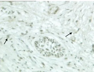
in the areas of the stromal cells close to epithelial cells (Fig. 2). CD10 was considered positive in 14 of 23 SA. SA cases tended to have a stromal cell positivity for CD10 ⱖ 10% and a moderate and strong degree of
Table I. CD10 expression in peritumoral stromal cells
in the 3 ameloblastoma variants (UA, PA, and SA)
CD10 immunoreactivity in stromal cells UA PA SA Percentage of positive cells (mean⫾ SD) 9.05⫾ 7.71 9.25 ⫾ 4.03 19.60 ⫾ 13.93 Intensity of staining Weak 15 3 10 Moderate 3 1 7 Strong 0 0 6
No. cases with CD10 expressionⱖ10% of cells
4/18 1/4 14/23
OOOOE
staining more frequently compared to UA and PA cases (chi-square test: P⫽ .0231 and P ⫽ .010, respectively). CD10 immunoreactivity was found also in epithelial cells (both peripheral and central cells) in all amelo-blastoma variants without significant differences.
DISCUSSION
Proliferation of stromal cells is commonly seen when cancer cells invade and metastasize. The invasive and metastatic potential of several types of neoplastic cells is regulated by interactions with stromal cells, which involve stimulatory and inhibitory factors that regulate such functions as cellular adhesion, migration, and gene expression.24
CD10 is associated with differentiation and growth of neoplastic cells,4and CD10 expression is found to be increased with the increase of tumor dysplasia.4Ogawa et al.,4in fact, found that there was no expression of CD10 in the stromal cells of normal colorectal tissue, while CD10⫹ stromal cells were present adjacent to tumor cells in 16 of 73 adenomas with mild or moderate dysplasia, in 12 of 17 adenomas with severe dysplasia, in 10 of 16 intramucosal carcinomas, and in 50 of 63 invasive carcinomas. Iwaya et al.1found that there was no staining in the stromal cells of noninvasive ductal carcinoma or normal breast tissue, while the frequency of positive stromal staining increased in cases with axillary lymph node metastases. Moreover, the stromal expression of CD10 was strongly associated with ac-cumulation of p53 and with a large tumor size.4In the study of Makretzov et al.,25 stromal CD10 positivity was associated with epidermal negativity, higher tumor grade, and decreased survival in breast carcinoma. The fact that CD10 positive cells are frequently seen at the invasive front suggests that tumor-stromal interactions
exist between breast neoplastic cells and CD10-positive stromal cells. Ogawa et al.4concluded that CD10 ex-pression seemed to be an integral part of colorectal carcinogenesis, and that its expression seemed to play a relevant role in the invasion, probably facilitating me-tastases. In a study of CD10 in oral squamous cell carcinoma, it was found that CD10 positivity in stromal cells was an indicator of worse prognosis; a significant correlation was found with lymph node metastases, local recurrences, and histologic grade.26In the present study, the SA variant of ameloblastomas associated with a high risk of recurrence was correlated with a high immunoreactivity for CD10 of the peritumoral stromal cells. SA cases were all immunoreactive for CD10 in the stroma, which exhibited a uniformly strong and intense positivity in the areas of infiltrating odontogenic epithelium and a percentage of positive cells ⱖ10% in 60% of the cases. Moreover, CD10 stromal positivity was focally present also in UA and PA, but in almost 80% of the cases it was below the cutoff selected for positivity. These data strongly sug-gest that CD10 expression in the stromal cells is asso-ciated with local tumor invasion and that the prolifer-ation of CD10-positive stromal cells is part of the mechanism of invasive growth in ameloblastoma vari-ants. CD10 immunostaining may be useful to identify areas with locally aggressive behavior also in low-risk ameloblastomas. In our series, however, we could not correlate CD10 stromal positivity in ameloblastomas with the real potential of recurrence, as we do not have follow-up data of our patients. Moreover, all our cases of UA and PA were removed by tumorectomy, whereas SA were removed by partial mandibular resection and
Fig. 2. SA: CD10 positivity of the stromal component in the invasive portion of the tumor: positive stromal cells are indicated by black arrows and epithelial staining is shown by red arrows (CD10,⫻160).
Fig. 1. PA: Focal CD10 positivity of the stromal cells, indi-cated by black arrows (CD10,⫻200).
OOOOE
the type of surgery would have an impact on the risk of recurrence. Therefore, further investigations are needed to verify the potential prognostic value of CD10 stro-mal immunoreactivity in predicting the local aggres-siveness in this group of benign odontogenic tumors, by correlating immunostaining results with follow-up data in larger series.
As an additional finding, we also observed CD10 immunoreactivity in the epithelial cells in a proportion of cases, without finding differences in the different types of ameloblastomas. We think this observation should be further investigated in larger series.
REFERENCES
1. Iwaya K, Ogawa H, Izumi M, Kuroda M, Mukai K. Stromal expression of CD10 in invasive breast carcinoma: a new predic-tor of clinical outcome. Virchow Arch 2002;440:589-93. 2. Reyes-Botella C, Montes MJ, Abadia-Molina AC,
Vallecillo-Capilla MF, Ruiz C. CD10 expression in cultured human osteo-blast-like cells. Folia Biol 1999;45:257-60.
3. Xiao SY, Wang HL, Hart J, Fleming D, Beard MR. cDNA arrays and immunohistochemistry identification of CD10 (CALLA) ex-pression in hepatocellular carcinoma. Am J Pathol 2001;159: 1415-21.
4. Ogawa H, Iwaya K, Izumi M, Kuroda M, Serizawa H, Koyanagi Y, et al. Expression of CD10 by stromal cells during colorectal tumor development. Hum Pathol 2002;33:806-11.
5. Albrecht M, Gillen S, Wilhelm B, Doroszewicz J, Aumuller G. Expression, localization and activity of neutral endopeptidase in cultured cells of benign prostatic hyperplasia and prostate cancer. J Urol 2002;168:336-42.
6. Borscheri N, Roessner A, Rocken C. Canalicular immunostain-ing of neprilysin (CD10) as a diagnostic marker for hepatocel-lular carcinomas. Am J Surg Pathol 2001;25:1297-303. 7. Ordi J, Nogales FF, Palacin A, Marquez M, Pahisa J, Vanrell JA,
et al. Mesonephric adenocarcinoma of the uterine corpus. CD10 expression as evidence of mesonephric differentiation as evi-dence of mesonephric differentiation. Am J Surg Pathol 2001;25: 1540-5.
8. Bahrami S, Malone JC, Lear S, Martin AW. CD10 expression in cutaneous adnexal neoplasms and a potential role for differenti-ating cutaneous metastatic renal cell carcinoma. Arch Pathol Lab Med 2006;130:1315-9.
9. Moritani S, Kushima R, Sugihara H, Bamba M, Kobayashi TK, Hattori T. Avaiulability of CD10 immunohistochemistry as a marker of breast myoepithelial cells on paraffin sections. Mod Pathol 2002;15:397-405.
10. Groisman GM, Amar M, Livne E. CD10, a valuable tool for the light microscopic diagnosis of microvillous inclusion disease (familial microvillous atrophy). Am J Surg Pathol 2002;26: 902-7.
11. Granados R, Carrillo R, Najera L, Garcia-Villanueva M, Patròn M. Osammomatoid ossifying fibromas: immunohistochemical analysis and differential diagnosis with psammomatous menin-giomas of craniofacial bone. Oral Surg Oral Med Oral Pathol Oral Radiol Endod 2006;101:614-9.
12. Koutlas IG, Pambuccian SE, Jessurum J, Manivel JC, Go-palakrishnan R. Perivascular epithelioid cell tumor of the oral mucosa. Arch Pathol Lab Med 2005;129:690-3.
1 3 . Butnor KJ, Nicholson AG, Allred DC, Zander DS, Henderson DW, Barrios R, et al. Expression of renal cell carcinoma-associated markers erythropoietin, CD10, and renal cell carcinoma marker in diffuse malignant mesothelioma and metastatic renal cell carci-noma. Arch Pathol Lab Med 2006;30:823-7.
14. Xu Y, McKenna R, Kroft SH. Assessment of CD10 in the diagnosis of small B-cell lymphomas. A multiparameter flow cytometric study. Am J Clin Pathol 2002;117:291-300. 15. Xu Y, McKenna RW, Kroft SH. Comparison of multiparameter
flow cytometry with cluster analysis and immunohistochemistry for the detection of CD10 in diffuse large B-cell lymphomas. Mod Pathol 2002;15:413-9.
16. Cutrona G, Leanza N, Ulivi M, Melioli G, Burgio VL, Mazza-rello G, et al. Expression of CD10 by human T cells that undergo apoptosis both in vitro and in vivo. Blood 1999;94:3067-76. 17. Cutrona G, Tasso P, Dono M, Roncella S, Ulivi M, Carpaneto
EM, et al. CD10 is a marker for cycling cells with propensity to apoptosis in childhood ALL. Brit J Cancer 2002;86:1776-85. 18. Go JH, Yang WI, Ree HJ. CD10 expression in primary intestinal
large B-cell lymphomas: its clinical significance. Arch Pathol Lab Med 2002;126:956-60.
19. Tokuhara T, Adachi M, Hashida H, Ishida H, Taki T, Higash-iyama M, et al. Neutral endopeptidase/CD10 and aminopeptidase N/CD13 gene expression as a prognostic factor in non-small cell lung cancer. Jpn J Thorac Cardiovasc Surg 1991;49:489-96. 20. Ohshima K, Kawasaki C, Muta H, Muta K, Deyev V, Haraoka S,
et al. CD10 and Bcl 10 expression in diffuse large B-cell lym-phoma: CD10 is a marker of improved prognosis. Histopathol-ogy 2001;39:156-62.
21. Xu Y, McKenna RW, Molberg KH, Kraft SH. Clinicopathologic analysis of CD10⫹ and CD10⫺ diffuse large B-cell lymphoma. Identification of a high-risk subset with coexpression of CD10 and bcl-2. Am J Clin Pathol 2001;116:183-90.
22. Uherova P, Ross CW, Schnitzer B, Singleton TP, Finn WG. The clinical significance of CD10 antigen expression of diffuse large B-cell lymphoma. Am J Clin Pathol 2001;115:582-8.
23. Gardner DG, Heikinheimo K, Shear M, Philipsen HP, Coleman H. Ameloblastomas. In: Barnes L, Eveson JW, Reichart P, Sidransky D, editors. World Health Organization Classification of Tumours: Pathology and Genetics of Head and Neck Tu-mours. Lyon, France: IARC Press; 2005. p. 296-300. 24. Tran NL, Nagle RB, Cress AE, Heimark RL. N-Cadherin
ex-pression in human prostate cancer cell lines. An epithelial-mes-enchymal transformation mediating adhesion with stromal cells. Am J Pathol 1999;155:787-98.
25. Makretzov NA, Hayes M, Carter BA, Dabiri S, Gilks CB, Hunts-man DG. Stromal CD10 expression in invasive breast carcinoma correlates with poor prognosis, estrogen receptor negativity, and high grade. Mod Pathol 2007;20(1):84-9.
26. Piattelli A, Fioroni M, Iezzi G, Perrotti V, Stellini E, Piattelli M, et al. CD10 expression in stromal cells of oral cavity squamous cell carcinoma: a clinic and pathologic correlation. Oral Dis 2006;12:301-4.
Reprint requests: Francesco Carinci, MD Chair of Maxillofacial Surgery Department of DMCCC University of Ferrara Corso Giovecca, 203 44100 Ferrara Italy [email protected] OOOOE