In vitro and ex vivo evaluation of the
anti-Giardia duodenalis activity of the supernatant
of Slab51 (SivoMixx)
Stefania PerrucciID1*, Gianluca Fichi2, Enrica Ricci2, Livio Galosi3, Marco Lalle4, Giacomo Rossi3
1 Department of Veterinary Sciences, University of Pisa, Pisa, Italy, 2 Istituto Zooprofilattico Sperimentale
delle Regioni Lazio e Toscana, Pisa, Italy, 3 School of Biosciences and Veterinary Medicine, University of Camerino, Matelica (MC), Italy, 4 Department of Infectious Diseases, Unit of Foodborne and Neglected Parasitic Diseases, European Union Reference Laboratory for Parasites, Istituto Superiore di Sanità, Roma, Italy
Abstract
The effects on Giardia duodenalis of Slab51 probiotic supernatants were evaluated in vitro and ex vivo. In vitro, Slab51 (101UFC) was cultured and the obtained supernatant was fil-tered, adjusted at pH 7, and added (100μl/ml) as such (Slab51 FS) or after heat-treatment, to G. duodenalis cultures to evaluate its effects on G. duodenalis trophozoites growth and adherence. For comparison, negative and metronidazole (20μg/ml) treated controls were used. The morphological and ultrastructural alterations of G. duodenals trophozoites follow-ing treatment with Slab51 FS supernatant were investigated by transmission electron microscopy. Ex vivo, mice duodenal portions were cultivated in standard conditions with 5x10⁵G. duodenalis trophozoites/ml, while to further five duodenal portions similarly
cul-tured and infected, Slab51 FS 200μl was added. After 12 and 18h, samples were fixed in 10% buffered formalin and histologically processed to score Giardia infection and cell dam-age. Cell proliferation/apoptosis was scored by Ki67, TUNEL and Caspase–3 tests. All experiments were conducted in triplicate throughout the study. All data were statistically evaluated (P<0.05). Results showed that Slab51 FS significantly reduced Giardia growth and adherence respect to negative controls, but its efficacy was overall lower than that of metronidazole. Moreover, the effects of Slab51 FS were significantly lowered by heat-treat-ment and this reduction was statistically higher at 90˚C than at 56˚C, indicating a heat-sensi-tive nature of acheat-sensi-tive Slab51 FS compounds. At the ultrastructural level, Slab51 FS treated
Giardia trophozoites were swelling, increased in size and showed alterations of their cellular
membrane and vacuole patterns, loss of the nuclear envelope and nuclear architecture. In
ex vivo trials, viable G. duodenalis trophozoites and enterocyte TUNEL+ and Caspase-3
expression were significantly reduced in intestinal sections added with Slab51 FS, while enterocyte Ki67 expression was significantly increased, confirming the anti-G. duodenalis activity of Slab51 FS observed in vitro. In conclusion, results from this study showed that the fresh culture supernatant of the commercial probiotic Slab51 has anti-G. duodenalis proper-ties both in vitro and ex vivo in a mouse model.
a1111111111 a1111111111 a1111111111 a1111111111 a1111111111 OPEN ACCESS
Citation: Perrucci S, Fichi G, Ricci E, Galosi L, Lalle
M, Rossi G (2019) In vitro and ex vivo evaluation of the anti-Giardia duodenalis activity of the supernatant of Slab51 (SivoMixx). PLoS ONE 14 (3): e0213385.https://doi.org/10.1371/journal. pone.0213385
Editor: Alessandro Giuffrè, National Research Council, ITALY
Received: October 5, 2018 Accepted: February 19, 2019 Published: March 7, 2019
Copyright:© 2019 Perrucci et al. This is an open access article distributed under the terms of the
Creative Commons Attribution License, which permits unrestricted use, distribution, and reproduction in any medium, provided the original author and source are credited.
Data Availability Statement: All relevant data are
within the paper.
Funding: The authors declare that this study was
partly supported by Actial Farmaceutica Lda, Prac¸a Severino Ferraz, 258 082 Funchal, Madeira, Portugal. The funder had no role in study design, data collection and analysis, decision to publish, or preparation of the manuscript and no authors received a salary from this funder.
Competing interests: The authors declare that this
Introduction
Flagellated protozoans of the genusGiardia are found in the digestive tract of vertebrate hosts
worldwide in which they are the cause of giardiasis [1].Giardia duodenalis (syn. Giardia intes-tinalis, Giardia lamblia) is the only species found in humans and in many other wild and
domestic mammals worldwide [1,2]. Based on genetic analysis,G. duodenalis is considered a
species complex, which includes at least eight distinct genetic groups or assemblages, from A to H [1]. Assemblages A and B are usually isolated from humans but can also infect other ani-mals, being considered zoonotic [1,3].
The localization site ofG. duodenalis is the small intestine, mainly duodenum and jejunum,
and it may be responsible for asymptomatic, acute and chronic clinical forms [4,5]. Diarrhea, malabsorption and weight loss, as well as numerous post-infectious pathologies and extra-intestinal complications are the main clinical signs of symptomatic infections [4–6]. The life cycle ofG. duodenalis is direct and involves two stages, the trophozoite and the cyst. Mammal
hosts may acquireG. duodenalis infections via ingestion of infectious cysts in contaminated
food or water sources, or directly via the fecal-oral route [1,2].
Giardiasis is one of the most common intestinal protozoal infections reported in humans, pet and farm animals [7,8]. Moreover, human giardiasis has been included in the World Health Organization’s (WHO) Neglected Diseases Initiative since 2006, estimating that 280 million people are infected each year [9,10]. The control of giardiasis is dependent on chemotherapy, and treatments are based mainly on the use of nitroimidazoles, such as metronidazole and tini-dazole, and benzimidazoles, mainly fenbendazole and albendazole; furazolidone, acridine, quin-acrine, nitazoxanide and paromomycin are also used in some situations [5;11–15]. However, most of the therapeutically used anti-Giardia drugs, including metronidazole, may cause severe
side effects and are not well tolerated by many human and animal patients or cannot be used in farm animals [11,16]. Moreover, the use of these drugs is often associated with clinical failure and drug resistance [16–18]. Hence, identifying new anti-Giardia agents is an important
con-sideration for the control of giardiasis in human and veterinary medicine [16,19].
Some data from recentin vitro and in vivo studies, largely from mice and humans, have
shown that probiotic treatment may possibly ameliorateG. duodenalis symptoms or reduce
infection withG. duodenalis [6,20]. These compounds have attracted the attention as potential substitutes for, or as combined therapy to currently used anti-Giardia drugs due to their
pow-erful activity, stability and low toxicity to humans and other mammal hosts [11,21].
In the present study, potential negative effects of the supernatant of a commercial probiotic onG. duodenalis were evaluated in vitro and ex vivo.
Materials and methods
Slab51 (SivoMixx)
Slab51 (sold in Europe today under the trademark SivoMixx, Ormendes SA, Jouxtens-Me´zery, CH) is a commercial multi-strain probiotic containing 200 billion lactic acid bacteria per 1.5 grams of product, comprised of the following strains:Streptococcus thermophilus DSM 32245, Bifidobacterium lactis DSM 32246, Bifidobacterium lactis DSM 32247, Lactobacillus acidophi-lus DSM 32241, Lactobacilacidophi-lus helveticus DSM 32242, Lactobacilacidophi-lus paracasei DSM 32243, Lac-tobacillus plantarum DSM 32244, LacLac-tobacillus brevis DSM 27961.
Parasite and cultures
Trophozoites (5x10⁴) ofG. duodenalis WB strain (genotype A1) were maintained in axenic
culture at 37˚C in 8 ml of TYI-S-33 medium in screw-cap cell culture vials. Penicillin G
Lda, Prac¸a Severino Ferraz, 258 082 Funchal, Madeira, Portugal. The funder had no role in study design, data collection and analysis, decision to publish, or preparation of the manuscript and no authors received a salary from this funder. This does not alter our adherence to PLOS ONE policies on sharing data and materials.
(250μg/ml), streptomycin sulfate (250μg/ml), gentamicin sulfate (50μg/ml) and amphotericin B (0.25μg/ml) were added during routine culture [22,23]. After two days, log-phase cultures were harvested after cooling the culture vials at 4˚C for 15 min and centrifugation at 700× g for 10 min. Trophozoites were washed three times, counted by using a Neubauer cell-counter chamber under light microscope (Nikon Eclipse 80i) and used as inoculum to study thein vitro effects of fresh and heat-treated Slab51 supernatants on growth and adherence of G. duo-denalis trophozoites, to evaluate the morphological and ultrastructural alterations of G. duode-nalis trophozoites following treatment with fresh Slab51 supernatant and to evaluate possible ex vivo differences between mice intestinal portions cultured with G. duodenalis trophozoites
and withG. duodenalis trophozoites plus 200μl of fresh Slab51 supernatant.
Effects of Slab51 supernatant
in vitro
The effects of Slab51 supernatant on growth and adherence ofG. duodenalis trophozoites were
evaluatedin vitro by using previously reported methods [23–25].
The supernatant was obtained by culturing Slab51 (at 101UFC) in TYI-S-33 medium with-out antibiotics at 37˚C for 24h and the supernatant (Slab51S) obtained from these cultures was collected, centrifuged at 4,000g x 10min [24], filtered by using filters with pore size of 0.22μm Pes and adjusted at pH 7 by using 5N NaOH. The supernatant was used as fresh (Slab51 FS) and after heat-treatment at 56˚C (Slab51S 56˚C) and at 90˚C (Slab51S 90˚C) for 30 minutes. In all assays, 100μl of Slab51 supernatants were added to 900μl of fresh TYI-S-33 medium in 1.5 ml eppendorf vials with 5x10⁴log-phase trophozoites (FS-treated groups).
Negative controls (NC) were performed in similar experimental conditions without any supernatants, while positive controls (PC) were performed in similar conditions but adding metronidazole at 20μg/ml to G. duodenalis culture medium.
To verify the growth of Slab51 lactobacilli in TYI-S-33 medium, Slab51 (101UFC) was cul-tured in this medium without antibiotics at 37˚C. After 24 h, 100μl of bacterial colonies grown onto TYI-S-33 media were cultured on MRS agar plates at 37˚C. After 24 h, 5 colonies for each plate were identified with API 50CHL (Biomerieux, France).
Growth inhibition assay. The growth ofG. duodenalis trophozoites was evaluated at 24
and 48h in cultures treated with fresh (Slab51 FS) and heat-treated (Slab51S 56˚C and Slab51S 96˚C) Slab51 supernatants, and in negative and positive controls. After each different incuba-tion periods, the culture vials were placed at 4˚C for 15min, the trophozoites were resuspended and the total number of cells was counted using a Neubauer cell-counter chamber under light microscope in triplicates (Nikon Eclipse 80i).
Adhesion inhibition assay. The effects of Slab51 supernatants (Slab51 FS, Slab51S 56˚C
and Slab51S 96˚C) on the adhesion ability ofG. duodenalis trophozoites were evaluated after
24 and 48h and compared to that observed in negative and positive controls. After inverting to mix, from each culture 10μl of the medium were removed and the number of unattached cells was counted using a Neubauer cell-counter chamber under light microscope in triplicates (Nikon Eclipse 80i). After exposure to 4˚C for 15min, the total cell number was calculated as described in the growth assay. Results were expressed as the percentage of attached trophozo-ites in relation to the total number ofG. duodenalis trophozoites counted in each culture.
More specifically, these percentages were obtained by dividing the difference between the number of trophozoites counted in the medium after exposure to 4˚C for 15min (total cells) and the number of trophozoites counted in the medium after mixing at 37˚C (non-adhering cells) on the total cells [23].
Transmission electron microscopy. After the treatment with the Slab51 supernatant for
investigated by transmission electronic microscopy (TEM). To this aim, trophozoite samples were fixed in phosphate-buffered 0.1M of 2% glutaraldehyde (pH 7.4), post-fixed in phos-phate-buffered 1% OsO4 and, after dehydration, embedded in Epon/Araldite (Polyscience Inc., Warrington, PA, USA). Ultrathin sections (70 nm) were placed on 200-mesh nickel grids supplied with formvar-carbon film (Agar Scientific Ltd, Stansted, UK). Grids were then stained with lead citrate and uranyl acetate and examined with a JEOL 1200-EX transmission electron microscope (JEOL, Peabody, MA, USA).
Effects of Slab51
ex vivo
With the aim to evaluate the anti-Giardia activity of Slab51 FS supernatant in controlled
con-ditions but with the minimum alteration of natural concon-ditions, someex vivo trials were
con-ducted on mice gut. Intestinal tracts were taken from healthy mice used as negative control in a study approved by the institutional research ethics board of the Italian Ministry of Health (authorization n˚244/2017-PR). More specifically, thirty– 1 cm long duodenal portions were taken from CD-1(ICR)BR mice obtained from Charles River GmbH, Sulzfeld, Germany. Ani-mals were kept according to the Italian regulations of animal experiments with free access to germfree food and sterile water. All mice were considered negative forGiardia spp. infection
based on the absence ofGiardia trophozoites, cysts and fecal antigens in three fecal samples
collected from each mouse in three non-consecutive days and examined by fresh and Lugol stained fecal smears, flotation test and a commercial rapid immune-chromatographic assay (RIDA QUICKCryptosporidium/Giardia Combi, R-Biopharm, Darmstadt, Germany) [26]. Mice duodenal portions were cultivatedin vitro for 12 to 18h in RPMI 1640 medium
contain-ing 10% v/v heat-inactivated fetal bovine serum added with 100 units/ml of an antibiotic-anti-mycotic solution (Antibiotic Antiantibiotic-anti-mycotic Solution, Sigma-Aldrich, Saint Louis, MO, USA). Specimens were then placed in 25ml Falcon’s tubes and incubated at 37˚C with 5% CO2until
examination. Five intestinal fragments were cultured with 1.8ml of medium containing 5x10⁵
G. duodenalis WB strain trophozoites/ml plus 200μl of ultrafiltered Slab51 FS, while further
five intestinal fragments were cultured with 1.8ml of the same medium containing 5x10⁵ tro-phozoites/ml plus 200μl of sterile saline solution (negative controls, NC). Samples were stopped at different times (12 and 18h, respectively) and the tissues fixed in 10% buffered for-malin for a period of 8h, then washed in sterile saline solution, dehydrated and paraffin embedded.
Histological examination. Two-μm paraffin sections were placed on Superfrost Plus slides (Histoline, Milan, Italy). The slides were then dewaxed and stained with hematoxylin & eosin stain (H&E) for microscopic examination, primarily to score the intensity of infection and the morphology of the intestinal mucosa at different time periods both in Slab51 FS-treated and in unFS-treated samples, as reported afterwards. Histological examination included assessment of inflammation by scoring the number of inflammatory cells (mononuclear cells, such as macrophages, lymphocytes, and plasma cells, and neutrophils) at a magnification of ×400. The number of inflammatory cells was evaluated by using a visual analogue scale modi-fied for murine gastrointestinal specimens, and results were reported as the mean for the entire specimen. When considerable variation of intensity of infiltration was evident in the same specimen, the mean for several areas was determined and the specimen was scored accord-ingly. Neutrophils and mononuclear cells were classified as absent (score of 0) when at a mag-nification of×400, there were no or fewer than 5 cells per high-power field (HPF), mild (score of 1) for 5 to 19 cells per HPF, moderate (score of 2) for 20 to 49 cells per HPF, marked (score of 3) for 50 to 99 cells per HPF, and severe (score of 4) for 100 to 200 cells or more per HPF.
Histological criteria for normal intestinal characteristics included detection of no or only a few mononuclear cells per HPF and no or only a few scattered neutrophils in sub-epithelial areas and/or in peri-glandular area of duodenal mucosa, without tissue changes, i.e. no inter-stitial thickening, Peyer’s patches/gut associated lymphoid tissue [GALT] enlargement or epi-thelial-associated lymphocytes increase).
Immuno-histochemical tests. Paraffin sections were used for immuno-histochemical
tests. Rehydrated sections were treated for endogenous peroxidases neutralization with 3% hydrogen peroxide for 1h followed by rinsing for 5min in deionized water. Antigen retrieval was achieved by incubating slides in antigen retrieval solution in a steamer (Black & Decker, Towson, MD, USA) for 20min. Nonspecific immunoglobulin binding was blocked by incuba-tion of slides for 10min with a protein-blocking agent (Dako, Carpinteria, CA, USA) before application of the primary antibody. Slides were incubated overnight in a moist-chamber with polyclonal rabbit anti-human Ki67 antibody (Santa Cruz Biotechnology, Inc., Dallas, TX, USA), used as primary antibody at dilutions of 1:50. A goat anti-rabbit byotinilated antibody (Dako), was used as secondary antibody at standard dilution of 1:250 in buffer. A streptavi-din–immunoperoxidase staining procedure (Dako, Carpinteria, CA, USA) was used for immunolabeling. The immunoreaction was observed with 3,3’-diaminobenzidine (DAB) or VIP substrate (Vector Laboratories, Inc., Burlingame, CA, USA). Sections were counterstained with Mayer’s hematoxylin. Positive immunohistochemical controls included mouse mammary carcinoma sections. Negative immunohistochemical controls were known mouse mammary carcinoma or intestinal sections, treated identically as routine sections with 20min antigen retrieval and 10min protein blocking, except that the overnight incubation with primary anti-bodies was replaced by an overnight incubation with buffer. Expression of cleaved Caspase-3 in paraffin-embedded tissue sections was investigated using the Anti-active Caspase-3 anti-body (Promega Corporation, Madison, WI, USA) directed against a peptide from the p17 frag-ment of the active (cleaved) human Caspase-3, and after an O/N incubation with this
antibody, sections were treated routinely as described above. Sections from mouse mesenteric lymph nodes were subsequently selected as the positive control for further tests. The primary antibody was replaced by phosphate buffered saline solution (PBS) as a negative control. In small intestinal sections, pro-apoptotic effect induced by Giardia in crypts, and mucosal lining epithelial cells were highlighted through a TUNEL colorimetric staining (DeadEnd, Promega Corporation, Madison, WI, USA) according to the manufacturer’s instructions. To score the intensity ofG. duodenalis trophozoites, and Ki67 positive cells at different times in treated and
untreated samples, ten random fields of the sample were examined under a dry-X40 objective. The total number of protozoans and Ki67 stained epithelial cells was recorded. The mean value obtained per histological section per time was considered. To enumerate the TUNEL positive nuclei and cleaved Caspase-3 expression, tissues were graded in five categories by two independent blinded observers according to the number of detected apoptotic cells as follows: 0: without any apoptotic signal; 1: low level of apoptotic signal (<5%); 2: moderate level of apo-ptotic signal (5–10%); 3: high level of apoapo-ptotic signal (10–20%); 4: very high level of apoapo-ptotic signal (>20%). For the evaluation of these parameters indicating the apoptotic rate, 10 random fields of the sample were examined under a dry-x40 objective. The number of positive entero-cytes was normalized to the number of enteroentero-cytes per field and expressed as a percentage of these values. Similarly, Ki-67 slides were visually scanned and scored: 0, negative (<5% posi-tive cells); 1, sporadic (5%-20% posiposi-tive cells); 2, moderate (20–50%); 3, diffuse (50–75%), and 4, strongly diffuse (>75% cells). For all parameters, cells on the margins of the tissue sections were not considered for evaluation to avoid possible artifactual staining.
Statistical analysis
Allin vitro experiments were repeated in triplicate in two independent assays. Values were
expressed as mean± sd and compared by repeated measures analysis of variance, followed by Bonferroni’s multiple comparison [23].
Allex vivo experiments were repeated in triplicate. Descriptive and comparative statistical
analyses ofex vivo tests were performed and results (mitotic and apoptotic cell numbers and
number ofG. duodenalis trophozoites in fresh supernatant-treated or untreated
tri-dimen-sional culture cells) were described and tested for normal distribution with the Kolmogorov– Smirnov test and normal probability plots. As they were not normally distributed, the non-parametric Wilcoxon Signed-Rank test was used to compare median values for these variables between the treated and untreated control biopsies. Correlations between degrees of expres-sions of these different variants in the two groups of samples were analysed with Spearman rank tests [27].
The level of statistical significance was set at P <0.05 throughout the study.
Results
Effects of Slab51
in vitro
Slab51FS was able to inhibit the growth and the adhesion ability ofG. duodenalis trophozoites
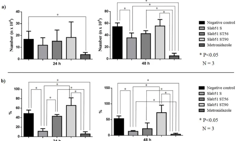
respect to untreated controls, but its effects were generally lower than that of metronidazole, the reference drug. In fact, in the growth inhibition assay the number of trophozoites (35.48 ±8.16x10⁴) found after 48h in cultures treated with fresh Slab51 supernatant was significantly (P <0.05) reduced respect to that counted in untreated cultures (54.15±6.58x10⁴). However, this reduction was significantly lower (P <0.05) respect to that observed in cultures treated with metronidazole where the number of trophozoites was extremely low (5.05±4.47x10⁴) (Table 1;Fig 1). In the adhesion assays, after 24h the number of adherentG. duodenalis
tro-phozoites (9.51± 7.08%) observed in Slab51 FS-treated cultures was significantly reduced (P <0.05) respect to that observed in untreated cultures (48.58± 7.31%) but similar to that counted in culture treated with metronidazole (5.64±4.75). However, after 48h the number of adherentG. duodenalis trophozoites (12.85±2.26%) counted in Slab51 FS-treated cultures was
still significantly lower (P<0.05) respect to that observed in untreated cultures (52.91±8.64%), but significantly higher (P<0.05) than in culture treated with metronidazole (3.43±3.44%) (Table 2;Fig 1). On the contrary, the cultures treated with metronidazole showed a significant
Table 1. Growth inhibition ofGiardia duodenalis trophozoites by fresh (Slab51 FS) and 56˚C (Slab51S 56˚C) and 90˚C (Slab51S 96˚C) heat-treated Slab51
superna-tants. The number (n x104) ofGiardia duodenalis trophozoites are expressed as average and standard deviation of trophozoites counted in three replicates after 24 and 48 h observation periods. Growth assay 24 h 48h Mean SD Mean SD Negative control 16.74b 6.88 54.15c 6.58 Slab51 FS 11.82ab 6.26 35.48b 8.16 Slab51 S 56˚C 15.14ab 9.46 42.54bc 5.02 Slab51 S 90˚C 18.32ab 13.16 55.42bc 10.96 Metronidazole 3.82a 1.82 5.05a 4.47 a,b,c: P < 0.05 https://doi.org/10.1371/journal.pone.0213385.t001
reduction of the growth and adhesion ofG. duodenalis trophozoites in comparison with
untreated cultures, both at 24h and at 48h (Tables1and2;Fig 1).
The heat-treatment reduced the negative effects onG. duodenalis growth and adhesion
abil-ity of fresh Slab51 supernatant. In fact, while after 48 h the inhibiting activabil-ity of 56˚C heat-treated Slab51 supernatant (42.54± 5.02%) was significantly different (P<0.05) both from treated (5.05±4.47x10⁴) and untreated controls (54.15±6.58 x10⁴
), as well as from Slab51 FS-treated cultures (35.48±8.16x10⁴). After the same time-period, results observed for 90˚C heat-treated Slab51 supernatant cultures (55.42±10.96 x10⁴
) were comparable to that of untreated controls (Table 1,Fig 1). Moreover, in the adhesion assay no statistical difference with untreated cultures (52.91±8.64%) was observed both for 56˚C (21.15±18.33%) and 90˚C heat-treated Slab51 supernatant cultures (72.06±10.96%) (Table 2).
All the colonies from Slab51 cultures in TYI-S-33 medium with and without antibiotics and cultured in MRS agar plates were identified asL. plantarum.
At the ultrastructural level, untreated trophozoites showed normal structure and morphol-ogy (Fig 1A and 1C), while treated parasites were swelling and increased in size (Fig 2B and 2D). Moreover, trophozoites showed alterations of their cellular membrane and vacuole pat-terns. Inside the cells, an ostensibly low electron density and granules grouped in clusters were evidenced. In the nucleus, the loss of the nuclear envelope and nuclear architecture and the presence of structures resembling holes or lacunas were clearly visible (Fig 2B and 2D).
Fig 1. (a) Growth inhibition ofGiardia duodenalis trophozoites by fresh (Slab51 FS) and 56˚C (Slab51S 56˚C) and 90˚C (Slab51S 96˚C) heat-treated Slab51 supernatants. The number (n x104) of
G. duodenalis trophozoites are expressed as average and standard deviation of trophozoites counted in three replicates after 24 and 48 h observation periods.; (b) Adhesion inhibition ofG. duodenalis trophozoites by fresh (Slab51 FS) and 56˚C (Slab51S 56˚C) and 90˚C (Slab51S 96˚C) heat-treated Slab51 supernatants. Attached trophozoites have been expressed as the mean percentage of attachedG. duodenalis trophozoites in relation to the total number ofG. duodenalis trophozoites counted after 24 and 48 hours in each culture and in three replicates.
Effects of Slab51
ex vivo
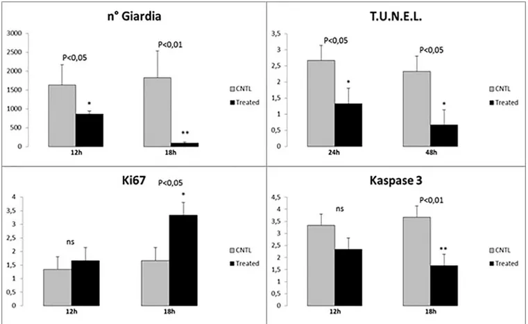
Inex vivo trials, significant results were observed in treated samples at 18h post-infection (PI).
Indeed, at this time treated with Slab51 ultrafilterd fresh supernatant showed a significant reduction of viableG. duodenalis trophozoites at the end of the observation period, as
evi-denced inFig 3. In these same samples, a progressive and significant decrease in TUNEL + enterocytes was observed (Fig 4C), while at the same time apoptotic activity peaked in untreated samples (Fig 5C) when compared to Slab51 FS-treated samples. Similar results were obtained in sections stained for Caspase-3 (Figs4Dand5D). In fact, as shown inFig 4D, also for Caspase-3 the peak in decrease of expression was observed inex vivo intestinal tissue
cul-tures after 18h of incubation with Slab51 ultrafilterd fresh supernatant. In untreated controls,
G. duodenalis trophozoites showed an intact morphology also after 18h (Fig 6A) while the apo-ptosis rate of enterocytes in untreated samples increased progressively in these groups
throughout the experiment (Fig 6B). As shown in Figs3Band4B, Ki67 enterocyte nuclear staining was observed in mice intestinal mucosa still after 18h in both Slab51 FS-treated groups and untreated control groups, suggesting that even after this period of cultivation, the epithe-lium covering the intestinal mucosa is still able to live and proliferateex vivo. These
observa-tions were also supported by the general morphology of bioptic samples. In fact, even if well preserved, untreated samples showed a clear loss of epithelial cells with a different pattern of inflammatory cell distribution throughout the mucosal corion, as evidenced in haematoxylin-eosin stained tissues (Figs4Aand5A).
Discussion
G. duodenalis is a common fecal-oral parasite of the small intestine and one of the most
impor-tant causes of human and animal diarrheal disease worldwide. Indeed,G. duodenalis infection
can be asymptomatic, or cause acute or chronic diarrhea, dehydration, intestinal malabsorp-tion, malnutrition and steatorrhea [2,4–7]. Chronic fatigue, post-infectious irritable bowel syndrome and intestinal dysbiosis have also been documented in humans as possible conse-quences ofG. duodenalis infections, [28,29], while growth retardation and cognitive malfunc-tion have been reported in children from endemic areas [2,30]. Probiotics may interfere with
G. duodenalis infection through different mechanisms, including competition for limited
adhesion sites, competition for nutrients that would otherwise be utilized byG. duodenalis,
stimulation of the host immune response and by producing substances that may inhibitG. duodenalis [6,7,11]. Probiotic bacteria can produce compounds, which have inhibitory effects directed against pathogens, as viruses, bacteria, fungi, parasites, as well as against cancer cells
Table 2. Adhesion inhibition ofGiardia duodenalis trophozoites by fresh (Slab51 FS) and 56˚C (Slab51S 56˚C) and 90˚C (Slab51S 96˚C) heat-treated Slab51
super-natants. Attached trophozoites have been expressed as the mean percentage of attachedG. duodenalis trophozoites in relation to the total number of G. duodenalis tropho-zoites counted after 24 and 48 hours in each culture and in three replicates.
Adhesion assay 24 h 48h Mean SD Mean SD Negative control 48.58b 7.31 52.91c 8.64 Slab51S 9.51a 7.08 12.85b 2.26 Slab51S 56˚C 42.84b 3.70 21.15bc 18.33 Slab51S 90˚C 65.65b 16.10 72.06c 23.79 Metronidazole 5.64a 4.75 3.43a 3.44 a,b,c: P < 0.05 https://doi.org/10.1371/journal.pone.0213385.t002
[11]. Among them, the anti-G. duodenalis activity of probiotic compounds, mainly derived
fromLactobacilli, has been demonstrated [11,20,24]. In fact, bacteriocins derived from Lacto-bacillus acidophilus were found able to inhibit in vitro the adhesion and the growth of G. duo-denalis trophozoites [11]. Moreover, these negative effects were found associated with severe morphological changes ofG. duodenalis trophozoites, a decline of the intestinal parasite
den-sity and amelioration of intestinal pathology in infected mice treated withL. acidophilus
bacte-riocins [11]. More recently, results from some studies suggested that the ability to deconjugate bile salts showed by some lactobacilli, asL. johnsonii strain LA1 and Lactobacillus gasseri
CNCM I-4884, may represent a further mechanism contributing to the inhibition ofGiardia
trophozoite growthin vitro [31,32].
Fig 2. Ultrastructure ofGiardia duodenalis by TEM. Untreated G. duodenalis trophozoites showing normal structure and morphology (a). Trophozoite coronal section (c). A coronal view of a trophozoite demonstrates the nuclei (N), endoplasmic reticulum (ER), flagella (F), vacuoles (V), ventral disk (VD), lateral crest (LC) and ventrolateral flange (VLF). The same sections (b, d) of treated parasites show swelling trophozoites, with an increased size, and evident alterations of their cellular membrane and with a vacuolar degenerative pattern (x6,700). Note in the coronal section the severely damaged nuclei, nuclear membrane rupture, loss of the chromatin, flanges and ventral disk rupture (x6,700).
Negative effects onG. duodenalis showed by the fresh supernatant of the commercial
probi-otic evaluatedin vitro and ex vivo in the present study, are similar or higher to those reported
in most of these previous studies. In fact, the fresh Slab51 supernatant was able to inhibitin vitro the adhesion and the growth of G. duodenalis trophozoites, although this inhibition was
significantly lower than that of metronidazole. In the study of Perezet al. [24], the culture supernatant of the probiotic strain LA1 ofLactobacillus johnsonii was able to control G. duode-nalis growth in vitro but it was unable to inhibit the adhesion of the parasite, while six Lactoba-cillus acidophilus strains tested did not show any noticeable effects. These data could be
indicative that Slab51 constituent probiotic strains, mainlyL. plantarum DSM 32244, possibly
produce more effective active anti-G. duodenalis compounds with respect to those produced
byL. johnsonii LA1. Negative effects on G. duodenalis adherence here observed are consistent
with the ability reported for some lactobacilli to modulateG. duodenalis infection in vivo by
minimizing or preventing the adherence of trophozoites to the intestinal mucosal surface [33,
34].
In agreement with previous studies in which some probiotic compounds were found able to induce morphological changes ofG. duodenalis trophozoites [11,24], important morpho-logical alterations of this protozoan parasite were herein observed inG. duodenalis
trophozo-ites fromin vitro cultures treated with Slab51 fresh supernatant, including profound
Fig 3. Statistical evaluation of different parameters inex-vivo duodenal tissue cultures, before and after treatment. (a) Significant reduction in the number of viableGiardia cells counted in biopsies treated with Slab51 ultrafiltered supernatant at 12h and 18h. (b) Statistical comparison of TUNEL positive nuclei before and after the same treatment at 24h and 48h. (c) Statistical comparison of level of cellular viability and replication by Ki67 nuclear assessment at 12h and 18h. (d) Statistical confrontation of Caspase3 positive cells before and after the same treatment at 12h and 18h. Caspase3 expression in association with the level of the TUNEL expression, as showed inFig 2B, indicate the entire fraction of apoptotic cell because these two apoptotic markers are expressed in subsequent time.
alterations of cellular and nuclear membranes, nuclear disorganization and formation of intra-cytoplasmic cavities. Thesein vitro cytopathic effects are very similar to those caused by L. aci-dophilus bacteriocins in vivo, possibly indicating a similar mode of action [11] and may be one of the main factors responsible for the inhibition of the trophozoite proliferation and adhesion here observedin vitro.
Trials performed in this study showed that thein vitro inhibiting effects on G. duodenalis
showed by Slab51 fresh supernatant were greatly reduced by heat treatment at 56˚C and completely annulled at 90˚C, indicating that active metabolites contained in Slab51 superna-tant are likely term labile compounds. These results agree with those of a previous report [24] and encourage further studies aimed to identify the extracellular factors responsible for the anti-Giardia effects of fresh Slab51 supernatant observed in the present study.
Results fromex vivo trials confirmed the inhibiting effects of the fresh supernatant of
Slab51 onG. duodenalis trophozoites observed in vitro. The best observations were obtained
after an 18h incubation period. In fact, after this period a significant reduction ofG. duodenalis
Fig 4.Ex-vivo intestinal tissue, from mice treated with Slab51 ultrafiltered fresh supernatant. At 18h post-infection with Giardia duodenalis, biopsies showed a preserved morphology and viability as demonstrated by H&E stain (a) and Ki67 enterocytes expression (b). In these samples, a low number of TUNEL+ enterocytes is observed (c). A similar pattern of expression of Caspase-3 indicates a low apoptotic rate in these samples (d). Presence of inflammatory cells with a diffuse and non-polarized pattern of infiltration is also observed in these biopsies (H&E, and IHC with Mayer Haematoxilin nuclear counterstain, scale bar 400μm).
trophozoites and a significantly greater vitality of intestinal epithelial cells was evidenced in treated intestinal cultures respect to the untreated controls. Moreover,G. duodenalis was
found capable of slowing or damaging the intestinal epithelial cell turnover in untreatedex vivo cultures, since in these cultures the apoptotic rate was increased. On the other side,
obtained results showed that the number ofG. duodenalis trophozoites was significantly
low-ered by the fresh Slab51 supernatant. Moreover, inex vivo cultures the apoptosis and the death
of intestinal epithelial cells was higher inG. duodenalis-inoculated cultures respect to those
inoculated withG. duodenalis and the fresh supernatant of Slab51, indicating that the damage
to the epithelial cells induced byG. duodenalis was reduced by the fresh supernatant of this
commercial probiotic. Considering that the increase in the rate of enterocyte apoptosis and enterocyte damage are included among the main pathogenic mechanisms ofG. duodenalis
[34], results obtained inex vivo trials are promising about possible in vivo protective effects of
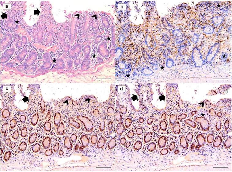
Fig 5.Ex-vivo intestinal tissue, from negative control (NC) group. Section stained with H&E revealed a diffuse epithelial loss with inflammatory cells infiltration polarized under the destroyed intestinal epithelium. Note the reinforcement of inflammatory cells around intestinal glands (a). Villi are partially damaged (arrows), while in part they are totally flat or in any case strongly tuned (arrowheads) due to the effect of the strong colonization-adhesion ofGiardia duodenalis trophozoites on the surface of the epithelium, which has become detached in many areas of the mucosa. Note the reinforcement of inflammatory cells around intestinal glands (stars). Ki67 nuclear staining evidenced an apparently higher number of positive cells because many inflammatory cells showed a strongly nuclear positivity (b). Note that TUNEL (c) and Caspase-3 (d) are over-expressed in these explanted intestinal samples. (H&E, and IHC with Mayer Haematoxilin nuclear counterstain, scale bar 400μm).
the fresh culture supernatant of Slab51 againstG. duodenalis. This is the first study in which
negative effects onG. duodenalis by metabolites from lactobacilli were demonstrated both in vitro and by using a murine ex vivo model. In previous studies, a variety of different systems
have been used to evaluate the adherence and growth ofG. duodenalis, including synthetic
sur-faces, human cells and non-human cells, as isolated rat enterocytes and rat enterocyte cell lines [35–37]. Among them, the human colonic adenocarcinoma derived epithelial cell line Caco-2, functionally and structurally may resemble small bowel enterocytes [38]. Therefore, this cell line model is considered useful and appropriate for studies of host intestine-pathogen interac-tions, and it is frequently used as a model to study the attachment and other effects ofG. duo-denalis trophozoites under different conditions [35]. However, this model does not allow a proper evaluation of the damages caused byG. duodenalis trophozoites to the intestinal
mucosa and of associated inflammatory cells, while the murineex vivo model performed in
this study allowed these evaluations, by preserving the tissue architecture and the cellular com-plexity over several days. Indeed, alterations, damage and inflammation herein observed inex vivo negative controls, i.e. intestinal tracts inoculated with G. duodenalis trophozoites only,
were not so different from that observed inin vivo rodent models and at histopathological
examination of intestinal biopsies taken from symptomatic human patients [30,35]. More-over, theex vivo mouse model used in this study allowed to assess the anti-Giardia activity of
the fresh supernatant of Slab51 by the evaluation of several positive effects on intestines inocu-lated withG. duodenalis trophozoites, although this model cannot be able to mimic the
com-plexity of whole living organisms.
In conclusion, results from this study showed that the fresh culture supernatant of the com-mercial probiotic Slab51 has negative effects onG. duodenalis both in vitro and ex vivo in a
mouse model. These antagonistic effects may suggest that this probiotic may likely represent a further and interesting approach for the prevention of giardiasis and/or the reduction of the pathogenic effects and proliferation of this protozoan parasite in infected hosts. However, fur-ther studies aimed to evaluate its efficacyin vivo on experimentally and/or naturally infected
animals, are needed.
Fig 6.Giardia duodenalis in negative control (NC) group. G. duodenalis trophozoites showed an intact morphology also after 18h (a), and the apoptosis rate of enterocytes (arrows), as demonstrated in b, increased progressively during the experiment, by the combined effect of the infection and theex-vivo condition. (H&E, scale bar: a, 10μm–b, 50 μm).
Author Contributions
Conceptualization: Stefania Perrucci, Gianluca Fichi, Giacomo Rossi.
Data curation: Stefania Perrucci, Gianluca Fichi, Livio Galosi, Marco Lalle, Giacomo Rossi. Formal analysis: Stefania Perrucci, Gianluca Fichi, Giacomo Rossi.
Funding acquisition: Stefania Perrucci, Giacomo Rossi.
Investigation: Stefania Perrucci, Gianluca Fichi, Enrica Ricci, Livio Galosi, Giacomo Rossi. Methodology: Stefania Perrucci, Gianluca Fichi, Enrica Ricci, Giacomo Rossi.
Project administration: Stefania Perrucci. Resources: Stefania Perrucci.
Supervision: Stefania Perrucci, Gianluca Fichi, Giacomo Rossi. Validation: Stefania Perrucci, Gianluca Fichi, Giacomo Rossi.
Writing – original draft: Stefania Perrucci, Gianluca Fichi, Giacomo Rossi.
Writing – review & editing: Stefania Perrucci, Gianluca Fichi, Enrica Ricci, Livio Galosi,
Marco Lalle, Giacomo Rossi.
References
1. Ryan U, CacciòSM. Zoonotic potential of Giardia. Int J Parasitol. 2013; 43: 943–956.https://doi.org/10. 1016/j.ijpara.2013.06.001PMID:23856595
2. Manko A, Motta JP, Cotton JA, Feener T, Oyeyemi A, Vallance BA, et al. Giardia co-infection promotes the secretion of antimicrobial peptides beta-defensin 2 and trefoil factor 3 and attenuates attaching and effacing bacteria-induced intestinal disease. PloS ONE, 2017; 12: e0178647.https://doi.org/10.1371/ journal.pone.0178647PMID:28622393
3. Marangi M, Berrilli F, Otranto D, Giangaspero A. Genotyping of Giardia duodenalis among children and dogs in a closed socially deprived community from Italy. Zoonoses Public Health 2010; 57: 54–58.
4. Allain T, Amat CB, Motta JP, Manko A, Buret AG. Interactions of Giardia sp. with the intestinal barrier: Epithelium, mucus, and microbiota, Tissue Barriers 2017; 5:1, e1274354.https://doi.org/10.1080/ 21688370.2016.1274354PMID:28452685
5. Minetti C, Chalmers RM, Beeching NJ, Probert C, Lamden K. Giardiasis. BMJ 2016;27: 355:i5369.
https://doi.org/10.1136/bmj.i5369PMID:27789441
6. Hawrelak J. 2003. Giardiasis: Pathophysiology and Management. Altern Med Rev. 2003; 8: 129–142. PMID:12777159
7. Tangtrongsup S, Scorza V. Update on the diagnosis and management of Giardia spp. infections in dogs and cats. Top Companion Anim Med. 2010; 25: 155–162.https://doi.org/10.1053/j.tcam.2010.07. 003PMID:20937499
8. Geurden T, Vanderstichel R, Pohle H, Ehsan A, von Samson-Himmelstjerna G, Morgan ER, et al. 2012. A multicentre prevalence study in Europe on Giardia duodenalis in calves, with molecular identifi-cation and risk factor analysis. Vet Parasitol. 2012; 190: 383–90.https://doi.org/10.1016/j.vetpar.2012. 06.039PMID:22824061
9. Lalle M. Giardiasis in the Post Genomic Era: Treatment, Drug Resistance and Novel Therapeutic Per-spectives. Infect Disord Drug Targets. 2010; 10: 283–294. PMID:20429863
10. Savioli L, Smith H, Thompson A. Giardia and Cryptosporidium join the ’Neglected Diseases Initiative’. Trends Parasitol. 2006; 22: 203–208.https://doi.org/10.1016/j.pt.2006.02.015PMID:16545611 11. Amer EI, Mossallam SF, Mahrous H. Therapeutic enhancement of newly derived bacteriocins against
Giardia lamblia. Exp Parasitol. 2014; 146: 52–63.https://doi.org/10.1016/j.exppara.2014.09.005PMID:
25300763
12. Morrone F, Carneiro J, Reis C, Cardozo C, Ubal C, De Carli G. Study of enteroparasites infection fre-quency and chemotherapeutic agents used in pediatric patients in a community living in Porto Alegre, RS, Brazil. Rev Inst Med Trop Sao Paulo. 2004; 46: 77–80. PMID:15141275
13. Sullayman I, Nolder D, Warchurst D, Rossignol J. In vitro activity of nitazxanide and related compounds against isolates of Giardia intestinalis, Entamoeba histolytica and Trichomonas vaginalis. J Antimicrob Chemother. 2002; 49: 103–111. PMID:11751773
14. Escobedo AA, Cimerman S. Giardiasis: a pharmacotherapy review. Expert Opin Pharmcother. 2007; 8: 1885–902.
15. Fiechter R, Deplazes P, Schnyder M. Control of Giardia infections with ronidazole and intensive hygiene management in a dog kennel. Vet Parasitol. 2012; 187: 93–98.https://doi.org/10.1016/j.vetpar.2011. 12.023PMID:22240238
16. Hart CJS, Munro T, Andrews KT, Ryan JH, Riches AG, Skinner-Adams TS. A novel in vitro image-based assay identifies new drug leads for giardiasis. Int J Parasitol Drugs Drug Resist. 2017; 7: 83–89.
https://doi.org/10.1016/j.ijpddr.2017.01.005PMID:28171818
17. Tian HF, Chen B, Wen JF. Giardiasis, drug resistance, and new target discovery. Infect Disord Drug Targets. 2010; 10: 295–302. PMID:20429862
18. Ansell BR, McConville MJ, Ma’ayeh SY, Dagley MJ, Gasser RB, Svard SG, Jex AR. Drug resistance in Giardia duodenalis. Biotechnol. Adv. 2015; 33: 888–901.https://doi.org/10.1016/j.biotechadv.2015.04. 009PMID:25922317
19. Tejman-Yarden N, Eckmann L. New approaches to the treatment of giardiasis. Curr Opin Infect Dis. 2011; 24: 451–456.https://doi.org/10.1097/QCO.0b013e32834ad401PMID:21857510
20. Travers MA, Florent I, Kohl L, Grellier P. Probiotics for the control of parasites: an overview. J Parasitol Res. 2011: 610769.https://doi.org/10.1155/2011/610769PMID:21966589
21. Hassan M, Kjos M, Nes IF, Diep DB, Lotfipour F. Natural antimicrobial peptides from bacteria: charac-teristics and potential applications to fight against antibiotic resistance. J. Appl. Microbiol. 2012; 113: 723–736.https://doi.org/10.1111/j.1365-2672.2012.05338.xPMID:22583565
22. Keister DB. Axenic culture of Giardia lamblia in TYI-S-33 medium supplemented with bile. Trans. R. Soc. Trop. Med. Hyg. 1983; 77: 487–488. PMID:6636276
23. Machado M, Dinis AM, Salgueiro L, Cavaleiro C, Custo´dio JBA, Sousa CM. Anti-Giardia activity of phe-nolic-rich essential oils: effects of Thymbra capitata, Origanum virens, Thymus zygis subsp. sylvestris, and Lippia graveolens on trophozoites growth, viability, adherence, and ultrastructure. Parasitol. Res. 2010; 106: 1205–1215.https://doi.org/10.1007/s00436-010-1800-7PMID:20217133
24. Perez PF, Minnard J, Rouvet M, Knabenhans C, Brassart D, De Antoni GL, et al. Inhibition of Giardia intestinalis by extracellular factors from lactobacilli: an in vitro study. Appl. Environ. Microbiol. 2001; 67: 5037–5042.https://doi.org/10.1128/AEM.67.11.5037-5042.2001PMID:11679323
25. Be´ne´re´ E, da Luz RA, Vermeersch M, Cos P, Maes L. A new quantitative in vitro microculture method for Giardia duodenalis trophozoites. J Microbiol Methods. 2007; 71: 101–106.https://doi.org/10.1016/j. mimet.2007.07.014PMID:17888535
26. Sauda F, Malandrucco L, Macrı` G, Scarpulla M, De Liberato C, Terracciano G, et al. Leishmania infan-tum, Dirofilaria spp. and other endoparasite infections in kennel dogs in central Italy. Parasite. 2018; 25: 2.https://doi.org/10.1051/parasite/2018001PMID:29388550
27. Rossi G, Pengo G, Caldin M, Palumbo Piccionello A, Steiner JM, Cohen ND, et al. Comparison of Microbiological, Histological and Immunomodulatory Parameters in Response to Treatment with Either Combination Therapy with Prednisone and Metronidazole or Probiotic VSL#3 Strains in Dogs with Idio-pathic Inflammatory Bowel Disease. PLOS ONE 2014; 9: e94699.https://doi.org/10.1371/journal.pone. 0094699PMID:24722235
28. Persson R, Wensaas KA, Hanevik K, Eide GE, Langeland N, Rortveit G. The relationship between irrita-ble bowel syndrome, functional dyspepsia, chronic fatigue and overactive bladder syndrome: a con-trolled study 6 years after acute gastrointestinal infection. BMC Gastroenterol. 2015; 15: 66.https://doi. org/10.1186/s12876-015-0296-0PMID:26058591
29. Beatty JK, Akierman SV, Motta JP, Muise S, Workentine ML, Harrison JJ et al. Giardia duodenalis induces pathogenic dysbiosis of human intestinal microbiota biofilms. Int J Parasitol. 2017; 47: 311– 326.https://doi.org/10.1016/j.ijpara.2016.11.010PMID:28237889
30. Halliez MC, Buret AG. Extra-intestinal and longterm consequences of Giardia duodenalis infections. World J Gastroenterol. 2013; 19: 8974–8985.https://doi.org/10.3748/wjg.v19.i47.8974PMID:
24379622
31. Travers MA, Sow C, Zirah S, Deregnaucourt C, Chaouch S, Queiroz RM. et al. Deconjugated bile salts produced by extracellular bile-salt hydrolase-like activities from the probiotic Lactobacillus johnsonii La1 Inhibit Giardia duodenalis In vitro Growth. Front. Microbiol. 2016; 7:1453.https://doi.org/10.3389/fmicb. 2016.01453PMID:27729900
32. Allain T, Chaouch S, Thomas M, Travers MA, Valle I, Langella P, Grellier P, Polack B, Florent I, Bermu´-dez-Humara´ n LG. Bile salt hydrolase activities: a novel target to screen anti-Giardia Lactobacilli? Front. Microbiol. 2018; 9:89.https://doi.org/10.3389/fmicb.2018.00089PMID:29472903
33. Shukla G, Devi P, Sehgal R. Effect of Lactobacillus casei as a probiotic on modulation of giardiasis. Dig Dis Sci. 2008; 53: 2671–2679.https://doi.org/10.1007/s10620-007-0197-3PMID:18306038
34. Certad G, Viscogliosi E, Chabe´ M, CacciòSM. Pathogenic Mechanisms of Cryptosporidium and Giar-dia. Trends Parasitol. 2017; 33: 561–576.https://doi.org/10.1016/j.pt.2017.02.006PMID:28336217 35. Kraft MR, Klotz C, Bu¨cker R, Schulzke JD, Aebischer T. Giardia’s Epithelial Cell Interaction In Vitro:
Mimicking Asymptomatic Infection? Front. Cell. Infect. Microbiol. 2017; 7: 421.https://doi.org/10.3389/ fcimb.2017.00421PMID:29018775
36. Inge PMG, Edson CM, Farthing MJG. Attachment of Giardia lamblia to rat intestinal epithelial cells. Gut 1988; 29: 795–801. PMID:3384364
37. Favennec L, Chochillon C, Meillet D, Magne D, Savel J, Raichvarg D, et al. Adherence and multiplica-tion of Giardia intestinalis on human enterocyte-like differentiated cells in vitro. Parasitol Res 1990; 76: 581–4. PMID:2217120
38. Pinto M, Robine-Leon S, Appay MD, Kerdinger M, Triadou N, Dussaulx E, et al. Enterocyte-like differen-tiation and polarisation of the human colon carcinoma cell line Caco-2 culture. Biol Cell 1983; 47: 323– 30.