Università degli Studi del Piemonte Orientale
Dipartimento di Scienze e Innovazione Tecnologica
Dottorato di Ricerca in Chemistry & Biology
curriculum: Drug Discovery and Development
XXIX ciclo a.a. 2015-2016
SSD: CHIM/03
Passive drug targeting and delivery of antitumor
Pt(IV) prodrugs
Elena Perin
Supervised by Prof. Mauro Ravera
Università degli Studi del Piemonte Orientale
Dipartimento di Scienze e Innovazione Tecnologica
Dottorato di Ricerca in Chemistry & Biology
curriculum: Drug Discovery and Development
XXIX ciclo a. a. 2015-2016
SSD: CHIM/03
Passive drug targeting and delivery of antitumor
Pt(IV) prodrugs
Elena Perin
Supervised by Prof. Mauro Ravera
i
Contents
Chapter I: The Tumor
. . . .11.1 The Origin of the Tumor . . . 2
1.2 Antitumor Therapies . . . .6
1.3 New Generation Therapies . . . .9
1.4 Cell Proliferation . . . 13
1.5 Cell Cycle . . . .13
1.6 Cell Death . . . .15
References . . . 19
Chapter II: The Platinum Chemistry
. . . 212.1 The Chemistry of Platinum Complexes . . . .22
2.2 History of Cisplatin . . . 23
2.3 Cisplatin Mechanism of Action . . . 26
2.4 Second Generation of Platinum Complexes: Features and Cytotoxic Activity . . . .31
2.5 Platinum(IV) Prodrugs: Characteristics and Mechanism of Action . . . . . . 35
2.6 Drug Targeting and Delivery . . . 39
2.6.1 Active Strategies . . . 40
2.6.2 Passive Strategies . . . .41
2.6.3 The Choice of Passive DTD Vectors . . . .42
2.6.4 Nanoparticles Properties . . . 44
2.6.5 The Drug Release . . . 46
ii
Chapter III: Outline of the Thesis
. . . .51Chapter IV: Synthesis and Characterization of Pt(IV) Complexes
and Coupling Reactions with Amino-Functionalized Fluorescent
Core-Shell Silica Nanoparticles
. . . 534.1 Introduction . . . 54
4.2 Instrumental Information . . . 56
4.3 Synthesis of the Nonporous Core-Shell Silica Nanoparticles . . . 58
4.3.1 Quantification of the Fluorophore . . . .61
4.3.2 Quantification of the Amino Functionalities . . . .62
4.3.3 Size and Stability Investigations . . . 63
4.4 Synthesis of Cisplatin . . . .66
4.4.1 Synthesis of (SP-4-2)-diamminediiodidoplatinum(II) . . . .67
4.4.2 Synthesis of (SP-4-2)-diamminedichloridoplatinum(II) . . 68
4.4.2.1 Characterization of the Complex . . . 68
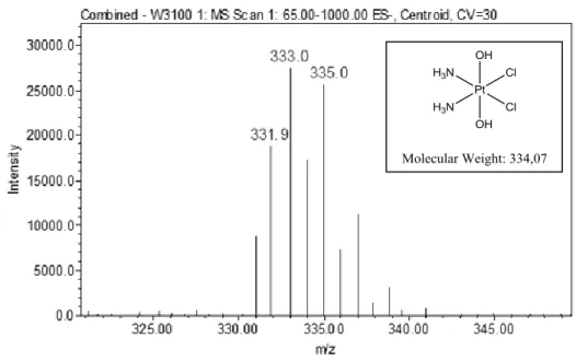
4.5 Synthesis of (OC-6-33)-diamminedichloridodihydroxidoplatinum (IV) (1) . . . 70
4.5.1 Characterization of the Complex . . . .71
4.6 Synthesis of (OC-6-33)-diamminebis(4-carboxypropanoato) dichloridoplatinum(IV) (2) . . . .74
4.6.1 Characterization of the Complex . . . .74
4.7 Synthesis of the Activated N-hydroxysuccinimidyl Diester of 2 (3) . . . . . 79
4.7.1 Characterization of the Complex . . . .79
4.8 Synthesis of (OC-6-33)-diamminebis(4-oxo-4-(propylamino) butanoato)dichloridoplatinum(IV) (4) . . . 82
iii
4.9 Synthesis of Conjugate 5a . . . 87
4.9.1 Characterization of Conjugate 5a . . . 89
4.9.2 Quantification of Pt Loading on Conjugate 5a . . . 90
4.10 Synthesis of Conjugates 5b-5d. . . 91
4.10.1 Characterization of Conjugates 5b-5d . . . .92
4.11 Synthesis of (OC-6-44)-diamminedichloridoethanolatohydroxido platinum(IV) (6) . . . .93
4.11.1 Characterization of the Complex . . . .93
4.12 Synthesis of (OC-6-44)-diammine(4-carboxypropanoato)dichlorido ethanolatoplatinum(IV) (7) . . . .97
4.12.1 Characterization of the Complex . . . .98
4.13 Synthesis of the Activated N-hydroxysuccinimidyl Ester of 7 (8) . . . . . . 103
4.13.1 Characterization of the Complex . . . .103
4.14 Synthesis of (OC-6-44)-diamminedichloridoethanolato(4-oxo-4- (propylamino)butanoato)platinum(IV) (9) . . . .106
4.14.1 Characterization of the Complex . . . .107
4.15 Synthesis of Conjugates 10a-10d . . . .110
4.15.1 Characterization of Conjugates 10a-10d . . . .111
4.15.2 Quantification of Pt Loading on 10a-10d . . . 112
4.16 Spontaneous Release of Platinum from Conjugates . . . .113
4.17 The Mechanism of Action of the Pt(IV) Complexes: the Activation by Reduction . . . .116
4.17.1 Study of the Reduction Process of the Synthesized Pt(IV) Complexes. . . .119
4.17.1.1 Reduction Kinetics of Complex 7 . . . .120
4.17.1.2 Study of the Behavior with L(+) Ascorbic Acid . . . . . 120
iv
4.17.1.3 Study of the Behavior with L-Reduced
Glutathione (GSH) . . . 123
4.17.2.1 Reduction Kinetics of Complex 9 . . . .124
4.17.2.2 Study of the Behavior with L(+) Ascorbic Acid . . . . . 124
4.17.2.3 Study of the Behavior with L-Reduced Glutathione (GSH) . . . 126
4.17.3 Conclusions of the Reduction Kinetics of Pt(IV) Complexes . . . .126
4.18 Platinum Release from Conjugates in Reducing Conditions . . . . . . .126
4.19 Biological In Vitro Studies . . . .127
4.19.1 The Antiproliferative Activity (IC50) . . . .127
4.19.2 The Cellular Accumulation . . . 133
4.19.3 Confocal Microscopy . . . .136
4.19.4 Studies on the NPs Internalization Mechanisms . . . .141
4.20 Conclusions . . . 146
References . . . 147
Chapter V: Synthesis, Characterization of Platinum Complexes
and Coupling Reactions with New Amino-Functionalized Silica
Nanoparticles
. . . 1515.1 Introduction . . . 152
5.2 Synthesis of (OC-6-44)-(acetylamido-N)diamminedichlorido hydroxidoplatinum(IV) (11) . . . .154
5.2.1 Characterization of the Complex . . . .154 5.3 Synthesis of (OC-6-44)-(acetylamido-N)diammine(4-carboxy
v
propanoato)dichloridoplatinum(IV) (12) . . . 159
5.3.1 Characterization of the Complex . . . .159
5.4 Synthesis of the Activated N-hydroxysuccinimidyl Ester of 12 (13) . . . . . .164
5.4.1 Characterization of the Complex . . . .165
5.5 Synthesis of NPs Decorated with APTES (e) . . . .167
5.5.1 Quantification of the Amino Functionalities . . . .168
5.5.2 Size and Stability Investigations . . . 169
5.5.3 Solid-State NMR Characterization . . . 171
5.6 Synthesis of Conjugates 14e and 15e . . . .175
5.6.1 Quantification of the Pt Content in 14e and 15e . . . 175
5.6.2 Characterization of Conjugates 14e and 15e . . . .176
5.6.3 Platinum Release from Conjugates 14e and 15e in Reducing Conditions . . . 178
5.6.4 Spontaneous Platinum Release from Conjugates 14e and 15e . . . .178
5.7 Synthesis of NPs Decorated with AHAMTES (f - i) . . . .180
5.7.1 Quantification of the Amino Functionalities . . . .181
5.7.2 Size and Stability Investigations of NPs f - i . . . .184
5.7.3 Solid-State NMR Characterization . . . 186
5.8 Synthesis of Conjugate 14h at Different Reaction Times . . . 187
5.8.1 Quantification of the Pt Content in Conjugates 14h (at 1, 2, 4 and 8 hours) . . . 188
5.9 Synthesis of Conjugate 14h and 15h at Fixed Reaction Time but with Different Pt/Total Amino Groups Ratios . . . .189
5.9.1 Quantification of the Pt Content in 14h and 15h (Different Pt/Amino Groups Ratios) . . . 189
vi
Pt/Total Amino Groups Ratio . . . 191
5.9.3 Releases from Conjugates 14h and 15h: Spontaneous and in Reducing Conditions . . . .193
5.10 Biological In Vitro Studies . . . .194
5.10.1 The Antiproliferative Activity (IC50) . . . .194
5.10.2 The Cellular Accumulation . . . 197
5.11 Conclusions . . . 200
5.12 Perspectives . . . 201
5.12.1 Synthesis of the (SP-4-3)-dichlorido((1Z,5Z)-cycloocta- 1,5-diene)platinum(II) (16) . . . 203
5.12.2 Characterization of the Complex . . . .204
5.13 Synthesis of the (SP-4-3)-chlorido(2,2':6',2''-terpyridine) platinum(II) Chloride (17) . . . 207
5.13.1 Characterization of the Complex . . . .207
5.14 Synthesis of the (OC-6-33)-chloridodihydroxido(2,2':6',2''- terpyridine)platinum(IV) Chloride (18) . . . 212
5.14.1 Characterization of the Complex . . . .212
5.15 Synthesis of the (OC-6-33)-(4-carboxypropanoato)chlorido hydroxidoplatinum(IV) (19) . . . .217
5.15.1 Characterization of the Complex . . . .217
References . . . 221
Chapter VI: Coupling Reactions with Chitosan and Chitosan
Derivatives
. . . .2256.1 Introduction . . . 226
6.2 Synthesis of the (OC-6-44)-diamminedichloridoethanolato(4-oxo-4- (((3S,4S,5S)-2,4,5-trihydroxy-6-(hydroxymethyl)tetrahydro-2H- pyran-3-yl)amino)butanoato)platinum(IV) (20) . . . .231
vii
6.2.1 Characterization of the Complex . . . .231
6.3 Conductometric Titration of Chitosan . . . 235
6.4 Acid-Base Titration of Chitosan . . . .236
6.5 Synthesis of the Chitosan Conjugates (21a-21f) . . . 238
6.6 Synthesis of the Chitosan Modified with (3-carboxypropyl) trimethylammonium Chloride (22) . . . .241
6.6.1 Size and Stability Investigations . . . 244
6.7 Synthesis of the Modified Chitosan Conjugates at Different Reaction Times (22a - 22e) . . . .245
6.7.1 Synthesis of the Modified Chitosan Conjugates at Fixed Reaction Time but by using Different Prodrug Amounts (22b, 22f - 22h) . . . .246
6.7.2 Size and Stability Investigations . . . 248
6.8 Synthesis of the Activated N-hydroxysuccinimidyl Ester of 4- (methylsulphonyl)benzoic Acid (23) . . . 249
6.8.1 Characterization of the Compound . . . 250
6.9 Further Coupling of the Modified Chitosan Conjugate 22b with Compound 23 (24) . . . 254
6.9.1 Size and Stability Investigations . . . 254
6.10 Coupling of the Modified Chitosan Conjugate 22b with the 4- (bis(quinolin-2-ylmethyl)amino)butanoic Acid (25) . . . 255
6.10.1 Size and Stability Investigations . . . 257
6.11 Chelation Reaction of the Re(I) Complex with Diethylenetriamine (26) . . . .258
6.11.1 Characterization of the Complex . . . .259
6.12 Chelation Reaction of the Re(I) Complex with the 4-(bis(quinolin- 2-ylmethyl)amino)butanoic Acid (27) . . . 263
viii
6.13 Chelation Reaction of Conjugate 25 with the Re(I) Complex (28) . .
. . . 265
6.13.1 Size and Stability Investigations . . . 266
6.13.2 Characterization of Conjugate 28 . . . 267
6.14 Conclusions . . . 268
References . . . 269
Chapter VII: Coupling Reaction with Iron Oxide Nanoparticles
(IONPs)
. . . 2717.1 Introduction . . . 272
7.2 Synthesis of the (Z)-4-((carboxymethyl)amino)-4-oxobut-2-enoic Acid (29) . . . .276
7.2.1 Characterization of the Compound . . . 277
7.3 Synthesis of the 2-(2,5-dioxo-2,5-dihydro-1H-pyrrol-1-yl)acetic Acid (30) . . . .281
7.3.1 Characterization of the Compound . . . 282
7.4 Synthesis of the (SP-4-3)-diamminechlorido(2-(2,5-dioxo-2,5- dihydro-1H-pyrrol-1-yl)acetate)platinum(II) (31) . . . 285
7.4.1 Characterization of the Complex . . . .286
7.5 Synthesis of the (OC-6-44)-diamminechloridodihydroxido(2-(2,5- dioxo-2,5-dihydro-1H-pyrrol-1-yl)acetate)platinum(IV) (32) . . . .292
7.5.1 Characterization of the Complex . . . .293
7.6 Synthesis of the (OC-6-44)-diamminedichlorido(2-hydroxy ethanolato)(2-(2,5-dioxo-2,5-dihydro-1H-pyrrol-1-yl)acetate) platinum(IV) (33) . . . .293
7.6.1 Characterization of the Complex . . . .294 7.7 Synthesis of the (OC-6-44)-diammine(4-carboxypropanoato)
ix
platinum(IV) (34) . . . .295
7.7.1 Characterization of the Complex . . . .296
7.8 Synthesis of the (OC-6-44)-diamminedichloridoethanolato(2-(2,5- dioxo-2,5-dihydro-1H-pyrrol-1-yl)acetate)platinum(IV) (35) . . . .298
7.8.1 Characterization of the Complex . . . .299
7.8.2 Stability in Aqueous Solution of Complex 35 . . . .303
7.9 Diels-Alder Reaction with Furan . . . 305
7.9.1 Characterization of Furan . . . 305
7.9.2 Synthesis of the (OC-6-44)-diamminedichloridoethanolato (2-(1,3-dioxo-3a,4,7,7a-tetrahydro-1H-4,7-epoxyisoindol- 2(3H)-yl)acetate)platinum(IV) (36): I Method . . . 306
7.9.3 Synthesis of the (OC-6-44)-diamminedichloridoethanolato (2-(1,3-dioxo-3a,4,7,7a-tetrahydro-1H-4,7-epoxyisoindol- 2(3H)-yl)acetate)platinum(IV) (36): II Method . . . 309
7.10 Diels-Alder Reaction with the Furan-Functionalized Phosphonic Acid-Terminated POE Monomethyl Ether . . . .314
7.10.1 Characterization of the Ligand . . . 314
7.10.2 Diels-Alder Reaction: I Method . . . 315
7.10.3 Diels-Alder Reaction: II Method . . . .319
7.11 Diels-Alder Reaction with IONPs. . . .322
7.11.1 Main Features of the IONPs . . . 322
7.11.2 Diels-Alder Reaction for 24 Hours . . . 323
7.11.3 Diels-Alder Reaction for 5 Days . . . .323
7.12 Conclusions. . . .323
References . . . 324
Chapter VIII: Drugs Encapsulation into Liposomes
. . . 327x
8.2 Synthesis of the (OC-6-33)-diamminedichloridobis(2-propyl
pentanoate)platinum(IV) (39) . . . .331
8.2.1 Characterization of the Complex . . . .332
8.3 Preparation of the Liposomes . . . .335
8.3.1 Size and Stability Investigations . . . 338
8.4 Spontaneous Drug Release from Liposomes . . . 340
8.5 Biological In Vitro Studies . . . .341
8.5.1 The Antiproliferative Activity (IC50) . . . .341
8.5.2 The Cellular Accumulation . . . 345
8.5.3 The DNA Platination . . . .348
8.6 Conclusions . . . 350
References . . . 351
Conclusions and Perspectives
. . . .355List of Publications
. . . 3591
Capitolo 1
Chapter I
2
1.1 The Origin of the Tumor
A tumor (from the Latin tumor, “swelling”), also defined as neoplasia (from the Greek néos, “new”, and plàsis, “formation”), is an abnormal mass of tissue growing upon a pathological proliferation of the cells in a part of the body. The main feature of these cells is the uncontrolled and uncoordinated growth with respect to the healthy ones and this behavior is due to the fact that they do not respond to the mechanisms of cellular control and, in turn, this is due to alterations of their genetic heritage.
Although tumors are characterized by a common origin mechanism, they can undergo different evolution and symptoms pathways while sharing a steady and progressive cancerous cells increase, owing to a more rapid cellular reproduction. Therefore, a continuous multiplication of the majority of tumor cells is observed and only few cells do not survive and die.
A neoplasia usually grows and develops obeying a geometric law: in the early stages, the growth is slow and then it accelerates when the tumor mass increases. In addition, when the tumor reaches a critical size (about 1 cm3), it grows more and more rapidly and the symptoms can be felt (Figure 1.1).
3
Figure 1.1: Scheme of the growth phases of a tumor process.
The picture is a modification of that reported in literature [1]
The term cancer (from the Latin cancer, “crab”) was introduced by observing neoplastic cells behavior. In fact, in the multiplication phase, they form ramifications that entrap and destroy the surrounding healthy cells; metaphorically, in the same way, a crab destroys its prey with its claws.
The genes responsible for cellular control are located inside each cell and they have to prevent diseased cells to survive; the beginning of a tumoral process is the result of a failure of these genes. Therefore, since the mechanism responsible for the replication does not work anymore, the cells behave abnormally and multiply themselves forming daughter cells characterized by the same problems. Into the cells a series of changes happens, in particular as regards the genes. The neoplasia can have a benign or malignant biological behavior, depending on the features of the neoplastic cells. A benign tumor has a structure in which the cells mostly preserve the morphological and functional original tissue features, it has a growth speed usually slow and this is an expansive but not infiltrative growth. The tumor, indeed, is localized and enclosed in a wrapping of connective fibrous tissue, which allows it to distinguish itself from the other tissues, and, although
4
expanding, it does not invade the surrounding tissues but it only compresses them. On the contrary, a malignant tumor, commonly defined cancer, consists of cells which are morphologically and functionally different from the original tissue and this kind of neoplasia rapidly grows, also by infiltrating into the surrounding tissues, thus destroying them. The infiltration of these cancerous cells does not stop and continues its way into the human body through blood and lymphatic systems, giving rise to the metastasis (or secondary tumors). Moreover, the cancer has a high risk of reformation after surgery, unlike a benign tumor.
Finally, some anomalies characterize the neoplastic cells: an excess of sodium;
a lack of potassium; an excess of water.
In particular, water is about 90%, unlike 66% of the healthy cells: this excess is due, in turn, to the high sodium content in the blood, indeed sodium, being aggressive, attracts protective liquid around itself.
The medical nomenclature of tumors provides their classification on the basis of the tissue where they start growing and of their biological behavior (i.e. benign or malignant) (Table 1.1).
Tissue of origin Benign tumor Malignant tumor
Connective tissue, Cartilage, Bone, Muscle Fibroma, Chondroma, Osteoma, Leiomyoma, Rhabdomyoma Sarcoma Leukocytes Leukemia Glandular epithelial
and coating Adenoma, Papilloma Carcinoma
5
Melanocytes Melanocytic nevus Melanoma
Lymphatic glands Lymphoma
Table 1.1: Classification and medical nomenclature of tumors
Moreover, there are several methods employed to classify a neoplasia according to its stage. In particular, some methods consider the tumor size (T), the degree of regional spread or node involvement (N), or distant metastasis (M): this is the TNM staging method [2]. The T and N stages range from 0 to 4, e.g. T 0 means no cancer evidence, N 0 no nodal involvement, and T and N increase with the tumor size and the degree of lymph nodes involvement increasing, respectively. As regards metastasis, M 0 and M 1 are used and indicate no evidence or evidence of metastasis.
The neoplasia formation is divided into different phases, such as hyperplasia and dysplasia.
The hyperplasia is characterized by an increase of the tissue volume due to an abnormal multiplication of the cells that compose it: this is the expression of the adaptive response of a tissue to those stimuli that lead to an increase of its functional activity.
An anomaly of the development of a tissue or a system is observed in the dysplasia and this behavior is often accompanied by deviations of the function and deformity. Therefore, the anomalous proliferative process involves a volume increase and an alteration of the affected area architecture. Thus, these diseases can be distinguished because the former leads to the increase of the cells number, while the latter to variations of their shape and organization.
6
1.2 Antitumor Therapies
The immune system of the patient is not able to distinguish cancerous cells from healthy ones or the reaction against them is not enough. Antibiotics and antiviral drugs administered have no effect on tumor cells because from a structural point of view, they are still substantially human cells; neoplastic drugs must be highly selective and able to recognize a cancerous tissue respect to healthy ones. Therefore, a tumor needs to be cured by means of one of the following treatments:
radiotherapy; surgical excision; chemotherapy; hormone therapies.
The rapid reproduction of tumor cells makes them more vulnerable to radiations than the healthy ones and this aspect can be exploited for the treatment of some kinds of tumor with gamma rays (radiotherapy) to physically destroy the majority of malignant cells.
By means of early diagnosis, some types of still embryonic tumors can be identified and therefore, in these cases, it may be necessary only surgical excision or the irradiation with high energy sources.
In the case of advanced stage diseases and, in particular, when malignant cells have already spread in the body, giving rise to metastasis, chemotherapy is exploited. This treatment is based on specific chemical compounds (antiproliferative or antineoplastic chemotherapeutics). The antineoplastic (or antiblastic) drugs are substances able to stop or to slow down tumoral processes. Although physical therapy and surgery are the best tumor treatments, antineoplastic drugs, especially in particular cases, allow to obtain remarkable
7 results, even though not definitive. Among them, antimitotic and antimetabolites must be considered.
Since its first administration, in half a century, the antitumor chemotherapy saw the study of thousands chemical compounds. However, only few reached the stage of trials in animals (in vivo) and a lower number showed to be tolerable and effective in order to allow clinical trials in patients. A small part of the drugs tested in patients demonstrated to be useful in cancer therapy.
Moreover, usually, radiotherapy and chemotherapy were employed after a surgical excision as a preventive measure, in order to reduce the risk of a relapse.
Another possible cure can be obtained by means of hormone therapies. It is known that the occurrence of some kinds of neoplasia (such as breast and prostate tumors) is stimulated by hormones (such as estrogen and androgen). Therefore, hormone therapy aims to limit/avoid hormone production or their proliferative actions on cancer.
The requirements of an effective antitumoral treatment are numerous and some of them are mentioned below. In particular, the drug must be selectively sent to the tumor tissue and a reasonable amount of it must remain into the cells for a defined time, without degrading. The tumor itself must be sensitive to the drug treatment and the patient must tolerate the drug side effects. The employed strategy of administration should be specific for each case, being monochemotherapy, which includes the use of a single drug, or combination chemotherapy (or polychemotherapy), when a combination of more antineoplastics is exploited. It is known that a combination of antitumor drugs is more effective than the use of single ones because these latter, with rare exceptions, are not able to completely eradicate cancer. The clinical superiority of combination chemotherapy can be ascribed to several causes:
8
a single drug leads to the maximum cellular destruction within the tolerated dose;
the efficacy against resistant cells is larger;
the development of resistant clones, from cells which initially were sensitive to the therapy, can be prevented or slowed down.
Over the years, some useful principles have been identified in order to guide the choice of drugs to be combined for the development of new therapeutic combinations, even though the drugs are still combined on mainly empirical basis. In general, however, only singularly active drugs, which do not show overlapping toxicities, must be chosen.
In today therapy, the dose is administered for a period ranging from one to five days per cycle and the interval between two cycles of chemotherapy is established according to the recovery time from the toxicity of normal tissues or organs, sensitive to the drug. These considerations have induced to define a two-weeks free period between two cycles of treatment: the new cycle begins in the 21st day, if the drugs are administered on the 1st and the 8th days, and on the 29th day, if the administration is on the 1st and the 14th days. In some rapidly growing tumors (such as lymphomas and leukemias), however, this therapeutic scheme is not appropriate because the tumor can grow again during the period without treatment and different schedules of drugs administration must be used. Actually, in the clinical practice is often necessary to change the drugs dose in order to adapt the treatment to the tolerability of the individual patient. In doing so, it should be considered that a decrease of the dose intensity can result in a significant decrease of the treatment efficacy. For this purpose, it can be useful to use hematopoietic growth factors in order to maintain the dose intensity and, anyway, to make any modifications of dose in a standardized way, according to the commonly accepted guidelines, in order to avoid alterations of the chemotherapy regimen.
9 Another modality of therapy involves the use of combinations with variable drug composition, which is defined alternating or sequential polychemotherapy. Among the side effects that may occur, fever, nausea and vomit can be kept under control by means of the most modern drugs, but the most widespread adverse effect is myelosuppression. This medical condition occurs when the bone marrow produces blood components in smaller quantities: this is defined anemia when concerns the red blood cells, leukopenia as regards the white blood cells, and thrombocytopenia in the case of platelets. The effects that may occur are characteristic of the individual patient but, if the chemotherapy is accompanied by hyperthermia, the side effects can be made less pronounced and the positive ones for the tumor treatment are enhanced.
1.3 New Generation Therapies
Other therapeutic methods allow to reduce the neoplastic mass and some of them are mentioned below. In the radiotherapy field, a new methodology defined Boron Neutron Capture Therapy (BNCT) is gradually earning importance (Figure 1.2). This particular technique involves the penetration of a thermal neutrons beam in the brain tissue thus reaching the tumor mass: the low energy neutrons (about 0.025 eV), therefore, react with 10B (previously introduced into the neoplastic cells by means of drugs), giving origin to 11B, able to emit alpha radiations that destroy the tumor.
Similarly to boron, in the organism there are many other elements able to capture neutrons, with consequent radiations emission. Their impact section (i.e.
10
the probability of capturing neutrons), however, is several orders of magnitude lower than the boron one (Table 1.2).
Element Impact section
(expressed in Barn) boron 3838.00 oxygen 0.0002 carbon 0.0037 magnesium 0.069 phosphorus 0.19 hydrogen 0.332 calcium 0.44 sulfur 0.52 sodium 0.536 nitrogen 1.75 potassium 2.07 iron 2.62 chlorine 33.8
11
Figure 1.2: Application and advantages of Boron Neutron Capture Therapy.
The picture is a modification of that reported in literature [3]
This method is appropriate for surface tumors: indeed, the penetration of the thermal neutrons is limited and, therefore, this technique is ideal for cutaneous neoplasias, such as melanomas. Neoplastic cells, then, must be enriched with boron and this can be carried out by using boron compounds, bound to antibodies, or by using boronic acid, injected near the melanoma itself.
Other developed techniques are Immunoscintigraphy (ISG) and Radioimmunotherapy (RIT), which are able to early identify neoplastic cells and to block the tumor, by means of a targeted action, avoiding the damage to the surrounding tissues. The tumoral tissues are characterized by the alteration of some biochemical processes, among which an increase of both cellular metabolism and nucleic acids synthesis must be recalled. Furthermore, the cancerous cells have antigens that are not expressed by normal tissues or are
12
present only in smaller amount. In this regard, the use of radiolabelled antibodies, specific for each antigen, allows the identification of diseased tissues by means of external detection; this is the purpose of Immunoscintigraphy. This treatment enables the detection of different types of tumor, among which melanoma, gastrointestinal, brain, breast, ovarian and lung tumors.
The principle on which the previously discussed method is based can also be extended to Radioimmunotherapy. It directs its action towards specific targets (cancerous cells) by identifying them and by stopping their progress. With this purpose, it employs radiolabelled antibodies and specific peptides and, thanks to the radiations produced in situ, it makes possible the damage to the DNA of neoplastic cells.
Another form of non-surgical treatment is Photodynamic Therapy (PDT), the principle of which is a photodynamic reaction able to selectively destroy tumoral cells. A photodynamic reaction involves the absortion of laser light by a photosensitive substance, based on metal-porphyrin groups, administered to the patient, and the subsequent localized formation of singlet oxygen 1O2, i.e.
oxygen in its reactive form (which is one of the so-called ROS, Reactive Oxygen Species). The species thus formed are toxic for the cell that generated them and cause its death; therefore, according to the purpose of this method, these tissues are destroyed. Among the requirements for the choice of a photosensitizer, it is important that it must be a molecule which does not show any toxic effects in humans, with limited size in order to allow its penetration through the skin, and able to distinguish and, therefore, to select diseased cells from healthy ones.
13
1.4 Cell Proliferation
The reproduction of human cells occurs by means of the duplication of their DNA, which is followed by the division of the parental cell into two daughter cells. In physiological conditions, the equilibrium of the cells number of the tissues is guaranteed by another equilibrium, the harmonic one among cell proliferation, senescence and cell death. The cell constantly receives signals of proliferation or death from the microenvironment, elaborates them by means of mechanisms of signals transduction, and, ultimately, transmits them to the nucleus, where the last phase of the cell cycle regulation occurs. In order to ensure the identity of the genetic heritage transmitted to the daughter cells, every individual cell includes appropriate mechanisms of DNA repair that, in the case of the occurrence of spontaneous mutations or induced by mutagens, provides for the correction. After the completion of a predefined number of mitotic cycles, the cells, except those stem, enter a phase of irreversible senescence. In contrast to normal cells homeostasis, which is regulated by a complex net of molecular pathways that supervise a rigid control of cell cycle, DNA repair and cell death, the neoplasias are characterized by specific genetic lesions which cause a genomic instability and alter the control of cell cycle and apoptosis.
1.5 Cell Cycle
The administration of cytotoxic drugs has the main purpose of preventing cell division and, in particular, the substances can act in a targeted way on a phase or indiscriminately on several cycle phases.
The cell cycle is an event of great importance and possible mistakes could compromise the life of the cells themselves: for this reason, it is regulated in all
14
its phases. In particular, the cycle consists of five distinct phases [4] (Figure 1.3 and Table 1.3):
Figure 1.3: Cell division cycle of a eukaryotic cell [5]
G0 phase
resting state, in which cells have stopped dividing (temporarily or irreversibly)
(not shown in Figure 1.3)
G1 phase
state that follows mitosis and in which it is possible to assist to the synthesis of proteins and RNA, necessary to duplicate
chromosomes
S phase chromosomes duplication occurs and, therefore, DNA synthesis G2 phase state that precedes cell division and in which proteins are synthesized again
M phase
divided into mitosis and cytodieresis (or cytokinesis), in which it is possible to assist to the division of chromosomes and cytoplasm of
the cell
Table 1.3: Main phases of the cell cycle and their biological function
The points where the process control occurs in order to avoid mistakes during the cycle, defined as “checkpoints”, are located in correspondence with G1-S
15 The genetic information must be correctly transmitted from mother to daughter cells (the latter are genetically identical to the progenitor cell) and, for this reason, at first the genome must be duplicated during the S phase and then chromosomes must be confined into the two daughter cells during the M phase. This latter phase is divided into two processes, closely related: mitosis and cytodieresis (or cytokinesis). During the first phase, cell chromosomes are divided between the two daughter cells, while in the second one it is possible to assist to the physical division of the cell cytoplasm.
At the end of M phase and at the beginning of G1 phase, cells can leave the cell
cycle and enter a quiescence phase (G0 phase), in which they can remain for an
indefinite time, following a path that leads to the terminal differentiation, or for a limited period of time, returning to be a part of the cycle. At this point a strict genetic control exists and tumoral cells, which are less differentiated than healthy ones, can be induced to terminal differentiation, thus reducing the malignancy of the tumor.
1.6 Cell Death
The cytotoxicity of a molecule can be defined as the ability to kill a cell. A cell that starts to die modifies its structure. Below, two different types of cell death are described: apoptosis and necrosis (Figure 1.4).
16
Figure 1.4: Schematic representation of apoptosis and necrosis mechanisms
The necrosis is an accidental cell death, which involves at the same time more or less large groups of cells that are part of an organ or a tissue. This pathological phenomenon occurs when a cell undergoes violent chemical or physical attacks or violent variations of the physiological conditions, due, for example, to hypothermia or hypoxia, which result in a irreversible damage to the cell membrane.
The necrosis may occur due to considerable osmotic variations, to the stopping of oxygen or nutrients supply, or to the proteins denaturation.
The necrosis is a passive process (ATP-independent) and it begins when a loss of the homeostatic abilities occurs. We can assist to an unregulated influx of water and extracellular ions, to an increase of the cellular and mitochondrial size until reaching the rupture (Figure 1.5). This necrotic cells explosion is followed
17 by the overflow of their content on the nearby cells and by an inflammatory response (phlogosis), loading to the area occupied by the debris of dead cells. Then this is followed by the chemotactic recall of leukocytes and this has a double function: the debris phagocytosis and the release of lysosomal enzymes, which act on the dead cells favoring their dissolution.
Figure 1.5: Main steps which lead a healthy cell to necrosis [6]
The apoptosis is an active process (ATP-dependent), which affects isolated cells in viable tissues, during which the cell begins a specific program which determines the death (it is defined, in fact, as a suicide of the cell itself).
The term apoptosis derives from the Greek and is employed to describe the fall of the flowers petals or the leaves. This etymology, however, is related to the characteristics of the cell death process: apoptotic cells condense and detach from the tissue support structures on which they are growing, as well as the tree leaves which fall in autumn.
It can manifest in a spontaneous way or by induction and the resulting events may be a cellular contraction, a change in the distribution of membrane phospholipids (in fact, phosphatidylserine (PS) moves from the cytoplasmic to
18
the extracellular area of the plasma membrane), and the chromatin condensation (due to its organization collapse), during which bubbles are formed (phenomenon defined Blebbing) that, separating from the cell itself, give rise to apoptotic bodies (which contain cytoplasm and organelles) that are subsequently phagocyted (Figure 1.6).
The process begins when an alteration of the mitochondrial membrane permeability occurs and this is followed by the release of cytochrome c into the cytosol, which caused the activation of enzymatic substances (the Caspases) giving rise to the cutting of cytoplasmic and nuclear substrates. The DNA fragmentation is not random, unlike what occurs in necrosis.
As regards the apoptotic bodies phagocytosis, it occurs very rapidly and efficiently so that avoids any inflammatory response because it prevents the release of their content outside.
Figure 1.6: Main step which lead a healthy cell to apoptosis [7]
It is a fundamental phenomenon in physiological processes because it removes not functional or structurally not useful cells. The apoptosis, in fact, has a complementary and reverse function respect to that of mitosis and cell proliferation in the regulation of cell populations.
19 In the embryonic phase, it proceeds to the removal of the interdigital membranes, to the formation of the lumen of hollow organs, to the numeric scaling of nerve cells and to the opening of the eyelids during the development of nervous and immune systems.
In adult it is responsible for the atrophy observed in some tissues and for the endometrial cells destruction during the menstrual cycle, for the atresia of the ovarian follicles during the menopause and for the breast regression after the weaning.
The apoptosis, therefore, is involved in many physiological cellular processes, not only in the pathological ones, unlike necrosis, which has mainly a pathological nature.
References
[1] Liquidarea.com website, Tumore del colon retto: è difficile guarire senza gli screening.
[2] A. Mandal, Cancer Classification, News Medical, Life Science & Medicine. [3] National Cancer Center Singapore website, The next big thing in radiotherapy.
[4] GM. Cooper, The Cell: A Molecular Approach. The Eukaryotic Cell Cycle, 2000.
[5] epertutti.com website, Divisione Cellulare negli eucarioti - Ciclo cellulare, Regolazione della divisione cellulare.
[6] SlidePlayer, Patologia cellulare. Cause di danno Virchow (il padre della patologia moderna) nell’800: tutte le forme di danno di organo, cominciano con alterazioni.
21
Capitolo 2
Chapter II
22
2.1 The Chemistry of Platinum Complexes
Platinum was discovered in 1748 by the Spaniard Antonio de Ulloa in the gold mines of Colombia and the name with which it is currently known was disparangingly attributed to it: in fact, “platinum” is the short for “plata”, i.e. small silver. The explanation for this name is that, although it looked like silver, it had not its properties.
It is a not very abundant metal on the Earth crust (0.015 ppm) and it can be found in its native state or associated with sulfur compounds of copper and nickel.
Although it is noble (resistant to chemical attack), platinum can be dissolved in
aqua regia (a mixture of nitric and hydrochloric concentrated acids in a
volumetric ratio 1:3) and at elevated temperature it can also react with F2 to
obtain PtF4 or PtF6, according to the reaction conditions, and with Cl2 in order to
obtain PtCl2 or PtCl4, according to the temperature at which the process is
carried out.
It may form compounds in the oxidation states 0, II, IV, V and VI, and the last two, in particular, with oxygen and fluorine derivatives.
The most important oxidation states in aqueous solution and, therefore, in the biological environments are II and IV: in the +2 oxidation state the complexes show a square-planar geometry (that corresponds to a coordination number of 4), whereas in the +4 oxidation state the geometry is octahedral (the coordination number is 6) and this is the most stable.
Since the platinum in the +2 oxidation state, according to the Hard Soft Acid Base (HSAB) classification of Pearson, is a “soft” acid, the most stable complexes will be realized in the presence of “soft” bases, such as S, Se, P and As-donor ligands and also the complexes with N-donor ligands are very significant.
23 The most important anionic compound of platinum(II) for the preparative chemistry is [PtCl4]2-, which is synthesized by reduction of [PtCl6]2- with
hydrazine, and, in turn, [PtCl6]2- is prepared by means of the reaction of the
metal with aqua regia.
Among the cationic complexes, [Pt(H2O)4]4+ is relevant and it can be easily
obtained by treating [PtCl4]2- with a solution of AgNO3.
Moreover, there are numerous neutral complexes with a stoichiometry [PtXYLL’], where X and Y are anions such as halides, hydrides and alkyls, whereas L and L’ are neutral ligands such as NH3, amines, nitrogen heterocycles
and phosphines.
In the IV oxidation state, the compounds can be prepared by means of substitution reaction of the halide ion in the most significant anionic complex [PtCl6]2-, by oxidative reactions of the corresponding Pt(II) compounds (by
using oxidants such as hydrogen peroxide, chlorosuccinimide [1], N-bromosuccinimide [2], iodobenzene dichloride [3, 4], etc.), by substitution of hydroxyl groups in axial position in neutral complexes of general formula [PtA2L(OH)X2] (A = amine, L = -OH, -OR, halide, -COOH, and X = -COOR,
halide).
2.2 History of Cisplatin
The merit of having synthesized and, therefore, discovered one of the main drugs employed in antitumoral therapy, the complex
cis-diamminedichloroplatinum(II) (cisplatin), is assigned to Michele Peyrone; for this reason, cisplatin was originally defined “Peyrone’s chloride” (1845). After nearly half a century, it was possible to distinguish the compound into two subgroups, defined isomers. The isomers are species with the same chemical formula but different arrangements of the atoms in the space or different ways in
24
which the atoms are bound to each other. The isomers can be classified in many types and, in this regard, it is possible to introduce the cis-trans geometric isomerism concept, according to which the two species are defined geometric isomers. Alfred Werner, a theoretical of coordination chemistry, attributed the prefix cis to the Peyrone’s chloride to indicate the presence of two identical ligands on the same side of the complex. In doing this, he distinguished this geometric isomer from the other one (trans, Figure 2.1).
Figure 2.1: Geometric isomers of the diamminedichloroplatinum(II).
a) Cisplatin and b) Transplatin
The structure of cisplatin, however, was determined by means of X-ray diffraction in 1966.
In the 60s, Barnett Rosenberg, physics professor at the Biophysics Laboratory of the Michigan State University, by observing the similarity between the force lines of the electric fields and the mitotic spindles (i.e. microtubules chains that go from the centrioles, which are located at the poles, till the chromosomes located at the equator of a cell, during the mitosis), dealt with the influence of the electric fields on the cell growth in Escherichia coli bacteria.
In his experiment, current was made to pass into the growth medium of the bacterial population (a solution also containing ammonium chloride as electrolyte) by means of a pair of metal electrodes (in particular, platinum, characterized by a high chemical inertness in normal conditions).
One or two hours after the application of the electric field, the bacterial cells interrupted their normal division process, even if they continued their growth until they formed long filaments. After the removal of the previously applied
25 electric field, the elongation of filaments continued for about two hours, at the end of which the cells division began again. Therefore, Rosenberg observed that bacteria were not able to reproduce themselves because the division (and, therefore, the replication of their DNA) was impossible, but they were subject to a growth leading to a length of more than three orders of magnitude higher than in normal conditions.
Rosenberg, therefore, focused on the possible cause of the process and, in doing this, he realized that it was not due to the presence of the electric field but to new chemical compounds generated in solution.
Afterwards, the responsible species for this process was identified and attributed to a platinum complex: cis-diamminedichloroplatinum(II).
Since the cancer is a pathology derived from the uncontrolled division process, that discovery allowed Rosenberg to think that the effect of the platinum compounds, observed on Escherichia coli, could probably occur also in tumor cells.
The antitumor efficacy of cisplatin was then tested on tumors, such as sarcoma 180 implanted in mice. The results obtained were striking: the tumor growth stopped and, in some cases, also the regression of the tumor itself occurred as a result of the administration of the platinum complex [5].
In the following decade (in particular, in 1972), the trials of cisplatin on humans began. However, its clinical use was not authorized immediately: in fact, there was the risk of no longer being employed because, within a short time, the antitumor action of the drug was accompanied by serious toxic effects (especially if the administration provides high doses). Among the side effects, alterations in renal function till the nephrotoxicity, neuropathologies, peripheral neuropathy and alterations in liver function may occur. Due to these negative aspects, its approval and clinical application occurred only after the emanation
26
of a therapeutic scheme, which can mitigate the problem by using pre- and post-hydration of the patient and induced diuresis.
The search for the best methods is still an active investigation field that tries to develop new protective agents to be administered with cisplatin, able to reduce its side effects but not its efficacy.
In 1978 the clinical use of this drug was approved in the United States of America and, only later, in Japan and Europe. Since then, it represents the most employed antineoplastic drug and it shows its efficacy, in particular, in testicular cancer. It is successful also for other types of neoplasia, such as prostate, ovarian, bladder, head and neck tumors.
The trans-diamminedichloroplatinum(II) has no significant antitumor effects. It is kinetically more reactive and more labile than the cis isomer and, therefore, it has an average life shorter than cisplatin: this explains the fact that it is not employed as antitumor drug.
2.3 Cisplatin Mechanism of Action
Cisplatin is administered intravenously. In the bloodstream, the complex does not dissociate due to the high concentration of chloride ions (about 0.1 M). In the blood plasma, cisplatin can react with human serum albumin (HSA). This latter is a single-chain protein of 64 kDa that contains several methionines (Met) and one cysteine (Cys), which can form stable N,S-chelates with cisplatin inactivating it. This ability is due to the high affinity of platinum(II) for sulfur. The cisplatin is able to cross the cell membrane by means of a passive diffusion mechanism of the neutral molecule, although some facilitated transport mechanisms (CTR1) seem to be operative [6, 7].
When the cisplatin molecule arrives in the cytoplasm, where the chloride ions concentration in very low (about 3 mM), hydrolysis processes occur in order to
27 obtain and hydroxido-complexes and, in particular, the cisplatin aqua-complexes represent its active forms.
The rate constant for the substitution of the first chloride ion, at 37 °C, is 7 × 10
-3 min-1, which corresponds to a half-life time of about 2 hours. The hydrolysis of
the first chloride ligand is twice faster than the substitution of the second chloride ion.
At acidic pH (< 6), about 50% of cisplatin is in the diaquacomplex form.
At neutral and alkaline pH, the equilibrium is shifted towards the deprotonation of the coordinated water molecule: therefore, hydroxido and aquahydroxidocomplexes are obtained.
Once into the cell, the complex is in its hydrolyzed form and its final pharmacological target is the DNA; moreover, it is able to interact with several fundamental proteins for the DNA replication itself and for the mitosis.
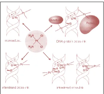
The hydrated active species is a bifunctional electrophilic reagent, therefore able to bind a nucleophilic site present on the DNA strand. The antitumoral function of cisplatin depends on its activation inside the cell as diammineplatinum species, by the dissociation of the two chloride ions and by the formation of inter- and intra-strand crosslinks, respectively on the opposite strands and on the same strand of DNA (Figure 2.2). These crosslinks cause the DNA replication arrest and, as a result, the cell death, if a mechanism of repair does not occur.
28
Figure 2.2: Crosslink processes of the hydrolyzed cisplatin with the DNA
The characteristics of the main DNA platination sites are (Figure 2.2):
1. monofunctional bond with a Guanine (Figure 2.3), defined as monoadduct,
2. bifunctional bond with both a Guanine and a donor atom of a protein, 3. inter-strand crosslink by means of two Guanines,
4. intra-strand crosslink by two adjacent Guanines,
5. intra-strand crosslink by means of two Guanines, separated by a third nitrogenous base,
6. intra-strand crosslink by a Guanine and an adjacent Adenine (Figure
29
Figure 2.3: The Purines Adenine and Guanine represent the nucleophilic binding sites for the
active form of cisplatin
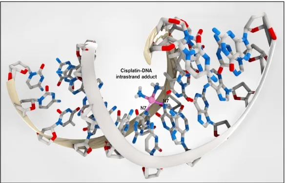
In the case of cisplatin, the type 4 adducts represent 50-60% of metallated sites, the type 5 adducts 20-30%, the type 5 adducts only 10% of the coordinated platinum. As regards the types 1, 2 and 3 adducts, they represent a percentage lower than 4%. Therefore, the main coordination way of cisplatin to DNA is by intra-strand crosslinks with two adjacent Guanines or with a Guanine and an adjacent Adenine (Figure 2.4).
30
Figure 2.4: Intra-strand adduct of cisplatin with DNA, by means of the N-7 of two adjacent
Guanines [8]
The mechanism of cisplatin activation is summed below. In the first phase, it loses the first chloride ligand, which is replaced by a water molecule, and it undergoes the first aquation reaction or “hydrolysis”. Afterwards, the aquacomplex can bind the nitrogen in the 7th position (N-7) of a Guanine. At this stage, the complex undergoes a second “hydrolysis” and bind a second adjacent Purine.
When cisplatin binds DNA, it is possible to assist to a deformation of the hydrogen bonds present between the two DNA helices, which are weaker than the coordination bond, and this leads to a consequent distortion of the platinated helix.
In the final phase, the part of the complex that does not bind to DNA is excreted, and therefore degraded, through the liver: only about 50% of the initially
31 administered cisplatin is eliminated from the organism within 48 hours; the remaining part is excreted in a period that goes from 2 to 8 weeks.
2.4 Second Generation of Platinum Complexes: Features and
Cytotoxic Activity
The administered dose of an antitumoral drug is related to different factors. At first, it is necessary to evaluate the activity of the drug itself and, therefore, the maximum dose tolerated by the organism. Another very important requirement is the solubility of the compound: a solubility of about 1 mg/mL, as in the case of cisplatin, is close to the limit of chemotherapeutic agent parenterally administrable, i.e. by injection.
The increase of the solubility in water of the cisplatin derivatives was the first target for the creation of a second generation of chemotherapeutic drugs.
The most of the platinum complexes containing two chlorides as “leaving groups”, defined “leaving” because they detached before the compound reaches its biochemical target, is usually not much soluble in water. For this reason, it was decided to replace them with a carboxylate anion, preferably a chelating agent, i.e. a dicarboxylate, such as oxalate, glycolate, lactate or 1,1’-cyclobutanedicarboxylate.
The leaving groups of a complex must be coordinated to the metal by means of medium strength bonds. If this does not occur, the ligands weakly retained will lead to the formation of very toxic complexes, whereas those strongly bound will generate inactive compounds. For this reason, the carboxylates are the leaving groups preferred to make longer the half-life times of the resulting complexes.
The oxidation state of the metal is another important factor: in general, the Pt(IV) complexes are less active than those of Pt(II) but are more soluble in
32
water and can be activated only when they are reduced to platinum(II) species by biological reducing agents, such as cysteine, ascorbic acid, reduced glutathione, cytochrome c, etc.
Another fundamental requirement for a cytotoxic complex to be employed consists in being neutral so that it can more easily cross the cell membrane by passive diffusion.
It is possible to summarize the SAR (Structure-Activity Relationship) rules, enunciated in 1973 by Cleare and Hoeschele [9], which should allow a more focused design of new drugs:
the complex must be neutral (in order to avoid interactions with charged species present into the cells and to facilitate the crossing through the cell membrane),
the oxidation state must be +2,
the geometric isomer must be the cis one,
the complex must have two leaving groups with modulable hydrolysis rate (e.g. halides or dicarboxylates) and two carrier groups with modulable steric hindrance (e.g. aliphatic or aromatic N-donors, often chelating agents such as diamines).
For the cancer treatment, two dicarboxylate Pt(II) complexes are worldwide
employed: carboplatin (or
cis-diammine-(1,1’-cyclobutanedicarboxylato)platinum(II), CBDCA) and oxaliplatin (or cis-(1R,2R-cyclohexanediammine)oxalatoplatinum(II)). On the contrary, the approval of other Pt(II) complexes is restricted to some States: in particular, lobaplatin (or (1,2-di(aminomethyl)cyclobutane)lactatoplatinum(II)) in China, nedaplatin (or
diammineglycolatoplatinum(II)) in Japan, and heptaplatin (or
cis-malonato[(4R,5R)]-4,5-bis(aminomethyl)-2-isopropyl-1,3-dioxolane] platinum(II)) in Korea (Figure 2.5).
33
Figure 2.5: Main platinum complexes of II generation
The choice of carboplatin is related to several favorable characteristics that allow to reduce side effects. This compound reacts like cisplatin does: it forms intra-strand and inter-strand crosslinks with the nucleophilic sites of DNA. The replacement of the two chloride ligands has the main function of making carboplatin more resistant (even hundred times) to the hydrolysis reactions and this allows the drug to have a higher life time. In fact, the cyclobutanedicarboxylate ring makes the ligand more stable than the chloride groups in cisplatin: as a consequence, the nephrotoxic potential of the molecule is significantly reduced, due to the decrease of the complex reactivity.
In vivo tests, carried out in order to determine the antitumoral characteristics of
the several derivatives of cisplatin, led to the conclusion that carboplatin gives rise to a series of side effects, such as nephrotoxicity, myelosuppression, vomiting, neurotoxicity but, anyway, it shows a minimal non-haematologic toxicity if compared to cisplatin.
34
The more limited toxicological profile of carboplatin allows to perform studies in which a much higher chemotherapeutic drug dose, than the standard cisplatin dose, was employed.
Pharmacokinetic studies indicate that cisplatin and carboplatin behaviors are different and this difference is clearly correlated to the different chemical stability of the two drugs.
In vitro test with plasma showed that cisplatin rapidly decomposes with a
half-life time of about 2 hours at 37 °C. Carboplatin, instead, is much more stable and has a half-life time of about 30 hours. Therefore, cisplatin is inactivated mainly by the binding to plasma proteins which, after about 4 hours from the intravenous infusion, bind more than 90% of the administered cisplatin. On the contrary, carboplatin binds much less to blood proteins and, after 4 hours from the infusion, only 24% of the total platinum is bound to the proteins.
The drug elimination occurs by glomerular filtration through the kidneys. For a period ranging from 4 to 6 hours after the administration, the carboplatin present in plasma remains intact and, only later, there is the formation of crosslinks between platinum and proteins. Moreover, about 65% of the administered carboplatin dose can be found in the urine within 24 hours, whereas for cisplatin only about 15%: this demonstrates that cisplatin-proteins crosslink can be formed more easily than the one between carboplatin and proteins.
The introduction of carboplatin in clinical trials minimized the necessity of hyper-hydration or the use of antiemetics, and the consequence of this was a decrease of nephrotoxicity, neurotoxicity and ototoxicity. The suppression of the spinal cord activity, however, is its main side effect and this restricts the doses of this drug.
After several studies, it is possible to state that carboplatin is 10-40 times less effective than cisplatin: in fact, in order to obtain tumoral cell growth inhibition
35 effects similar to those of cisplatin, carboplatin requires a 10 times higher concentration.
However, carboplatin inhibits the hemopoietic cells growth as effectively as cisplatin does: therefore, carboplatin is a drug with a higher antiproliferative activity on hemopoietic cells, although the mechanism that explains this higher sensitivity is not completely clear.
Clinically carboplatin can be replaced by cisplatin in the treatment of ovarian and lung cancers but is less effective than cisplatin in genitourinary system, head, neck and esophagus tumors.
Oxaliplatin is a platinum complex different from both cisplatin and carboplatin and it shows no cross-resistance on the Pt-resistant tumoral cell lines. The oxaliplatin toxicity profile, more similar to cisplatin than to carboplatin, includes nausea, vomiting, diarrhea and hematological effects. Alopecia and nephrotoxicity, instead, are not associated with oxaliplatin. Neurological toxicity is the dose-limiting effect. As regards the clinical activity, oxaliplatin is the first platinum complex that shows cytotoxic activity towards colorectal cancer; moreover, it is also effective to the Pt-resistant ovarian tumors.
2.5 Platinum(IV) Prodrugs: Characteristics and Mechanism of
Action
Even though the early SAR rules (see paragraph 2.4) pointed out that Pt(II) complexes are preferred, the antitumoral drugs based on Pt(IV) show several advantages, if compared to their Pt(II) analogues. In particular, they are less reactive and, therefore, they can undergo fewer side reactions, produce fewer side effects and they may be orally administered (because of their stability in biological fluids). Furthermore, they make possible many structural modifications, which are reflected on important physical and chemical
36
properties for the activity of these compounds, such as reduction potential, lipophilicity and, therefore, the cell uptake.
Platinum(IV) compounds have an octahedral geometry and an electronic configuration [Xe] 4f14 5d6 and their higher inertness is due to the fact that they remain intact until their arrival into the cells and, only in the hypoxic and reducing tumor site, they can be activated by biological reducing agents. In particular, the generally accepted intracellular mechanism by which Pt complexes exert their antitumoral effects can be divided into four main phases [10]:
cell membrane crossing and entrance into the cells,
activation, i.e. reduction for Pt(IV) compounds and following aquation reactions of the Pt(II) metabolites,
interaction with the main biological target, i.e. DNA, cell response to the DNA damage.
The axial ligands (L) are released only at a cellular level, where substances such as ascorbate and reduced glutathione (GSH), present in higher concentration than in the extracellular fluid, reduce platinum from +4 to +2 oxidation states. For this reason, Pt(IV) mechanism of action is defined as “activation by reduction” and platinum(IV) compounds are defined as “prodrugs” because they are reduced to the more reactive Pt(II) species. Afterwards, the corresponding Pt(II) complexes, after their aquation reaction, can rapidly bind DNA [11] (Figure 2.6).
37
Figure 2.6: Mechanism of action of Pt(IV) complexes, the “activation by reduction”
(A = carrier ligands, X = leaving ligands, L = axial ligands). The picture is a modification of that reported in literature [11]
The ease with which a complex may be reduced will affect its biological activity and, in order to ensure the activity, the analogue Pt(II) compound must be active [12].
As regards the biological reducing agents, there is a higher concentration of these species into the cells: in particular, ascorbic acid concentration in blood plasma is 50-150 M, whereas in the intracellular site is about 1 mM. The same considerations can be made also for GSH: about 900 M in the extracellular fluid and about 2 mM into the cells [13].
The direct crosslink of the Pt(IV) complex to DNA was observed in vitro in a reductant free environment, but mechanistically these reactions are negligible because the half-life times are much higher than those of the reduction: in fact, it is unlikely that a platinum(IV) compound can resist the large amount of reducing agents present in vivo and arrive intact to the nucleus [14].
38
If the reduction process occurs in the extracellular site, the Pt(II) complex, after the replacement of the leaving groups with two water molecules, binds to proteins and, therefore, it is deactivated.
Although Pt(IV) compounds have been investigated less than those of Pt(II), some of them proved to be promising as antitumoral drugs for clinical trials. The most studied were iproplatin, tetraplatin and JM216 (satraplatin) (Figure 2.7).
Figure 2.7: The most studied Pt(IV) complexes
Iproplatin (JM9 or cis,trans,cis-[PtCl2(ipa)2OH2], where ipa is isopropylamine)
was selected for its high solubility from a series of compounds synthesized by Tobe. It is well tolerated, entered the II and III phases [15] but then was abandoned because it is less active than cisplatin [16].
Tetraplatin (or [PtCl4(DACH)], where DACH is 1R,2R-diminocyclohexane)
demonstrated to be very promising in preclinical studies but it was abandoned in the I phase, due to a severe induced neurotoxicity [17].
JM216 (satraplatin or cis,trans,cis-[Pt(c-C6H11NH2)Cl2(NH3)(OCOCH3)2]) has
recently arrived in the III phase [18]. It was selected because it represents a compromise among the oral activity (similar to the intravenously administered carboplatin in most of the models), few emetic properties (studied on ferrets, since mice do not have any emetic responses) and favorable physicochemical properties, such as a good solubility in water. Furthermore, the complex shows a good stability in acidic solutions, with a half-life time of some hours in 1 M
39 HCl: this fact tends to decrease the risk of possible transformations in the stomach before the absorption.
The fact that none of the clinically tested compounds revealed to be significantly more active than cisplatin was particularly disappointing for researchers, since the in vitro results obtained by Kelland et al. [19] demonstrated that JM216 analogues are up to 840 times more active than cisplatin. This high activity was put in relation with the high cell uptake, but the in vivo reduction alters the pharmacological properties and, therefore, also the efficacy. Probably, the studied drugs are reduced too early in the bloodstream.
2.6 Drug Targeting and Delivery
The Drug Targeting and Delivery (DTD) methods [20] aim at synthesizing drugs selective towards tumoral tissues and administrable at lower doses with fewer side effects and with a high therapeutic index (TI, i.e. the ratio between the average lethal dose (LD50) and the effective dose, which causes a 90%
reduction in tumor mass (ED90)).
The DTD allows to achieve these aims by using vectors able to selectively lead cytotoxic agents to the tumoral cells, thus avoiding damages to the healthy ones, by exploiting the biochemical differences between neoplastic and normal cells/tissues, and limiting drug resistance.
A century ago Paul Ehrlich introduced the concept of drug targeting and it considered a “magic bullet” as an entity composed of two components: the former recognizes and binds the target, whereas the latter provides a therapeutic action. At present, the concept of magic bullet includes a combined action of three components: the drug, the targeting moiety and the pharmaceutical carrier, employed in order to increase the number of drug molecules per single targeting moiety [21].

![Figure 2.9: Main passive DTD vehicles and their general stage of development [22]](https://thumb-eu.123doks.com/thumbv2/123dokorg/4805108.49524/60.774.104.617.436.888/figure-main-passive-dtd-vehicles-general-stage-development.webp)
![Table 2.1: NPs size and their presumed bioactivity in the body [29]](https://thumb-eu.123doks.com/thumbv2/123dokorg/4805108.49524/63.774.120.699.148.970/table-nps-size-presumed-bioactivity-body.webp)