A te che sei stato la luce nel buio,
a te che mi hai accompagnato sempre,
a te che hai mi hai riservato il tuo caldo sorriso quando
ero in difficoltà,
a te che hai gioito per ogni mio attimo di felicità,
a te che hai tenuto stretta la mia mano per sempre,
a te che mi hai lasciato troppo presto,
a te papà
e
Al senso della mia vita…..
ai miei figli Lorenzo e Rodolfo
I
ABSTRACT
1RATIONALE
6Background
121. Human adrenal gland
131.1 The adrenal gland: general structure 13
1.2 Embriology and development 14
1.3 Histology 16
1.4 Adrenalcortical steroidogenesis 17
1.5 The steroidogenic regulatory protein 20
1.5.1 StAR protein 21 1.5.2 P450SCC 21 1.5.3 P450C17 22 1.5.4 P450C21 22 1.5.5 P450C11 - P450C18 23 1.5.6 P450arom: Aromatase 23 1.5.7 Isozymes of 5α-Reductase 23 1.5.8 3βHSD 24 1.5.9 β-steroid-sulfotransferase-sulfatase 24 1.5.10 17-chetosteroid-reductase 24
2. Adrenocortical cancers
25 2.1 Introduction 25 2.2 Adrenocortical adenoma 26 2.3 Adrenocortical carcinoma 26 2.3.1 Treatment options 28 2.3.2 Surgery 29 2.3.3 Mitotane 30 2.3.4 Chemotherapy 31 2.3.5 Radiotherapy 33 2.3.6 Targeted therapy 33 2.3.7 Conclusion 392.4 Cancer cell metabolism and ACC 40
3. Molecular markers involved in adrenocortical tumor cell
proliferation and progression: PELP1, EGR-1 and ERRα
47
3.1 The nuclear receptor coregulator PELP1 (PROLINE-, GLUTAMIC-,AND LEUCINE-RICH PROTEIN 1)
47
3.1.1 Introduction 47
II
3.1.5 Biological functions of PELP1 51
3.1.6 Functions of PELP1 in cancer 58
3.2 The Early growth response-1 (EGR1) 60
3.2.1 Introduction 60
3.2.2 EGR1's discovery and function 61
3.2.3 Biological function and role in tumors 64
3.3 The Estrogen-Related Receptor Alpha 66
3.3.1 Estrogens 66
3.3.2 The role of the estrogen receptor 67
3.3.3 Domain organization of estrogen receptors 70
3.3.4 Estrogen-related receptor alpha 75
3.3.5 Estrogen receptor family DNA-binding domains 72
3.3.6 Estrogen receptor family ligand-binding domains 76
3.3.7 Physiological functions of estrogen-related receptor alpha 81 3.3.7.1 Role of estrogen-related receptor alpha in
metabolism
81 3.3.7.2 Role of estrogen-related receptor alpha in
osteogenesis
82 3.3.7.3 Genes induced by estrogen-related receptor alpha 82 3.3.7.4 Activation of estrogen-related receptor alpha 83
3.3.8 ERRα Agonists 84
3.3.9 Cholesterol: The First Endogenous ERRα Agonist 85
3.3.10 Cholesterol and ERRα in Breast, Prostate, and Adrenocortical Cancer
86
3.3.11 ERRα in invasion, angiogenesis and metastasis 90
4. MATERIALS AND METHODS
934.1 Cell culture and tissues 93
4.2 Protein extraction and Western-blotting 95
4.3 MTT assay and [3H] thymidine incorporation 96
4.4 RNA interference 96
4.5 Immunoprecipitation assay 97
4.6 Microarray 97
4.7 RNA extraction, reverse transcription and real time PCR 97
4.8 Egr-1 gene silencing 98
4.9 ROS detection 99
4.10 Immunofluorescence and immunohistochemistry 99
4.11 Scoring system 100
4.12 Glucose uptake assay 100
4.13 Stable transfection 100
4.14 Trypan blue exclusion test 101
4.15 Colony formation 101
4.16 Wound-Healing Assay 102
4.17 Boyden chamber assay 102
4.18 Spheroids culture 103
4.19 Seahorse XFe96 metabolic flux analysis 104
4.19.1 Mitochondrial Stress Analysis 104
III 5.1 Role of Scaffold Protein Proline-, Glutamic Acid-, and Leucine-Rich
Protein 1 (PELP1) in the Modulation of Adrenocortical Cancer Cell Growth
108
5.1.1 PELP1 is expressed in Human ACC samples and in H295R cells 108 5.1.2 PELP1 is recruited to form a multiprotein complex in H295R cells
after E2 and IGF-II treatment
109 5.1.3 PELP1 knockdown decreases ERK1/2 phosphorylation in H295R
cells
109 5.1.4 PELP1 knockdown decreases IGFIR expression in H295R cells 110 5.1.5 PELP1 knockdown decreases Cyclin D1 expression in H295R cells 111
5.1.6 PELP1 knockdown reduces proliferation of H295R cells 112
5.2 GPER-independent inhibition of adrenocortical cancer growth by G-1 involves ROS/Egr-1/BAX pathway
113 5.2.1 G-1-inducible genes in H295R cells defined by microarray analysis 113
5.2.2 G-1 induces Egr-1 nuclear translocation in H295R cells 115
5.2.3 G-1 induces ROS-dependent Egr-1 upregulation 116
5.2.4 G-1 activates Egr-1/BAX signaling in H295R cells through ERK signaling
118 5.2.5 Egr-1 gene silencing abolishes G-1-mediated effects on H295R cells 119 5.3 Estrogen Related Receptor α (ERRα) a key metabolic factor as target
to prevent Adrenocortical Cancer progression
120
5.3.1 Serum total cholesterol levels in 5 ACC patients 120
5.3.2 ERRα increases H295R cell viability in the presence of cholesterol 121 5.3.3 ERRα overexpression increases in vitro glucose uptake in H295R 122 5.3.4 ERRα modulates H295R cells motility and EMT markers expression 123 5.3.5 ERRα overexpression increases resistance to anoikis in H295R cells 124 5.3.6 Long serial 3D-spheroid culture of H295R cells enhances EMT
markers expression
125 5.3.7 Stable suppression of endogenous ERRα gene expression in H295R
cancer cells
126
5.3.8 ERRα silencing reduces H295R cell growth 127
5.3.9 ERRα knockdown causes a defect in colony formation 128
5.3.10 ERRα knockdown suppress cell migration 129
5.3.11 ERRα knockdown decreases EMT markers 130
5.3.12 Effect of ERRα gene silencing on mitochondrial activity in H295R cells
131
5.3.13 Depletion of ERRα reduces mitochondrial mass 132
5.3.14 ERRα is crucial for H295R cells 3D-spheroid formation and propagation
133 5.3.15 Treatment with the inverse agonist, XCT790, reduces mitochondrial
respiration activity in H295R cells
134 5.3.16 Treatment with the inverse agonist, XCT790, reduces the
mitochondrial mass
IV with XCT790
5.3.19 Metabolic effects induced by Simvastatin treatment 140
5.3.20 Effects on the rate of extracellular acidification induced by treatment with Simvastatin
143
6. DISCUSSION
1451 I Carcinomi surrenalici (ACC) sono tumori rari e altamente aggressivi, associati a una prognosi molto sfavorevole, principalmente a causa di un alto rischio di recidiva e di opzioni terapeutiche limitate (Stojadinovic et al. 2002). L'escissione chirurgica completa offre le migliori possibilità di sopravvivenza a lungo termine, ma abbastanza spesso, nonostante la resezione completa, l’incidenza della recidiva è molto frequente (Glover et al. 2013). L’eziologia del cancro surrenale rimane ancora ignota, ma studi negli ultimi 10 anni suggeriscono che alcune mutazioni genetiche nella ghiandola surrenale sono alla base dell'iniziazione di un tumore maligno (Libe et al. 2007; Soon et al. 2008; Giordano et al. 2009). Tuttavia, l'ACC è una malattia estremamente eterogenea e sta diventando chiaro che la patogenesi dell'ACC determina l’alterazione di diversi segnali cellulari e l’interazione di alcuni pathways. Tra questi, il sistema dell’IGF e le vie estrogeno-dipendenti sembrano essere implicati. L’individuazione di fattori coinvolti negli eventi regolatori di tali pathways cellulari può contribuire a chiarire i meccanismi molecolari alla base delle alterazioni ed a individuare potenziali target terapeutici. Il laboratorio di Biologia Applicata e Cellulare dell’Unical, sta lavorando da alcuni anni per chiarire il coinvolgimento dei pathway IGF dipendenti ed estrogeno dipendenti nell’insorgenza e nella progressione del carcinoma surrenalico.
In questo lavoro di tesi si è voluto approfondire il ruolo di tre fattori coinvolti in questi pathways:
PELP1 (Scaffold Protein Proline-, Glutamic Acid-, and Leucine-Rich Protein 1); EGR-1 (Early growth response gene-1);
ERRα (Estrogen Related Receptor α).
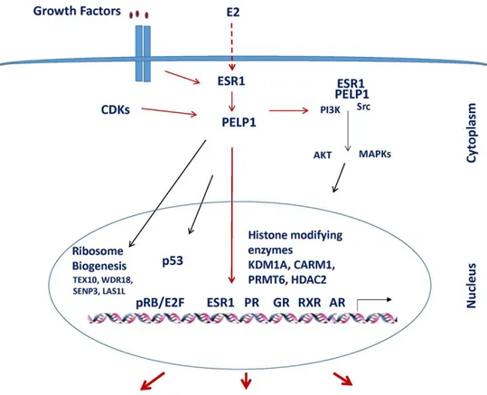
PELP1 agisce come un coattivatore del recettore dell'estrogeno (ER) e quindi esercita un ruolo essenziale nella modulazione delle funzioni di ER. I coregolatori di ER hanno un ruolo fondamentale nella progressione e nella risposta al trattamento ormonale dei tumori estrogeno-dipendenti. In precedenza il laboratorio di Biologia Applicata e Cellulare dell’Unical ha dimostrato che, nel carcinoma adrenocorticale (ACC), ER è up-regolato e l'estradiolo attiva le vie di segnalazione IGF-II/IGF1R definendo il ruolo di questo cross-talk funzionale nella proliferazione delle cellule di ACC, H295R. Lo scopo della prima parte di questo studio è stato determinare se PELP1 è espresso nell’ACC e se possa giocare un ruolo nel promuovere l'interazione tra ER e IGF1R che consente
2 l'attivazione di vie importanti per la crescita delle cellule di ACC. L'espressione di PELP1 è stata rilevata tramite analisi Western blot nei tessuti di ACC e nelle cellule H295R. La riduzione della proliferazione cellulare delle H295R è stata valutata mediante il saggio A3-(4,5-Dimetilthiaoly)-2,5-difeniltetrazolio bromuro (MTT) e mediante l’incorporazione della timidina [3H]. PELP1 è espresso nei tessuti di ACC e nelle cellule H295R. Inoltre, il trattamento delle H295R con E2 o IGF-II ha indotto la una formazione di un complesso multiproteico costituito da PELP1, IGF1R, ER e Src, coinvolto nell'attivazione rapida di ERK1/2. Il silenziamento di PELP1 spegne tale via di segnalazione e riduce la crescita delle cellule H295R indotta da E2 e da IGF-II. L’identificazione del complesso PELP1/ER/IGF1R/c-Src come parte della via di segnalazione E2- e IGF-II-dipendente nell’ACC suggerisce che PELP1 è un nuovo ed efficiente potenziale bersaglio, per ridurre la crescita dell'ACC.
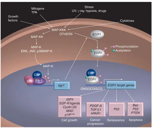
Nella seconda parte di questo lavoro di tesi abbiamo focalizzato la nostra attenzione sul recettore EGR-1, perché in uno studio precedente si è dimostrato che il trattamento della linea cellulare di carcinoma adrenocorticale, H295R con G-1, l’agonista non steroideo, ad alta affinità per GPER (G protein-coupled estrogen receptor 1) ha ridotto la crescita tumorale in vitro ed in vivo attraverso un meccanismo GPER-indipendente. Inoltre, abbiamo osservato che il trattamento con G-1 induce arresto del ciclo cellulare ed apoptosi a seguito di un'attivazione sostenuta di ERK1/2. A partire dai nostri risultati pubblicati, abbiamo eseguito uno studio di microarray che ha chiaramente evidenziato una forte e significativa up-regolazione del gene EGR-1 nelle cellule H295R trattate per 24 ore con la concentrazione 1 micromolare di G-1. I risultati di microarray sono stati confermati dalla RT-PCR e dall’analisi Western-blot, nonché dall’ immunofluorescenza che ha rivelato una significativa colorazione nucleare per EGR-1 dopo il trattamento con G-1. EGR-1 è un punto di convergenza di molte cascate di segnali intracellulari che controllano la crescita e la proliferazione delle cellule tumorali così come altri segnali che riguardano il meccanismo di morte cellulare. Abbiamo rilevato che l'aumento dell'espressione di Egr-1 era una conseguenza dell'attivazione di ERK, ROS-dipendente mediata da G-1, prontamente invertita dalla presenza dell'antiossidante n-acetil-cisteina. Infine, abbiamo osservato che il silenziamento dell'espressione del gene EGR-1 ha invertito gli effetti principali indotti da G-1 nelle cellule di ACC, inclusa la up-regolazione del regolatore negativo del ciclo cellulare, p21Waf1/Cip1 e del regolatore positivo della via apoptotica mitocondriale, BAX, così come l'inibizione della crescita
3 potenziale effetto off-target del G-1 potrebbe essere utile nell'implementazione dell'approccio farmacologico per la terapia dell’ACC.
Nell’ultima parte di questo lavoro di tesi abbiamo concentrato la nostra attenzione sul ruolo dell’ERRα, un target a valle di diversi pathways molecolari coinvolti nella patogenesi dell’ACC, la cui attività può essere modulata da diversi co-regolatori, tra cui il colesterolo. In un recente studio del laboratorio del Prof. Pezzi stati testati gli effetti dell'agonista inverso ERRα, XCT790, sulle H295R. Il trattamento con XCT790 (1-5-10 μM) ha diminuito i livelli della proteina ERRα in modo dose-dipendente causando anche un'inibizione dose-dipendente della crescita cellulare dopo 48-72 ore di trattamento. E’ stato inoltre studiato l’effetto in vivo dell’XCT790 su xenotrapianti di H295R. Topi trattati con XCT790 (2,5 mg / Kg) presentavano una significativa riduzione della crescita del tumore rispetto al gruppo di controllo trattato col veicolo. Inoltre, un aumento dei livelli di colesterolo nel siero è stato riscontrato in uno studio condotto su 152 pazienti con ACC. La maggior parte di questi pazienti sono stati trattati con mitotano, noto per indurre epatotossicità e disregolazione del metabolismo dei lipidi, eventi che potrebbero determinare l'aumento di Chol. Inoltre, i nostri dati sul siero di 28 pazienti con ACC trattati con il mitotano mostrano livelli di Chol superiori ai normali valori fisiologici (248 ± 11 mg / dl, media ± se). L'analisi di questi campioni ha mostrato che i livelli di Chol totali in terapia erano significativamente superiori a quelli prima della terapia (da 167 ± 21 a 229 ± 25 mg / dl). l nostri dati in vitro mostrano che la sovraespressione di ERRα in un modello di cellule tumorali corticosurrenali (H295RERRα) è in grado di aumentare la proliferazione cellulare solo quando il siero conteneva lipoproteine. Questi dati suggerisco che ERRα richiede Chol come un agonista funzionale nelle cellule di ACC. Basandoci su questi dati, abbiasmo ipotizzato che il trattamento a lungo termine con mitotano, aumentando i livelli di Chol, favorisca l'attivazione di ERRα selezionando un fenotipo di ACC più invasivo e metastatico. Questi dati combinati con i nostri risultati suggeriscono che il complesso Chol/ERRα/PCG1α è un target idoneo a controllare la crescita dell'ACC. Noi ipotizziamo che l’ERRα potrebbe essere un "regolatore principale" di riprogrammazione del metabolismo nell’ACC. In effetti, la deplezione di ERRα nelle cellule H295R ha causato una riduzione della massa e della funzione mitocondriale. Inoltre, i nostri risultati dimostrano che la sovraespressione di ERRα aumenta drasticamente
4 l'assorbimento di glucosio nelle cellule H295R, supportando l'ipotesi che ERRα sia coinvolto nell'adattamento metabolico dell'ACC. Inoltre il trattamento delle cellule H295R con XCT790 per 24 ore (un tempo inferiore a quello richiesto per causare la morte cellulare), riduce significativamente la migrazione cellulare, al contrario, la sovraespressione di ERRα aumenta questo processo. Di conseguenza, l'espressione ectopica di ERRα aumenta i livelli dei marker dell’EMT (N-Cadherin, Vimentin, Slug), mentre l’XCT790 blocca la loro up-regulazione. Un'altra osservazione interessante è l'aumento delle cellule anoikis-resisistenti in presenza della sovraespressione di ERRα. Inoltre la sovraespressione di ERRα nelle H295R aumenta la crescita in condizioni di aderenza indipendente, consente la formazione e aumenta il numero di sferoidi 3D (H295RSph). Abbiamo sviluppato H295R come sferoidi per 5 giorni, dissociati e riseminati settimanalmente nel mezzo per gli sferoidi per 5 settimane (H295RSph-5), prima di testare le cellule per la motilità. La migrazione di queste cellule era superiore alle cellule aderenti. Le H295RSph-5 hanno una maggiore espressione di marcatori
mesenchimali come Vimentin, N-cadherin e Slug se confrontati con le H295R coltivate in adesione. Abbiamo valutato gli effetti metabolici indotti dal trattamento con l’agonista inverso del recettore ERRα, XCT790, sulle cellule H295R, tramite l’analisi del tasso di consumo di ossigeno (OCR) e del tasso di acidificazione extracellulare (ECAR). Abbiamo dimostrato che il trattamento con XCT790 influenza profondamente il metabolismo ossidativo e glicolitico delle cellule H295R, infatti è in grado di ridurre il consumo di ossigeno, la capacità glicolitica e la riserva glicolitica, in maniera dose-dipendente. Similmente agli effetti metabolici indotti dall’XCT790, abbiamo osservato che anche le cellule H295R trattate con Simvastatina presentato una funzione mitocondriale ridotta come dimostrato dalla diminuzione dei valori OCR ed ECAR. In conclusione, nel nostro studio abbiamo valutato gli effetti del trattamento con l'agonista inverso di ERRα e del silenziamento stabile di ERRα in un modello sperimentale di carcinoma adrenocorticale. Complessivamente, questi risultati suggeriscono che ERRα svolge un ruolo fondamentale nella transizione epiteliale-mesenchimale e nella resistenza all’anoikis nelle cellule tumorali di ACC. I risultati ottenuti hanno confermato, nel nostro modello sperimentale, il ruolo funzionale di ERRα nell'attività mitocondriale e nel metabolismo ossidativo e glicolitico cellulare, suggerendo che ERRα svolge un ruolo fondamentale nella riprogrammazione
5 terapeutico del carcinoma adrenocorticale.
Il lavoro è stato oggetto di due pubblicazioni scientifiche:
1. CASABURI, IVAN*, AVENA, PAOLA*, DE LUCA, ARIANNA*, SIRIANNI, ROSA, RAGO, VITTORIA, CHIMENTO, ADELE, TROTTA, FRANCESCA, CAMPANA, CARMELA, RAINEY, WILLIAM E., PEZZI, VINCENZO (2017). GPER-independent inhibition of adrenocortical cancer growth by G-1 involves ROS/Egr-1/BAX pathway. ONCOTARGET, vol. 8, p. 115609-115619, ISSN: 1949-2553, doi: 10.18632/oncotarget.23314
*These authors contributed equally to this work.
2. DE LUCA ARIANNA, AVENA PAOLA, SIRIANNI ROSA, CHIMENTO ADELE, FALLO FRANCESCO, PILON CATIA, CASABURI IVAN, PEZZI VINCENZO. (2017). Role of Scaffold Protein Proline-, Glutamic Acid-, and Leucine-Rich Protein 1 (PELP1) in the Modulation of Adrenocortical Cancer Cell Growth. CELLS, vol. 6, ISSN: 2073-4409, doi: 10.3390/cells6040042 Inoltre, questo lavoro ha contribuito alla realizzazione di due REVIEWS:
1. CASABURI IVAN, CHIMENTO ADELE, DE LUCA ARIANNA, NOCITO MARTA, SCULCO SARA, AVENA PAOLA, TROTTA FRANCESCA, RAGO VITTORIA, SIRIANNI ROSA, PEZZI VINCNZO. (2018). Cholesterol as an Endogenous ERRα Agonist: A New Perspective to Cancer Treatment. Front Endocrinol (Lausanne). Sep 11;9:525. doi: 10.3389/fendo.2018.00525. eCollection 2018. Review.
2. CHIMENTO ADELE, CASABURI IVAN, AVENA PAOLA, TROTTA FRANCESCA, DE LUCA ARIANNA, RAGO VITTORIA, PEZZI
VINCENZO, SIRIANNI ROSA. (2018). Cholesterol and Its Metabolites in Tumor Growth: Therapeutic Potential of Statins in Cancer Treatment. Front Endocrinol (Lausanne). 2019 Jan 21;9:807. doi: 10.3389/fendo.2018.00807. eCollection 2018. Review.
6
RATIONALE
It is known that the pathogenesis of adrenal carcinoma is due to an abnormal expression of different genes that can determine the dysregulation of many cellular pathways promoting tumor initiation and progression. The identification of molecule/s involved in the regulatory events of these cellular pathways can help to clarify the altered molecular mechanisms that could also become potential therapeutic targets. The Laboratory of Applied and Cellular Biology of Unical has been working for some years to clarify the involvement of IGF1R and estrogen-dependent pathways in the onset and progression of adrenal carcinoma.
In this thesis work we wanted to investigate the role of three main factors involved in these pathways:
PELP1 (Scaffold Protein Proline-, Glutamic Acid-, and Leucine-Rich Protein 1); EGR-1 (Early growth response gene-1);
ERRα (Estrogen Related Receptor α).
During the first part of the PhD program, it was investigated the role of Scaffold Protein Proline-, Glutamic Acid-, and Leucine-Rich Protein 1 (PELP1) in the modulation of adrenocortical cancer cell growth.
The second part of the program was focused on the involvement of ROS/Egr-1/BAX pathway in independent inhibition of adrenocortical cancer growth by GPER-agonist, G-1.
In the last part of the program it was elucidated the molecular mechanisms activated by the Estrogen Related Receptor α (ERRα), a key cellular metabolic factor, that represent a novel potial target to prevent adrenocortical cancer progression. A part of the final stage of the work was completed at the University of Bari Aldo Moro, Department of Biosciences, Biotechnologies and Biopharmaceuticals (Bari, Italy), where a set of different experiments was performed by using the Seahorse XFe96 Extracellular Flux Analyzer, able to monitor in living cells and, at the same time, cellular oxygen consumption rate (OCR), as a measure of mitochondrial respiration capability, and the extracellular acidification rate, (ECAR), as a measure of glycolytic flux within live cells.
7 with a very poor prognosis, mostly due to a high risk of recurrence and limited therapeutic options (Stojadinovic et al. 2002). Complete surgical excision offers the best chance of long term survival but quite often, despite the complete resection, the tumor reinstates very recurrently (Glover et al. 2013). The cause of adrenal cancer remains elusive, but studies in the past 10 years suggest genetic mutations in the adrenal gland lead to the initiation of a malignant tumor (Libe et al. 2007; Soon et al. 2008; Giordano et al. 2009). However, ACC is an extremely heterogeneous disease and the majority of currently published studies have analyzed only single pathways of signal transduction. It is becoming clear that ACC pathogenesis involves integration of signals and the interplay of downstream pathways. Among these, the IGF system and estrogen-dependent pathways appear to be of particular interest. It has been demonstrated that the insulin-growth factor 2 (IGF-II) gene is strongly over-expressed in adrenocortical carcinomas, representing one of the most commonly identified mutations in ACC (Lafemina and Brennan 2012). IGF-II indicates a reliable prognostic marker in this disease, suggesting that it can be used to identify patients with a high risk of recurrence (Giordano et al. 2003; Samani et al. 2007; Ribeiro and Latronico 2012). In ACC cells, IGF-II induces mitogenic effects through the interaction with the IGF1 receptor (IGF1R), resulting in the activation of the PI3K/AKT/mTOR signaling cascade, as well as RAS/MAPK and the PLC/PKC pathways (Pollak 2008). It is known that estrogens are produced by the enzyme aromatase using androgens as substrate, and we have already shown that ACC is characterized by aromatase overexpression (Barzon et al. 2008). Thus, we can speculate that, in ACC patients, despite the normal circulating estrogen levels, a higher local estrogen production can occur. The classical mechanisms of estrogen action are mainly mediated by two members of the nuclear receptor superfamily, the estrogen receptor (ER) α and β. In ACCs, we previously demonstrated that ERα is upregulated (Barzon et al. 2008) and that estradiol enhances H295R cell proliferation (Montanaro et al. 2005). Recently, the team of Prof. Pezzi demonstrated, both in vitro and in vivo, the potential involvement of G-coupled-estrogen receptor (GPER) in H295R cell growth that was strongly inhibited by GPER agonist, G1 (Chimento et al. 2015). Furthermore, the growth inhibitory effect was also achieved using XCT790, the ERRα inverse agonist (Casaburi et al. 2015). These results suggest that estrogen signaling and the related nuclear receptors are involved in ACC cell growth. Prof. Pezzi’lab also demonstrated that the existence of a functional interplay
8 between the IGF-II/IGF1R axis and the estrogen signaling which turned out to be essential in controlling intracellular pathways crucial for ACC proliferation. In particular, it has been demonstrated that IGF-II caused ERα phosphorylation on serine 118 and serine 167 residues, activating ERα in a ligand-independent manner and increasing cell proliferation. On the other hand, activation of IGF-II/IGF1R pathways could be also triggered by ERα. Accordingly, ERα knock-down was more effective than an IGF1R antibody in controlling H295R cell proliferation (Sirianni et al. 2012). However, the molecular mechanisms involved in IGF-II-induced ERα phosphorylation and in E2/ERα activation of IGF-II/IGF1R-dependent pathways in ACC are not completely clearified.
A large number of studies highlighted that, in cancer cells, ER coregulators play a critical role in hormonal responsiveness and tumor progression (Mishra et al. 2004). PELP1/MNAR is a novel ER coactivator that exerts an essential role in ER’s actions and its expression is deregulated in hormone-driven cancers. PELP1 appears to function as a scaffolding protein, coupling ER with several proteins including growth factors supporting oncogenesis in terms of cell proliferation and metastasis (Vadlamudi et al. 2004; Migliaccio et al. 2005; Vadlamudi et al. 2005b; Manavathi and Kumar 2006; Rajhans and Vadlamudi 2006). These regulatory interactions have important functional implications in the cross-talk of ER and growth factors signaling (Nagpal et al. 2008; Chakravarty et al. 2010b). Taking into account all of these observations, the main purpose of the first part of this study was to define if PELP1 is expressed in ACC and if it is able to play a role in ACC growth by promoting cross-talk between ERα and IGF1R.
Limited therapeutic options are the main features to deal with when addressing adrenocortical cancer (ACC). Mitotane is the drug that is currently used for the treatment of advanced and metastatic ACC (Glover et al. 2013). However, toxicity, narrow therapeutic window and unwanted side effects represent major limitations to its use as well as therapeutic success (Else et al. 2014; Ronchi et al. 2014). As above reported, we found that ERα expression is up-regulated in ACC and estradiol enhances proliferation of H295R cells (Montanaro et al. 2005; Barzon et al. 2008). Moreover, tamoxifen, a selective estrogen receptor modulator (SERM), inhibits estrogen-and IGF-II-stimulated H295R adrenocortical cancer cell proliferation in vitro and reduces H295R xenografts growth (Sirianni et al. 2012). However, in addition to ERα modulation, it has been demonstrated that tamoxifen can act as full agonist on the G protein-coupled
9 3]dioxol-5yl)-3a,4,5,9b-tetrahydro-3H-cyclopenta-[c]quinolin-8-yl]-ethanone), a non-steroidal GPER agonist, has been developed to dissect GPER-mediated estrogen responses from those mediated by ERα and β (Bologa et al. 2006). Since its discovery, G-1 has been used in a large number of studies to investigate the role of GPER in several systems including the nervous, immune, reproductive and vascular systems as well as cancer (Chimento et al. 2013; Chimento et al. 2014; Prossnitz and Barton 2014). It is worth mentioning that the biological activities triggered by G-1-mediated GPER activation, such as cell proliferation (Vivacqua et al. 2006; Albanito et al. 2015) and/or cell death (Chen et al. 2005; Chimento et al. 2013), appear to be cell type specific and dependent on the ERs expression pattern (Ariazi et al. 2010). The picture becomes even more complex considering the effects elicited by G-1 in a GPER-independent manner (Wang et al. 2012). According to a Prof.’s Pezzi team previous study, G-1 is able to inhibit ACC cell growth both in vitro and in vivo (Chimento et al. 2015). In particular, cell cycle arrest and activation of the intrinsic apoptotic pathway were triggered by G-1 via long-term sustained ERK phosphorylation in a GPER-independent fashion. The aim of the second part of this study was to define in the G-1-activated pathways in adrenocortical cancer. Transcription analysis defined the gene expression alternations in H295R cells exposed to G-1, that were here investigated.
The study of De Martino et al. (De Martino et al. 2013) investigated a large cohort of advanced ACC and confirmed the presence of a large number of potentially targetable molecules such as mutation of TP53 and CTNNB1. These studies suggest that ACC is a disease extremely heterogeneous and that ACC pathogenesis involves integration of signals and the interplay of downstream pathways. Consequently, one useful strategy to develop an effective therapy for ACC will be to identify downstream target of multiple pathways. A good target could be the ERRα. ERRα is an orphan member of the superfamily of hormone nuclear receptors and it is expressed in several high energy demanding tissues, including heart, skeletal muscle and brain. In addition to its control of energy metabolism and mitochondrial biogenesis, ERRα has recently been associated with cancer progression in which it requires an elevated cell metabolism (Chang and McDonnell 2012). Notably, increased expression of ERRα has been shown in several cancerous tissues, including breast, ovary and colon (Bernatchez et al. 2013). An association between elevated expression of ERRα and a poor clinical outcome in both breast and ovarian tumors was observed in several independent studies (Ariazi et al.
10 2002; Fujimoto et al. 2007). Several studies suggested that peroxisome proliferator-activated receptor γ coactivator-1 α and β (PGC-1α or PGC-1β) expression level and/or activity could regulate the transcriptional activity of ERRα. The ERRα/PGC-1 complex is a downstream target of multiple signaling pathways in cancer. Several signaling pathways relevant to cancer pathogenesis have been shown to converge upon and regulate the expression and activity of PGC-1α and β, two key ERRα coactivators. It has been shown recently that activation of HER2 and insulin-like growth factor (IGF)-I receptor signaling pathways increase the expression of PGC-1β through induction of c-MYC. ERRα has also been shown to interact with the β-cat/TCF complex and with HIF-1 and reciprocally modulate each other's transcriptional activities to affect cell migration and angiogenesis (Chang and McDonnell 2012). In a recent work (Wei et al. 2016) cholesterol (Chol) has been identified as potential natural ERRα ligand able to increase the recruitment of PGC-1s to ERRα up-regulating its transcriptional activity. This new scenario, characterized by the ability of Chol ability to regulate ERRα/PGC1α complex activity, needs to be investigated particularly in the adrenal gland, a high Chol demanding tissue for steroidogenesis. Moreover, it has been demonstrated in breast cancer that ERRα expression can be regulated by estrogen through ERα and it contributes to regulate aromatase expression (Rajhans et al. 2008). ERRα shares significant sequence homology and structural similarity to ER (Giguere et al. 1988) and recognizes the same responsive elements (Johnston et al. 1997). It was initially considered, therefore, that ERRα might exhibit similar activities as ER and that it would play a role in breast cancer. However, a comprehensive evaluation of the impact of ERRα activation on ERα-dependent transcriptional regulation in MCF-7 breast cancer cells revealed surprisingly few genes that were coregulated by these receptors (Stein et al. 2008). Several studies have reported that ERRα inverse agonist, XCT790, can induce cell growth arrest in different tumors cell lines (Chisamore et al. 2009b; Wang et al. 2010). Few studies have investigated the role of ERRα in adrenal and ACC. ERRα is expressed in normal adult adrenal and regulates the expression of enzymes involved in steroidogenesis (Seely et al. 2005). Moreover, ERRα seems to be more expressed in ACC respect to normal adrenal and adenoma (Felizola et al. 2013). Interestingly, ERRα recognizes the same responsive elements bound by SF-1, a nuclear receptor overexpressed in 90% of ACC (Faria and Almeida 2012). In our recent study (Casaburi et al. 2015) we demonstrated that a dose dependent decrease of ERRα protein content caused also a dose-dependent inhibition of cell growth after 48-72h treatment. We also
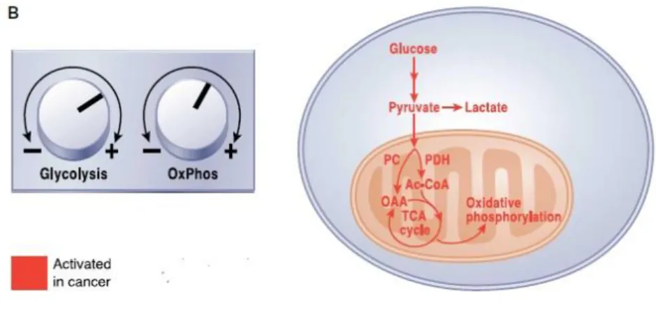
11 XCT790 (2.5 mg/Kg) displayed a significant tumor growth reduction compared to the vehicle treated control group. These data suggest that Chol/ERRα/PCG1α complex is an eligible target to control ACC growth. We hypothesize that ERRα could be a “master regulator”of reprogramming metabolism in ACC. In fact, ERRα depletion in H295R cells caused a reduction of mitochondrial mass and function (Casaburi et al. 2015). Tumor metastasis involves a series of interrelated events including angiogenesis, epithelial to mesenchymal transition (EMT) and invasion. The ability of cancer cells to switch from a predominantly oxidative metabolism to glycolysis, even when oxygen is plentiful, causes lactate production and consequently an acidic environment (Gatenby and Gawlinski 2003). This leads to extracellular matrix degradation by proteolytic enzymes (Lardner 2001) and normal cell death (Williams et al. 1999) enhancing cancer cell migration and invasion. Experimental observations support the hypothesis for a role of ERRα in invasion, angiogenesis and metastasis in several tumors (Deblois et al. 2013). The absence of ERRα is able to impair tumorigenic potential in aggressive xenografted breast cancer cells (Stein et al. 2009). In addition, ERRα/PGC-1α complex binds to the promoter of VEGF regulating its expression, promoting tumor angiogenesis and invasion (Stein et al. 2009).
The aim of the last part of this study is to elucidate the role of ERRα in the reprogramming cellular metabolism associated with ACC cell motility, invasion, angiogenesis and metastases.
12
Background
13
1. Human adrenal gland
1.1 The adrenal gland: general structure
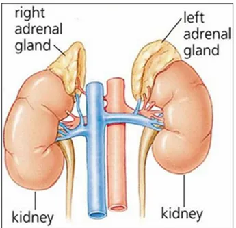
In mammals, the adrenal glands (also known as suprarenal glands) are endocrine glands that sit at the top of the kidneys (Figure 1.1).
Fig. 1.1 Human adrenal gland.
The adrenal glands are located bilaterally in the retroperitoneum superior and slightly medial to the kidneys. In humans, the right adrenal gland is triangular in shape, whereas the left adrenal gland is semilunar in shape; in non-humans, they are quadrilateral in shape. The combined weight of the adrenal glands in an adult human ranges from 7 to 10 grams. They are surrounded by an adipose capsule and renal fascia.
It is now known that the adrenal gland consists of two ontogenetically, structurally and functionally distinct endocrine tissues, the cortex and the medulla. The cortex is mesodermal in origin and derived from proliferation of the coelomic epithelium. It produces various steroids with specific functions as will be described later. The medulla, on the other hand, is ectodermal in origin and neural crest-derived. It secretes
14 catecholamines, i.e., adrenaline and noradrenaline, that facilitate the acute mammalian stress or “fight-or-flight” response.
The adrenal glands affect kidney function through the secretion of aldosterone, and recent data suggest that adrenocortical cells under pathological as well as under physiological conditions show neuroendocrine properties; within normal adrenal glands, this neuroendocrine differentiation seems to be restricted to cells of the zona glomerulosa and might be important for an autocrine regulation of adrenocortical function.
1.2 Embriology and development
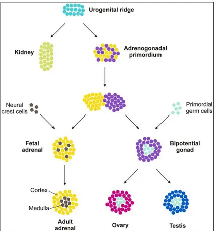
The adrenal gland is two distinct endocrine organs that have separate embryological origins and physiologic functions; the mesoderm-derived cortex secretes steroid hormones while the neural crest-derived medulla secretes catecholamines (Else and Hammer 2005). Formation of the adrenal gland occurs in several distinct developmental events (Else and Hammer 2005; Kim and Hammer 2007) (Figure 1.2). During the 4th week of gestation in humans (E9.0 in mice), proliferation of mesoderm-derived cells of the coelomic epithelia and underlying mesonephros results in coalescence of the adrenogonadal primordium (AGP), defined by expression of the nuclear receptor NR5a1 (Steroidogenic factor 1, Sf1) (Hatano et al., 1996; Luo et al., 1994). At the 8th week of gestation in humans (E10.5 in mice), the bipotential AGP separates into discrete adrenal primordia (fetal adrenal zone) and gonadal primordial (Hatano et al. 1996; Kim and Hammer 2007). The segregation of a discrete adrenal primordia from the AGP involves a Wilm’s tumor 1 (Wt1) and Cited2-mediated upregulation of Sf1 expression (Val et al. 2007). Once separated from the AGP, the adrenal primordial activates Sf1 expression through an entirely different mechanism – the recruitment of the homeobox protein PKNOX1 (Prep1), homeobox gene 9b (Hox) and pre B-cell leukemia transcription factor 1 (Pbx1) to a fetal adrenal-specific Sf1 enhancer (FAdE) (Zubair et al. 2008). Sf1 itself maintains FAdE-dependent expression of Sf1 in the adrenal primordia over time through autoregulation of Sf1 expression. Proliferation of fetal adrenocortical cells is believed to be under control of fetal pituitary-derived adrenocorticotropic homormone (ACTH) (Mesiano et al. 1993). However, insulin like growth factor 2 (IGF2) is expressed throughout the fetal adrenal cortex and several studies have suggested ACTH mediates some of its effects on proliferation through
15 activation of FAdE-driven Sf1 expression at embryonic day E11.5-12.5 in mice (equivalent to 8–9th week of gestation in humans), neural-crest-derived chromaffin
progenitor cells migrate into the central fetal gland. These cells form the adrenal medulla followed by the coalescence of the mesenchymal capsule around the fetal adrenal gland (Else and Hammer 2005). Before encapsulation is complete, the development of the definitive cortex (definitive zone or adult cortex) is initiated between the capsule and the fetal zone. While the fetal cortex ultimately regresses in all species, the timing of regression is species-specific; in humans the fetal zone regression occurs at birth while in mice the zone persists until puberty in males or the first pregnancy in females (Kim et al. 2009). In humans, functional zonation of the adult cortex into unique concentric steroidogenic regions initiates at birth concurrent with the coalescence of the adrenal medulla (Beuschlein et al. 2002).
16
1.3 Histology
The adrenal cortex is composed of three functionally distinct regions, the zona
glomerulosa (ZG) lying immediately below the capsule and corresponding to
approximately 15% of cortical volume characterized by cells organised in rounded clusters around capillary coils or glomeruli, zona fasciculata (ZF) corresponding to up to 75% of cortical volume, characterized by cells arranged in radial rows separated by trabeculae and by blood vessels and zona reticularis (ZR) that lies next to the medulla, in which cells are located within a uniform reticular net of connective tissue and blood vessels (Miller WL, 2008). The ZG synthesizes mineralocorticoids; the ZF produces cortisol and the ZR secretes the so called adrenal androgens, DHEA and DHEA-sulfate. Each zone is preferentially regulated by different circulating factors that include angiotensin II (Ang II) and potassium (K+) for the ZG, adrenocorticotropic hormone (ACTH) for the ZF, and ACTH plus other yet to be determined factors for the ZR (Wang and Rainey 2012) (Figure 1.3). It has been established that the reason each zone secretes a unique set of steroids is related to the selective expression of steroid-metabolizing enzymes within each zone (Rainey 1999; Rainey et al. 2002; Nguyen and Conley 2008). However, the molecular mechanisms that cause zone-specific expression patterns of enzymes are yet to be resolved. Adrenal steroid production remains an area of active research, which supports the need to develop appropriate cell models that can mimic adrenal physiology or pathology. Primary cultures of adrenocortical cells have proven to be useful for examining the mechanisms controlling many aspects of adrenal physiology (Chen et al. 2006; Kuulasmaa et al. 2008; Cardoso 2009; Xing et al. 2010; Xing et al. 2011). However, several issues have limited the use of primary adrenal cells as in vitro models. The most common limitations are the constant requirement for fresh tissue and the difficulties associated with the isolation of adequate cortical cells. In addition, cells from different human donors are subject to considerable variability; whereas cells from rodents do not produce cortisol or adrenal androgens due to the lack of steroid 17ahydroxylase (CYP17)expression. To overcome the problems with tissue accessibility and quality, many groups have attempted to establish cell lines from adrenocortical carcinomas. This approach has been somewhat successful leading to adrenal cell lines from several species and we have previously reviewed the overall development of these models (Mountjoy et al. 1994; Rainey et al. 2004).
17 Fig. 1.3 Adrenal cortex regions.
1.4 Adrenalcortical steroidogenesis
Steroid hormones regulate a wide variety of developmental and physiological processes from fetal life to adulthood. Steroid hormones are all synthesized from cholesterol and hence have closely related structures based on the classic cyclopentanophenanthrene 4-ring structure. The human adrenal can synthesize cholesterol de novo from acetate (Mason and Rainey 1987), but most of its supply of cholesterol comes from plasma low-density lipoproteins (LDLs) derived from dietary cholesterol (Gwynne and Strauss 1982). By contrast, rodent adrenals derive most of their cholesterol from high-density lipoproteins via a receptor termed scavenger receptor B1, but this pathway appears to play a minor role in human steroidogenesis. The intracellular cholesterol economy is largely regulated by the sterol response element binding protein (SREBPs), a group of transcription factors that regulate genes involved in the biosynthesis of cholesterol and fatty acids (Horton et al. 2002). Adequate concentrations of LDL will suppress 3-hydroxy-3-methylglutaryl co-enzyme A reductase, the rate-limiting enzyme in cholesterol synthesis. ACTH also stimulates the activity of 3-hydroxy-3-methylglutaryl co-enzyme A reductase, LDL receptors, and uptake of LDL cholesterol. LDL cholesterol esters are taken up by receptor-mediated endocytosis, and are then stored directly or converted to free cholesterol and used for steroid hormone synthesis (Brown and Goldstein 1979). The first step in steroidogenesis takes place within mitochondria. The mechanisms by which cholesterol is transported to and loaded into the outer mitochondrial membrane (OMM) remain an active area of research (Chang et al. 2006;
18 Miller 2007) the principal action of StAR is to facilitate the movement of cholesterol from the OMM to the inner mitochondrial membrane (IMM). Some cholesterol may be incorporated into vesicular membranes that then fuse with other membranes, thus delivering cholesterol from one intracellular compartment to another, but this appears to be a minor pathway (Soccio and Breslow 2004). Instead, cholesterol is solubilized by binding to proteins. A steroidogenesis abnormality can often be life threatening. Congenital adrenal hyperplasia (CAH) is one of the most common disorders caused by deficiency of any enzyme involved in steroidogenesis in adrenal glands (Claahsen-van der Grinten et al. 2011; White and Bachega 2012), Impaired cortisol and aldosterone production increases adrenocorticotropic hormone (ACTH) secretion from the pituitary gland, leading to adrenal hyperplasia and accumulation of adrenal androgens. Female patients are prenatally virilized because of excess androgen and neonates of both genders may suffer from a life-threatening Addisonian crisis. Steroid hormone deficiency also occurs in aging people by hypogonadism. Most enzymes involved in steroid biosynthesis are either cytochrome P450s (CYPs) or HSDs. These steroidogenic enzymes are functionally, if not absolutely, unidirectional, so the accumulation of products does not drive flux back to the precursor. All P450-mediated hydroxylations and carbon-carbon bond cleavage reactions are mechanistically and physiologically irreversible (Hall et al. 1986) (Figure 1.4). Cytochrome P450 is a generic term for a group of oxidative enzymes, all of which have about 500 amino acids and contain a single heme group (Gonzalez 1988). The human genome includes genes for 57 cytochrome P450 enzymes (Lander et al. 2001; Venter et al. 2001). The genes are now formally termed CYP genes. Seven human cytochrome P450 enzymes are targeted to the mitochondria and are termed “type 1”; the other 50 human P450 enzymes are targeted to the endoplasmic reticulum and are termed “type 2.” All P450 enzymes activate molecular oxygen using their heme center and add electrons from the reduced form of nicotinamide adenine dinucleotide phosphate (NADPH). Type 1 enzymes receive electrons from NADPH via a flavoprotein termed ferredoxin reductase and a small iron-sulfur protein termed ferredoxin, whereas type 2 P450 enzymes receive electrons from NADPH via a single 2-flavin protein termed P450 oxidoreductase (POR) (Miller 2005). Six P450 enzymes are involved in steroidogenesis.
19 Fig. 1.4 Pathway of steroid biosynthesis in adrenal cortex.
Mitochondrial P450scc is the cholesterol side-chain cleavage enzyme catalyzing the series of reactions formerly termed “20,22 desmolase.” The two isozymes of mitochondrial P450c11, P450c11β (11β-hydroxylase) and P450c11AS (aldosterone synthase), catalyze 11β-hydroxylase, 18-hydroxylase, and 18-methyl oxidase activities. In the endoplasmic reticulum, P450c17 catalyzes both 17α-hydroxylase and 17,20-lyase activities, P450c21 catalyzes 21-hydroxylation in the synthesis of both glucocorticoids
20 and mineralocorticoids, and P450arom catalyzes aromatization of androgens to estrogens. The HSDs have molecular masses of about 35 to 45 kDa, do not have heme groups, and require nicotinamide adenine dinucleotide (phosphates) (NADH/NAD+ or NADPH/NADP+) as cofactors; based on their activities, it is physiologically more useful to classify the HSDs as dehydrogenases or reductases. The dehydrogenases use NAD+ as their cofactor to oxidize hydroxysteroids to ketosteroids, and the reductases mainly use NADPH to reduce ketosteroids to hydroxysteroids (Agarwal and Auchus 2005; Sherbet et al. 2007).
1.5 The steroidogenic regulatory protein
Unlike cells that produce polypeptide hormones, which store large amounts of hormone in secretory vesicles ready for rapid release, steroidogenic cells store very little steroid. Thus, a rapid steroidogenic response (e.g., adrenal secretion of aldosterone and cortisol in response to stress or the “pulsing” of sex steroids in response to an LH surge) requires rapid synthesis of new steroid. ACTH promotes adrenal steroidogenic cell growth. This growth occurs primarily by ACTH stimulating the production of cAMP, which in turn promotes the synthesis of IGF-II (Mesiano et al., 1993; Voutilainen and Miller, 1987), basic fibroblast growth factor (Mesiano et al. 1991), and epidermal growth factor (Coulter et al., 1996). Together, these growth factors stimulate adrenal cellular hypertrophy and hyperplasia, determining the amount of steroidogenic tissue. Second, acting over days, ACTH acts through cAMP, and angiotensin II acts through the calcium/calmodulin pathway to promote the transcription of genes encoding various steroidogenic enzymes and electron-donating cofactor proteins, thus determining the amount of steroidogenic machinery in the cell. Third, ACTH rapidly stimulates StAR gene transcription (Stocco et al., 2005) and phosphorylation of Ser195 in extant StAR (Arakane et al., 1997) to increase the flow of cholesterol from the OMM to the IMM, where it becomes substrate for the first and rate-limiting enzyme, P450scc. This acute response occurs within minutes and is inhibited by inhibitors of protein synthesis (e.g., puromycin or cycloheximide), indicating that a short-lived protein species mediates this process. All microsomal (type 2) cytochrome P450 enzymes, including steroidogenic P450c17, P450c21, and P450aro, receive electrons from POR, a membrane-bound flavoprotein that is a different protein from the mitochondrial flavoprotein, ferredoxin reductase (Miller, 2005). Nuclear magnetic resonance and x-ray scattering data have
21 changes while receiving and then transferring electrons (Ellis et al., 2009).
1.5.1 StAR protein
For steroidogenesis, free cholesterol transport across mitochondrial membranes into the mitochondria is facilitated by the steroidogenic acute regulatory protein (STAR). The role of this protein has been well demonstrated in patients with mutations in the gene encoding STAR in the disorder termed congenital lipoid adrenal hyperplasia wherein the mitochondria from the adrenals and gonads of these patients are unable to convert cholesterol to pregnenolone (Lin et al. 1995). It has been suggested that the protein StAR put directly in contact, in the mitochondria, the inner membrane with the external one, allowing the passage of cholesterol, according to the concentration gradient (Bose et al. 1999).
1.5.2 P450SCC
The initial and rate-limiting step in the pathway leading from cholesterol to steroid hormones is the cleavage of the side chain of cholesterol to yield pregnenolone. This step is catalysed by the inner mitochondrial membrane bound cholesterol side chain cleavage enzyme (P450scc, CYP11A1) (Morohashi et al. 1987), and involves three distinct chemical reactions: 20α-hydroxylation, 22-hydroxylation, and scission of the cholesterol side-chain to yield pregnenolone and isocaproic acid (Lambeth and Pember 1983). In human adrenal gene transcription is regulated by ACTH, by gonadotropins in testis and ovary and by unknown factcors in placenta all activated through cAMP as intracellular second messenger (Kimura and Suzuki 1967). Each catalytic cycle requires a molecule of NADPH and one molecule of oxygen (Figure 1.5).
22 Fig. 1.5 Electron transport to mitochondrial forms of cytochrome P450.
1.5.3 P450C17
P450C17 is the steroidogenesis qualitative regulator. It presents both 17--hydroxylase
and C-17, 20-lyasic activities and represents a strategic point in the synthesis of steroid hormones as it can direct pregnenolone toward mineralocorticoids, glucocorticoids or sex steroids synthesis. Pregnenolone and progesterone can form respectively, 17--hidroxy-pregnenolon (17-OH-Preg) and 17--hydroxyprogesterone (17-OH-Prog) after 17--hydroxylation. These 17-hydroxylated steroids then can be cleaved to give C17/20 DHEA and androstenedione, respectively. When the P450C17 is absent, as in the zona
glomerulosa, the products are C-21 17-deoxy steroids such as aldosterone. When the activity of 17--hydroxylase is present products are C-21 17-hydroxysteroids such as cortisol. Instead, when there are 17--hydroxylase and 17, 20 P450C17 liasica activities
the products are C-19 precursors of sex steroid hormones.
1.5.4 P450C21
Progesterone and 17-OH-Prog, once synthesized, are hydroxylated in position 21 to give rise respectively to DOC (deoxycorticosterone) and 11-deoxycortisol. The P450 reductase P450C21 uses the same used by P450C17 for the transport of electrons from
23 P450C11 and P450C18 are located in the inner mitochondrial membrane. The human genome has two P450 genes located on chromosome 8 between bands q13 and q22 (Kawainoto et al. 1990). These two genes encode P450 proteins that have 93% amino acid sequence identity. P450C11 is encoded by the gene CYP11B1; it is significantly expressed in the fasciculata zone and is the only with 11--hydroxylase activity. The related gene, the P450C18, is encoded by CYP11B2 and expressed, at very low level, only in the zona glomerulosa. The P450C18 has 11--hydroxylase, 18-hydroxylase and 18-oxidase activities (Malee and Mellon 1991). The gene CYP11B1, which encodes for P450C11, required for the synthesis of cortisol, is regulated by ACTH, whereas CYP11B2 gene, which encodes the P450C18 required for the synthesis of aldosterone, is regulated by angiotensin II (Ang II), sodium and potassium.
1.5.6 P450arom: Aromatase
Estrogens are produced by the aromatization of androgens by a complex series of reactions catalyzed by a single microsomal aromatase, P450aro (Grumbach and Auchus, 1999; Simpson et al., 2002; Simpson et al., 1994). This typical cytochrome P450 is encoded by a single gene on chromosome 15q21.1. This gene uses several different promoter sequences, transcriptional start sites, and alternatively chosen first exons to encode aromatase mRNA in different tissues under different hormonal regulation. The
CYP19A1gene for P450aro spans over 75 kb (Mahendroo et al., 1991) and contains five
different transcriptional start sites (Mahendroo et al., 1993) with individual promoters that permit the tissue-specific regulation of its expression in diverse tissues. P450aro is a glycoprotein, but glycosylation per se does not appear to affect activity (Shimozawa et al., 1993). The p450aro oxidative demethylation action of C19 steroids, mainly androstenedione and testosterone, consumes three equivalents of molecular oxygen and NADPH, yielding formic acid and C18-steroids with an aromatic A-ring (Simpson et al., 1994).
1.5.7 Isozymes of 5α-Reductase
The 5α-reductases are important beyond the context of male genital differentiation and androgen action because both isozymes reduce a variety of steroids in degradative pathways. Progesterone, 17OHP, and related C21steroids are excellent substrates for both 5α-reductases, particularly the type 1; cortisol, cortisone, corticosterone, and
24 related compounds are also good substrates (Frederiksen and Wilson, 1971). Such 5α- (and 5β-) reduced steroids may be metabolized further and conjugated for excretion in the urine. Inhibitors of the type 2 enzyme have been developed for the treatment of prostatic hyperplasia and the prevention of its recurrence after surgery(McConnell et al., 1998): finasteride selectively inhibits human 5α-reductase type 2, whereas dutasteride inhibits both isoenzymes. These drugs are approved for treatment of prostatic hyperplasia in the United States.
1.5.8 3HSD
Once formed, pregnenolone can be converted into 17- idrossipregnenolone by P450C17 or in progesterone by 3--hydroxysteroid dehydrogenase 4-5 isomerase, encoded by the HSD3B gene.
This enzyme presents two activities: 3--hydroxysteroid dehydrogenase and isomerase activities. In humans there are at least two forms of HSD3B, encoded by different genes:
- the gene for HSD3B type I (HSD3B1) is expressed in placenta, skin, mammary gland; - the gene for HSD3B type II (HSD3B2) is expressed in adrenal glands and gonads. Both genes are on band p13 of chromosome 1 (Berube et al. 1989).
1.5.9 -steroid-sulfotransferase-sulfatase
The steroid sulfates can be synthesized directly from cholesterol sulfate or may be formed by sulfation of steroids such as DHEA, by means of a cytosolic sulphate transferase leading to DHEA-S, encoded by the gene SULT2A1.
The steroid sulfates can also be converted by hydrolysis in native form using a steroid-sulfatase.
1.5.10 17-chetosteroid-reductase
In adrenal, DHEA can be converted in 5-androstenediol and 4-androsterone in testosterone through 17-chetosteroid-reductase (17-CHSR) activity. 5-androstenediol, testosterone and estradiol can also be converted respectively in DHEA, 4-androstenedione and estrone by the same enzyme, tank to a reversible activity known as 17-- hydroxysteroid dehydrogenase (17--HSD). So this enzyme presents both androgenic and estrogenic 17-CHSR characteristics.
25
2. Adrenocortical cancers
2.1 Introduction
Tumors that originate from the adrenal cortex can be divided into benign adenomas and malignant adenocarcinomas. They differ from other cancers because the cancer may be associated to an endocrine component (Allolio and Fassnacht 2006).
Secreting forms are responsible for the onset of endocrine syndromes which vary depending on the type of hormone produced in excess: • Cushing's syndrome, caused by hypersecretion of cortisol; • Conn's syndrome, caused by aldosterone hypersecretion; • hirsutism and virilization, caused by hypersecretion of androgens.
ACC can be asymptomatic or can present with symptoms of hormone excess or complaints referable to the mass (Brennan 1987; Schulick and Brennan 1999a). Generally ACC present an immature steroidogenesis and almost all of these tumors exhibit hormonal precursor excess but, approximately, 60% of all ACC patients will present with hormone-related signs and symptoms (so-called “functional tumors”) (Schulick and Brennan 1999a; Schulick and Brennan 1999b).
Differential diagnosis between ACA and ACC is of pivotal clinical relevance, as the prognosis and clinical management of benign and malignant ACTs is entirely different. Imaging techniques including computed tomography, magnetic resonance imaging and positron emission tomography with 18F-2-fluoro-2-deoxy-D-glucose (FDG-PET) can be used for assessing malignancy, but none of these techniques are absolutely reliable (Terzolo et al. 2011; Morelli et al. 2013). It is very difficult to establish malignancy in small adrenal tumors and to exclude it in large tumors with the available imaging techniques. Currently used guidelines propose to remove adrenal tumors with a diameter of >6 cm, as they are associated with a risk of malignancy >25% (Aron et al. 2012). Some hormonal features (eg, androgen secretion characteristic for malignant tumors) can also be exploited in diagnosis. Most recent data using urinary steroid hormone metabolomics showed characteristic patterns of steroid secretion and metabolism in ACC samples (Arlt et al. 2011). The histological diagnosis of malignancy is also often difficult (Patalano et al. 2009) and novel markers of
26 malignancy are intensively searched for using bioinformatics approaches to establish an early and specific differential diagnosis between ACC and ACA.
2.2 Adrenocortical adenoma
It is a benign neoplastic proliferation of adrenocortical cells almost always associated with clinical, histological and instrumental evidences of hyperfunction.
Dimensions are variable depending on the hormone produced:
adenoma with hyperaldosteronism is usually unilateral and of yellowish color, around 1.5 cm of size and non-enveloped;
adenoma with hypercortisolism is unilateral, has dimensions of about 4 cm, is yellow-brown and is encapsulated;
adenoma with virilization is unilateral, has dimensions of about 5 cm, is red-brown and is encapsulated.
In many patients, adrenal adenomas is asymptomatic, except for begning hormones producing tumors. In case of large tumors, patients may have symptoms due to the compression of other organs, such as feeling of abdominal fullness or localized abdominal pain. More frequent with advancing age, adrenocortical adenomas have a peak between 50 and 70 years and the most affected are women (58%) and the right side.
2.3 Adrenocortical carcinoma
People per year with an increased incidence in the first and fourth-fifth decades of life. By gender, females are the most affected (55-60%) (Else et al. 2014). ACC is burdened by a poor prognosis with a mean year survival rate between 16 and 47%, falling to 5-10% in the advanced stages (Barlaskar and Hammer 2007). The cornerstones in the pathogenesis of ACC are considered to be the genetic alterations of the IGF-2, p53 and β-catenin molecular pathways (Barlaskar and Hammer 2007; Ragazzon et al. 2011). Additionally, other genes, such as ZNFR3, identified by a genome-wide study, appear potentially involved in the tumorigenesis of ACC (Assie et al. 2014b). Comparative genomic hybridization (CGH) demonstrated several complex mutations in ACC with chromosomal gains at 4q, 4p16, 5p15, 5q12-13, 9q34, 12q13, 12q24, 19p and losses at 1p, 2q, 11q, 17p, 22p and 22q. Genes within these regions that are potentially involved
27 dependent kinase 4 (CDK4), and cyclin E1 (CCNE1) (Else et al. 2014). A recent epigenetic study performed on 51 ACCs identified a promoter hypermethylation of the
H19, GOS2, PLAGLI and NDRG2 genes (Else et al. 2014). However, it has been
recently observed that the dysregulation of some miRNAs, such as the upregulation of miR-483 and the downregulation of miR-195 and miR-335, could play a substantial role in the ACC tumorigenesis (Assie et al. 2014a).
When ACC manifests as a condition of steroid hormone excess, the clinical picture is dominated mainly by hypercortisolism and/or hyperandrogenism, whereas symptoms of estrogen hypersecretion such as gynaecomastia and testicular atrophy are pathognomonic in male patients (Fassnacht et al. 2011). DHEA-S represents a possible hormonal marker of ACC, conversely a decreased serum DHEA-S concentration likely indicates an adrenal adenoma (Fassnacht et al. 2004). Mineralocorticoid excess is a rare event, that occurs with severe hypertension and hypokalemia. Notably however, an excess of glucocorticoids could produce a similar effect (Fassnacht et al. 2011). Although few ACC appear non-secreting, they may produce an excessive amount of adrenal precur- mass spectrometry methods revealed that >95% of ACC patients are able to autonomously secrete steroids or steroid precursors (Arlt et al. 2011). Imaging plays a key role in the diagnosis of primary ACC, in the involvement of surrounding tissues and in its spread to distance sites. Either computerized tomography (CT) or magnetic resonance imaging (MRI) exploiting particular features such as Hounsfield unit (HU) values chemical shift imaging, respectively allow adequate diagnostic accuracy to be achieve (Blake et al. 2006; Terzolo et al. 2011) as suggested by a recent analysis of the German ACC registry showing that the value of 13 HU may be considered as the threshold for benign from malignant adrenal masses (Petersenn et al. 2015). More recently, the fluorine 18 fluorodeoxyglucose (18F-FDG) positron emission tomography (PET) or PET/CT was introduced as a diagnostic tool for ACC (Wong et al. 2011). 11C-metomidate, due to its particular ability to bind 11 β-hydroxylase, has been proposed for the identification of tumors of adrenocortical origin (Hennings et al. 2006). The introduction of [123I]IMTO for single photon emission computed tomography (SPECT) and planar scintigraphy has provided a diagnostic alternative to PET for the discrimination of adrenal masses from non-adrenal tissues (Hahner et al. 2013).The first official TNM classification for ACC was established only in 2004 by the International
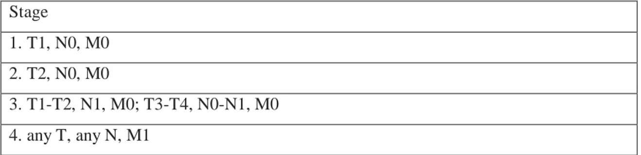
28 Union Against Cancer (UICC) and the World Health Organization (WHO). It was based on the criteria described by MacFarlane (Macfarlane 1958) and later modified by (Sullivan et al. 1978). A significant improvement in the prognostic assessment was due to the adoption of the ENSAT (European Network for the Study of Adrenal Tumors) ACC staging system, which proposes a careful prognostic differentiation among the stages (Fassnacht et al. 2009) (Table I). The use of this system in recent years has greatly improved the diagnostic accuracy and the prediction of survival for stage compared to the criteria previously adopted (Lughezzani et al. 2010).
Stage
1. T1, N0, M0 2. T2, N0, M0
3. T1-T2, N1, M0; T3-T4, N0-N1, M0 4. any T, any N, M1
Table I. Staging system for ACC proposed by the European Network for the Study of Adrenal Tumors (ENSAT).
Classification criteria of the tumor stage according to the TNM system: Stage 1: T1, tumor ≤5 cm; N0, no positive lymph nodes; M0, no distant metastases. Stage 2: T2, tumor >5 cm; N0, no positive lymph nodes; M0, no distant metastases. Stage 3: T1, tumor ≤5 cm - T2, tumor >5 cm; N1, positive lymph node(s); M0, no distant metastases; T3, tumor infiltration into surrounding tissue - T4, tumor invasion into adjacent organs or venous tumor thrombus in vena cava or renal vein. N0, no positive lymph nodes - N1, positive lymph node(s). M0, no distant metastases. Stage 4: T1, tumor ≤5 cm - T2, tumor >5 cm; T3, tumor infiltration into surrounding tissue - T4, tumor invasion into adjacent organs or venous tumor thrombus in vena cava or renal vein; N0, no positive lymph nodes - N1, positive lymph node(s). M1, presence of distant metastasis.
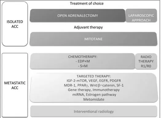
2.3.1 Treatment options
ACC is a neoplastic disease with a poor prognosis. Current studies in this field have indicated the need for a multidisciplinary approach in the management of this tumor (Creemers et al. 2016; Stigliano et al. 2016). Surgery remains the most effective treatment choice for the primary tumor or in for the removal of isolated metastases (Crucitti et al. 1996; Else et al. 2014). The experience that at least one-third of patients show loco-regional recurrence or distant metastases even after a radical surgical excision introduced the concept of adjuvant therapy in these patients (Donatini et al.
29 estimated as ~50% after 5 years (Vaughan 2004). Although these data support the need for an adjuvant cancer therapy, the therapeutic options in ACC currently remain under debate. At present, mitotane represents the only drug approved in Europe and in the United States for ACC treatment; however opinions regarding its use in adjuvant settings are still highly discordant (Huang and Fojo 2008). Currently, chemotherapy is reserved for those cases of advanced disease with evidence of distant metastases unresponsive to mitotane treatment. Many efforts are directed to the development of targeted therapy in ACC. Several strategies have been developed in vitro and some clinical trials have been conducted with small molecules, such as inhibitors of tyrosine kinase receptors or serine/threonine kinase receptors and monoclonal antibodies.
2.3.2 Surgery
Surgery is the only truly effective therapy in the treatment of ACC. A complete surgical resection (R0) is the treatment of choice, avoiding tumor spread that is considered an adverse prognostic factor. The achievement of R0 resection status often requires a radical surgery with a wide dissection of the neighboring organs. It represents a predictor of long-term survival (Icard et al. 2001). The choice of an open approach vs laparoscopic approach is debated. Open adrenalectomy is classically the more secure treatment recommended in patients with localized (stage I-II) and locally advanced (stage III) ACC. Comparative data concerning the two surgical techniques are lacking and originate from retrospective data that involved selection bias (Porpiglia et al. 2010; Lombardi et al. 2012). It is likely that laparoscopic surgery might be reserved only for selected cases with masses of small size. However, these statements must be confirmed by prospective trials. Regardless of the surgical option chosen, the surgical team must have proven experience in the oncologic ACC surgery. Although lymphadenectomy has never been considered as a standard procedure in the adrenalectomy, recent studies show that lymph nodes dissection is significantly associated with a reduction of the relapse rate in patients with localized disease (Gaujoux and Brennan 2012; Reibetanz et al. 2012). However, confirmatory data are needed in order to standardize the surgical procedure. The therapeutic option of removing metastases is founded on the observation that their excision is associated with longterm survival (Kemp et al. 2011; Ripley et al. 2011) and the consideration that many ACC are metastatic at the onset (Stojadinovic et al. 2002). Encouraging results from several retrospective studies show that the