CASE REPORT
Liver Biopsy Discloses a New Apolipoprotein A-I Hereditary
Amyloidosis in Several Unrelated Italian Families
LAURA OBICI,* GIOVANNI PALLADINI,*,‡ SOFIA GIORGETTI,*,§ VITTORIO BELLOTTI,*,§
GINA GREGORINI,¶ ELOISA ARBUSTINI,㛳 LAURA VERGA,㛳 SABRINA MARCIANO,* SIMONA DONADEI,* VITTORIO PERFETTI,‡ LAURA CALABRESI,# CESARE BERGONZI,** FRANCESCO SCOLARI,¶ and GIAMPAOLO MERLINI*,§
*Biotechnology Research Laboratories,‡Department of Internal Medicine, and㛳Institute of Human Pathology, IRCCS Policlinico San Matteo, Pavia;§Department of Biochemistry, University of Pavia, Pavia;¶Nephrology Unit, Spedali Civili, Brescia;#E. Grossi Paoletti Centre,
Department of Pharmacological Sciences, University of Milan, Milan; and **Haematology Unit, Ospedale Civile, Cremona, Italy
Background & Aims: Hereditary systemic amyloidoses
are autosomal dominant, late-onset disorders caused by mutations in the genes for a group of plasma proteins including transthyretin, lysozyme, fibrinogen A␣ chain, gelsolin, apolipoprotein A-I, and apolipoprotein A-II. We investigated both phenotypic and genotypic aspects of apolipoprotein A-I amyloidosis unexpectedly disclosed by liver biopsy in 13 unrelated individuals with asymp-tomatic, persistent elevation of alkaline phosphatase and ␥-glutamyltransferase levels. Methods: Immuno-electron microscopy was used for in situ characteriza-tion of amyloid deposits on liver biopsy specimens. Mu-tation analysis was performed by sequencing of the apolipoprotein A-I gene in all patients. Wild-type/variant apolipoprotein A-I ratio in plasma high-density lipopro-teins was assessed by a peptide mass fingerprinting approach after purification of total apolipoprotein A-I of 2 patients. Results: Family history was informative in 5 cases. Renal failure developed in 9 cases. Hypogonad-ism due to testicular involvement was observed. Amy-loid fibrils specifically stained with anti–apolipoprotein A-I antibody. A novel (Leu75Pro) heterozygous mutation in the apolipoprotein A-I gene was present in affected individuals but not in controls. Variant apolipoprotein A-I was about 10% of the total protein in high-density li-poproteins. Conclusions: The high number of individuals with apparently sporadic disease might reflect wide-spread occurrence of this mutation in the population and a milder phenotype of this variant compared with other apolipoprotein A-I amyloidogenic mutants. These findings suggest that specific staining for amyloid should be performed on liver biopsy of individuals with asymptomatic chronic elevation of alkaline phospha-tase and␥-glutamyltransferase levels.
A
myloidosis is a heterogeneous group of diseases characterized by the pathologic aggregation of pro-teins into insoluble fibrils that accumulatepredomi-nantly extracellularly in tissues and organs, causing dam-age and eventually death. Systemic forms of amyloidosis include hereditary amyloidoses, a group of autosomal dominant, late-onset disorders caused by variant trans-thyretin, apolipoprotein (apo) A-I, apoA-II, lysozyme, fibrinogen A␣ chain, cystatin C, and gelsolin.1
ApoA-I hereditary amyloidosis (AApoAI, MIM 107680) is a rare disease characterized by progressive deposition of amyloid fibrils mainly constituted by N-terminal polypeptide fragments of this protein. Ten amyloidogenic apoA-I variants have been identified thus far,2,3 and most of these are private. Amyloid deposits predominantly affect the kidneys, heart, and liver, caus-ing either progressive nephropathy or cardiomyopathy and, rarely, hepatopathy. Although an extensive visceral amyloid load is frequently observed as a postmortem finding or by means of serum amyloid P component scintigraphy in patients with progressive kidney or heart disease,4,5 deterioration of liver function is seldom ob-served. In a large Spanish kindred with an apoA-I dele-tion/insertion mutant, however, a severe hepatopathy progressing to fatal liver failure was the unique clinical presentation of apoA-I amyloidosis.6In this family, the disease course was characterized by a long-lasting asymp-tomatic phase with only biochemical evidence of liver damage, followed by progressive deterioration of organ function leading to frank cholestasis, portal hyperten-sion, hepatic encephalopathy, and death.6,7
Here we describe a novel apoA-I variant, Leu75Pro, associated with systemic amyloidosis predominantly in-volving the liver and kidneys. The disease was
unexpect-Abbreviation used in this paper: apo, apolipoprotein.
©2004 by the American Gastroenterological Association 0016-5085/04/$30.00
edly diagnosed in 13 unrelated patients who underwent a liver biopsy for chronic liver test abnormalities of unknown origin.
Materials and Methods
Thirteen unrelated patients (8 men and 5 women; age range, 50 –70 years; Table 1) were independently referred to our center for biopsy-proven liver amyloidosis. The biopsy was performed for asymptomatic chronic liver test abnormalities in the absence of a history of drug and alcohol use, specific biochemical markers, and abnormal imaging in 11 patients and for chronic hepatitis C in the remaining 2 patients. Common histologic features included conserved lobular archi-tecture with anisocytosis of the hepatocytes and the presence of enlarged portal areas, filled with variable amounts of an amor-phous, hyaline material that showed green birefringence under polarized light after Congo red staining. Amyloid deposits were localized both in the stromal and perivascular regions and occasionally resulted in bile duct compression. A mild, mono-nuclear inflammatory infiltration without evidence of piece-meal necrosis and a limited periportal fibrosis were also present. All patients underwent complete clinical and labora-tory evaluation.
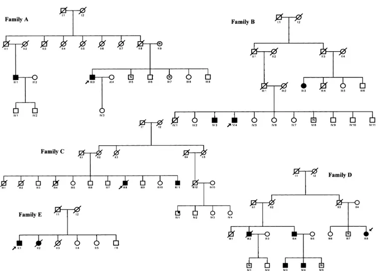
All subjects are of Italian ancestry and originate from the Lombardy region in northern Italy, where our hospital is located. Pedigree analysis showed a significant family history for possible amyloid-related liver or kidney disease in 5 of 13 patients (patients 1–3, 10, and 11). Patient 1 (Figure 1; family A, III:3) was the only known affected member of his family at presentation. Later, his first-degree cousin (III:1) was found to have amyloidosis on liver biopsy performed for chronic hepa-titis C. Their fathers (II:1 and II:8), who were siblings, died in their 80s from apparently unrelated causes. The patient’s mother (II:9) is alive and well and is aged 89 years. Patient 2 (Figure 1; family B, IV:4) had a similarly affected brother
(IV:3). Later, their 76-year-old cousin (III:3) presented with renal failure and amyloid deposits on abdominal fat biopsy. The proband’s father (III:1) and grandfather (II-1) died in their 80s from undefined causes. Patient 3 (Figure 1; family C, III:8) had a brother (III:4) who presented with hepatomegaly, ab-normal liver function tests, and renal failure in his 60s. Amy-loid deposits were shown on liver and adrenal gland biopsy specimens. He died at the age of 76 years from stroke. A younger brother (III:11) developed mild renal failure and hepatopathy at the age of 60 years. A second-degree cousin (IV:1) had amyloid deposits on a testicular biopsy performed at the age of 40 years for gynecomastia and testicular failure, but he was not available for study. The proband’s mother (II:2) died at the age of 61 years from stroke.
Patient 10 (Figure 1; family D, III:8) was retrospectively found to have a first-degree cousin (III:2) with amyloid depos-its documented on a liver biopsy performed at the age of 58 years during cholecystectomy. Mild renal failure had been documented since the age of 53 years. He died at the age of 70 years from apparently unrelated reasons. Later, a younger brother (III:4) and his 2 sons (IV:3 and IV:4) were similarly found to be affected by renal failure of unknown origin.
Revision of medical records showed that patient 11 (Figure 1; family E, II:1) had a sister (II:2) who underwent a liver biopsy for chronic liver test abnormalities and hepatomegaly at the age of 58 years. The biopsy specimen showed the presence of amyloid deposits that were not further characterized. Type 2 diabetes mellitus and mild renal failure were also docu-mented on that occasion. She died at the age of 67 years from undefined reasons.
Histology and Immunohistochemistry
Sections of a formalin-fixed, paraffin-embedded liver biopsy specimen from patient 1 were stained with Congo red and examined under polarized light. Immunoelectron
micros-Table 1. Characteristics of Patients at Presentation
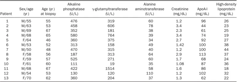
Patient Sex/age (yr ) Age (yr ) at biopsy Alkaline phosphatase (U/L) ␥-glutamyltransferase (U/L) Alanine aminotransferase (U/L) Creatinine (mg/dL) ApoA-I (mg/dL) High-density lipoprotein (mg/dL) 1 M/55 55 476 319 60 1.2 96 26 2 M/63 53 458 606 78 3.4 44 23 3 M/69 67 352 181 38 2.3 61 25 4 M/68 65 580 764 39 3.4 74 19 5 F/64 46 360 375 34 2.7 92 27 6 M/53 52 313 158 49 1.42 100 38 7 M/50 48 470 315 40 1.2 100 44 8 F/58 56 187 313 64 2.9 113 37 9 F/59 57 525 271 60 1.7 68 24 10 F/61 60 111 19 35 1.08 87 36 11 M/68 67 422 1514 58 1.6 86 18 12 M/54 53 130 120 110 1.2 102 32 13 F/70 62 360 204 37 1.3 62 22
NOTE. Reference ranges are as follows: alkaline phosphatase, 60 –260 U/L;␥-glutamyltransferase, 11–53 U/L; alanine aminotransferase, ⬍40 U/L; creatinine,⬍1.2 mg/dL; apoA-I, 110–205 mg/dL in men and 125–215 mg/dL in women; HDL, 55–140 mg/dL in men and 55–125 mg/dL in women.
copy typing of amyloid deposits was performed as described8
using antibodies against and light chains, transthyretin, fibrinogen, lysozyme, serum amyloid P, serum amyloid A, and apoA-I.
Paraffin-embedded liver biopsy specimens from 6 patients were also studied by immunoelectron microscopy. Paraffin-embedded specimens were rehydrated through a series of graded ethanol solutions, fixed in osmium tetraoxide, and re-embedded in Epon araldite resin. Immunohistochemical analysis was then performed as previously described.
Mutation Search in the apoA-I Gene
DNA was obtained using standard procedures from peripheral blood mononuclear cells of 13 patients, 8 affected relatives (family A: III:1; family B: III:3 and IV:3; family C: III:11; family D: III:2, III:4, IV:3, and IV:4), 9 healthy family members, and 100 controls. For affected individual II:2 in family E, genomic DNA was extracted from a paraffin-embed-ded liver biopsy specimen as previously described.9
Mutation detection was performed in all patients on exons and exon-intron boundaries of the apoA-I gene. Amplification
conditions for exons 3 and 4 were as described.9 A single
514 – base pair amplicon containing the first 2 exons and the 5⬘ region of the gene was also obtained using the following pair of primers: 5⬘-AGGGACAGAGCTGATCCTTG-3⬘ and 5⬘-GTGAGAAACCTGCTGCCTCTG-3⬘. Amplification condi-tions consisted of an initial denaturation step (at 95°C for 10 minutes) followed by 30 cycles of denaturation (95°C for 2 minutes), annealing (63°C for 1 minute), and elongation (72°C for 1.5 minutes), with a final step of elongation (72°C for 5 minutes).
Polymerase chain reaction products were sequenced on both strands using the same forward and reverse primers as used in the amplification reactions. Sequence reactions were performed with the Big Dye Terminator Cycle Sequencing kit (Applied Biosystems, Foster City, CA), and the products were analyzed on an ABI PRISM 377 DNA sequencer (Applied Biosystems).
Restriction Analysis of the Leu75Pro Mutation
Restriction fragment length polymorphism analysis with the enzyme HpaII, for which the mutation introduces a
Figure 1. Family trees of 5 kindreds with apoA-I amyloidosis (families A–E). Probands are indicated by arrows. Clinically affected individuals and known carriers of the mutation are indicated by closed symbols. Dead individuals are indicated by a diagonal line through the symbols. Open symbols indicate asymptomatic individuals not tested. Partially filled symbols indicate amyloid confirmed histologically but genetic test not performed.N, tested individuals but no mutation found. See text for detailed clinical information on single members in families.
new restriction site, was performed in patients, their relatives, and 100 controls by digestion of a 392– base pair DNA fragment, corresponding to the 5⬘ of exon 4, previously am-plified for sequencing analysis.
To avoid possible problems expected from DNA fragmen-tation, genomic DNA extracted from paraffin was tested for the presence of the mutation after polymerase chain reaction amplification of a 134 – base pair DNA fragment using the same conditions and the following primers: 5 ⬘-CAGCGT-GACCTCCACCTTC-3⬘ and 5⬘-TGGCCTTCACCTGGTG-GAG-3⬘. The same restriction enzyme HpaII was used on this product to detect the mutation.
Characterization of Wild-Type and Variant apoA-I in Plasma High-Density Lipoprotein
Lipid-free apoA-I was purified from plasma high-den-sity lipoproteins and digested by Staphylococcus V8 protease; separation of peptides and identification by mass spectra anal-ysis was performed by reversed-phase chromatography coupled with an LCQ ion trap spectrometer as previously reported.10
Results
Clinical Features
Patients were symptom-free at presentation. Their clinical characteristics are shown in Table 1. On physical examination, hepatomegaly without spleen en-largement was present in 10 patients. Liver function tests showed increased alkaline phosphatase and/or ␥-glu-tamyltransferase levels, more than twice the upper refer-ence limit, in all but one patient. Aminotransferase levels were within reference range in 6 individuals and showed values less than twice the upper reference limit in 6 patients. In one patient with chronic hepatitis C (patient 12), the alanine aminotransferase level was markedly altered (110 U/L; upper reference limit, ⬍40 U/L). Se-rum bilirubin level, albumin level, and prothrombin time were within reference range in all cases. Serum creatinine level was increased in 9 patients, with concen-trations ⱖ2 mg/dL in 5 cases. In contrast to what is usually observed in amyloid kidney involvement, only 2 patients (patients 2 and 4) had a significant proteinuria (1 g/24 h and 2.43 g/24 h, respectively). No patient had cardiac symptoms, and echocardiography at presentation showed no evidence of heart involvement in all cases. Two patients (patients 6 and 7) had evidence of testicular failure, with low testosterone levels and elevated plasma gonadotropin levels. Patient 6 underwent surgical cor-rection of gynecomastia at the age of 46 years and was then treated with testosterone for 4 years.
A long follow-up since identification of amyloid de-posits was available for patient 5. Liver biopsy was performed at the age of 46 years because of mild
hepa-tomegaly and elevated alkaline phosphatase and ␥-glu-tamyltransferase levels. Eighteen years later, we observed severe hepatomegaly (10 cm below the costal margin), elevation of alkaline phosphatase (360 U/L) and ␥-glu-tamyltransferase (375 U/L) levels, and renal failure (se-rum creatinine level, 2.7 mg/dL).
A mild to moderate reduction of apoA-I and high-density lipoprotein levels, with normal total cholesterol and triglyceride levels, was observed in all patients (Ta-ble 1) and their affected relatives.
Unfortunately, patient 3 suddenly died from stroke at the age of 70 years, 3 years after biopsy and 1 year after diagnosis.
Liver Histology
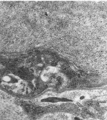
Histologic examination of the liver biopsy speci-men from patient 1 showed conserved lobular architec-ture and the presence of amorphous material in portal areas, presenting typical green birefringence under po-larized light, after Congo red staining. Amyloid deposits corresponded to about 20% of the sample area and were mainly localized at the perivascular level and as nodular aggregates. A mild, mononuclear inflammatory infiltra-tion was also present in portal areas. On electron micros-copy, the liver appeared heavily infiltrated by amyloid fibrils that strongly reacted with anti–apoA-I (Figure 2) and anti–serum amyloid P antibodies but not with the other antibodies tested.
Figure 2. Electron micrograph showing 10-nm immunogold labeling of liver amyloid fibrils with anti–apoA-I antibody.
Immunohistochemical analysis was also performed on 6 paraffin-embedded liver biopsy specimens on which amyloidosis was diagnosed by Congo red staining. Elec-tron microscopic examination of these samples showed the presence of variable amounts of amyloid fibrils spe-cifically immunostained by anti–apoA-I antibody.
DNA Analysis
ApoA-I gene sequencing showed in all patients a T to C transition at position 1772 of the nucleotide sequence, resulting in a proline for leucine substitution at codon 75 of the polypeptide chain. No other sequence variations, apart from at known polymorphic sites, were detected in the gene regions analyzed. All patients and their 9 affected relatives were heterozygous for this mu-tation, as confirmed by restriction fragment length poly-morphism analysis with the enzyme HpaII, for which the mutation introduces a specific cleavage site (Figure 3). No mutation was found among healthy relatives tested. Screening of 100 controls did not show any carrier.
Plasma apoA-I Studies
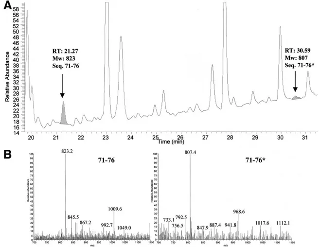
Total apoA-I, purified from 2 patients (patients 7 and 10), was digested by Staphylococcus V8 protease. Peptides were separated by reversed-phase chromatogra-phy. Wild-type and mutant peptides, encompassing codon 75, were eluted at different retention times (Fig-ure 4A) and were recognized by their characteristic mass
Figure 3. Restriction fragment length polymorphism analysis with the enzyme HpaII, for which the mutation creates a new restriction site, shows the presence of 2 additional fragments in patients (lanes 1, 3, 5, and 8) compared with healthy relatives (lanes 2, 4, 6, and 7).M, markerX174 HaeIII.
Figure 4. (A ) Reversed-phase chromatography of V8 protease digestion of apoA-I purified from patient 7. The peptide indicated as 71–76* has Pro in position 75 and peptide 71–76 has Leu in position 75. The 2 integrated areas of the related peaks are shaded in gray. (B) Mass spectrum of peptide 71–76 (eluted at 21.27 minutes) and peptide 71–76* (eluted at 30.59 minutes) shown in A.
spectra (Figure 4B). The wild-type/mutant ratio, quan-tified on the basis of the integrated area of the correspon-dent chromatographic peak, indicates that the variant represents approximately 10% of the total apoA-I.
Discussion
The definition of the pathology underlying asymptomatic, persistent elevation of alkaline phospha-tase and␥-glutamyltransferase levels is a thorny issue in gastroenterology practice. Here we report that, in several patients, this condition was sustained by liver amyloid deposits caused by a new hereditary apoA-I variant.
The presence of amyloid fibrils specifically immuno-reacting with anti–apoA-I antibody in 7 patients and the demonstration that this mutation segregates with the disease strongly support its pathogenic role. Moreover, no other sequence variations in the apoA-I gene were observed.
This is the first apoA-I amyloidogenic variant identi-fied in so many unrelated patients. The absence of a significant family history for the disease in most cases is an unexpected and previously unreported finding, prob-ably related to the relatively milder disease associated with the Leu75Pro variant compared with all other known amyloidogenic apoA-I mutations.
With respect to derangement of liver function tests, the onset takes place in the fifth to sixth decade of life in all patients observed. The disease progresses through a long asymptomatic phase in which the only manifesta-tions are a mild to moderate hepatomegaly and persistent elevation of alkaline phosphatase and ␥-glutamyltrans-ferase levels. This feature is common to all amyloido-genic apoA-I variants associated with massive liver dep-osition of amyloid fibrils4 – 6 and reflects the slow infiltration of portal areas by amyloid deposits without hepatocyte damage, inflammatory infiltration, and fi-brous distortion of the lobular architecture. Compared with the deletion/insertion mutation reported in a large Spanish kindred,6,7 no evidence of progression to liver failure has been observed to date in individuals carrying the Leu75Pro variant, including obligate mutation car-riers in family A (II:1 and II:8) and B (II:1 and III:1).
Two patients had gynecomastia and testicular failure a few years before evidence of liver dysfunction. Although bioptic proof is not available, it is likely that testicular failure is caused by amyloid deposits because localization of amyloid fibrils into the testis has been documented in another apoA-I variant.9 In most patients, a slow gressive increase in serum creatinine levels without pro-teinuria appeared within a few years of liver biopsy. In one patient, in whom a positive liver biopsy specimen
was obtained 18 years before diagnosis, renal function has deteriorated slowly over time, with the serum creat-inine level increasing to 2.7 mg/dL. The lack of protein-uria is a characteristic feature of this type of amyloidosis because, in general, amyloid kidney involvement is as-sociated with significant proteinuria, frequently in the nephrotic range. The structural basis of this peculiar renal dysfunction remains to be elucidated. Further data obtained from kidney biopsy specimens are warranted to assess the presence of amyloid fibrils and their pattern of deposition.
Presently, the lack of tissue samples for chemical characterization of the protein from natural fibrils of patients carrying the Leu75Pro variant does not allow us to characterize the precise size and sequence of the polypeptide that accumulates in the amyloid deposits.
Similar to other apoA-I amyloidogenic mutants,5,6,9 all patients had a mild to moderate decrease in serum apoA-I concentration. Determination of wild-type and mutant species in total circulating apoA-I in 2 patients showed a 10:1 ratio or even less. This severe imbalance is in agreement with previously reported data for other amyloidogenic variants,10 suggesting abnormal metabo-lism of the mutant compared with the wild-type pro-tein.10 –12
Although the role of liver biopsy in the evaluation of abnormal liver enzyme results in asymptomatic patients is still controversial,13–15 in this case it was essential to establish a correct diagnosis, avoid further diagnostic procedures and unnecessary medications, and offer ge-netic counseling. Our findings indicate that a search for amyloid deposits using the Congo red method should be routinely performed in individuals with unexplained chronic elevation of cholestasis indices.
References
1. Westermark P, Benson MD, Buxbaum JN, Cohen AS, Frangione B, Ikeda S, Masters CL, Merlini G, Saraiva MJ, Sipe JD. Amyloid fibril protein nomenclature: 2002. Amyloid 2002;9:197–200. 2. Andreola A, Bellotti V, Giorgetti S, Mangione P, Obici L, Stoppini
M, Torres J, Monzani E, Merlini G, Sunde M. Conformational switching and fibrillogenesis in the amyloidogenic fragment of apolipoprotein A-I. J Biol Chem 2003;278:2444 –2451. 3. Lachmann HJ, Booth DR, Booth SE, Bybee A, Gilbertson JA,
Gillmore JD, Pepys ME, Hawkins PN. Misdiagnosis of hereditary amyloidosis as AL (primary) amyloidosis. N Engl J Med 2002; 346:1786 –1791.
4. Soutar AK, Hawkins PN, Vigushin DM, Tennent GA, Booth SE, Hutton T, Nguyen O, Totty NF, Feest TG, Hsuan JJ, Pepys MB. Apolipoprotein AI mutation Arg 60 causes autosomal dominant amyloidosis. Proc Natl Acad Sci U S A 1992;89:7389 –7393. 5. Persey MR, Booth DR, Booth SE, van Zyl-Smit R, Adam BK,
Fattaar AB, Tennent GA, Hawkins PN, Pepys MB. Hereditary nephropathic systemic amyloidosis caused by a novel variant apolipoprotein AI. Kidney Int 1998;53:276 –281.
JJ, Totty NF, Truong O, Soutar AK, Hawkins PN, Bruguera M, Caballeria J, Sole´ M, Campistol J, Pepys MB. Hereditary hepatic and systemic amyloidosis caused by a new deletion/insertion mutation in apolipoprotein AI gene. J Clin Invest 1996;97:2714 – 2721.
7. Caballeria J, Bruguera M, Sole´ M, Campistol JM, Rode´s J. Hepatic familial amyloidosis caused by a new mutation in the apolipopro-tein AI gene: clinical and pathological features. Am J Gastroen-terol 2001;96:1872–1876.
8. Arbustini E, Morbini P, Verga L, Concardi M, Porcu E, Pilotto A, Zorzoli I, Garini P, Anesi E, Merlini G. Light and electron micros-copy immunohistochemical characterization of amyloid deposits. Amyloid 1997;4:157–170.
9. Obici L, Bellotti V, Mangione P, Stoppini M, Arbustini E, Verga L, Zorzoli I, Anesi E, Zanotti G, Campana C, Vigano` M, Merlini G. The new apolipoprotein A-I variant Leu174Ser causes hereditary car-diac amyloidosis and the amyloid fibrils are constituted by the 93-residue N-terminal polypeptide. Am J Pathol 1999;155:695– 702.
10. Mangione P, Sunde M, Giorgetti S, Stoppini M, Esposito G, Gianelli L, Obici L, Asti L, Andreola A, Viglino P, Merlini G, Bellotti V. Amyloid fibrils derived from the apolipoprotein A1 Leu174Ser variant contain elements of ordered helical structure. Protein Sci 2001;10:187–199.
11. Rader DJ, Gregg RE, Meng MS, Schafer JR, Zech LA, Benson MD, Brewer HB. In vivo metabolism of mutant apolipoprotein apoAIIowa, associated with hypoalphalipoproteinemia and
heredi-tary systemic amyloidosis. J Lipid Res 1992;33:755–763. 12. Genshel J, Haas R, Propsting MJ, Schmidt HHJ. Apolipoprotein A-I
induced amyloidosis. FEBS Lett 1998;430:145–149.
13. Sorbi D, McGill DB, Thistle JL, Therneau TM, Henry J, Lindor KD. An assessment of the role of liver biopsies in asymptomatic patients with chronic liver test abnormalities. Am J Gastroenterol 2000;95:3206 –3210.
14. Pratt DS, Kaplan MM. Evaluation of abnormal liver-enzyme re-sults in asymptomatic patients. N Engl J Med 2000;342:1266 – 1271.
15. Skelly MM, James PD, Ryder SD. Findings on liver biopsy to investigate abnormal liver function tests in the absence of diag-nostic serology. J Hepatol 2001;35:195–199.
Received March 28, 2003. Accepted February 11, 2004.
Address requests for reprints to: Giampaolo Merlini, M.D., Biotech-nology Research Laboratories, IRCCS Policlinico San Matteo, Viale Golgi, 19-27100 Pavia, Italy. e-mail: [email protected]; fax: (39) 0382-502990.
Supported by grants from Ministero della Sanita` (ricerca finalizzata Malattia di Alzheimer cod. 020ALZ00/01), IRCCS Policlinico San Mat-teo, and Telethon-Italia (164-11477).
The authors thank Dr. Luisa Musaio (Department of Medicine, Os-pedali Riuniti, Bergamo, Italy) and Dr. Gian Paolo Balestrieri (Depart-ment of Medicine, Spedali Civili, Brescia, Italy) for referring their patients, Monica Concardi (Institute of Human Pathology, IRCCS Poli-clinico San Matteo, Pavia, Italy) for valuable technical assistance in immunoelectron microscopy studies, and Dr. Regina Tardanico (De-partment of Pathology, Spedali Civili, Brescia, Italy) and Prof. Giovanni Bertoli (Department of Pathology, Ospedale Civile, Cremona, Italy) for providing the liver biopsy specimens.