Benedetta Svezia
Study on the effects of Vitis vinifera L.
miRNAs on the heart following
Life is too short to wake up with regrets. Love the people who treat you right.
Forget about the ones who don’t. Believe everything happens for a reason. If you get a second chance, grab it with both hands.
If it changes your life, let it. Nobody said life would be easy. They just promised it would be worthy it.
1
Table of contents
Abstract ... 2
Chapter 1: Introduction ... 4
Aim and outline of the thesis ... 14
References ... 15
Chapter 2: Materials and Methods ... 22
Bioinformatics approaches ... 23
Biological Samples ... 25
Experimental protocols ... 28
Statistical analysis of data ... 38
References ... 38
Chapter 3: Results ... 41
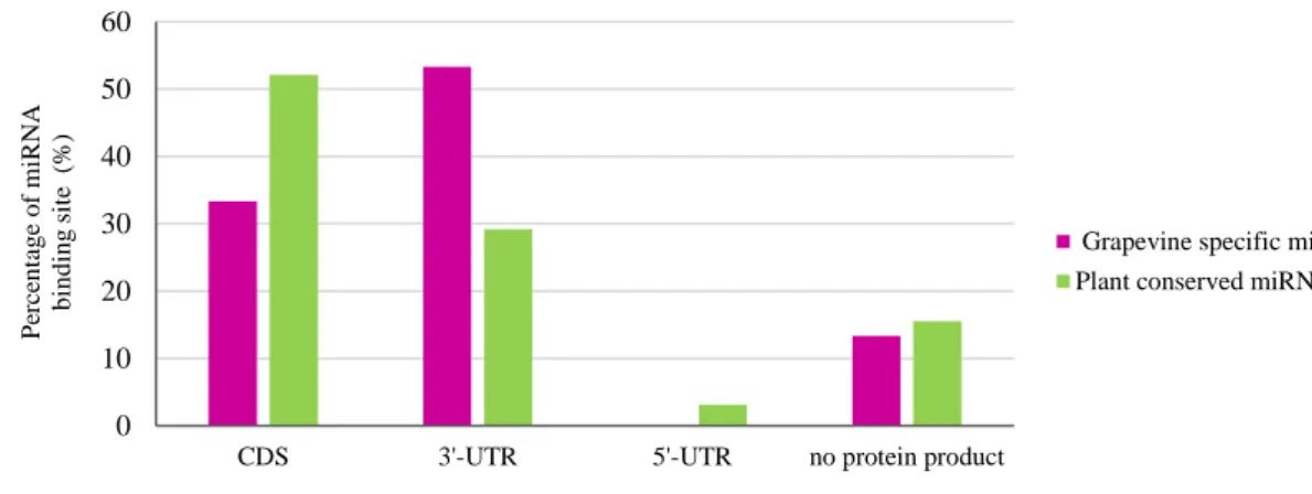
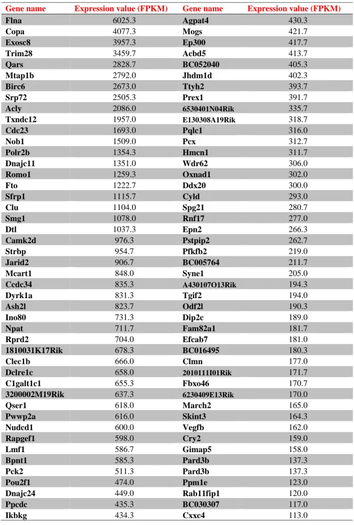
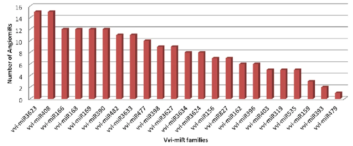
In silico prediction of grapevine miRNAs target genes in Mus musculus ... 42
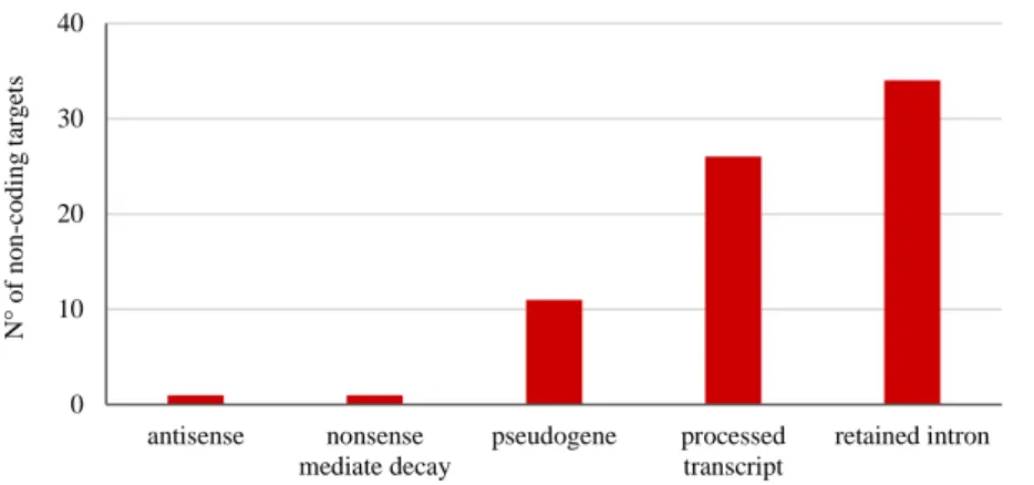
Non-coding RNAs interactions ... 52
Exploring the effect of grapevine small RNAs on MCEC-1 cell line. ... 62
Transcriptomic analysis of MCEC-1 exposed to Sangiovese grape juice. ... 69
Expression profile of the natriuretic peptides system in the heart of mice with myocardial infarction exposed to Sangiovese grape juice. ... 79
References ... 85
Chapter 4: Discussion and Conclusions ... 88
Discussion... 89
Conclusion and future perspective ... 94
2
Abstract
The term Nutraceutical, a portmanteau between the words nutrition and pharmaceutical, was coined in 1989 by Dr. Stephen De Felice, chairman of the Foundation for Innovation in Medicine. Food able to provide natural bioactive compounds with demonstrated physiological effects to support human health are defined as nutraceutical. Few years ago, it emerged that plant-based diet could contribute to maintaining a state of health and wellbeing not only by nutrient supply but also through the horizontal transfer of microRNAs (miRNAs) from plant to mammals (Jiang et al., 2012; Wagner et al., 2015; Yarmarkovich and Hirschi, 2015; Zempleni et al., 2015). MiRNAs are a class of small RNAs that play a key role in post-transcriptional regulation in both plants and animals (Ambros, 2004; Rubio-Somoza and Weigel, 2011; Vidigal and Ventura, 2015). In 2012 a study was published (Zhang et al., 2012a) demonstrating that miRNAs from rice could be absorbed by gastrointestinal tract and were stable in tissue and serum of mammals, including human. The authors also showed that plant miRNAs, after being released in the bloodstream, are able to regulate the expression of mammalian target genes. Thanks to this work, miRNAs ingested with food have been considered as novel bioactive molecules that could play an active role in human health. Due to the contradictories results reported by numerous papers, this attractive topic on cross-kingdom regulation mediate by miRNAs is still highly debated. Recently, several works have shown that miRNAs are abundant in different tissue of grapevine (Vitis vinifera L.), including berries. In light of the studies that confirmed the ability of plant miRNAs to survive in the bloodstream and to modulate mammalian genes (Zhang et al., 2012a; Baier et al., 2014; Zhou et al., 2014), we hypothesized that grapevine miRNAs might contribute to the protective effect of red wine consumption against cardiac disease. In this PhD research project, we studied whether the intake of miRNAs from berries could protect cardiac cells and improve heart performance. Different approaches have been used. First, we evaluated the biological response in an immortalized murine coronary endothelial cell line (MCEC-1) treated with small RNAs extracted from fully mature berries in physiological and ischemic condition through in
vitro assay and transcriptomic analysis. Concurrently we also applied bioinformatics tools to predict
potential targets of grapevine microRNAs in mouse. We evaluated the interaction among different classes of plant and mammalian non-coding RNA. Our results revealed that grapevine small RNAs treatment significantly increased the viability of MCEC-1 cells as compared to control, and this protective effect was present also under oxidative stress. Real-time PCR analysis showed that grapevine miRNAs were detectable in treated MCEC-1 cells.
To investigate the protective effects of Sangiovese grape juice on heart following myocardial infarction, we focused on the natriuretic peptides system. Administration of Sangiovese grape juice increased MCEC-1 viability at rest and under stress condition and, furthermore, reduced the amount
3 of radical oxygen species in stressed cells. Transcriptomic analyses showed a similar trend in C-type
natriuretic peptide (CNP) and endothelial nitric oxide synthase gene expression. Sangiovese
treatment increased the expression of these genes in both physiological condition and oxidative stress. In addition, we investigate whether Sangiovese juice modulate endothelial miRNAs and long non-coding RNAs (lncRNAs).
Concerning the in vivo approach, heart samples from a murine model of myocardial infarction fed with Sangiovese grape juice in addition to standard diet were analyzed. Transcriptomic data showed that Sangiovese grape juice could modulate the natriuretic peptides system following myocardial infarction. In particular, we observed an activation of CNP in contrast to its specific receptor NPR-B;
atrial-natriuretic peptide showed an opposite regulation as compared to the natriuretic peptide receptor A. B-type natriuretic peptide expression increased in infarcted mice exposed to Sangiovese
juice. Histological analysis and heart functional parameters indicated the beneficial effect of Sangiovese grape juice.
KEYWORDS: Vitis vinifera, small RNA, miRNA, nutraceutical, natriuretic peptide system, Sangiovese, C-type natriuretic peptide, myocardial infarction.
4
Chapter 1
5 “Let food be thy medicine and medicine be thy food”. Thanks to this famous quote from Hippocrates, the father of Western medicine, it is immediately clear that the connection between what we eating and human health or disease was already established some 2.500 years ago (Capozzi and Bordoni, 2013). Over the centuries, increasing knowledge on nutrition has changed the concept about food and wellbeing, moving from the prevention and control of nutrient deficiency to focusing on the maintenance of health in order to reduce the risk of chronic diseases. Food or parts of food able to provide health benefits, modulate immunity and prevent or treat disease are defined as nutraceuticals (DeFelice, 1995; Kalra, 2003). The term was coined in 1989 by Dr. Stephen DeFelice, founder and chairman of the Foundation for Innovation in Medicine (FIM), an American organization located in Cranford, New Jersey (Brower, 1998). The nutraceutical term combines the words nutrition and
pharmaceutical, and refers to food that provide natural bioactive compound to support human health,
in addition to their nutritional value (Ganapathy and Bhunia, 2015). The plant kingdom is a natural, rich and diversified source of bioactive substances that are important especially from a medical point of view (Balandrin et al., 1993; Cragg and Newman, 2014; Dias et al., 2012). Epidemiological and nutraceutical studies suggest that a regular and continuing intake of vegetables has a long-term effect in preventing a wide range of disease, thanks to the presence of secondary metabolites with demonstrated physiological benefits (Bhatena and Velasquez, 2002; Kris-Etherton et al., 2002; Liu, 2003; Ramaa et al., 2006; Zhao et al., 2007). For instance, resveratrol from nuts and red grape has strong antioxidant, antithrombotic, anti-inflammatory and anti-carcinogenesis activities (Gülçin, 2010; Gusman et al., 2011); hydroxytyrosol from olives and olives oil is a potent antioxidant (Gordon et al., 2001); lycopene from tomatoes is a carotenoid antioxidant protective again cancers (Martí et al., 2016); isothiocyanates in cruciferous vegetables have anti-carcinogenesis effects (Gupta et al., 2014). Over the last years, an interesting new topic on nutrition has emerged. Plant and animal derived foods seem to be able to provide not only carbohydrates, fats, proteins, vitamins and other molecules, but also genetic material that might influence gene expression in the recipient organism.
Recent studies suggest that this level of gene regulation is mediated by microRNAs (miRNAs) (Zhang et al., 2012a; Baier et al., 2014; Yang et al., 2015a). MiRNAs are a class of single stranded non-coding RNAs that control gene expression at post-transcriptional level by cleaving messenger RNAs (mRNA) or by repressing their translation (He and Hannon, 2004; Petersen et al., 2006; Brodersen et al., 2008; Beauclair et al., 2010). To date it is well established that endogenous miRNAs play a crucial role in almost all biological processes in both plants and animals, including cell differentiation and proliferation, development, metabolism, signalling pathways and diseases (Ambros, 2004; Rubio-Somoza and Weigel, 2011; Vidigal and Ventura, 2015).
Relating to this new level of post-transcriptional gene regulation, an intriguingly paper was published in 2012 when Zhang et al. (2012a) showed that miRNAs derived from plant food assumption could
6 be absorb and survive inside the gastrointestinal system, and were stable in serum and tissues of mammals. Applying high-throughput sequencing technologies, the authors showed that rice (Oryza
sativa) miRNAs including miR168a, miR166a and miR156a, were detectable in the serum of healthy
Chinese men and women. Unlike animal miRNAs, the identified miRNAs were 2’-O-methyl modified (Yu et al., 2005; Yu and Chen 2010) and was not degraded after exposition to sodium periodate, to confirm its plant origin.
More specifically, according to in vitro and in vivo results, plant miRNAs were absorbed by intestinal epithelial cells and packaged into micro-vesicles, which were released in the bloodstream to target mammalian organs. Low-density lipoprotein receptor adapter protein 1 (LDLRAP1) mRNA was identified as a putative target for miR168a, precisely finding a target site into exon 4. LDLRAP1gene encodes for a protein involved in the low-density lipoprotein (LDL) uptake from blood circulation (Gotthardt et al., 2000). Its protein levels were reduced by the exogenous miR168a in an in vitro model cell line. In in vivo studies, mice fed with rice (main source of miR168a) showed an increase of LDL level in plasma due to the reduction of the amount of LDLRAP1 in the liver, caused by
miR168a acquired through rice ingestion. The intravenous injection of an anti-miR168a antisense
oligonucleotide (ASO) during mice feeding reduced miRNA levels and increased LDLRAP1 expression in liver, to confirm the specificity of the inhibitory effect of exogenous plant miR168a on mammalian LDLRAP1.
The available data provided by Zhang et al. (2012a), have revealed a cross-kingdom transfer of plant-derived miRNAs, which are able to regulate gene expression in mammals in a sequence specific manner. Starting from this study, plant miRNAs (or more in general diet- derived miRNAs) ingested with food have been considered as a novel functional compound that could play an active role in human health and might have potential effect on the post-transcriptional regulation of human genes (Jiang et al., 2012; Hirschi et al., 2015; Wagner et al., 2015; Yarmarkovich and Hirschi, 2015; Zempleni, 2015).
Supporting evidence for diet-derived miRNA in mammals
Following Zhang et al.’s pilot study (2012a), several researchers investigated and effectively showed that plant miRNAs acquired by food ingestion could be absorbed by animals and exert biological functions. In this paragraph, the scientific papers concerning the current knowledge about
diet-derived miRNAs are briefly described following a chronological order.
Shortly after the publication of Zhang research group (2012a), Wang et al. (2012a) demonstrated that a significant amount of exogenous small RNAs was detected in human plasma of healthy people and patients with colorectal cancers or ulcerative colitis. In both groups, RNA sequences mapped to several species of the microbiome, such as bacteria, archaea, and fungi were present. The authors
7 were able to identify a high number of sequenced reads related to common dietary plants. Using their bioinformatics pipeline, the exogenous RNA reads corresponded to approximately 12% of the total sequences in plasma samples. Among the food-derived RNAs, the most abundant sequences belonged to Zea mays (maize), followed by Oryza sativa (rice). They also observed RNA molecules from
Solanum lycopersicum (tomato), Glycine max (soybean) and Vitis vinifera (grapevine). The authors
compared their data from Caucasian people with available sequencing data derived from the serum of healthy Chinese individual (SRR332232; Zhang et al., 2012a). Curiously, they found that the sequence abundance between maize and rice was reversed, according to the different diet and this fact indirectly could support the hypothesis that these plant RNAs were acquired from food intake (Wang et al., 2012a). In agreement with Zhang et al.’s work (2012a), this research demonstrated that small RNAs could circulate inside the bloodstream, binding to protein or lipids complex in order to avoid degradation from RNAse (Wang et al., 2012a).
Lukasik and Zielenkiewicz (2014) were focused on breast milk exosomes as a potential reservoir of plant miRNAs derived from the diet. They applied a bioinformatics approach analyzing public datasets derived from small RNA sequencing data in human and porcine (Sus scrofa) breast milk exosomes. In human exosomes 35 plant miRNAs from 25 MIR families were found, whereas in porcine exosomes the authors recognized 17 exogenous plant miRNAs belonging to of 11 MIR families. More specifically, miR166, miR951, miR472 and miR168 were the most represented miRNA families in the human sample, while in porcine exosomes they found plant miRNAs belonging to
miR168, miR156, miR166 and miR319 families. Moreover, the authors selected five plant miRNAs
(miR319b, miR167, miR166a, miR156a, miR444b.2) to predict their potential effects on human gene expression. Combining the results obtained by using three different algorithms (miRanda, PITA and
RNAhybrid) and mapping the common targets of miRNAs predicted on the Kyoto Encyclopedia of
Genes and Genome (KEGG) database, the authors showed that putative targets could have been involved in pathways related to starch, sphingolipids, purine drug and amino acid metabolism and oxidative phosphorylation (Lukasik and Zielenkiewicz, 2014).
Liang and colleagues confirmed the presence of exogenous plant miRNAs in several tissues of mouse after being fed with total RNAs extracted from Brassica oleracea L. (cabbage). The investigated plant specific miR172 was detected along the mouse gastrointestinal tract (stomach, intestine, fecal matter) for 72 hours after feeding, whereas in blood, spleen, liver and kidney the miRNA persisted for 2 hours. The authors also revealed that mature miR172 was more stable in the stomach than the miRNA precursor form (Liang et al., 2014).
Afterwards, the first study on human volunteers was carried out (Liang et al., 2015). After drinking watermelon juice or eating fruits (watermelon, banana, apple, orange, grape, mango, cantaloupe), the
8 researchers could identify 10 selected plant miRNAs in human plasma by using quantitative reverse transcriptase PCR (qRT-PCR) and the results were validated by northern blot.
Similarly to the articles previously cited, also an American research group demonstrated that humans could absorb meaningful amount of miRNAs from nutritionally relevant doses of cow’s milk (Baier et al., 2014). Assuming that a large amount of milk miRNAs was encapsulated in exosome (Howard et al., 2015; Alsaweed et al., 2016), the authors hypothesized that exosomes acted as a cargo able to protect miRNAs from degradation and, consequently, to deliver miRNAs and other molecules to target cells. Orally administered cow’s milk was given to healthy volunteers and milk-based miRNAs (bos tauros-miR-29b and bos tauros-miR-200c) showed a dose-dependent response (Baier et al., 2014). Furthermore, the authors confirmed the effects of milk miRNAs on the expression of human genes in vitro. Emerging studies from the same research group focused on the human absorption of microRNAs containing in boiled chicken eggs. (Zempleni et al., in press).
By employing deep-sequencing approaches, Zhou et al. (2014) showed that a specific small RNA
MIR2911 was particularly abundant in honeysuckle (Lonicera japonica), a plant using in the
traditional Chinese medicine to treat influenza infection mainly as decoction (Shang et al., 2011). When honeysuckle was boiled in water for 30 minutes the authors reveled that, surprisingly, MIR2911 was still highly detectable compare with other small RNAs. According to Zhou et al.’s hypothesis, MIR2911-enrichment honeysuckle decoction could be transferred inside the mammalian organism and acts as anti-viral against influenza. This small RNA appeared to remain stable due to unique sequence 5'-GGCCGGGGGACGGACUGG-GA-3' (Zhou et al., 2014). MIR2911 was previously
reported in miRBase as a miRNA in Popolus euphratica (Li et al., 2012), Nicotiana tabacum (Tang et al., 2012) and Helianthus annuus (Barozai et al., 2012) but currently is considered an atypical miRNA because it derives from 26S ribosomal RNA degradation and does not follow miRNA biogenesis (Yang et al., 2017). High levels of MIR2911 were observed in peripheral blood and lungs of mice after single gavage with honeysuckle decoction or the synthetic miRNA. By luciferase assay and western blot, the authors showed that MIR2911 could block the expression of two key proteins involved in viral replication, polymerase basic protein 2 (PB2) and non-structural protein 1 (NS1), previously identified through bioinformatics approaches. Honeysuckle decoction or synthetic
MIR2911 oral administration reduced the mortality in mice infected with the influenza A virus
subtype H5N1. According to the authors, these data confirmed the ability of some plant miRNAs to pass through the gastrointestinal tract of mammals and be transferred in the circulation to exert their biological activity in mammal’s organs (Zhou et al., 2014).
Based on the data on MIR2911, two studies performed by an American research group (Yang et al., 2015a; Yang et al., 2015b) investigated the presence of this small RNA in biological fluids of animals exposed to controlled diet. In their first article, they reported that MIR2911 was detectable in serum
9 and urine of mice 3 days fed with a chow diet containing honeysuckle. In addition, MIR2911 could remain in the biological fluids for 48 hours after the end of the administration by the diet. The authors also claimed that injured gastrointestinal tract resulting from chemotherapeutic treatment could enhanced the uptake of dietary-miRNAs, by modulating gut cellular structure (Yang et al., 2015a). In the second study, mice were fed with a chow diet supplemented with herbs or flower containing different concentration of MIR2911 (e.g. honeysuckle, sophora, chamomile, blue mellow). Previous data were confirmed and it was found a correlation between the level of MIR2911 in blood and urine samples and its abundance in the various herbs. Although most of miRNAs in blood circulation were usually detected inside cell-derived micro-vesicles and/or associated to RNA-binding proteins, such Argonaute (AGO) (Hunter at al., 2008; Arroyo et al., 2011), MIR2911 was not associated with the RNA-induced silencing complex (RISC). According to the authors, MIR2911 acquired through food ingestion was stabilized in biological fluids without binding with AGO, but perhaps due to its secondary structure (Yang et al., 2015b). Differently, Zhou and collaborators identified the majority of MIR2911 in the micro-vesicle fraction and it was associated with the AGO complex (Zhou et al., 2014).
A recent article reported that exogenous plant-derived miRNAs were detected by deep sequencing analysis in umbilical cord blood and amniotic fluid of healthy Chinese pregnant women (Li et al., 2015). To investigate whether small RNAs could pass through the placenta, a gavage administration of MIR2911 in pregnant mice was performed. After gavage feeding, the level of MIR2911 was elevated in maternal plasma and placenta, but also in the liver of the fetus, suggesting a hypothetical biological effect of dietary-miRNAs on its growth and development.
Mlotshwa and collaborators (2015) carried out an in vivo study to examine the therapeutic role of miRNAs. The ApcMin/+ mouse model of colon cancer was used (Leclerc et al., 2004). Mice were fed with a cocktail of three synthetic tumor suppressor miRNAs (miR-34a, miR-143 and miR-145) with a 2’-O-methyl group on the ribose of the 3’ terminal nucleotide, to mimic miRNAs of plant origin, mixed with purified plant miRNAs isolated from Arabidopsis thaliana. Two negative control groups were fed with total plant RNAs or water. After 28 days of gavage, experimental mice significantly reduced their tumor burden as compared to water- treated mice. In addition, a small tumor burden reduction was observed also in mice fed with total plant RNAs, suggesting a therapeutic effect of plant RNAs in general (Mlotshwa et al., 2015).
Chin and collaborators (2016) recently confirmed the beneficial effect of plant miRNAs against cancer. Plant miR159 was detected in the serum Western donor and miR159 abundance was inversely correlated with breast cancer incidence and progression in patients (Chin et al., 2016). In addition, the authors identified Trascription factor 7 (TCF7) as a putative miR159 target. In vitro, both synthetic miR159 and human serum extracellular vesicles suppressed breast cancer cell proliferation
10 by binding TCF7 3’ untranslated region, confirming that miRNAs absorbed by food could circulate in blood associated with micro-vesicles. A new model mouse for the study of human breast cancer (female NOD-scid IL2Rgnull) was used in vivo experiments (Shultz et al. 2014). Breast tumor was created by injection of MDA-MB-231 cells into the mammary fat pad (Kocatürk and Versteeg 2015). Gavage administration of synthetic miR159 caused a significant reduction of tumor weight in mice, while mice that received scrambled oligonucleotides did not show tumor reduction (Chin et al. 2016). Chen et al. (2016) published an animal study focus on Brassica campestris (rapeseed) bee pollen miRNAs. The authors found 132 reads of plant miRNAs in the serum of mice fed with rapeseed bee pollen. Among plant miRNAs reads, miR166a and miR159 were the most abundant in the mouse serum, which were abundant also in the rapeseed bee pollen. In order to investigate whether plant
miR166a detected in the mice serum derived from pollen absorption, the authors carried out a
comparison between rapeseed bee pollen feeding mice and control mice. Based on RT-qPCR, the data confirmed that the levels of plant miR166a in the were elevated in pollen feeding mice, concluding that mice gastrointestinal tract were able to absorb not only rapeseed bee pollen nutritional substance but also plant miRNAs.
Recently an Italian team studied the immune-modulatory capacity of plant miRNAs (Cavalieri et al., 2016). It was reported that dendritic cells (DC) pre-treated with synthetic strawberry miRNAs (Fragaria vesca) were able to change their capacity to respond to inflammatory stimuli. More specifically, fve-miR168 could bind to Toll receptor 3 (TLR3) on the surface of DC to avoid the activation of the TIR-domain-containing-adapter-inducing interferon-β (TRIF) signaling, which, in normal condition, stimulates the release of inflammatory cytokines and chemokine and regulates immune cells mode of action (Mogensen, 2009). Interestingly, the anti-inflammatory effect was also reproduced by treating DC with other synthetic plant miRNAs (bol-miR874 and osa-miR168 from cabbage and rice respectively) or isolated from plants, before the identification of inflammatory stimuli. On the other hand, DC pre-treated with miRNAs extracted from beef or human culture cells did not show the same results of plant small RNAs treatment. Assuming that in this context plant miRNAs did not act in a sequence specific manner, the authors hypothesized that the immune-modulatory effects could partly be explain by the presence of the 2’-OH-methylation at their 3’ end. The therapeutic potential of plant miRNAs was also confirmed in vivo. Using a multiple sclerosis mouse model (experimental Autoimmune Encephalomyelitis, EAE), the researchers reported that intravenous injection of plant small RNAs (extracted from cabbage leaves, fern fronds and apple peel) at the early stages of the syndrome were able to reduce the severity of the clinical expression of the disease. As a matter of act, plant small RNA-treated mice revealed a significant decrease of pro-inflammatory cytokines compared to control mice and a diminution of spinal cord pro-inflammatory infiltrations. (Cavalieri et al. 2016).
11 The most recent article supporting the evidence for diet-derived miRNA in mammals is that of Luo
et al. (2017). Maize miRNAs were detected in blood and tissue of pigs fed for seven days with fresh
maize. Using an ex vivo experimental protocols (everted gut sac), the researchers showed that maize miRNAs might pass through the gastrointestinal tract in physiological condition and, as previously reported, these exogenous diet-derived miRNAs were packaged into exosomes to avoid RNA degradation and then transferred in the bloodstream. In addition, by duel-luciferase assay was showed a possible cross-kingdom regulation of three putative mammalian targets by oza-miR164a-5p (Luo et al., 2017).
Contradictory evidence for diet-derived miRNAs in mammals
Despite mammalian plant-derived miRNAs uptake concept was investigated and further expanded as for the above mentioned studies, several research groups have reported negative results, including technical criticism of the Zhang et al.’s article (Witwer, 2012; Petrick et al., 2013; Witwer and Hirschi, 2014). Kenneth Witwer, who used the term “xenomiRs” to indicate diet-derived exogenous miRNAs, pointed out that the ten plasma pools used in Zhang et al. (2012a) for investigate global miRNA expression profile derived from healthy donors that showed a high variability in plant miRNA abundance (Witwer, 2012).
Considering that each pool was composed of 10-11 individuals, it has been suggested that not all the donors enrolled in the study might have detectable plant miRNA in plasma. For instance,
osa-miR168a was detected in all pools but the sequence reads varied more than 2,000-fold (Witwer, 2012;
Witwer and Hirschi, 2014). Another critical issue concerned the amount of rice administered to mice. Zhang et al. (2012a) reported that mouse consumed about 7 g of raw rice. According to Petrick et al. (2013), in order to compare dietary regimen between mouse and human a 55 kg Chinese adult would have to eat 33 kg of cooked rice per day (Petrick et al., 2013: Witwer and Hirschi, 2014). Zhang et
al. (2012a) used huge amounts of raw rice that were not reflective of a normal food intake level, as it
has been confirmed by the authors themselves (Zhang et al., 2012a; Petrick et al., 2013). Furthermore, the Chinese population that normally consumed a rice-based diet had low levels of LDL instead of what reported in Zhang et al.’s article about mice feeding study. This element suggests that
osa-miR168a activity would not had any impact on cholesterol-metabolism in human (Witwer, 2012;
Witwer and Hirschi 2014) compare to other molecular mechanism.
Monsanto and miRagen researchers failed to replicate Zhang et al.’s work (Dickinson et al. 2013). In
their study, mice were divided in groups based on three dietary formulations: standard chow, chow supplemented with 41% rice or raw rice diet. At the end of the feeding regimen, small RNAs were sequenced from mouse liver and plasma; however, the authors were unable to demonstrate plant miRNAs uptake in mice, more in particular osa-miR168a. Consistent with the result of Zhang et al.
12 (2012a), mice feeding with row rice had significantly increased plasma LDL level but, on the other hand, this increase was not linked with the reduction of the LDLRAP1, suggesting that plant miRNAs were unable to affect mammalian gene expression. Moreover, increase in plasma LDL was not observed in mice that ate standard chow and chow supplemented with 41% rice. Dickinson et al. (2013) proposed that the increase in LDL levels could be caused by the release of endogenous cholesterol in response to low cholesterol intake in mice fed with only rice. At the end of 2016, Witwer (2016) published a critical paper about packaging and stoichiometry for plant miRNAs uptake by mammals.
Generally, the lack of detectable plant miRNAs absorbed after controlled diet and the possibility of cross-contamination during libraries preparation and RNA sequencing, were two of the many subjects discussed in this highly critical studies.
Researchers from the Monsanto Company (Zhang et al., 2012b) published one of the first contradictory studies. They analyzed available public small RNAs dataset derived from chicken, insect and mammalian biological fluids and tissues (including cell lines) in order to understand how common would be plant miRNAs absorption and to verify the presence of these diet-derived miRNAs in animals. In addition, a controlled insects feeding experiment was carried out. According to authors, 63 public datasets out of the 83 showed at least one read that perfectly matched plant RNA sequences and plant miR168 was the most abundant exogenous small RNA sequence present among all the dataset analyzed, except for the pig abdominal dataset. The authors stated that the overexpression of plant miR168 was unlikely in animals with different digestive anatomy and physiology, and, furthermore, miR168 abundance was low in food compared to other miRNAs. Moreover, identified plant miR168 had a sequence typical of monocot plant species, like Oriza sativa and Sorghum bicolor, but, concerning the feeding experiment, it was detected in insects that were not reared on monocot species. Based on their results, the researchers assumed that plant miRNAs observed in some available small RNA dataset could be artifact derived from sequencing process or due to contamination among samples (Zhang et al., 2012b; Witwer, 2015). Tosar et al. (2014) were focused on cross-contamination as the most probable source of exogenous RNAs, especially during RNA libraries preparation. According to the authors, exogenous miRNAs detected in several human small RNA sequencing dataset were the result of a contamination from the laboratory environment where samples were being processed (Tosar et al., 2014). The authors produced experimental evidence supporting their assumption and, in order to reinforce the concept, the available dataset published by Zhang’s group (2012a) derived from Branchiostoma lanceolatum (lancelet) were analyzed (Chen et al. 2009). Despite lancelets were being fed with algae, Tosar and collaborators identified raw reads corresponding to plant miRNAs that were not expected. They showed that the relative abundance of plant miRNAs detected in lancelet samples was similar to the relative abundance in the libraries from
13 sera of the healthy Chinese donors analyzed in Zhang et al. (2012a), strongly supporting contamination between sample as the likely explanation for the detection of plant miRNAs in human samples (Tosar et al., 2014). Recently, another research group aimed to analyze the presence of exogenous miRNAs in more than 800 public database from human tissues and body fluids (Kang et al., 2016). Using a new computational pipeline to normalize the raw sequences data, they argue that the low abundance of exogenous miRNAs present in humans did not originate from food absorption and, more specifically, tissues and fluids exposed to food such as liver and serum did not show a significant enrichment of diet-derived miRNAs. Actually, the most abundant xenomiRs detected in body fluid samples dataset belonged to insects and rodents that clearly did not originate from food sources, but probably from laboratory animal models. In addition, it was demonstrated through
principal component analysis that body fluid samples from the same studies tend to group together
when clustered by xenomiR compositions, suggesting technical batch effects. To investigate whether diet-derived miRNAs could be transferred in mammalian circulation, two different controlled animal feeding studies were carried out but neither plant miRNAs in rat blood or bovine milk sequences in piglets were detected by high-throughput sequencing technology (Kang et al., 2016).
Several research groups were unable to detect plant miRNAs in body fluids or tissue when conducting controlled feeding studies in insect and mammals. Snow and collaborators (2013) evaluated plant miRNAs absorption in human, mouse and honeybees (Snow et al., 2013). MiR156a, miR159a and
miR169a were selected based on their abundance in fruit normally consumed like apple and banana,
and in honey, nectar and pollen. Plasma of healthy athletes analyzed after routinely fruit intake did not show neither of plant miRNAs selected, suggesting an inability of human to maintain steady-state levels of plant miRNAs in bloodstream circulation. The feeding study was replicated in mice and honeybees obtaining similar negative results. Recently the same research group published a further research on honeybee feeding study (Masood et al. 2016). They confirmed that ingested pollen-derived miRNAs were not detected in tissue of honeybees at biologically relevant concentration, despite the midguts showed high levels of miRNAs after pollen ingestion.
A non-human primate model (Macaca nemestrina, the pig-tailed macaque) was used by Witwer et
al. (2013) to evaluate the presence of plant miRNAs in plasma after feeding by gavage with a
commercially plant-based substance, containing soy and fruits but no animal products. Plant miRNAs level was evaluated in plasma pre- and 1, 4, 12 hours post- gavage through RT-qPCR. In their research, the authors measured plant miR160, miR166, miR168 and miR172 that resulted undetectable in plasma despite their relative abundance in the plant-based substance that was administered to the macaque. Droplet digital PCR (ddPCR) was used to increase the detection of extremely low level of plant miRNAs in plasma. Detection by this method revealed the non-reproducibility of the results, that suggested variable and non-specific plant miRNAs amplification.
14 In their work concerning milk miRNAs absorption, Baier and collaborators evaluated plant miRNAs in plasma samples derived from a broccoli sprout (Brassica oleracea) feeding study in human, in order to analyze plant miRNAs bioavailability (Baier et al., 2014). Healthy men were fed with different doses of broccoli (34 g, 68 g, 102 g) and plasma was collected before and after consumption. Plasma samples were analyzed for the brassica-specific miR824 and plant-specific miR167a by RT-qPCR, but neither was detected.
Also the study performed by Micó et al. (2016) failed to detect plant miRNA in plasma of healthy volunteers after the ingestion of an acute dose (40 ml) of extra virgin olive oil (EVOO). According to the authors, miRNAs from beer and EVOO, two type of food consumed in the Mediterranean area, could be absorbed by human gut and exert beneficial effects on health. However, Micó et al. did not identified miRNAs in beer and olive oil as well as in human plasma.
Aim and outline of the thesis
Based on the literature above mentioned, the emerging theory concerning the ability of diet-derived
miRNAs from plant kingdom to affect gene expression in mammals is highly controversial. To the
best of our knowledge, the putative role of grapevine miRNAs into cardiovascular system has not been investigated yet. Thanks to the completion of Vitis vinifera genome sequence (Jaillon et al., 2007), several researchers began to characterize grapevine miRNAs in order to investigate their role on the molecular regulatory networks underlying plant development and stress responses (Carra et al., 2009; Mica et al., 2010; Pantaleo et al., 2010; Wang et al., 2012b; Han et al., 2014; Ren et al., 2014; Leng et al., 2015; Sun et al., 2015).
By employing next-generation sequencing approach, Belli Kullan et al. (2015) has significantly contributed to describe miRNAs abundance in several tissues of the cultivar Corvina, including berries. Furthermore, Pinto et al. (2016) evaluated small RNAs distribution, by focusing on miRNAs, in grapevine berries of two cultivars (Cabernet Sauvignon and Sangiovese) grown in three different vineyards (Bolgheri, Montalcino and Riccione).
Grapevine products are typical component of the Mediterranean Diet intended for direct consumption and for juice and wine production. According to the definition of nutraceutical food (DeFelice, 1995; Kalra, 2003), grapes contain numerous bioactive components relevant for human health and its consumption is linked to beneficial effects in the prevention of treatment of various diseases (Markoski et al., 2016; Imran et al., 2017). Despite a large amount of published contradictory studies and emerging questions that remain to be clarified as reported above, a number of researchers consider
diet-derived miRNAs as a novel bioactive compounds able to exert their action in mammals (Jiang et
15 could be conceivable that also miRNAs expressed in grapes may play a modulatory role in human physiology. Specifically, the hypothesis we wish to test in this PhD thesis is that miRNAs contained in grapevine berries could be involved the so-called French Paradox, i.e. the protective effects of red wine consumption against cardiovascular disease (Renaud and De Lorgeril, 1992). The research here described is part of a nutraceutical project funded by the Regione Toscana, called CardioMiRSanTo (“Cardioprotezione con MiRNA di Sangiovese Toscano”) that aims at developing a functional grape juice from Vitis vinifera berries cultivar Sangiovese with a high concentration of cardio-protective miRNAs, whose target are patients who have suffered a myocardial infarction, in addition to pharmacological therapy. Using a multimodal approach and integrating different expertise, the present project aims to clarify whether miRNAs from Sangiovese berries can protect cardiac cells and improve heart performance. Taking advantage of small RNA-sequencing data previously produced by our laboratory (Belli Kullan et al., 2015; Pinto et al., 2016), two bioinformatics algorithms were applied in order to identify putative targets of grapevine miRNAs in mammalian organism; the interaction among different classes of plant and mammalian non-coding RNAs are also investigated. An immortalized endothelial cell line was exposed to small RNAs extracted from fully mature
Sangiovese berries and grapevine juice to evaluate cellular response in physiological and ischemic
condition through in vitro assay and transcriptomic analysis. This research includes an in vivo approach using a mouse model (C57BL/6J) to study the protective effect of Sangiovese juice on infarcted heart. More specifically, Real-time PCR analysis was performed to assess the expression profile of the natriuretic peptides family, including atrial, B-type and C-type natriuretic peptides. These cardiac hormones, as well as having diuretic, natriuretic and vasodilator effects (Volpe et al., 2014), act as anti-fibrotic and anti-hypertrophic agents and play a key role to prevent cardiac remodeling after myocardial infarction (Nishikimi et al., 2006; Calvieri et al.,2012; Del Ry et al., 2013).
References
Alsaweed M, Lai CT, Hartmann PE, Geddes DT, Kakulas F (2016). “Human Milk Cells and Lipids Conserve Numerous Known and Novel miRNAs, Some of Which Are Differentially Expressed during Lactation”. Plos One 11(4): e0152610.
Ambros V (2004). “The functions of animal microRNAs”. Nature 431:350-355.
AR Chin, MY Fong, G Somlo, J Wu, P Swiderski, X Wu, … Wang SE (2016). “Cross-Kingdom Inhibition of Breast Cancer Growth by Plant miR159”. Cell Research 26(2):1–12.
Arroyo JD, Chevillet JR, Kroh EM, Ruf IK, Pritchard CC, Gibson DF, Mitchell PS, Bennett CF, Pogosova-Agadjanyan EL Stirewalt DL, Tait JF,Tewari M (2011). “Argonaute2 Complexes Carry a Population of Circulating microRNAs Independent of Vesicles in Human Plasma.” Proceedings of
16 Baier SR, Nguyen C, Xie F, Wood JR, Zempleni J (2014). “MicroRNAs are absorbed in biologically meaningful amounts from nutritionally relevant doses of cow milk and affect gene expression in peripheral blood mononuclear cells, HEK-293 kidney cell cultures, and mouse livers.” Journal of
Nutrition 144(10):1495-500.
Balandrin MF, Kinghorn AD, Farnsworth NR (1993). “Plant-derived natural products in drugs discovery and development”. Human Medicinal Agents from Plants, chapter 1: 1-12.
Barozai K, Younas M, Baloch IA, Din M (2012). “Identification of microRNAs and Their Targets in
Helianthus.” Molecular Biology Reports 39(3):2523–32.
Beauclair L, Yu A, Bouché N (2010). “microRNA-directed cleavage and translational repression of the copper chaperone for superoxide dismutase mRNA in Arabidopsis”. The Plant Journal 62(3):454-462.
Bhathena SJ and Velasquez MT (2002). “Beneficial role of dietary phytoestrogens in obesity and diabetes”. American Society for Clinical Nutrition 76:1191-1201.
Brodersen P, Sakvarelidze-Achard L, Bruun-Rasmussen M, Dunoyer P, Yamamoto YY, Sieburth L, Voinnet O (2008). “Widespread translational inhibition by plant miRNAs and siRNAs”. Science 320(5880):1185-1190.
Brower V (1998). “Nutraceuticals: Poised for a healthy slice of the healthcare market?” Nature
Biotechnology 16(8): 728-31.
Calvieri C, Rubattu S, Volpe M (2012). “Molecular mechanisms underlying cardiac antihypertrophic and antifibrotic effects of natriuretic peptides”. Journal of Molecular Medicine 90(1): 5-13.
Capozzi F and Bordoni A (2013). “Foodomics: a new comprehensive approach to food and nutrition”.
Genes & Nutrition 8:1-4.
Carra A, Mica E, Gambino G, Pindo M, Moser C, Pè ME, Schubert A (2009). “Cloning and Characterization of Small Non-Coding RNAs from Grape”. Plant Journal 59(5):750–63.
Cavalieri D, Rizzetto L, Tocci N, Rivero D, Asquini E, Si-Ammour A, Bonechi E, Ballerini C,Viola R (2016). “Plant microRNAs as Novel Immunomodulatory Agents”. Scientific Reports 6:25761. Chen X, Dai G, Ren Z, Tong Y, Yang F, Zhu Y (2016). “Identification of Dietetically Absorbed Rapeseed (Brassica Campestris L.) Bee Pollen MicroRNAs in Serum of Mice”. BioMed Research
International 2016.
Chen X, Li Q, Wang J, Guo X, … Zhang CY (2009). “Identification and Characterization of Novel Amphioxus microRNAs by Solexa Sequencing”. Genome Biology 10(7):R78.
Chin AR, Fong MY, Somio G, Wu J, Swiderski P, Wu X, Wang SE (2016). “Cross-Kingdom Inhibition of Breast Cancer Growth by Plant miR159.” Cell Research 26(2): 1–12.
DeFelice S (1995). “The nutraceutical revolution: its impact on food industry R&D”. Trends in Food
Science & Technology 2:59-61.
Del Ry S, Cabiati M, Clerico A (2013). “Recent advances on natriuretic peptide system: new promising therapeutic targets for the treatment of heart failure.” Pharmacological Research 76:190-8
17 Dias DA, Urban S, Roessner U (2012). “A historical overview of natural products”. Metabolites 2(2):303-336.
Dickinson B, Zhang Y, Petrick JS, Heck G, Ivashuta S, Marshall WS (2013). “Lack of Detectable Oral Bioavailability of Plant microRNAs after Feeding in Mice” . Nature Biotechnology 31(11):967– 69.
Ganapathy M and Bhunia S (2016). “Nutraceuticals: The new generation therapeutics”. Advanced
Techniques in Biology & Medicine 4(2):1-4.
Gordon MH, Paiva-Martins F, Almeida M (2001). “Antioxidant Activity of Hydroxytyrosol Acetate Compared with That of Other Olive Oil Polyphenols”. Journal of Agricultural and Food Chemistry 49(5):2480-2485.
Gotthardt M, Trommsdorff M, Nevitt MF, Shelton J, Richardson JA, Stockinger W, Nimpf J, Herz J (2000). “Interactions of the low density lipoprotein receptor gene family with cytosolic adaptor and scaffold proteins suggest diverse biological functions in cellular communication and signal transduction”. Journal of Biological Chemistry 275(33):25616:25624.
Gülçin I (2010). “Antioxidant properties of resveratrol: A structure–activity insight”. Innovative Food
Science & Emerging Technologies 11(1):210-218.
Gusman J, Malonne H, Atassi G (2001). “A reappraisal of the potential chemopreventive and chemotherapeutic properties of resveratrol”. Carcinogenesis 22(8):1111:1117.
Han J, Fang J, Wang C, Yin Y, Sun X, ... Song C (2014). “Grapevine microRNAs Responsive to Exogenous Gibberellin.” BMC Genomics 15(1):111.
He L and Hannon GJ (2004). “MicroRNAs: small RNAs with a big role in gene regulation”. Nature
Review Genetics 5:522-531.
Hirschi KD (2012). “New foods for thought.” Trends Plant Sci 17(3): 123-5.
Hirschi KD, Pruss GJ, Vance V (2015). “Dietary delivery: a new avenue for microRNA therapeutics?” Trends in Biotechnology 33(8): 431-2.
Howard KM, Kusuma RJ, Baier SR, Friemel T, Markham L, Vanamala J, Zempleni J (2015). “Loss of miRNAs during processing and storage of cow’s milk”. Journal of Agricultural and Food
Chemistry 63(2):588-92.
Imran M, Rauf A, Imran A, Nadeem M, Amhad Z, Atif M, Awais M, Sami M, Imran M, Fatima Z, Waqar AB (2017). “Health benefits of grapes polyphenols”. Journal of environmental and
agricultural sciences 10:40-51.
J Li, Y Zhang, D Li, Y Liu, D Chu, X Jiang, D Hou, K Zen, … Zhang CY (2015). “Small Non-Coding RNAs Transfer through Mammalian Placenta and Directly Regulate Fetal Gene Expression.” Protein
and Cell 6(6):391–96.
Jiang M, Sang X, Hong Z (2012). “Beyond nutrient: food-derived microRNAs provide cross-kingdom regulation.” Bioessays 34(4):280-4.
18 Kang W, Bang-Bertelsen CH, Holm A, Houben A, Holt A (2016). “Survey of 800 + Datasets from Human Tissue and Body Fluid Reveals XenomiRs Are Likely Artifacts”. RNA 2017.
Kocatürk B and Versteeg HH (2015). “Orthotopic Injection of Breast Cancer Cells into the Mammary Fat Pad of Mice to Study Tumor Growth”. Journal of Visualized Experiments: JoVE (96):e51967. Kris-Etherton PM, Hecker KD, Bonanome A, Coval SM, Binkoski AE, Hilpert KF, Griel AE, Etherton TD (2002). “Bioactive compounds in food: their role in the prevention of cardiovascular disease and cancer”. The America Journal of Medicine 113(9):71-88.
Kullan JB, Pinto DLP, Bertolini E, Fasoli M, Zenoni S, Tornielli GB, Pezzotti M, Meyers BC, Farina L, Pè ME, Mica E (2015). “miRVine: A microRNA Expression Atlas of Grapevine Based on Small RNA Sequencing.” BMC Genomics 16:393.
Leclerc D, Deng L, Trasler J, Rozen R (2004). “ApcMin/+ Mouse Model of Colon Cancer: Gene Expression Profiling in Tumors”. Journal of Cellular Biochemistry 93(6):1242–54.
Leng X, Fang J, Pervaiz T, Li Y, Wang X, Liu D, Zhu X, Fang J. (2015). “Characterization of Expression Patterns of Grapevine MicroRNA Family Members Using MicroRNA Rapid Amplification of Complementary DNA Ends”. The Plant Genome 8(2):1–9.
Li T, Chen J, Qiu S, Zhang Y, Wang P, Yang L, Lu Y, Shi J (2012). “Deep Sequencing and Microarray Hybridization Identify Conserved and Species-Specific MicroRNAs during Somatic Embryogenesis in Hybrid Yellow Poplar.” PLoS ONE 7(8):1–12.
Lian GF, Zhu YL, Sun B, Shao YH, Jing AH, Wang AH, Xiao ZD (2014). “Assessing the survival of exogenous plant microRNA in mice.” Food Science & nutrition 2(4):380-388.
Liang H, Zhang S, Fu Z, Wang Y, Wang N, Liu Y, Zhao C, Wu J, Hu Y, Zhang J, Chen X, Zen K, Zang CY (2015). “Effective detection and quantification of dietically absorbed plant microRNAs in human plasma.” Journal of Nutritional Biochemistry 26(5): 505-12.
Liu RH (2003). “Health benefits of fruit and vegetables are from additive and synergistic combinations of phytochemicals”. American Journal of Clinical Nutrition 78(suppl):517S-20S. Lukasik A, Zielenkiewicz P (2014). “In silico identification of plant miRNAs in mammalian breast milk exosome-a small step forward?” PLoS One 16;9(6):e99963.
Markoski MM, Garavaglia J, Oliveira A, Olivaes J, Marcadent A (2016). “Molecular Properties of Red Wine Compounds and Cardiometabolic Benefits”. Nutrition and Metabolic Insight 9:51-57. Martí R, Roselló S, Cebolla-Cornejo J (2016). “Tomato as a Source of Carotenoids and Polyphenols Targeted to Cancer Prevention”. Cancers (Basel) 8(6):58-86.
Masood M, Everett CP, Chan SY, Snow JW (2016). “Negligible Uptake and Transfer of Diet-Derived Pollen microRNAs in Adult Honey Bees”. RNA Biology 13(1):109–18.
Mica E, Piccolo V, Delledonne M, Ferrarini A, Pezzotti M, Casati C, Del Fabbro C, Valle G, Policriti A, Morgante M, Pesole G, Pè ME, Horner DS (2010). “High throughput approaches reveal splicing of primary microRNA transcripts and tissue specific expression of mature microRNAs in Vitis
19 Micó V, Martín R, Lasunción MA, Ordovás JM, Daimiel L (2016). “Unsuccessful Detection of Plant MicroRNAs in Beer, Extra Virgin Olive Oil and Human Plasma After an Acute Ingestion of Extra Virgin Olive Oil”. Plant Foods for Human Nutrition 71(1):102–8.
Mlotshwa S, Pruss GJ, MacArthur JL, … Vance V (2015). “A Novel Chemopreventive Strategy Based on Therapeutic microRNAs Produced in Plants”. Cell Research 25(4):521–24.
Mogensen TH (2009). “Pathogen Recognition and Inflammatory Signaling in Innate Immune Defenses.” Clinical Microbiology Reviews 22(2):240–73.
MP Hunter, N Ismail, X Zhang, BD Aguda, EJ Lee, L Yu, Xiao T, Schafer J, Ting Lee ML, Schmittgn TD, Nana-Sinkam SP, Jarjoura D, Marsh CB, (2008). “Detection of microRNA Expression in Human Peripheral Blood Microvesicles.” PLoS One 3(11).
Newman DJ and Gragg GM (2016). “Natural products as sources of new drugs from 1981 to 2014”.
Journal of Natural Products 79(3):629-661.
Nishikimi T1, Maeda N, Matsuoka H (2006). “The role of natriuretic peptides in cardioprotection”.
Cardiovascular research 69(2):318-328.
Pantaleo V, Szittya G, Moxon S, Miozzi L, Moulton V, Dalmay T, Burgyan J (2010). “Identification of Grapevine microRNAs and Their Targets Using High-Throughput Sequencing and Degradome Analysis”. The Plant Journal : For Cell and Molecular Biology 62(6):960–76.
Petersen CP, Bordeleau ME, Pelletier J, Sharp PA (2006). “Short RNAs Repress Translation after Initiation in Mammalian Cells”. Molecular Cell 21:533-542.
Petrick JS, Brower-Toland B, Jackson AL, and Kier LD (2013). “Safety Assessment of Food and Feed from Biotechnology-Derived Crops Employing RNA-Mediated Gene Regulation to Achieve Desired Traits: A Scientific Review” . Regulatory Toxicology and Pharmacology 66(2):167–76. Pinto DLP, Brancadoro L, Dal Santo S, De Lorenzis S, Pezzotti M, Meyers BM, Pè ME, Mica E (2016). “The Influence of Genotype and Environment on Small RNA Profiles in Grapevine Berry.”
Frontiers in Plant Science 7:1459.
Ramaa CS, Shirode AR, Mundada AS, Kadam VJ (2006). “Nutraceuticals--an emerging era in the treatment and prevention of cardiovascular diseases.” Current Pharmaceutical Biotechnology 7:15-2.3.
Ren G, Wang B, Zhu X, Mu Q, Wang C, Tao R, Fang J (2014). “Cloning, Expression, and Characterization of miR058 and Its Target PPO during the Development of Grapevine Berry Stone.”
Gene 548(2):166–73.
Renaud S. and De Lorgeril M (1992). “Wine, Alcohol, Platelets, and the French Paradox for Coronary Heart Disease.” The Lancet 339(8808):1523–26.
Rubio-SomozI and Weigel D (2011). “MicroRNA networks and developmental plasticity in plants”.
Trend in Plant Science 16(5):258-264.
She Tang, Yu Wang, Zefeng Li, Yijie Gui, Bingguang Xiao, Jiahua Xie, Qian-Hao Zhu, Longjiang Fan (2012)“Identification of Wounding and Topping Responsive Small RNAs in Tobacco (Nicotiana
20 Shultz LD, Goodwin N, Ishikawa F, Hosur V, Lyons BL, Greineret DL (2014). “Human Cancer Growth and Therapy in NSG Mice”. Cold Spring Harbor Protocol 2014(7):694–708.
Snow JW, Hale AE, Isaacs SK, Baggish AL, Chan SY (2013). “Ineffective Delivery of Diet-Derived microRNAs to Recipient Animal Organisms”. RNA Biology 10(7):1107–16.
Sun X, Fan G, Su L, Wang W, Liang Z, Li S, Xin H (2015). “Identification of Cold-Inducible microRNAs in Grapevine.” Frontiers in Plant Science 6(August):1–13.
Tosar JB, Rovira C, Naya H, Cayota A (2014). “Mining of Public Sequencing Databases Supports a Non-Dietary Origin for Putative Foreign miRNAs: Underestimated Effects of Contamination in NGS” . RNA (New York, N.Y.) 20(6):754–57.
Vaucheret H, Chupeau Y (2012). “Ingested plant miRNAs regulate gene expression in animal.” Cell
Research 22(1):3-5.
Vidigal AV and Ventura A (2015). “The biological functions of miRNAs: lessons from in vivo studies”. Trends in Cell Biology 25(3):137-147.
Volpe M, Rubattu S, Burnett J Jr (2014). “Natriuretic peptides in cardiovascular diseases: current use and perspectives”. European Heart Journal 35(7):419-425.
Wagner AE, Piegholdt S, Ferraro M, Pallauf K, Rimbach G (2015). “Food derived microRNAs”.
Food & Functions 6(3):714-718.
Wang C, Han J, Liu C, Kibet KN, … Fang J (2012). “Identification of microRNAs from Amur Grape (Vitis Amurensis Rupr.) by Deep Sequencing and Analysis of microRNA Variations with Bioinformatics.” BMC Genomics 13(1):122.
Wang K, Li H, Yuan Y, Etheridge A, Zhou Y, Huang D, Wilmes P, Galas D (2012). “The complex exogenous RNA spectra in human plasma: an interface with human gut biota?” PloS One 7(12):e51009.
Witwer KW and Hirschi KD (2014). “Transfer and Functional Consequences of Dietary microRNAs in Vertebrates: Concepts in Search of Corroboration”. BioEssays 36(4):394–406.
Witwer KW (2012). “XenomiRs and miRNA Homeostasis in Health and Disease”. RNA Biology 9(9):1147–54.
Witwer KW (2015). “Contamination or Artifacts May Explain Reports of Plant miRNAs in Humans” . Journal of Nutritional Biochemistry 26(12):1685.
Witwer KW (2016). “Hypothetical Plant-Mammal Small RNA Communication: Packaging and Stoichiometry Considerations.” Non-Coding RNAs and Inter-Kingdom Communication (book) 161– 76.
Witwer KW, McAlexander MA, Queen SE, Adams RJ (2013). “Real-Time Quantitative PCR and Droplet Digital PCR for Plant miRNAs in Mammalian Blood Provide Little Evidence for General Uptake of Dietary miRNAs: Limited Evidence for General Uptake of Dietary Plant xenomiRs”. RNA
Biology 10(7):1080–86.
Shang X, Pan H, Li M, Miao X, Ding H (2011). “Lonicera japonica Thunb.: ethnopharmacology, phytochemistry and pharmacology of an important traditional Chinese medicine”. Journal of
21 Luo Y, Wang P, Wang X, Wang Y, Mu Z, Li Q, Fu Y, ... Li M (2017). “Detection of Dietetically Absorbed Maize-Derived microRNAs in Pigs”. Scientific Reports 7(1):645.
Yang J, Farmer LM, Agyekum AA, Hirschi KD (2015a). “Detection of dietary plant-based small RNAs in animals.” Cell Research 25(4):517-20.
Yang J, Hirschi KD, Farmer LM (2015b). “Dietary RNAs: New stories regarding oral delivery.”
Nutrients 7:3184-3199.
Yang J, Kongchan N, Primo Planta C, Neilson JR, Hirschi KD (2017). “The Atypical Genesis and Bioavailability of the Plant-Based Small RNA MIR2911: Bulking up While Breaking down.”
Molecular Nutrition & Food Research 1600974.
Yarmarkovich M, Hirschi KD (2015). “Digesting dietary miRNA therapeutics”. Oncotarget 6(16):13848-13849.
Yu B, Chen X (2010). “Analysis of miRNA Modifications”. Methods in Molecular Biology 592:137-48.
Yu B, Yang Z, Li J, Minahina S, Yang M, Padgett RW, Steward R, ChenX (2005). “Methylation as a crucial step in plant microRNA biogenesis.” Science 11;307(5711):932-5.
Zempleni J, Baier SR, Howard KM, Cui J (2015). “Gene regulation by dietary microRNAs.”
Canadian Journal of Physiology & Pharmacology 93(12): 1097-102.
Zhang L, Hou D, Chen X, Li D, Zhu L, Zhang Y, Li J, Bian Z, Liang X, Cai X, Yin Y, Wang C, Zhang T, Zhu D, Zhang D, Xu J, Chen Q, Ba Y, Liu J, Wang Q, Chen J, Wang J, Wang M, Zhang Q, Zhang J, Zen K, Zhang CY (2012a). “Exogenous plant MIR168a specifically targets mammalian LDLRAP1: evidence of cross-kingdom regulation by microRNA.” Cell Research 22(1): 107-26.
Zhang Y, Wiggins BE, Lawrence C, Petrick J, Ivashuta S, Heck G (2012b). “Analysis of Plant-Derived miRNAs in Animal Small RNA Datasets.” BMC Genomics 13(1):381.
Zhao J. (2007). “Nutraceuticals, nutritional therapy, phytonutrients, and phytotherapy for improvement of human health: a perspective on plant biotechnology application.” Recent Patents on
Biotechnology 1: 75-97.
Zhou Z, Li Xihan, Liu J, Dong L, Chen Q, Liu J, Kong H, Zang Q, Qi X, Hou D, Zhang L, Zhan G, Liu Y, Zhang Y, Li J, Wang J, Chen X, Wang H, Zhang J, Chen H, Zen K, Zhang CY (2015). “Honeysuckle-encoded atypical microRNA2911 directly targets influenza A viruses.” Cell Research 25:39-49.
22
Chapter 2
23
Bioinformatics approaches
Input data
Bioinformatics analyses were performed on the sequencing data of RNA-seq experiments conducted within a project previously carried out in our laboratory on the characterization of small RNA involvement in grapevine berry maturation in three different grapevines (Pinto et al., 2016). More specifically, small RNA libraries from berries of two grapevine cultivars (Sangiovese and Cabernet
Sauvignon) growing side-by-side in three different vineyards and collected in four developmental
stages (pea size, bunch closure, 19° Brix and harvest) were produced. Two biological replicates were taken for each sample. Libraries were sequenced on an Illumina Hiseq 2000 by IGA Technology Service (Udine, Italy). After removing the adaptor sequence, the reads with 18-34 nucleotides length were mapped to the reference Vitis vinifera L. genomic sequence V1 (PN40024, (Jaillon et al., 2007) using Bowtie software (Langmead et al., 2009). Reads perfectly matching to the genome were retained, those matching to transfer RNAs (tRNAs), ribosomal RNAs (rRNAs), small nuclear RNAs (snRNAs), small nucleolar RNAs (snoRNAs) were excluded.
In order to avoid sequencing bias, the data were normalized using the linear count scaling method TP5M (Transcript per 5 Million). The raw reads abundance of each library were calculated according to the following formula:
TPM abundance: [ raw
(total genome matches − t/r/sn/snoRNA/chloroplast/mitochondria matches)] x n_base
Where n_base is 5,000,000.
The identification of known and novel grapevine miRNAs was performed first using miRBase (Kozomara and Griffith-Jones, 2014; version 20. www.mirbase.org), in order to compare the reads obtained in the libraries to the annotated Vitis vinifera miRNAs (vvi-miR). Then, the small RNAs set were submitted to five filters according to the approach developed by Meyers and collaborators (Meyers et al., 2008).
Passing through the filters, miRNAs were recognized, for instance, based on the minimum abundance threshold (≥ 30 TP5M), size range (18-26 nucleotides), maximum hits to the grapevine genome (1-20), strand bias (sense/total ≥ 0.9) and abundance bias [(top1+top2)/total ≥0.7].
Each miRNA identified by the whole pipeline was defined as “expressed” if the value of two biological replicate were equal to or greater than the threshold of 10 TP5M. MiRNA abundance was considered as the average value of two normalized biological replicates.
24 In the following in silico analyses, miRNAs expressed in fully mature berries (harvest phase) of cultivar Sangiovese growing in three vineyards located in Bolgheri, Montalcino and Riccione were used.
In silico prediction of Vitis vinifera miRNA targets genes in Mus musculus
To gain insight into the possible role of grapevine miRNAs in mammals, two different bioinformatics algorithms were applied. According to our knowledge, this is the first time that putative mammalian targets of grapevine miRNAs have been identified.
As first approach, miRNA targets were predicted applying psRNA Target (Dai and Zhao, 2011), a software used to identify miRNA target sites in plant based on reverse complementarity between small RNA and target transcript (2011 release, http://plantgrn.noble.org/psRNATarget/). The online software allows the user to insert both small RNAs list and the transcript library of the organism of interest.
Mus musculus transcriptome downloaded from Ensembl website (www.ensembl.org) was used
(Mus_musculus.NCBIM37.67.cdna.all.fa). The mouse transcripts of the genome annotation NCBI37/mm9 (University of California, Santa Cruz, 2007) derived from the mouse strain C57BL/6J. Analyses were done setting the maximum expectation to 3.0 (complete range of maximum expectation: 0-5.0) in order to reduce false positive prediction rate. All other parameters were not changed. TargetScan (release 7.1, 2016; Lewis et al., 2015) was chosen as the second approach to carried out the in silico prediction of vvi-miRNA targets in mouse. TargetScan was developed for mammals (human and mouse), worms and zebrafish and it is able to identify target transcripts based on animal miRNAs mode of action. TargetScan predicts putative miRNA targets by searching for the presence of binding sites matching the seed region that includes nucleotides in the position 2-7 on the 5’ end of each miRNA sequence. According to the program, there are four type of binding sites in mouse based on the number of nucleotides involved:
6mer: an exact match to position 2-7 of the mature miRNA (the seed) but is considered as poorly conserved binding sites in mammals;
7mer-m8: an exact match to position 2-8 of the mature miRNA (the seed and the nucleotide at position 8);
7mer-A1: an exact match to position 2-7 of the mature miRNA (the seed) followed by an adenine; 8mer-1A: an exact match to position 2-8 of the mature miRNA (the seed and the nucleotide at
position 8) followed by an adenine.
25 a tab-delimited file with the seed sequences of our vvi-miRNAs,
and
a tab-delimited multiple sequence alignment of the 3’ UTRs of mouse messenger RNAs.
Mus musculus 3’ UTR sequences derived from the genome annotation NCBI38/mm10 (University of
California, Santa Cruz, 2016), released on June 2016. 3’ UTR sequences were downloaded from the fasta file Mus_musculus.GRCm38.dna.toplevel.fa.gz (www.ensembl.org).
Interaction among microRNAs and long noncoding RNAs
Post-transcriptional regulations in mammals can be even more complex whether considering synergistic regulations among different classes of noncoding RNAs. Starting from the suggestion of cross-kingdom regulation (Zhang et al., 2012a; Baier et al., 2014; Lian et al., 2014; Zhou et al., 2014; Chin et al., 2016), the mutual regulatory influence of grapevine miRNAs and human noncoding RNAs was investigated. Noncoding RNAs interactions were analyzed using RNAhybrid
(http://bibiserv.techfak.uni-bielefeld.de/rnahybrid), a bioinformatics tool able to find the
energetically most favorable hybridizations between two RNA molecules (Rehmsmeier et al., 2003). RNAhybrid output shows the position of the nucleotides involved in the RNAs interaction and how the structure looks like. Results also provide RNA sequences length and the minimal-free-energy required for the hybridization. Grapevine miRNAs obtained from the above analysis were used as “miRNA sequences” and uploaded in FASTA format. Human endothelial miRNAs and lncRNAs were downloaded from miRBase (Kozomara and Griffith-Jones, 2014; version 21. www.mirbase.org) and LNCipedia (Volders et al., 2013; version 4.0. http://www.lncipedia.org) respectively and were inserted in “target sequences” frame. RNAhybrid was run keeping all the parameters as default.
Biological Samples
Plant material
Vitis vinifera berries, cultivar Sangiovese, were collected from the vineyard Fattoria Viticcio
(www.viticcio.com) located in Greve in Chianti, province of Firenze, Italy (latitude 43° 35’ 46.064; longitude 11° 18’ 31.468) during 2014-2015 growing season. In vitro experiments were performed using berries samples corresponded to harvest phase, when the berries are completely ripened. Once have been harvested, berries were stored in liquid nitrogen and transferred to a freezer at -80°C after arrived in our laboratory.
26 pressed and the liquid obtained was filtered twice through a dry gauze in our laboratory, to block fermentation activity. Grape juice was conserved in a freezer at -80°C and was sterilized by 0.22 µm membrane filter before used. Sangiovese juice chemical analyses were carried out by “Analisi Service” laboratory, located in Pontedera, Province of Pisa, Italy.
Cell line
The in vitro experiments were performed using immortalized murine coronary endothelial cell line (MCEC-1) (Figure 1). MCEC-1 cells are obtained from murine cardiac endothelial cells belong to transgenic mice (H-2K–tsA58) which express a temperature-sensitive simian virus 40 large Tag gene under the control of the inducible major histocompatibility complex H-2K class I promoter (Lidington et al., 2002). Endothelial cells have cobblestone morphology when grown on gelatin-coated culture plastic at 37°C and are able to form the tubes when cultured on matrigel (Lidington et al., 2002). Cells were plated in a T75 flask (growth area 75 cm2) and maintained in Dulbecco’s modified medium low-glucose supplemented with 10% fetal bovine serum, 1% L-glutamine, 1% Penicillin-Streptomycin-Amphotericin B and 10mmol/L HEPES ((4-(2-hydroxyethyl)-1-piperazineethanesulfonic acid) (Sigma-Aldrich, Milano, Italy). Cells were maintained in an incubator (Thermo Scientific Forma® Series III) at 37°C, in a humidified atmosphere containing 5% CO2. MCEC-1 cells were left to grow up to 80-90% confluence, with appropriate washes and changes of the growth medium if necessary. When 80-90% confluence was achieved, the cells were removed from the flask using trypsin/phosphate-buffered saline (PBS) 1X (Sigma-Aldrich, Milano, Italy) and used for the experiments, or diluted cells (1:5, 1:10) were plated for further expansion.