Table of contents
Abstract…...………... 3
Introduction………...………..………. 5
Materials and Methods………...………. 6
Results………9 Discussion……….. 12 References………..……… 16 Figure legends………...…..………... 21 Figures………...………..………..……... 22 Tables………...……….………...…..……… 26
Abstract
Objectives. This study was performed to investigate the prevalence and impact on survival of baseline calcific mitral stenosis (MS) in patients undergoing transcatheter aortic valve replacement (TAVR) due to the presence of severe symptomatic aortic stenosis (AS).
Methods. This retrospective study included 928 consecutive patients with severe, symptomatic AS undergoing TAVR in two institutions, from January 2012 to August 2016. Mean follow-up was 40.8±13.9 months.
Results. Based on mean mitral gradients (MMG) at baseline, 3 groups were identified: normal-mild, MMG<5 mmHg (n=737, 81.7%); moderate MMG5 and <10 mmHg (n=147, 16.3%); severe MMG10 mmHg (n=17, 1.9%). These latter were more frequently women, with a smaller body surface area, a higher prevalence of atrial fibrillation, chronic obstructive pulmonary disease and previous history of CABG/PCI. At baseline, patients with MMG10 mmHg compared with 5 and <10 mmHg and normal patients exhibited a lower mitral valve area (2.4±0.94 vs 2.1±0.86 vs 1.5±0.44 cm2) a lower prevalence of MR2+ (5.9% vs 28.6% and 15.6%, p<0.0001), a higher prevalence of severe mitral annular calcium (70.6% vs 45.6% and 13.0%, p<0.0001) and a higher systolic pulmonary arterial pressure (50.6±12.1 vs 47.2±14.5 and 41.6±14.4, p<0.0001). Despite the low prevalence of MMG10 mmHg, these patients experienced higher 5-year mortality compared to the other groups (adjusted Hazard Ratio: 2.91, 95% Confidence Interval [CI]:1.17-7.20, p=0.02).
Conclusions. Severe calcific MS is uncommon in patients undergoing TAVR. However, its presence is associated with higher long-term mortality whereas moderate MS is not. The
presence of severe calcific MS might identify a subgroup of patients in whom a double valve intervention should be considered.
Introduction
Degenerative aortic stenosis (AS) is characterized by calcific thickening and fusion of the aortic leaflets which often involves the mitral annulus and leaflets, particularly in the elderly population1-4 . In some patients, this leads to the concomitant presence of aortic and mitral stenosis (i.e. variously termed calcific, non-rheumatic, or degenerative mitral stenosis -MS). It has been reported that the prevalence of MS in patients with severe AS is around 10%5. Current guidelines on the management of patients with concomitant aortic and mitral stenosis recommend a double valve intervention when the disease is judged to be moderate or severe. Transcatheter aortic valve replacement (TAVR) is the new standard of care for patients with symptomatic severe AS who are deemed at intermediate or higher risk for surgical aortic valve replacement (SAVR). Because double valve replacement increases operative risk compared to SAVR alone, patients with severe AS and MS are almost always at least intermediate risk. In this population, data on the prevalence of MS and its role on survival are limited. A recent report from the TVT registry identified mitral valve area (MVA) as a predictor of mortality at 1-year after TAVR6. So far this is the only available data on this topic, and it suffers from lack of long-term follow-up. Moreover, severe MS was defined by MVA < 1.5 cm2 derived from echocardiography or catheterization using various methods, which likely overestimated the proportion of patients in the severe MS group6. In this paper, using a different definition of MS based on resting mean mitral gradients (MMG), we sought to investigate the prevalence of MS in a cohort of TAVR patients from two centers and how the presence of MS impacted the 5-year survival of this population.
Methods
Study design
We retrospectively examined 928 patients with severe symptomatic AS undergoing TAVR at Baylor Heart and Vascular Hospital (Dallas, TX) and The Heart Hospital Baylor Plano (Plano, TX) from January 2012 to August 2016. Baseline demographics, echocardiographic and procedural data were retrospectively collected and analyzed. For the purpose of this analysis, data from both medical centers were pooled and a joint database was created. Only patients with complete echocardiographic information at baseline and post-TAVR were considered for this analysis. Primary outcome was all-cause mortality at 5-years follow-up, which was obtained through querying the National Death Index. The study was approved by the Baylor Institutional Review Board.
Two-dimensional echocardiography
Transthoracic echocardiography was performed using a commercially available system (iE33 or Epiq, Koninklijke Philips Electronics N.V.). Images of the standard parasternal and apical views were obtained with the patient in the left lateral decubitus position. Left ventricular (LV) dimensions and function, left atrium diameters were measured according to the current guidelines 7, 8.
MR was evaluated pre- and post-TAVR on the basis of the integration of multiple parameters, including color Doppler jet area, vena contracta width, and effective regurgitant orifice area and regurgitant volume by proximal isovelocity surface area and volumetric methods and graded as no/trivial, mild, moderate, or severe per guideline recommendations9. MMG was
determined pre- and post-TAVR from the Doppler diastolic mitral flow; based on MMG at baseline, 3 groups of patients were identified normal-mild, MMG<5 mmHg; moderate MMG≥5 and <10 mmHg; severe MMG≥10 mmHg 10. Mitral annular calcification (MAC) was defined by presence of echodense calcium deposits at the base of mitral leaflets between the left atrium and ventricle 11, 12. MAC location was specified as anterior and/or posterior. MAC grade was reported as none, mild (<25% of mitral annulus), moderate (25-50% of mitral annulus), severe (≥50% of mitral annulus).
Statistical analysis
Continuous variables were presented as mean±standard deviation. Categorical data were reported as frequencies and percentages. Differences in continuous variables between MMG groups were compared using the one-way analysis of variance (ANOVA) or the Mann-Whitney U-test, as appropriate. Differences in categorical variables between MMG groups were compared using the Chi-square test. Unadjusted, cumulative long-term mortality was compared across the three MMG groups using Kaplan-Meier approach and the log-rank test. Due to the small number of patients (n=17) in the largest MMG group, inclusion of additional covariates for adjustment would have been inappropriate due to the lack of adequate overlap in characteristics between comparison groups. Therefore, to adjust for potential confounding due to preoperative patient characteristics, we formed a risk-adjusted Cox Proportional Hazards time-to-mortality model by including the US-TAVR score (modeled using a three-knot restricted cubic spline function) as an adjustment covariate along with MMG group. The proportional hazards assumption was confirmed by the chi-squared test of the interaction between MMG group and survival time (p=0.67). For all tests, a p-value<0.05 was considered
statistically significant. Statistical analyses were performed using SPSS® software version 20.0 (IBM, Armonk, NY, USA) 13 and SAS 9.4 (SAS Inc., Cary, NC).13.
Results
Study Population
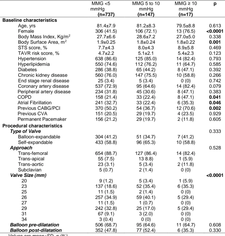
During the study period from January 2012 to August 2016, a total of 928 patients underwent TAVR. Of those, complete echocardiographic and survival data were available for 901 patients (97%). Table 1 displays the baseline characteristics for the study population according to baseline MMG. The majority had normal-mild MMG (< 5 mmHg) at baseline (n=737, 81.7%); 147 patients (16.3%) had a moderate increased MMG (≥5 and <10 mmHg), and only 17 patients (1.9%) showed a baseline MMG≥10 mmHg. As shown in Table 1, patients with a baseline MMG ≥10 mmHg tend to be more frequently female (76.5% vs 41.5% and 72.15, p<0.0001) and smaller in body size (1.8±0.22 vs 1.9±0.25 and 1.8±0.25, p=0.001). Compared to patients with normal-mild MMG and to those with a moderate increase in MMG, those with baseline MMG≥10 mmHg showed a higher prevalence of atrial fibrillation (35.3% vs 32.7% and 22.4%, p=0.046), chronic obstructive pulmonary disease (47.1% vs 21.4% and 22.4%, p=0.041) and previous history of CABG/PCI (70.6% vs 50.2% and 36.7%, p=0.002).
Procedural characteristics
The three groups were similar for procedural characteristics apart from the use of smaller aortic valve prosthesis in the group of patients with baseline MMG≥10 mmHg (p<0.0001; Table 1).
Echocardiographic characteristics
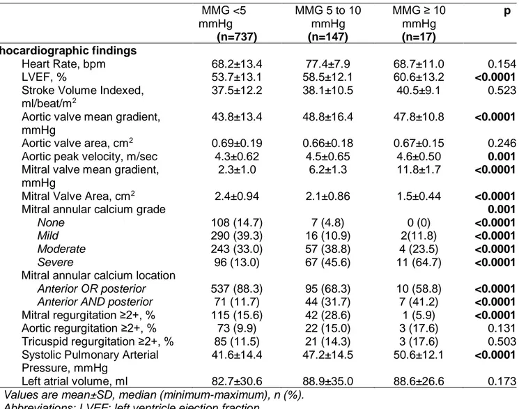
As reported in Table 2, patients with baseline MMG≥10 mmHg showed higher LV ejection fraction compared to the other two groups (LVEF, 60.6±13.2 vs 53.7±13.1 and 58.5±13.1,
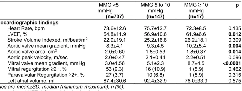
p<0.0001). This latter group also showed higher aortic valve mean gradients (47.8±10.8 vs 43.8±13.4 and 48.8±16.4, p<0.0001) and peak velocity (4.6±0.5 vs 4.3±0.62 and 4.5±0.65, p=0.001). Consistently, they had higher mitral mean gradients (11.8±1.7 vs 2.3±1.0 and 6.2±1.3, p<0.0001), smaller MVA (1.5±0.44 vs 2.4±0.94 and 2.1±0.86, p<0.0001) and a higher prevalence of severe MAC (64.7% vs 13.0% and 45.6%, p<0.0001), which was more frequently localized both on the anterior and posterior annulus (41.2% vs 11.7% and 31.7%, p<0.0001). Finally, patients with baseline MMG≥10 mmHg showed a significantly lower prevalence of MR≥2+ (5.9% vs 15.6% and 28.6%, p<0.0001) and a higher systolic pulmonary arterial pressure (sPAP, 50.6±12.1 vs 41.6±14.4 and 47.2±14.5, p<0.0001) compared to the other two groups. After TAVR, patients with baseline MMG≥10 mmHg showed a persistently higher LVEF (61.9±6.6 vs 54.8±11.9 and 56.9±10.6, p=0.012), slightly higher aortic mean gradient (10.2±5.4 vs 8.3±4.1 and 9.3±4.5, p=0.004). Patients starting with MMG≥10 mmHg also displayed higher MMG post-TAVR (8.7±4.5 vs 3.0±1.56 and 5.1±2.3, p<0.0001, Table 3).
Outcomes
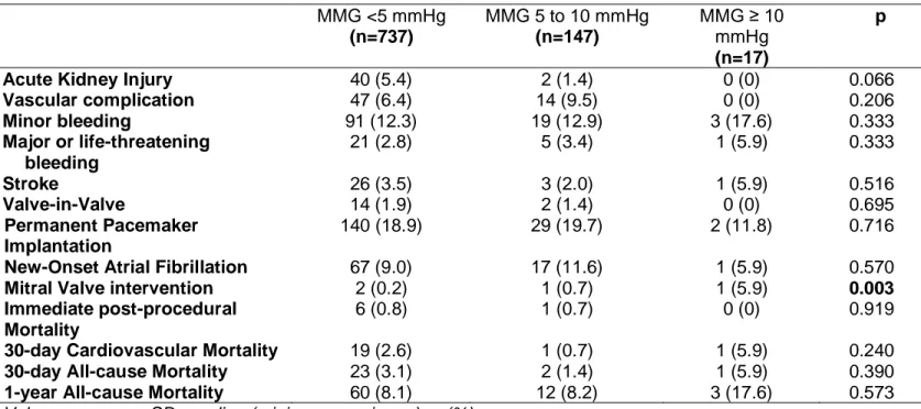
As reported in Table 4, the three groups did not differ for any of the listed outcomes, apart from the prevalence of mitral valve interventions at follow-up which occurred more frequently in the group of patients with baseline MMG≥10 mmHg (5.9% vs 0.2% and 0.7%, p=0.003).
Long-term Survival
Figure 1 shows unadjusted Kaplan-Meier cumulative survival (with shaded 95% Confidence Intervals) and TAVR score risk-adjusted Hazard Ratios by Mitral Mean Gradient [(MMG): <5, 5≤MMG<10, and MMG≥10] for 901 patients with complete MMG and follow-up data. There
were 102 total deaths matched with data from the National Death Index over 5 years (60 months) of follow-up. The mean follow-up was 40.8±13.9 months. Those with a baseline MMG≥10 experienced nearly 3 times the risk of long-term mortality compared to those with baseline MMG<5 (TAVR-risk Adjusted HR: 2.91, 95% CI: 1.17-7.20, p=0.02). When the population was stratified according to MVA, no difference in long-term mortality was observed between patients with MVA> 1.5 cm2 and those with MVA≤1.5 cm2 (TAVR-risk Adjusted HR: 1.08, 95% CI: 0.62-1.85, p=0.81, Figure 2). Mortality was additionally assessed by stratifying the population as follows: patients with baseline and post-TAVR MMG < 10 mmHg; patients with baseline MMG < 10 mmHg and post-TAVR MMG ≥10 mmHg; ; patients with baseline MMG ≥ 10 mmHg and post-TAVR MMG ≥10 mmHg; patients with baseline MMG ≥ 10 mmHg and post-TAVR MMG < 10 mmHg. Having a MMG ≥10 mmHg at baseline and post-TAVR was extremely rare (n=3) but portended a detrimental long-term survival (TAVR-risk Adjusted HR: 7.09, 95% CI: 1.74-28.9, p=0.03, Figure 3).
Discussion
To the best of our knowledge, this is the first registry assessing the prevalence and role on survival at 5-years after TAVR of MS. The main findings of this study are the following: 1.The prevalence of severe MS, defined as MMG ≥10 mmHg, in this cohort of severe AS patient is low, approximately 2%. 2. Patients with baseline MMG≥10 mmHg are frequently women, with a worse baseline risk profile compared to the other groups. 3. Despite the low prevalence of MMG≥10 mmHg prior to TAVR, patients in this group experience a nearly 3 times higher mortality at 5 years after the procedure compared to other groups.
The prevalence of MS in patients with AS has been reported to be around 10% and generally the outcome of these patients is very poor once they develop symptoms5. The ACC/AHA guidelines for valve disease recommend double valve intervention in these cases, although it carries a high operative risk 2, 14. Moreover, mitral valve replacement in cases of MS with severe MAC might be particularly challenging with a high risk of post-operative paravalvular leakage and complications. The development of TAVR in the last decade has drastically reduced the operative risk and improved survival of high to intermediate-risk or inoperable patients, and might represent a valid alternative also for patients with concomitant MS. However, data about MS in the TAVR era are scarce. A recent report form the TVT registry documented that severe MS (as defined by a MVA ≤1.5 cm2) is a predictor of mortality and re-hospitalization for heart failure at 1-year after-TAVR6. In this study, the authors reported a prevalence of MS around 3%, similar to what we found in our population (1.9%). However, this was based on site-reported MVA using variable methodologies and MMG was not site-reported. Although the latest U.S. and European guidelines on valvular disease define MS based on MVA as measured by
planimetry, this recommendation is based on rheumatic heart disease15, 16. When the mitral valve is calcified, planimetry is limited by shadowing and blooming artifact 17 (Figure 4). MVA by pressure half-time is strongly affected by the net compliance of the LV and LA, which are altered in AS18. By defining calcific MS as MMG ≥ 10 mmHg, we selected a group with a high specificity for severe MS and elevated LA pressures at rest with normal heart rate (68.7±11.0 bpm). Indeed, when we stratified our population according to MVA, the group with MS (MVA≤1.5 cm2, n=123) did not experience a higher mortality up to 5 years after TAVR compared to patients with MVA>1.5 cm2. Because MMG is flow and heart rate dependent, it is possible that more patients would have been classified as severe MS, had they undergone exercise testing; however; there was no clinical indication to do such in this population with severe symptomatic AS.
As to why patients with MMG≥10 mmHg experience a poor prognosis compared to the other two groups, some hypothesis might be generated. Firstly, it is likely that in our population the etiology of MS is degenerative given the older age, the higher prevalence of atherosclerosis in this group, as testified by the higher prevalence of CAGB/PCI and the higher prevalence of severe MAC located both in the anterior and posterior annulus. In turn, the higher atherosclerotic burden could explain, at least in part, the worse outcome. Previous studies have reported that patients with calcification of the aortic and mitral annulus frequently have calcified LV outflow tract which independently predict post-TAVR aortic regurgitation19. Although the presence of post-TAVR paravalvular leak has been associated with worse outcomes, we did not find differences in its prevalence in our population. Additionally, the group of patients with MMG≥10 mmHg had a higher prevalence of atrial fibrillation and higher sPAP; if, on the one hand, both conditions might be a direct result of the severe MS, on the other
hand, it has been widely shown that their presence is a marker of impaired prognosis 13, 20, 21. Taken together these observation suggest that the increased baseline risk profile for patients with severe MS undergoing TAVR could potentially explain the increased rates of 5-year mortality. The result of this study indicate that patients with severe MS are a minority of those undergoing TAVR and that these patients experience a bad outcome at 5 years follow-up compared to patients with normal MMG. This does not mean that patients with severe AS and MS should not be offered a TAVR, but that such patients will need a more comprehensive approach that possibly includes the discussion and timing for mitral valve intervention. Indeed, with advances in transcatheter valve therapies, a percutaneous approach may become a viable alternative to conventional open heart surgery in selected high-risk patients with concomitant severe AS and MS. Further studies are certainly needed at this regard to prove a survival benefit of mitral valve intervention following TAVR in patients with concomitant severe MS.
Limitations
First, this study suffers from the intrinsic limitations of a retrospective design. Second, complete echocardiographic data were not available or not accurate for 2.9% of the population, which was therefore excluded from this analysis. Third, in this study we categorized MS according to MMG, which has the advantage of being a direct measurement, unlike calculated MVA derived from pressure half-time measurement or continuity equation. MVA by pressure half-time is prone to error resulting from LV/LA compliance and aortic regurgitation,10 both of which are common in TAVR patients. Direct planimetry of MVA is recommended in rheumatic MS, but is challenging in degenerative MS due to shadowing and blooming artifact from annular and
leaflet calcium. Although MMG is influenced by heart rate and cardiac output, a high value reflects elevated LA pressure which may limit symptomatic improvement after TAVR. All of the echocardiographic measurements of the included patients have been done at a heart rate < 100 bpm; also, the group of patients with MMG≥10 mmHg showed the lower prevalence of MR≥2+, such the increased MMG cannot be explained by significant MR. Finally, the small number of patients with a MMG≥10 mmHg has to be acknowledged as a potential limitation of our study.
References
1. Barasch E, Gottdiener JS, Larsen EK, Chaves PH, Newman AB and Manolio TA. Clinical significance of calcification of the fibrous skeleton of the heart and aortosclerosis in community dwelling elderly. The Cardiovascular Health Study (CHS). American heart journal. 2006;151:39-47.
2. Nishimura RA, Otto CM, Bonow RO, Carabello BA, Erwin JP, 3rd, Fleisher LA, Jneid H, Mack MJ, McLeod CJ, O'Gara PT, Rigolin VH, Sundt TM, 3rd and Thompson A. 2017 AHA/ACC Focused Update of the 2014 AHA/ACC Guideline for the Management of Patients With Valvular Heart Disease: A Report of the American College of Cardiology/American Heart Association Task Force on Clinical Practice Guidelines. Circulation. 2017;135:e1159-e1195. 3. Nishimura RA, Otto CM, Bonow RO, Carabello BA, Erwin JP, 3rd, Guyton RA, O'Gara PT, Ruiz CE, Skubas NJ, Sorajja P, Sundt TM, 3rd, Thomas JD and American College of Cardiology/American Heart Association Task Force on Practice G. 2014 AHA/ACC guideline for the management of patients with valvular heart disease: executive summary: a report of the American College of Cardiology/American Heart Association Task Force on Practice Guidelines. Journal of the American College of Cardiology. 2014;63:2438-88.
4. Stewart BF, Siscovick D, Lind BK, Gardin JM, Gottdiener JS, Smith VE, Kitzman DW and Otto CM. Clinical factors associated with calcific aortic valve disease. Cardiovascular Health Study. Journal of the American College of Cardiology. 1997;29:630-4.
5. Baudet EM, Puel V, McBride JT, Grimaud JP, Roques F, Clerc F, Roques X and Laborde N. Long-term results of valve replacement with the St. Jude Medical prosthesis. The Journal of thoracic and cardiovascular surgery. 1995;109:858-70.
6. Joseph L, Bashir M, Xiang Q, Yerokun BA, Matsouaka RA, Vemulapalli S, Kapadia S, Cigarroa JE and Zahr F. Prevalence and Outcomes of Mitral Stenosis in Patients Undergoing Transcatheter Aortic Valve Replacement: Findings From the Society of Thoracic Surgeons/American College of Cardiology Transcatheter Valve Therapies Registry. JACC Cardiovasc Interv. 2018;11:693-702.
7. Lang RM, Badano LP, Mor-Avi V, Afilalo J, Armstrong A, Ernande L, Flachskampf FA, Foster E, Goldstein SA, Kuznetsova T, Lancellotti P, Muraru D, Picard MH, Rietzschel ER, Rudski L, Spencer KT, Tsang W and Voigt JU. Recommendations for cardiac chamber quantification by echocardiography in adults: an update from the American Society of Echocardiography and the European Association of Cardiovascular Imaging. Journal of the American Society of Echocardiography : official publication of the American Society of Echocardiography. 2015;28:1-39 e14.
8. Kircher BJ, Himelman RB and Schiller NB. Noninvasive estimation of right atrial pressure from the inspiratory collapse of the inferior vena cava. The American journal of cardiology. 1990;66:493-6.
9. Zoghbi WA, Adams D, Bonow RO, Enriquez-Sarano M, Foster E, Grayburn PA, Hahn RT, Han Y, Hung J, Lang RM, Little SH, Shah DJ, Shernan S, Thavendiranathan P, Thomas JD and Weissman NJ. Recommendations for Noninvasive Evaluation of Native Valvular Regurgitation: A Report from the American Society of Echocardiography Developed in Collaboration with the Society for Cardiovascular Magnetic Resonance. Journal of the
American Society of Echocardiography : official publication of the American Society of Echocardiography. 2017;30:303-371.
10. Baumgartner H, Hung J, Bermejo J, Chambers JB, Evangelista A, Griffin BP, Iung B, Otto CM, Pellikka PA, Quinones M, American Society of E and European Association of E. Echocardiographic assessment of valve stenosis: EAE/ASE recommendations for clinical practice. Journal of the American Society of Echocardiography : official publication of the American Society of Echocardiography. 2009;22:1-23; quiz 101-2.
11. Abramowitz Y, Kazuno Y, Chakravarty T, Kawamori H, Maeno Y, Anderson D, Allison Z, Mangat G, Cheng W, Gopal A, Jilaihawi H, Mack MJ and Makkar RR. Concomitant mitral annular calcification and severe aortic stenosis: prevalence, characteristics and outcome following transcatheter aortic valve replacement. European heart journal. 2017;38:1194-1203. 12. Sannino A, Losi MA, Giugliano G, Canciello G, Toscano E, Giamundo A, Scudiero F, Brevetti L, Scudiero L, Prastaro M, Perrino C, Perrone-Filardi P, Galderisi M, Trimarco B and Esposito G. Aortic and Mitral Calcification Is Marker of Significant Carotid and Limb Atherosclerosis in Patients with First Acute Coronary Syndrome. Echocardiography. 2015;32:1771-7.
13. Sannino A, Stoler RC, Lima B, Szerlip M, Henry AC, Vallabhan R, Kowal RC, Brown DL, Mack MJ and Grayburn PA. Frequency of and Prognostic Significance of Atrial Fibrillation in Patients Undergoing Transcatheter Aortic Valve Implantation. The American journal of cardiology. 2016;118:1527-1532.
14. Nishimura RA, Otto CM, Bonow RO, Carabello BA, Erwin JP, 3rd, Guyton RA, O'Gara PT, Ruiz CE, Skubas NJ, Sorajja P, Sundt TM, 3rd, Thomas JD and American College of Cardiology/American Heart Association Task Force on Practice G. 2014 AHA/ACC guideline
for the management of patients with valvular heart disease: a report of the American College of Cardiology/American Heart Association Task Force on Practice Guidelines. Journal of the American College of Cardiology. 2014;63:e57-185.
15. Nishimura RA, Otto CM, Bonow RO, Carabello BA, Erwin JP, 3rd, Fleisher LA, Jneid H, Mack MJ, McLeod CJ, O'Gara PT, Rigolin VH, Sundt TM, 3rd and Thompson A. 2017 AHA/ACC Focused Update of the 2014 AHA/ACC Guideline for the Management of Patients With Valvular Heart Disease: A Report of the American College of Cardiology/American Heart Association Task Force on Clinical Practice Guidelines. Journal of the American College of Cardiology. 2017;70:252-289.
16. Baumgartner H, Falk V, Bax JJ, De Bonis M, Hamm C, Holm PJ, Iung B, Lancellotti P, Lansac E, Rodriguez Munoz D, Rosenhek R, Sjogren J, Tornos Mas P, Vahanian A, Walther T, Wendler O, Windecker S, Zamorano JL and Group ESCSD. 2017 ESC/EACTS Guidelines for the management of valvular heart disease. European heart journal. 2017;38:2739-2791. 17. Smith MD, Handshoe R, Handshoe S, Kwan OL and DeMaria AN. Comparative accuracy of two-dimensional echocardiography and Doppler pressure half-time methods in assessing severity of mitral stenosis in patients with and without prior commissurotomy. Circulation. 1986;73:100-7.
18. Thomas JD, Wilkins GT, Choong CY, Abascal VM, Palacios IF, Block PC and Weyman AE. Inaccuracy of mitral pressure half-time immediately after percutaneous mitral valvotomy. Dependence on transmitral gradient and left atrial and ventricular compliance. Circulation. 1988;78:980-93.
19. Buellesfeld L, Stortecky S, Heg D, Gloekler S, Meier B, Wenaweser P and Windecker S. Extent and distribution of calcification of both the aortic annulus and the left ventricular
outflow tract predict aortic regurgitation after transcatheter aortic valve replacement. EuroIntervention : journal of EuroPCR in collaboration with the Working Group on Interventional Cardiology of the European Society of Cardiology. 2014;10:732-8.
20. Sannino A, Gargiulo G, Schiattarella GG, Perrino C, Stabile E, Losi MA, Galderisi M, Izzo R, de Simone G, Trimarco B and Esposito G. A meta-analysis of the impact of pre-existing and new-onset atrial fibrillation on clinical outcomes in patients undergoing transcatheter aortic valve implantation. EuroIntervention : journal of EuroPCR in collaboration with the Working Group on Interventional Cardiology of the European Society of Cardiology. 2016;12:e1047-e1056.
21. Masri A, Abdelkarim I, Sharbaugh MS, Althouse AD, Xu J, Han W, Chan SY, Katz WE, Crock FW, Harinstein ME, Kliner DE, Navid F, Lee JS, Gleason TG, Schindler JT and Cavalcante JL. Outcomes of persistent pulmonary hypertension following transcatheter aortic valve replacement. Heart. 2018;104:821-827.
Figure Legend
Figure 1. Meier curves for 5-year survival according to MMG. Unadjusted Kaplan-Meier cumulative survival (with shaded 95% Confidence Intervals) and TAVR score risk-adjusted Hazard Ratios by MMG (MMG<5, 5≤MMG<10, and MMG≥10) for 901 patients with complete MMG and follow-up data.
Figure 2. Kaplan-Meier curves for 5-year survival according to MVA. Unadjusted Kaplan-Meier cumulative survival (with shaded 95% Confidence Intervals) and TAVR score risk-adjusted Hazard Ratios by Mitral Valve Area [(MVA): ≤1.5 cm2, MVA>1.5 cm2] for 890 patients with complete MVA and follow-up data.
Figure 3. Meier curves for 5-year survival according to MMG. Unadjusted Kaplan-Meier cumulative survival (with shaded 95% Confidence Intervals) and TAVR score risk-adjusted Hazard Ratios by MMG (Both pre and TAVR MMG: <10, either pre and/or post-TAVR MMG≥10) for 901 patients with complete MMG and follow-up data.
Figure 4. Examples of short-axis view of the mitral valve. Quantification of the MVA by planimetry is compromised by the amount of calcium on the mitral annulus generating a blooming artifact and/or by the high acoustic thoracic impedance.
Table 1. Characteristics of the study population according to baseline mean mitral gradients (MMG). MMG <5 mmHg (n=737) MMG 5 to 10 mmHg (n=147) MMG ≥ 10 mmHg (n=17) p Baseline characteristics Age, yrs 81.4±7.9 81.2±8.3 79.5±8.8 0.613 Female 306 (41.5) 106 (72.1) 13 (76.5) <0.0001
Body Mass Index, Kg/m2 27.7±6.6 28.6±7.2 27.0±5.0 0.338
Body Surface Area, m2 1.9±0.25 1.8±0.24 1.8±0.22 0.001
STS score, % 7.7±4.3 8.0±4.3 8.9±5.8 0.469
TAVR risk score, % 4.7±2.2 5.1±2.1 5.4±2.3 0.123
Hypertension 638 (86.6) 125 (85.0) 14 (82.4) 0.793
Hyperlipidemia 550 (74.6) 112 (76.2) 11 (64.7) 0.585
Diabetes 286 (38.8) 65 (44.2) 8 (47.1) 0.392
Chronic kidney disease 560 (76.0) 147 (75.5) 10 (58.8) 0.266
End stage renal disease 25 (3.4) 5 (3.4) 0 (0) 0.742
Coronary artery disease 537 (72.9) 95 (64.6) 14 (82.4) 0.079 Peripheral artery disease 234 (31.8) 45 (30.6) 8 (47.1) 0.383
COPD 158 (21.4) 33 (22.4) 8 (47.1) 0.041 Atrial Fibrillation 241 (32.7) 33 (22.4) 6 (35.3) 0.046 Previous CABG/PCI 370 (50.2) 54 (36.7) 12 (70.6) 0.002 Previous CVA 151 (20.5) 29 (19.7) 4 (23.5) 0.929 Permanent Pacemaker 156 (21.2) 29 (19.7) 2 (11.8) 0.605 Procedural characteristics Type of Valve 0.333 Balloon-expandable 304 (41.2) 51 (34.7) 7 (41.2) Self-expandable 433 (58.8) 96 (65.3) 10 (58.8) Approach 0.528 Trans-femoral 654 (88.7) 127 (86.4) 14 (82.4) Trans-apical 55 (7.5) 13 8.8) 1 (5.9) Trans-aortic 23 (3.1) 5 (3.4) 2 (11.8) Subclavian 5 (0.7) 2 (1.4) 0 (0) Valve Size (mm) <0.0001 20 9 (1.2) 5 (3.4) 1 (5.9) 23 137 (18.6) 52 (35.4) 6 (35.3) 25 11 (1.5) 2 (1.4) 0 (0) 26 257 (34.9) 59 (40.1) 5 (29.4) 27 11 (1.5) 1 (0.7) 0 (0) 29 242 (32.8) 25 (17.0) 5 (29.4) 31 67 (9.1) 3 (2.0) 0 (0) 34 3 (0.4) 0 (0) 0 (0) Balloon pre-dilatation 506 (68.7) 95 (64.6) 11 (64.7) 0.608 Balloon post-dilatation 352 (47.8) 77 (52.4) 6 (35.3) 0.330
Values are mean±SD, n (%).
Abbreviations: CABG: coronary artery by-pass graft; COPD: chronic obstructive pulmonary disease; CVA: cerebrovascular accident; LVEF: left ventricle ejection fraction; PCI: percutaneous coronary intervention; SD: standard deviation.
Table 2. Baseline echocardiographic findings according to baseline mean mitral gradients. MMG <5 mmHg (n=737) MMG 5 to 10 mmHg (n=147) MMG ≥ 10 mmHg (n=17) p Echocardiographic findings Heart Rate, bpm 68.2±13.4 77.4±7.9 68.7±11.0 0.154 LVEF, % 53.7±13.1 58.5±12.1 60.6±13.2 <0.0001
Stroke Volume Indexed, ml/beat/m2
37.5±12.2 38.1±10.5 40.5±9.1 0.523 Aortic valve mean gradient,
mmHg
43.8±13.4 48.8±16.4 47.8±10.8 <0.0001 Aortic valve area, cm2 0.69±0.19 0.66±0.18 0.67±0.15 0.246 Aortic peak velocity, m/sec 4.3±0.62 4.5±0.65 4.6±0.50 0.001 Mitral valve mean gradient,
mmHg
2.3±1.0 6.2±1.3 11.8±1.7 <0.0001 Mitral Valve Area, cm2 2.4±0.94 2.1±0.86 1.5±0.44 <0.0001
Mitral annular calcium grade 0.001
None 108 (14.7) 7 (4.8) 0 (0) <0.0001
Mild 290 (39.3) 16 (10.9) 2(11.8) <0.0001
Moderate 243 (33.0) 57 (38.8) 4 (23.5) <0.0001
Severe 96 (13.0) 67 (45.6) 11 (64.7) <0.0001
Mitral annular calcium location
Anterior OR posterior 537 (88.3) 95 (68.3) 10 (58.8) <0.0001
Anterior AND posterior 71 (11.7) 44 (31.7) 7 (41.2) <0.0001
Mitral regurgitation ≥2+, % 115 (15.6) 42 (28.6) 1 (5.9) <0.0001 Aortic regurgitation ≥2+, % 73 (9.9) 22 (15.0) 3 (17.6) 0.131 Tricuspid regurgitation ≥2+, % 85 (11.5) 21 (14.3) 3 (17.6) 0.503 Systolic Pulmonary Arterial
Pressure, mmHg
41.6±14.4 47.2±14.5 50.6±12.1 <0.0001 Left atrial volume, ml 82.7±30.6 88.9±35.0 88.6±26.6 0.173
Values are mean±SD, median (minimum-maximum), n (%). Abbreviations: LVEF: left ventricle ejection fraction.
Table 3. Post-TAVR echocardiographic findings according to baseline mean mitral gradients. MMG <5 mmHg (n=737) MMG 5 to 10 mmHg (n=147) MMG ≥ 10 mmHg (n=17) p Echocardiographic findings Heart Rate, bpm 73.6±12.6 75.7±12.7 72.3±8.5 0.135 LVEF, % 54.8±11.9 56.9±10.6 61.9±6.6 0.012
Stroke Volume Indexed, ml/beat/m2 22.9±19.1 25.2±16.8 26.2±18.1 0.309 Aortic valve mean gradient, mmHg 8.3±4.1 9.3±4.5 10.2±5.4 0.004
Aortic valve area, cm2 2.0±0.60 1.8±0.53 1.8±0.37 0.014
Aortic peak velocity, m/sec 2.0±0.47 2.1±0.44 2.2±0.51 0.096 Mitral valve mean gradient, mmHg 3.0±1.56 5.1±2.3 8.7±4.5 <0.0001 Mitral regurgitation ≥2+, % 53 (9.3) 16 (10.9) 1 (5.9) 0.462 Paravalvular Regurgitation ≥2+, % 27 (3.7) 10 (6.8) 1 (5.9) 0.315
Left atrial volume, ml 87.4±30.6 92.4±32.9 76.0±33.9 0.575
Values are mean±SD, median (minimum-maximum), n (%). Abbreviations: LVEF: left ventricle ejection fraction.
Table 4. Post-TAVR outcomes according to baseline MMG. MMG <5 mmHg (n=737) MMG 5 to 10 mmHg (n=147) MMG ≥ 10 mmHg (n=17) p
Acute Kidney Injury 40 (5.4) 2 (1.4) 0 (0) 0.066
Vascular complication 47 (6.4) 14 (9.5) 0 (0) 0.206 Minor bleeding 91 (12.3) 19 (12.9) 3 (17.6) 0.333 Major or life-threatening bleeding 21 (2.8) 5 (3.4) 1 (5.9) 0.333 Stroke 26 (3.5) 3 (2.0) 1 (5.9) 0.516 Valve-in-Valve 14 (1.9) 2 (1.4) 0 (0) 0.695 Permanent Pacemaker Implantation 140 (18.9) 29 (19.7) 2 (11.8) 0.716
New-Onset Atrial Fibrillation 67 (9.0) 17 (11.6) 1 (5.9) 0.570
Mitral Valve intervention 2 (0.2) 1 (0.7) 1 (5.9) 0.003
Immediate post-procedural Mortality
6 (0.8) 1 (0.7) 0 (0) 0.919
30-day Cardiovascular Mortality 19 (2.6) 1 (0.7) 1 (5.9) 0.240
30-day All-cause Mortality 23 (3.1) 2 (1.4) 1 (5.9) 0.390
1-year All-cause Mortality 60 (8.1) 12 (8.2) 3 (17.6) 0.573