1
UNIVERSITA’ DEGLI STUDI DI VERONA
DEPARTMENT OF
Diagnostic and Public Health, Microbiology Section
GRADUATE SCHOOL OF Life and Health sciences
DOCTORAL PROGRAM IN Applied Life and Health Sciences Microbiology and Infectious Diseases
XXX Cycle Year 2014
Epidemiology, molecular and phenotypic typing of Methicillin Resistant Staphylococcus aureus (MRSA) strains isolated from multi-drug resistant
screening program and blood culture.
S.S.D. MED/07 Coordinator: Prof. Giovanni Malerba
Signature_______________ Tutor: Prof.ssa Annarita Mazzariol Signature_______________
Doctoral Student: Dr.ssa Liliana Galia
Signature______________________
2
This work is licensed under a Creative Commons Attribution-NonCommercial- NoDerivs 3.0 Unported License, Italy. To read a copy of the licence, visit the web
page:http://creativecommons.org/licenses/by-nc-nd/3.0/
Attribution — You must give appropriate credit, provide a link to the license, and indicate if changes were made. You may do so in any reasonable manner, but not in any way that suggests the licensor endorses you or your use.
NonCommercial — You may not use the material for commercial purposes.
NoDerivatives — If you remix, transform, or build upon the material, you may not distribute the modified material.
Epidemiology, molecular and phenotypic typing of Methicillin Resistant Staphylococcus aureus (MRSA) strains isolated from multi-drug resistant
screening program and blood culture-Liliana Galia
PhD Thesis Verona,11th May 2018
3 A Mamma
A Nonna Murru A Pietruccia Nel mio cuore.
4
I.
SOMMARIO
Staphylococcus aureus è un comune batterio presente sulla cute e sulle membrane
mucose nel 20-30% delle persone sane. Talvolta può causare infezioni nell’uomo, solitamente infezioni della cute e suppurative a livello locale, ma anche infezioni più gravi a carico di diversi distretti dell’organismo. Alcuni ceppi di questo batterio, tuttavia, hanno sviluppato una resistenza agli antibiotici ß-lattamici, tra cui le penicilline, che sono utilizzati nella cura di numerose infezioni. Questi ceppi sono noti con il nome di Staphylococcus aureus meticillino-resistente (MRSA).
L’MRSA si trasmette all’uomo prevalentemente mediante contatto diretto con la persona infetta o con strumenti medici e apparecchiature medicali. L’MRSA è problematico soprattutto negli ospedali, dove i pazienti con un sistema immunitario indebolito sono più esposti al rischio di infezione rispetto alla popolazione generale. La ricerca in questo ambito si occupa dello studio di nuove strumenti diagnostici e terapia salvavita che permettano di individuare velocemente ceppi multi-resistenti, in campioni clinici riducendo i tempi di refertazione, per dare garanzia di una corretta e immediata terapia.
Uno dei primi obiettivi di questo studio su ceppi di MRSA solati da campioni clinici è la tipizzazione molecolare della proteina A (specie-specifica) responsabile del legame con la porzione Fc delle immunoglobuline, rendendole cosi inattive al fine di individuare le caratteristiche epidemiologiche e molecolari di questi ceppi in pazienti colonizzati/infetti. La tipizzazione della proteina A è stata fatta tramite l’utilizzo della tecnica spa-typing.
Abbiamo inoltre caratterizzato la cassetta cromosomica mobile SCCmec e le rispettive correlazione con i diversi profili di antibiotico resistenza tra 6 spa-type più rappresentativi quali: t032 CC22, t1036 CC22, t1214 CC22 ,t022 CC22, t041 CC5
,t121 CC8, ricercati su 135 ceppi di MRSA provenienti da tamponi faringei, rettali e
5
128 ceppi sono stati ritrovati appartenenti al tipo SCCmec IV e 7 appartenenti al tipo
t041 appartenenti a SCCmec I, rispettivamente tutti tutti di classe B. Abbiamo notato
inoltre che nei 41 ceppi isolati da emocolture mancano gli spa-type t1036 e t022 considerati tra i “6 rappresentativi” ritrovati in questo studio. Il nostro pensiero, per le ricerche future sarà quello di aumentare il numero di ceppi isolati da emocolture. Abbiamo individuato un nuovissimo spa-type t16026 CC22 sottomesso nell’anno 2016.
Solo 29 ceppi su 135 testati sono sensibili all'eritromicina ma gli altri ceppi hanno un alto livello di resistenza ai fluorochinoloni e macrolidi. Questo studio ha dimostrato inoltre che i valori di Mic ed E-test del Ceftobiprole (Basilea Pharmaceutica), una nuova cefalosporina di quinta generazione, designata a trattare le infezioni associate a MRSA, ha valori che rientrano nei breakpoint di sensibilità secondo le linee guida EUCAST. Questo è di notevole importanza poiché è, fino ad ora l’unica cefalosporina sensibile. La descrizione della suscettibilità agli antibiotici e dei tipi di
spa è molto dettagliata e fornisce informazioni rilevanti sull'epidemiologia
molecolare e sui fattori di virulenza di MRSA in Italia. Utilizzando la tecnica dello spa-typing, quindi, abbiamo potuto individurare la presenza di un clone principale E-MRSA 15 in questa parte dell’Italia.
Possiamo quindi concludere che i ceppi di MRSA isolati presso i reparti di anestesia e rianimazione, terapia intensiva, medicina interna, oncologia, pediatria, centro ustioni del policlinico di Verona “G. Rossi” hanno un profilo molecolare attribuibile per definizione ai ceppi comunitari CA-MRSA ma un profilo di multi- resistenza tipico dei ceppi ospedalieri e solo 6 ceppi presentano il gene pvl.
Quindi possiamo ipotizzare ad un cambiamento epidemiologico e microbiologico di ceppi di MRSA isolati in questa parte dell’Italia e si potrebbe inoltre proporre un cambiamento di definizione dei CA-MRSA.
6
Un altro obiettivo è stato quello di studiare una Real-time PCR che rilevasse rapidamente la resistenza alla meticillina , il fattore di virulenza PVL direttamente da un campione clinico e il gene nuc (specie -specifico) . Questa tecnica presentata in questo studio può identificare e differenziare MRSA, MSSA, resistente alla meticillina, Stafilococchi negativi alla coagulasi (MR-CNS) con un significato diagnostico e terapeutico di notevole importanza e una riduzione dei tempi di refertazione dalle 48h ad 1h 30 minuti.
Questa tecnica automatizzata e standardizzata ha come risultato una concordanza del 100% con le tecniche molecolari standard , il 100% di specificità e una sensibilità di 514 UFC/ml.
7
II.
ABSTRACT
Staphylococcus aureus is a common bacterium found on the skin and mucous membranes in 20-30% of healthy people. Sometimes it can cause infections in humans, usually skin infections and suppuratives at the local level, but also more serious infections affecting different parts of the body. Some strains of this bacterium, however, have developed resistance to ß-lactam antibiotics, including penicillins, which are used in the treatment of numerous infections. These strains are known as
Methicillin-resistant Staphylococcus aureus (MRSA). MRSA is transmitted to
humans mainly through direct contact with the infected person or with medical instruments and medical equipment. MRSA is problematic especially in hospitals, where patients with a weakened immune system are more susceptible to infection than the general population. Research in this area deals with the study of new diagnostic tools and life-saving therapy that allow the rapid identification of multi-resistant strains, in clinical samples, reducing reporting times, to guarantee correct and immediate therapy. One of the first objectives of this study on MRSA strains solved by clinical samples is the molecular typing of protein A (species-specific) responsible for binding to the Fc portion of immunoglobulins, thus rendering them inactive in order to identify the epidemiological and molecular characteristics of these strains in colonized/infected patients. Protein A typing was done by using the spa-typing technique. We have also characterized the SCCmec mobile chromosomal cassette and the respective correlations with the different antibiotic resistance profiles among 6 more “representative” spa-types such as: t032 CC22, t1036 CC22, t1214
CC22, t022 CC22, t041 CC5, t121 CC8, sought from 135 strains of MRSA from
pharyngeal, rectal and blood cultures of which 94 from rectal and pharyngeal swabs and 41 from blood cultures respectively. 128 strains were found belonging to the
SCCmec IV type and 7 t041 strains belonging to SCCmec I, all of class B
respectively. We also noted that in the 41 strains isolated from blood cultures, the
8
study are missing. Our think, for future research will be to increase the number of isolates isolated from blood cultures. We have identified a brand new spa-type t16026 CC22 submitted in the year 2016. Only 29 out of 135 tested strains are sensitive to erythromycin but the other strains have a high level of resistance to fluoroquinolones and macrolides. This study also showed that the Mic and E-test values of Ceftobiprole (Basilea Pharmaceutica), a new fifth-generation cephalosporin, designed to treat infections associated with MRSA, have values that detect sensitivity breakpoints according to EUCAST guidelines. This is of considerable importance since it is, until now, the only sensitive cephalosporin. The description of susceptibility to antibiotics and spa types is very detailed and provides relevant information on the molecular epidemiology and virulence factors of MRSA in Italy. Using the spa-typing technique, therefore, we have been able to identify the presence of a main E-MRSA 15 clone in this part of Italy. We can therefore conclude that the MRSA strains isolated in the departments of anesthesia, intensive care, internal medicine, oncology, pediatrics, burns center of the "polyclinic G. Rossi" have a molecular profile attributable by definition to the CA-MRSA community strains but a multi-resistance profile typical of hospital strains with only 6 strains presenting the
pvl gene. Thereore we can hypothesize an epidemiological and microbiological
change of MRSA strains isolated in this part of Italy and it could also propose a change in the definition of CA-MRSA. Another objective was to study a Real-time PCR, which quickly detected the resistance to methicillin, the virulence factor PVL directly from a clinical sample and the nuc gene (species-specific). This technique presented in this study can identify and differentiate MRSA, MSSA, resistant to methicillin, coagulase-negative Staphylococci (MR-CNS) with a significant diagnostic and therapeutic significance and a reduction in reporting times from 48h to 1h 30 minutes.
This automated and standardized technique results in 100% agreement with standard molecular techniques, 100% specificity and a sensitivity of 514 CFU / ml.
9
10
I. SOMMARIO ... 4
II. ABSTRACT ... 7
III. OBJECTIVES ... 12
Background ... 13
IV. STAPHYLOCOCCUS AUREUS ... 14
V. Antibiotics: ... 17
VI. Antibiotic resistance: ... 24
II. Decreased Antibiotic Penetration and Efflux ... 28
III) Target sites modification ... 28
VII. BETA-LACTAMS RESISTANCE MECHANISMS IN S. AUREUS ... 31
VIII. METHICILLIN RESISTANCE ... 33
IX. Recommended methods for detection of methicillin resistance in S. aureus... 33
X. TYPING OF S. AUREUS ... 37
A. SPA TYPING ... 37
XI. STAPHYLOCOCCAL CASSETTE CHROMOSOME MEC ... 42
A. mec gene complex ... 44
B. ccr gene complex ... 46
C. Joining regions ... 47
XII. EPIDEMIOLOGY OF MRSA ... 49
A. Health Care Associated MRSA (HA-MRSA)... 50
B. Community Associated MRSA (CA-MRSA) ... 51
XIII. MATERIAL AND METHODS ... 53
11
Multiplex detection of mec, pvl, scn and spA genes... 59
PCR based SCCmec typing. ... 61
RESULTS ... 69
12
Staphylococcus aureus is a major human pathogen that causes a wide range of
clinical infections. It is the main cause of infectious bacteremia and endocarditis, as well as osteo-articular tissues, skin and soft tissue, and prosthetic devices. Over the last two decades there have been two net changes in the epidemiology of S. aureus infections: first, an increasing number of infections associated with healthcare, particularly observed in infectious endocarditis and prosthetic device infections, and second, a skin-related epidemic associated with the community and strain-driven soft tissue infections with certain virulence and resistance to β-lactam antibiotics. According to data from the European Antimicrobial Resistance Surveillance System (EARSS), there are a decrease in MRSA isolates from 18.8% in 2012 to 16.8% in 2015 in the European Union (EU) countries (EARSS 2015).
III. OBJECTIVES
Objectives of the study were: (i) molecular characterization of MRSA S. aureus clinical isolates by spa-typing; (ii) typing the SCC mec cassette; (iii) correlation between antibiotic resistance patterns and spa type t032 CC22, t1036 CC22, t1214 CC22 ,t022 CC22, t041 CC5 ,t121 CC8. antibiotic profile simultaneous identification by Real-time tecnique of nuc (specific species), mecA and pvl genes.
13
Background
New antibiotics are urgently needed due to the alarming development of resistance against all antibiotics on the market and in clinical use.
The pre-antibiotic era was the leading era of mortality and morbidity of humans and animals due to infectious diseases [1]. Among some of the successful pathogens was the Gram-positive Staphylococcus aureus, which had a mortality rate among infected patients that over 80%, while over 70% developed metastatic infections [2] MRSA infections are seldom eradicated by routine antimicrobial therapies. Evidence supports the use of S. aureus decolonization in surgical patients to prevent S. aureus infection, and this intervention has been associated with low rates of postoperative S.
aureus infection. The staphylococcal carriage is most commonly eradicated by
intranasal application of mupirocin either alone or in combination with antiseptic soaps or systemic antimicrobial agents. However, the major cause of nosocomial infection is methicillin-resistant S. aureus (MRSA), which is hard to eradicate despite reports of some cases treated by warming therapy.
Nosocomial infection is a major cause of surgical morbidity and mortality, and SSIs have a reported incidence rate of 2%–20%. More concerning, some strains have become resistant to the newest antibiotics of last resort.
Furthermore, the efficacy of eradication in patients with community-associated MRSA has not been established, and the necessity of routine decolonization is not supported by data. MRSA outbreaks have created a significant challenge for surgery and clinical practice in recent decades; the failure of traditional antimicrobial treatments has gradually become a worldwide problem, especially in the developing world. Thus, effective therapeutic options to combat S. aureus infection, with an emphasis on MRSA, are urgently needed.
14
IV. STAPHYLOCOCCUS AUREUS
S. aureus was discovered in 1880s by the German surgeon Anton J. Rosenbach and
since its discovery it has been emerged as an opportunist pathogen with the ability to cause a wide range of infections, ranging from mild-skin infections to a fatal outcome [3].
Staphylococci are catalase-positive and Gram-positive, with a diameter of 0.8-1.5 μm. no motile, no spore forming, without a capsule, grow well in common culture medium. On solid medium they produce colonies of 2-3 mm in diameter, rounded and marginal, convex, smooth, opaque and
golden-yellow pigmentation. They develop between 10 and 45 ° C, with an optimum temperature ranging from 30 to 37 ° C, at a pH between 4 and 9, with an optimum obetween 7.0 and
7.5. Staphylococci are facultative anaerobes. Among
the Staphylococcal genus, S. aureus is the organism that shows the highest pathogenic potential [5], [6]. The pathogenic potential of S. aureus lies in an array of factors, such as its great ability to establish successful infections independent of environmental conditions, its intrinsic virulence, quorum sensing mechanism, its genetic diversity, plasticity and ability to acquire exogenous DNA such as antibiotic resistance genes [4][7]. With the discovery of the antibiotic agent penicillin by A. Fleming, the success of this pathogen, and other pathogens, was dramatically reduced. In mid-1940s, penicillin was introduced into the clinical practice, which resulted in infections caused by these death-causing pathogens was easily treatable. However, the success of one of the greatest medical discovery did not last for long, as two years after its introduction to clinical practice, the first penicillin-resistant S.
aureus strain was isolated and described by Patricia Jevons [2], [4]. By 1960s, more
15
cope with the emerging number of penicillin-resistant S. aureus strains, the semi-synthetic antibiotic compound methicillin, among other, was discovered. However, as with penicillin, soon after the introduction of methicillin to clinical practice in 1961, the first methicillin-resistant S. aureus (MRSA) strain emerged [2]. Now, S. aureus, and especially MRSA is a major global health care concern, due to its ability to cause nosocomial infections and to its ability become resistance to multiple antibiotic compounds, and thus sought the importance of effective surveillance and control strategies of this pathogen is becoming more and more urgent.
The natural habitat of S. aureus is the skin and mucous membrane of humans and animals. It is estimated that approximately 30% of healthy individuals are asymptomatically carriers of S. aureus [8]. These patients do not have directly clinically relevance, but may act as a reservoir of S. aureus, from which S. aureus can transmit to other patients [9]. In fact, S. aureus have emerged as the leading pathogen that is the cause of more than 50% of healthcare associated infections, posing a great burden worldwide [5], [9], [10]. What might have helped S. aureus to emerge as leading pathogen is perhaps its capacity and capability to acquire antimicrobial resistance genes.
The prevalence of S. aureus and a high selective pressure of antibiotics in hospitals and healthcare institutions may have act as a dangerous cocktail. [8]. Especially the methicillin-resistant S. aureus (MRSA) become a global concern, as an infection caused by MRSA rather than a non-MRSA more often leads to a clinical infection. MRSA do not replace non-MRSA strains, but rather adds to the burden of infections caused by S. aureus [2] [11].
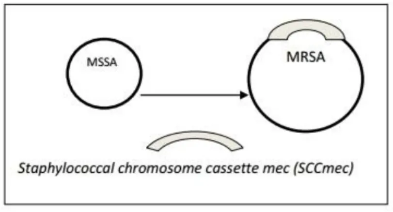
What differentiates methicillin-sensitive S. aureus (MSSA) strains from MRSA strains is the acquisition and insertion of the staphylococcal cassette chromosome mec (SCCmec) into orfX gene on the chromosome of MRSA strains (figure 1). The SCCmec element is a mobile genetic element, which harbors the single determinant for methicillin resistance, namely the mecA or mecC gene. Homology studies of the
16 mecA gene suggests that mecA may have its origin from Staphylococcus sciuri or Staphylococcus fleurettii with 88 % and 99.8 % nucleotide identity, respectively [3],
[4]. Although, S. fleurettii shows a higher nucleotide identity, it does not show an in
vitro resistance, which points to that S. sciuri as the prime candidate for the origin of mecA. However, the origin of the SCCmec still remains unclear. There have been
postulations about that the mecA have been introduced into coagulase negative S.
aureus (CoNS) isolates, together with other genes specific for the SCCmec element
and from which the SCCmec element has been formed, and thus CoNS may serve as the origin and reservoir for the SCCmec element [12].
Figure 1: Schematic representation of the insertion of SCCmec into methicillin-resistance S. aureus (MRSA).
Some newly developed antibiotics exhibit high effectiveness in combating MRSA infection, as do candidates under development. With a combination of debridement and modern wound dressings, these agents can successfully treat MRSA wound infections limiting their usage. However, antibiotic resistance rapidly spreads, resulting in increasing numbers of multidrug- and even pan-drug-resistant strains. In addition to the development of novel antimicrobials and antibiotic-free treatments, the verification and validation of ethnomedical drugs is a feasible and cost-effective approach to address this issue.
17
MRSA is resistant to penicillin-like ß-lactam antibiotics. However, some drugs still retain activity against MRSA, including glycopeptides (vancomycin and teicoplanin), linezolid, tigecycline, daptomycin, and even some new ß-lactams, such as ceftaroline and ceftobiprole. However, MRSA has shown outstanding versatility at emerging and spreading in different epidemiological settings over time like hospitals, community, and, more recently, in animals.
Moreover, although resistance to anti-MRSA agents usually occurs through bacterial mutation, there have been reports of the transfer of resistance to linezolid and glycopeptide antibiotics, which is cause for major concern [13].
V. Antibiotics:
The term in the current common use indicates a drug, of natural or synthetic origin able to slow down or stop the proliferation of the bacteria. Antibiotics are therefore distinguished in bacteriostatic, blocking the reproduction of the bacterium, preventing its splitting and bactericides, killing directly the micro-organism.
The bacterial cell target of antibiotics could be different:
1) bacterial cell wall: penicillins, cephalosporins, monobactams, carbapenems,
bacitracin, glycopeptides (vancomycin) and cycloserine;
2) cell membrane of the bacterium: polymyxins, daptomycin;
3)interfering with the synthesis of nucleic acids: quinolones, rifampicin,
nitrofurantoin, nitroimidazoles;
4) interfering with protein synthesis: aminoglycosides, tetracyclines,
chloramphenicol, macrolides, clindamycin, spectinomycin, mupirocin;
18 Figure 2: The bacterial cell target of antibiotics [14]
Antibiotics acting on Cell wall synthesis:
Peptidoglycan, a component of the bacterial cell wall, confers form and rigidity to the cell. This molecule is formed by units of N-acetylglucosamine and N-acetyl- muramic. The mature glycine peptide is held together by picoidal peptide chains that cross wise connect the long glycan chains. This cross-linking process is the target of two major groups of antibiotics, ß-lactams and glycopeptides (vancomycin and
teicoplanin). [15].
β-lactam antibiotics: are a class of broad-spectrum antibiotics, consisting of all
antibiotic agents that contain a ß-lactam ring in their molecular structures. This includes penicillin derivatives (penams), cephalosporins (cephems), monobactams, and carbapenems. Most β-lactam antibiotics work by inhibiting cell wall biosynthesis in the bacterial organism and are the most widely used group of antibiotics. Until
19
2003, when measured by sales, more than half of all commercially available antibiotics in use were β-lactam compounds [16].
They are bactericidal, and act by inhibiting the synthesis of the peptidoglycan layer of bacterial cell walls. The final transpeptidation step in the synthesis of the peptidoglycan is facilitated by DD-transpeptidases which are penicillin binding proteins (PBPs). PBPs vary in their affinity for binding penicillin or other β-lactam antibiotics. The amount of PBPs varies among bacterial species.
β-lactam antibiotics are analogues of D-alanyl-D-alanine the terminal amino acid residues on the precursor NAM/NAG-peptide subunits of the nascent peptidoglycan layer. The structural similarity between β-lactam antibiotics and D-alanyl-D-alanine facilitates their binding to the active site of PBPs. The β-lactam nucleus of the molecule irreversibly binds to (acylates) the Ser403 residue of the PBP active site. This irreversible inhibition of the PBPs prevents the final crosslinking (transpeptidation) of the nascent peptidoglycan layer, disrupting cell wall synthesis [17].
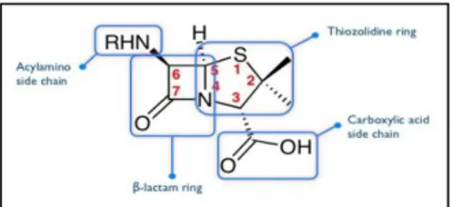
ß-lactams are classified according to their chemical structure: single ß-lactam ring (monobactam), or a fused ß-lactam ring with a 5 atoms penemic ring (penicillins and carbapenems) or fused with a cephalosporin ring 6 atoms (cephalosporins). Within these major groups, and differences in the site of attachment of the single or double-ring chains may have a significant effect on pharmacological properties and on the spectrum of ß-lactams
Penicillin is a group of antibiotics which include penicillin G (figure3)(intravenous
use), penicillin V (oral use), procaine penicillin, and benzathine penicillin (intramuscular use). Penicillin antibiotics were among the first medications to be effective against many bacterial infections caused by Staphylococci and Streptococci. Penicillins resistant to penicillinases enzymes are methicillin, nafcillin, oxacillin, have a narrower spectrum of action but are very active against S. aureus that produces penicillases.
20 Figure3: Penicillin chemical structure.
Ampicillin penetrate the gram-negative outer membrane, Piperacillin and ticarcillin are active also against Pseudomonas spp but are less effective than ampicillins given against gram-negative bacteria.
Cephalosporins (figure 4) are resistant to the hydrolysis of the penicillinases of the Staphylococci and ß-lactamases of gram negative bacilli. They are classified
according to generation I II III IV. The term generation indicates to discoveries that have historically allowed the expansion of the action spectrum by modifying the site
Figure 4: Cephalosporins chemical structure.
of attack of the chains. The last cephalosporin discovery of V generation is the ceftobiprole which is considered an anti-MRSA.
21 Carbapenems: Imipenem and meropenem are broad-spectrum antibiotics, that
penetrate gram positive and gram-negative bacteria and resist to ß-lactamase action. However, imipenem (figure 5) is hydrolyzed by renal dehydropeptidase-1 therefore it must be administered in association with cilastatin which allows an increase in the concentration of the drug.
Figure 5: imipenem chemical structure
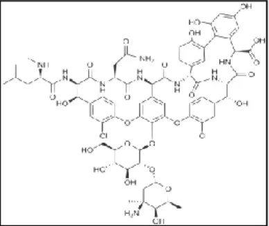
Glycopeptide antimicrobials are a class of drugs of microbial origin that are
composed of glycosylated cyclic or polycyclic non ribosomal peptides. Significant glycopeptide antibiotics include the anti-infective antibiotics vancomycin, teicoplanin, telavancin, ramoplanin and decaplanin, and the antitumor antibiotic bleomycin. Vancomycin (figure 6) is used if MRSA infection is suspected.
Some members of this class of drugs inhibit the synthesis of cell walls in susceptible microbes by inhibiting peptidoglycan synthesis. They bind to the amino acids within the cell wall preventing the addition of new units to the peptidoglycan. They bind to acyl-D-alanyl-D-alanine in peptidoglycan.
Figure 6: vancomycin chemical structure
22
2)Inhibitors of protein synthesis:
Macrolides: erythromycin (figure7) azitromycin claritromycin belong to a class
of natural products that consist of a large macrocyclic lactone ring to which one or more deoxy sugars, usually cladinose and desosamine, may be attached. The lactone rings are usually 14-, 15-, or 16-membered. The antimicrobial spectrum of macrolides is slightly wider than that of penicillin, and, therefore, macrolides are a common substitute for patients with a penicillin allergy. ß-hemolytic Streptococci,
pneumococci, Staphylococci, and Enterococci are usually susceptible to macrolides.
Figure 7: erythromycin chemical structure
Macrolides are protein synthesis inhibitors. The mechanism of action of macrolides is inhibition of bacterial protein biosynthesis, and they are thought to do this by preventing peptidyl transferase from adding the growing peptide attached to tRNA to the next amino acid as well as inhibiting ribosomal translation. [18]
Another potential mechanism is premature dissociation of the peptidyl-tRNA from the ribosome. [19]
Macrolide antibiotics do so by binding reversibly to the P site on the 50S subunit of the bacterial ribosome. This action is bacteriostatic.
23
Nucleic acid synthesis inhibitors:
Chinolones have a nucleus of two rings with six terms fused to each other and when the replacement in position 6 of the phenolic ring takes place with a fluorine atom they become fluochinolones, like ciprofloxacin, norflocacin, levofloxacin, ofloxacin Main target is DNA topoisomerase (gyrase) the enzyme responsible for cutting, supercoiling and welding of bacterial DNA during replication. DNA bacterial topoisomerase has four subunits, each of which is inhibited by every single quinolone.
Figure:.8 fluoroquinolones chemical structure
The increased activity at a lower frequency of occurrence of resistant strains seems due to the ability of the more recent fluoroquinolones to bind to different enzyme fingers. They are bactericidal antibiotics.
Have a wide spectrum of aerobic and facultative anaerobic action including
24
VI. Antibiotic resistance:
Resistance to antibiotics could be intrinsic or acquired.
Intrinsic or natural resistance is the constitutional insensitivity of a microorganism
to a certain antibiotic. Immutable over time, genetically determined. It manifests itself in all strains of the same species.
It depends on:
characteristics of the antibiotic
microorganism structures
lack of penetration of the drug in the microorganism.
Acquired resistance (informational variation) is the acquisition of new genetic
determinant of resistance for a specific strain originally sensitive to a chemotherapy, that bring to emergence of antibiotic resistance.
It can be divided into:
chromosomal or endogenous;
extrachromosomal or exogenous;
Chromosomal resistance
It is only 10-15% of all the acquired resistances (low frequency of onset)
It is achieved through a spontaneous mutational alteration of chromosome genetic information.
The antibiotic has a selective action (select the mutants resistant, inhibiting the sensitive cells. This resistance affects only antibiotic to which they are resistant mutants.
The same mutants can also be resistant to other antibiotics with similar characteristics (cross-resistance).
25 It is transmitted vertically through the offspring (from mother cell to
daughter cell).
Extrachromosomal resistance
It constitutes 90% of all resistances (high frequency of onset).
It originates for acquisition of new genetic information that it comes from other micro-organisms and enters the cell through the mechanisms of conjugation, transformation and transduction.
It concerns more antibiotics simultaneously (resistance multiple).
It is horizontal transmitted (genetic exchange).
It can also be transferred to microorganisms belonging to different species (contagious resistance).
It is due to genes present on plasmids or transposons (mobile genetics elements).
From an evolutionary perspective, bacteria use two major genetic strategies to adapt to the antibiotic “attack”:
A) mutations in gene(s) often associated with the mechanism of action of the compound;
B) acquisition of foreign DNA coding for resistance determinants through horizontal gene transfer (HGT);
A) In general, mutations resulting in antimicrobial resistance alter the antibiotic
action via one of the following mechanisms,
a) modifications of the antimicrobial target (decreasing the affinity for the drug, see below);
b) a decrease in the drug uptake;
26
d) global changes in important metabolic pathways via modulation of regulatory; networks. Thus, resistance arising due to acquired mutational changes is diverse and varies in complexity. [20]
B) Horizontal gene transfer: classically, bacteria acquire external genetic material
through three main strategies (figure 9);
a) transformation (incorporation of naked DNA) b) transduction (phage mediated)
c) conjugation
Figure 9: [21] Horizontal gene transfer Kennet todar, revew of bacteriology
Transformation is perhaps the simplest type of HGT, but only few clinically relevant bacterial species “naturally” incorporate naked DNA to develop resistance. Emergence of resistance in the hospital environment often involves conjugation, a very efficient method of gene transfer that involves cell-to-cell contact and is likely to occur at high rates in the gastrointestinal tract of humans under antibiotic treatment. Conjugation uses mobile genetic elements (MGEs) as vehicles to share valuable genetic information, although direct transfer from chromosome to chromosome has also been well characterized [22] .
The most important MGEs are plasmids and transposons, both of which play a crucial role in the development and dissemination of antimicrobial resistance among clinically relevant organisms.
27
Furthermore, this genetic exchange has been implicated in the dissemination of resistance to many frequently used antibiotics. Acquisition of foreign DNA material through HGT is one of the most important drivers of bacterial evolution and it is frequently responsible for the development of antimicrobial resistance. Most antimicrobial agents used in clinical practice are products naturally found in the environment. As mentioned before, bacteria sharing the environment with these molecules harbor intrinsic genetic determinants of resistance and there is robust evidence suggesting that such “environmental resistome” is a prolific source for the acquisition of antibiotic resistance genes in clinically relevant bacteria.
Finally, one of the most efficient mechanisms for accumulating antimicrobial resistance genes is represented by integrons, which are site-specific recombination systems capable of recruiting open reading frames in the form of mobile gene cassettes. Integrons provide an efficient and rather simple mechanism for the addition of new genes into bacterial chromosomes, along with the necessary machinery to ensure their expression; a robust strategy of genetic interchange and one of the main drivers of bacterial evolution. [23]
Antibiotic resistance mechanisms can be classified in: I) modifications of the antimicrobial molecule;
II) prevention to reach the antibiotic target (by decreasing penetration or actively extruding the antimicrobial compound);
III) changes and/or bypass of target sites;
IV) resistance due to global cell adaptive processes;
I) MODIFICATIONS OF THE ANTIMICROBIAL MOLECULE:
One of the most successful bacterial strategies to cope with the presence of antibiotics is to produce enzymes that inactivate the drug by adding specific chemical moieties to the compound or that destroy the molecule itself, rendering the antibiotic unable to interact with its target.
28 I.A) Chemical alterations of the antibiotic: the production of enzymes capable of
introducing chemical changes to the antimicrobial molecule is a well-known mechanism of acquired antibiotic resistance in both gram-negative and gram-positive bacteria. Interestingly, most of the antibiotics affected by these enzymatic modifications exert their mechanism of action by inhibiting protein synthesis at the ribosome level [24]
I.B). Destruction of the antibiotic molecule: The main mechanism of β-lactam
resistance relies on the destruction of these compounds by the action of β- lactamases. These enzymes destroy the amide bond of the β-lactam ring, rendering the antimicrobial ineffective.
Infections caused by penicillin-resistant S. aureus became clinically relevant after penicillin became widely available and the mechanism of resistance was found to be a plasmid-encoded penicillinase that was readily transmitted between S. aureus strains, resulting in rapid dissemination of the resistance trait. [25]
II. Decreased Antibiotic Penetration and Efflux II.A) Decreased permeability
Many of the antibiotics used in clinical practice have intracellular bacterial targets or, in case of gram-negative bacteria, located in the cytoplasmic membrane (the inner membrane) This mechanism is particularly important in gram-negative bacteria. [26]
II.B) Efflux Pumps many classes of efflux pumps have been characterized in both
gram-negative and gram-positive pathogens. These systems may be substrate-specific or with broad substrate specificity, which are usually found in MDR bacteria. The genes encoding efflux pumps can be in MGEs or in the chromosome.
III) Target sites modification
A common strategy for bacteria to develop antimicrobial resistance is to avoid the action of the antibiotic by interfering with their target site. To achieve this, bacteria
29
have evolved different tactics, including target protection and target modifications that result in decreased affinity for the antibiotic.
III.A) Target protection
Examples of drugs affected by this mechanism include tetracycline (Tet[M] and Tet[O]), fluoroquinolones (Qnr) and fusidic acid (FusB and FusC). One of the classic and best-studied examples of the target protection mechanism is the tetracycline resistance determinants Tet(M) and Tet(O). Tet(M) was initially described in
Streptococcus spp. TetO and TetM interact with the ribosome and dislodge the
tetracycline from its binding site in a GTP-dependent manner. These proteins belong to the translation factor superfamily of GTPases and act as homologues of elongation factors (EF-G and EF-Tu) used in protein synthesis. TetM directly dislodges and releases tetracycline from the ribosome by an interaction between the domain IV of the 16S rRNA and the tetracycline binding site. this interaction alters the ribosomal conformation, preventing rebinding of the antibiotic [27]
III.B.1) Target modification
Introducing target modifications is one of the most common mechanisms of antibiotic resistance in bacterial pathogens affecting almost all families of antimicrobial compounds. These target changes may consist of i) point mutations in the genes encoding the target site, ii) enzymatic alterations of the binding site (addition of methyl groups), and/or iii) replacement or bypass of the original target. As mentioned, regardless of the type of change, the final effect is always the same, a decrease in the affinity of the antibiotic for the target.
Fluoroquinolones kill bacteria by altering DNA replication through the inhibition of two crucial enzymes, DNA gyrase and topoisomerase IV. Development of chromosomal mutations in the genes encoding subunits of the above-mentioned enzymes (gyrA-gyrB and parC-parE for DNA gyrase and topoisomerase IV, respectively) is the most frequent mechanism of acquired resistance to these compounds. Importantly, since FQs interact with two enzymes (DNA gyrase and
30
topoisomerase), and both are essential for bacterial survival, the level of resistance achieved by developing changes in one of the enzymes will depend on the potency with which the antimicrobial inhibits the unaltered target.
III.B.2. Enzymatic alteration of the target
One of the best characterized examples of resistance through enzymatic target modification is the methylation of the ribosome catalyzed by an enzyme encoded by the erm genes (erythromycin ribosomal methylation), which results in macrolide resistance.
In Staphylococci, the most important erm genes are ermA (mostly distributed in a transposon in MRSA) and erm(C) (found in plasmids in methicillin-susceptible S.
aureus).
These enzymes are capable of mono- or dimethylating an adenine residue in position A2058 of the domain V of the 23rRNA of the 50S ribosomal subunit. [28]
III.B.3. Complete replacement or bypass of the target site
Using this strategy, bacteria are capable of evolving new targets that accomplish similar biochemical functions of the original target but are not inhibited by the antimicrobial molecule. The most relevant clinical examples include methicillin resistance in S. aureus due to the acquisition of an exogenous PBP (PBP2a) and vancomycin resistance in Enterococci through modifications of the peptidoglycan structure mediated by the van gene clusters.
Resistance to methicillin (a semisynthetic penicillin stable against the staphylococcal penicillinase) in S. aureus results from the acquisition of a foreign gene (likely from
Staphylococcus sciuri) designated mecA often located in a large DNA fragment
designated staphylococcal chromosomal cassette mec (SCCmec). The mecA gene encodes PBP2a, a PBP that has low affinity for all β-lactams, including penicillins, cephalosporins (except for last generation compounds) and carbapenems. Acquisition
31
of mecA renders most β-lactams useless against MRSA and alternative therapies need to be used in serious infections. Of note, PBP2a carries a transpeptidase domain, but it does not function as a transglycosylase (class B PBP), therefore, it requires the activity of other native PBPs to perform the latter function and fully crosslink peptidoglycan. Specifically, the penicillin-insensitive transglycosylase domain of PBP2 (a class A PBP) is particularly important to achieve transglycosylation of peptidoglycan in the presence of β-lactams in mecA-carrying MRSA isolate.
mecA gene is usually found as part of a gene cassette inserted into a larger MGE
(SCCmec), whose basic components include mecA, mecR1 (encoding the signal transducer protein MecR1), mecI (encoding the repressor protein Mecl), and ccr (encoding a recombinase; cassette chromosome recombinase). To date, 11 different SCCmec allotypes have been described with varying degrees of genetic homology and different sizes, insertion sequences and accompanying resistance genes [29] Importantly, SCCmec types seem to differ between different MRSA clones. Indeed, community-associated MRSA strains appear to harbor shorter SCCmec cassettes (SCCmec type IV) and carry less antibiotic resistance determinants, whereas hospital-associated (HA) isolates possess longer elements (SCCmec type II) and are usually multidrug resistant.
VII. BETA-LACTAMS RESISTANCE MECHANISMS IN S.
AUREUS
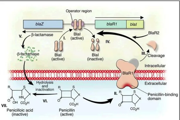
Mechanisms of resistance to lactam antibiotics is mediated in the production of β-lactamase, widely spread enzymes between Gram-positive and Gram-negative bacteria; they hydrolyze the amide linkage of the β-lattamic ring of penicillins and cephalosporins with the production of an lactam inactive derivative. [30] The β-lactamase production is plasmid encoded by the blaZ gene. blaZ is under the control of two adjacent regulatory genes, the blaR1 antirepressor and the blaI repressor. [31] Following exposure to β-lactams, BlaR1, a transmembrane sensor-transducer, cleaves itself. The hypothesis is that the cleaved protein functions as a protease that cleaves
32
the repressor BlaI, directly or indirectly (an additional protein, BlaR2, may be involved in this pathway) and allows blaZ to synthesize enzyme. [32] (Fig 9).
Figure 9: Induction of staphylococcal lactamase synthesis in the presence of the β-lactam antibiotic penicillin. Antimicrobial resistance [33]
The DNA-binding protein BlaI binds to the operator region, thus repressing RNA transcription from both blaZ and blaR1-blaI. In the absence of penicillin, β-lactamase is expressed at low levels. II. Binding of penicillin to the transmembrane sensor-transducer BlaR1 stimulates BlaR1 autocatalytic activation. III–IV. Active BlaR1 either directly or indirectly (via a second protein, BlaR2) cleaves BlaI into inactive fragments, allowing transcription of both blaZ and blaR1-blaI to commence. V–VII. β-Lactamase, the extracellular enzyme encoded by blaZ (V), hydrolyzes the β-lactam ring of penicillin (VI), thereby rendering it inactive (VII).
33
VIII. METHICILLIN RESISTANCE
The first MRSA strain appeared in 1961[34], one year after the introduction use of methicillin in therapy; MRSA strains subsequently spread to become a world-class problem. Several chromosomal genes are implicated in the phenotypic expression of methicillin-resistance, giving higher levels of staphylococcal resistance.
Level of resistance to β-lactams in MRSA is the result of the acquisition of the mecA gene, which encodes for penicillin-binding protein 2a (PBP2a).
The main mechanism of resistance is production of an auxiliary penicillin binding protein, PBP2a/PBP2c which renders the isolate resistant to all b-lactam except the novel class of specific ‘anti-MRSA’ cephalosporins. These agents have sufficiently hight affinity to PBP2a, and probably also the PBP encoded by mecC, to be active against MRSA. The auxiliary PBPs are encoded by the mecA gene or the recently described mecC gene. [35]
mec element is foreign to S. aureus and is not present in methicillin susceptible S. aureus. strains with marked heterogeneous expression of the mecA gene and
frequently low MICs of oxacillin hamper the accuracy of susceptibility testing. Some isolates express low level resistance to oxacillin, they are mecA and mecC negative and do not produce alternative PBPs (borderline susceptible S aureus BORSA.), these strains are relatively rare, and the mechanism of resistance is poorly characterized, but may include hyperproduction of b-lactamases or alteration of the pre-existing PBPs. [36]
IX. Recommended methods for detection of methicillin resistance in S.
aureus
Methicillin/oxacillin resistance can be detected phenotypically by MIC determination and by disk diffusion. Agglutination can be used to detect PBP2a, but it will not reliably to detect PBPc. Genotypic detection with PCR is reliable.
34 Detection by MIC determination or disk diffusion.
The heterogeneous expression of resistance particularly affects MICs of oxacillin, which can appear susceptible. Cefoxitin is a very sensitive and specific marker of mecA/ mecC mediated methicillin resistance including in heterogeneous expressing strains and is the agent of choice. Disk siffusion using oxacillin is discouraged and interpretative zone diameters are no longer included in the EUCAST breakpoint table due to poor correlation with the presence of mecA.
A. Broth microdiluition:
Strandard methodology (ISO 20776-1) is used and strains with cefoxitin MICs ≥4 mg/L should be reported ad methicillin resistant.
B. Disk diffusion:
The EUCAST disk diffusion method is used. Strains with cefoxitin 30μg disk zone diameter ≥22 mm should be reported as methicillin resistant.
Detection with genotipyc and latex agglutination methods.
Genotypic detection of the mecA and mecC genes by PCR [37] and detection of the PBP2a protein with latex agglutination kits is possible using commercial or “in house” assays. PBP2c is not detected by most of commercial assays
Particularly interesting is the mecA gene encoding for methicillin-resistance. The
mecA gene is part of a mobile genetic element, the SCC mec, which is incorporated
into the bacterial. MRSA clones possess the mecA gene, and its mecR1-mecI regulatory genes. They are allocated on a genomic mobile island called staphylococcus chromosomal cassette mec (SCCmec, about 21-67 kb). This chromosomal cassette combines the entire operon mec (about 28kb) to the ccr gene, a complex that encodes for specific recombinase sites responsible for the mobility of SCC mec [38]. This mobility is essential for resistance, as strains of S. aureus
35
methicillin-sensitive (MSSA) capture SCC mec from MRSA strains. The SCC mec elements contain:
- the mec genes complex includes insertion sequences (IS431mec), the mecA gene, and the mecR1 and mecI regulating genes;
- the complex of ccr genes encoding for recombinase (ccr) responsible for the precise excision and integration of SCCmec within the bacterial chromosome, and is responsible for its mobility;
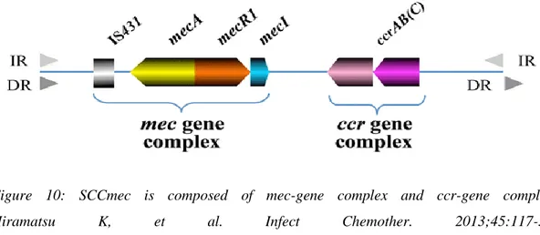
-the flattening regions the mec and ccr complexes are referred to as J (junkyard) regions, which do not appear to be essential or useful for bacterial cells, except where they contain genes for resistance to other antibiotics. The SCC mec elements are classified in types and subtypes. To date, 11 types of SCC mec have been identified [39,40, 41, 42].
Figure 10: SCCmec is composed of mec-gene complex and ccr-gene complex. Hiramatsu K, et al. Infect Chemother. 2013;45:117-36. Tsubakishita S, et al. Antimicrob Agents Chemother 2010;54:1469-75. [ 43]
36
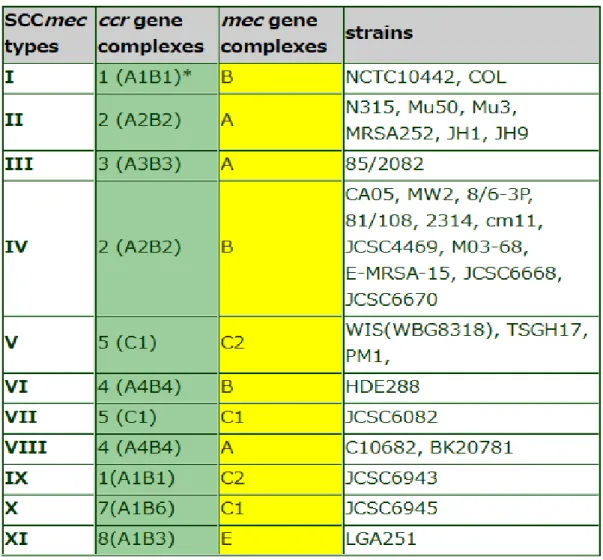
Most types of nosocomial MRSAs produce types I, II or III, while most of the EU-type CA-MRSA EU-types are EU-type IV or V, although EMRSA-15 codes Type IV. [44] Three classes of mec (A, B and C) and four subtypes of ccr complexes are known, which, by combining, generate five different SCCmec (I to XI) boxes (Tab. 3), distinct in various subtypes depending on differences in junkyard regions.
37
X. TYPING OF S. AUREUS
The spa typing technique uses the sequence of a polymorphic VNTR in the 3' coding region of the S. aureus-specific staphylococcal protein A (spa). Single locus DNA-sequencing of the repeat region of the Staphylococcus Protein A gene (spa) can be used for reliable, accurate and discriminatory typing of MRSA.Typing of S. aureus is crucial for preventing the spread of MRSA and for outbreak investigations [45]. A crucial factor in controlling MRSA is to know about the dissemination of MRSA and its clones, as S. aureus has a large clonal population structure, and thus a correct assignment of a strain to a clone is a highly important and essential part of the epidemiology and surveillance of MRSA. Typing of MRSA is used to support infection control measures. Different methods for S. aureus typing have been developed, all, which have their different strengths and weaknesses. A combination of different methods is necessary to obtain a correct assignment and have a high discriminatory power [45]. is the Genotypic methods consisting of multilocus sequence typing (MLST), spa typing, and SCCmec typing are among the mostly used typing method. The nomenclature is based on their sequence type (ST),
Staphylococcus protein A (spa) type and SCCmec type. The sequence type is a profile
of seven housekeeping genes, while the spa type is based on sequence polymorphism of the X-region in the spa gene. The sequence type is a multi-locus typing of S.
aureus, while spa typing is a single-locus typing. SCCmec type is the typing of the
mobile genetic element encoding the methicillin resistance in S. aureus strains [5]. A. SPA TYPING
The spa typing is based on sequencing of region X of the spa gene, a region that mainly consist of 24-bp repeats. These repeats are assigned a numerical code from which the spa type is determined. spa typing is much more simple and accessible than MLST, as it only requires sequencing of a single locus, which often can be performed by an in-house sequencing platform. Because of its higher discriminatory power, spa typing is more specific compared to MLST. Due to its high discriminatory
38
gene marker, spa typing can further differentiate a collection of ST. An ST can consist of several spa types, and thus spa typing can be used for evolutionary purpose as well as under outbreak situations [3]. The diversity of spa types is due to deletions, duplication or point mutations of the repeats in the spa gene [3].
DNA sequences of the spa gene therefore provide portable and biologically meaningful molecular typing data that have demonstrated their utility for macro- and micro-epidemiological purposes from surveillance through to outbreak investigations at various geographical levels [46][47].
www.spaserver.ridem. last visited OCTOBER 12, 2017
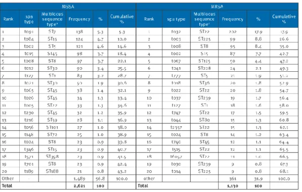
Table 3: the 20 most frequent spa types and multilocus sequence typing among meticillin-sensitive S. aureus and meticillin-resistant S. aureus isolates collected in 25 European countries in 2011. [46]
39
For MSSA, the top 20 ranking spa types included 43.2% of all MSSA isolates (Table 3). Importantly, there was very little difference among the first 11 ranking spa types between the 2011 and 2006 datasets. Only changes in rank order were observed. Ranks 12 to 20 contained four new spa types in 2011.
The figure 2 shows for MRSA the top 20 ranking MRSA spa types contained 68.1% of all MRSA isolates (73.4% in 2006). There were no differences in the top six spa types. [48]
Figure 2: comparison of meticillin-resistant Staphylococcus aureus spa-type frequencies, 2011 and 2006[46]
Among MRSA isolates, a dynamic expansion was demonstrated for several spa types. MRSA isolates with spa types belonging to ST22 increased most markedly making ST22 the most critically expanding MRSA clone in Europe. This lineage (designated EMRSA-15) was first described during hospital outbreaks in England. [49].
40 SCCmec TYPING :
SCCmec elements are classified by a hierarchical system into “types” and “subtypes”. “Types” are defined by the combination of (1) the type of ccr gene complex, which is represented by ccr gene allotype, and (2) the class of the mec gene complex. These are the key elements of the cassette responsible for integration and excision of SCCmec, and the beta-lactam resistance phenotype, respectively. To date, no excellent technique for SCCmec typing exists. After the SCCmec structure was recognized, different attempts for a SCCmec typing method have been developed. However, they are all not definitive typing methods as they often lack the ability to detect one or more types. The most promising SCCmec typing method was developed by Kondo et al. (2007) [50] and is based on conventional polymerase chain reaction (PCR), as conventional PCR remains the most convenient, common and easiest to implement in laboratories. This technique is based on a complex combination of multiplex-PCRs (M-PCR). The method is based on four M-PCRs; the first and second M-PCRs are used to recognize the SCCmec type, the third M-PCRs are used for subtyping purposes while the fourth M-PCRs is used for identification of transposons and plasmids. The primers are designed to target genes in the mec gene complex, ccr gene complex, genes relevant for subtyping, and for additional transposons and plasmids that are known to be found in SCCmec elements. Based on the amplicons from all four M-PCRs, a SCCmec typing can be determined. Often, using just M-PCR 1 and M-PCR 2 is sufficient as using these two M-PCRs can yield the SCCmec type. The advantage of the SCCmec typing method by Kondo et al. (2007) is that the nomenclature of identified SCCmec elements is based on the recommended nomenclature defined by The International Working Group on the Classification of Staphylococcal Cassette Chromosome Elements (IWG-SCC). The IWG-SCC was organized to: 1) form an intellectual network to contribute to the study of SCC elements; 2) establish a consensus on a uniform nomenclature system for SCC elements; 3) define minimum requirements for the description of new SCC
41
elements; and 4) establish guidelines for the identification of SCC elements for epidemiological study (i.e., SCCmec typing).
IWG-SCC was established for the development of a universal nomenclature system for SCCmec elements [5]. The group represented a nomenclature of SCCmec in which the SCCmec elements is designated by roman numerals followed with the mec gene complex and the ccr gene complex in parentheses. As an example, SCCmec type IV (2B), which indicates that it is a type IV SCCmec element, with a class 2 ccr gene complex and a class B mec complex (figure 11). The subtyping of SCCmec elements is based on the variation in the J1 region within the same SCCmec type. J1 region are designated based on the presence of specific DNA sequences, such as characteristic genes, pseudo genes, non-coding regions, and mobile genetic elements. However, a great disadvantage of this method is that it is rather time-consuming, quite sensitive and not fully developed, and should be further developed and evaluated. Another disadvantage is that, due to it is based on PCR, this method is unable to detect the presence of new alleles of the existing genes, and thus new SCCmec elements.
42
XI. STAPHYLOCOCCAL CASSETTE CHROMOSOME MEC
Since 1980s where the SCCmec element was recognized, it also being categorized as a genomic island. SCCmec, opposite to the other GIs, encodes genes for antibiotic resistance rather than virulence genes [6]. To this date, 11 different types of SCCmec elements have been identified in S. aureus (type I to XI) and are reported in figure 12.
Figure 12. Basic structure of SCCmec.
SCCmec is bracketed by direct repeats (DRs) that contain integration site sequence (ISS) recognized by cassette chromosome recombinase (CCR). A pair of inverted repeats (IRs) are present at the termini of SCCmec. Two critical gene complexes, ccr
43
and mec are present, and the other regions are designated J1, J2, and J3. The type of SCCmec is defined by the combination of the type of ccr-gene complex and the class of mec-gene complex. Subtype of the SCCmec is based on the difference in the J (standing for junkyard) regions. (B) Various types of SCCmec. Direct repeats that comprise integration site sequences of SCC are located at both extremities of SCCmec (the red arrowheads). The location of five (A-E) classes of mec-gene complexes is indicated by pink belt. The locations of ccr-gene complexes are indicated by blue belt. Insertion sequences and transposons are indicated in yellow. Representative genes related to heavy metal resistance and integrated plasmids located in the J regions are also indicated. Type XI is a newly identified SCCmec found in the MRSA strains of bovine source [52], [53]
They all contain the same backbone structure, which consist of a mec gene complex, a ccr gene complex and three joining (J) regions (figure 13). The different types of SCCmec elements is due to difference in the gene complexes, however they are still organized in the same way. The SCCmec elements are classified into types based on the combination of the mec gene complex and ccr gene complex, while subtyping of SCCmec is based on the variation in the J1 region.
www.sccmec.org – last visited June 14, 2016
Figure 13. Schematic representation of the organization of the backbone structure of SCCmec
The SCCmec element makes up approximately 1-2% of the total genome size of S.
aureus, and varies in size, ranging from approx. 0.1 kb to 34 kb3. They are composed
44
been characterized, and the ccr gene complex, of which eight different types have been characterized (table 4.2 and 4.3, respectively).
www.sccmec.org/Pages/SCC_ClassificationEN.html - last visited June 25, 2016 A. mec gene complex
The mec gene complex is composed of mecA, its regulatory genes, and associated insertion sequences. The class A mec gene complex (class A mec) is the prototype complex, which contains mecA, the complete mecR1 and mecI regulatory genes upstream of mecA, and the hyper-variable region (HVR) and insertion sequence IS431 downstream of mecA. The class B mec gene complex (class B mec) is composed of mecA, a truncated mecR1 resulting from the insertion of IS1272 upstream of mecA, and HVR and IS431 downstream of mecA. The class C mec gene complex (class C mec) contains mecA and truncated mecR1 by the insertion of IS431 upstream of mecA, and HVR and IS431 downstream of mecA. There are two distinct class C mec gene complexes; in the class C1 mec gene complex, the IS431 upstream of mecA has the same orientation as the IS431 downstream of mecA (next to HVR), while in the class C2 mec gene complex, the orientation of IS431 upstream of mecA is reversed. C1 and C2 are regarded as different mec gene complexes since they have likely evolved independently. The class D mec gene complex (class D mec) is composed of mecA and ΔmecR1, it does not carry an insertion sequence downstream of ΔmecR1 (as determined by PCR). mec gene complex is the complex responsible for the antibiotic resistance of MRSA stains. It encodes for the mecA gene, which is the single determinant for methicillin resistance or the mecALGA251, also known as the mecC gene. Both genes encode for a penicillin-binding protein (PBP2a or PBP2’), which has a low affinity towards β-lactam antibiotics. The mec gene complex additional encodes for the regulatory genes, mecR1 and mecI, and insertion sequence(s). mecR1 is a transmembrane β-lactam-sensing signal transducer, which senses the absence or presence of β-lactam antibiotics, while mecI is a repressor that represses the transcription of mecA and mecR1-mecI complex in the absence of the β-lactam antibiotics. Differences in the mec gene complex is due to the insertion of
45
insertion sequences, IS431 and IS1272, in the mecR1 and/or mecI genes resulting in truncated products. In total, there are six major classes of the mec gene complex (table 4.2), all which is a divergent type of the prototype gene complex (figure 14). The minor classes are variants within one of the major classes, such as class A3 and A4.
46
B. ccr gene complex
The ccr gene complex is composed of the ccr gene(s) and surrounding open reading frames (ORFs) several of which have unknown functions. Currently, three phylogenetically distinct ccr genes, ccrA, ccrB, and ccrC, have been identified in S.
aureus with DNA sequence similarities below 50%. To date, the ccrA and ccrB genes
that have been identified in S. aureus have been classified into four allotypes. These allotypes are also found in other staphylococcal species as well as other allotypes have been described for these species only. In general, ccr genes with nucleotide identities of more than 85% are assigned to the same allotype, whereas, ccr genes that belong to different allotypes have lower nucleotide identities of between 60% and 82%, each other. All ccrC variants identified to date in staphylococcal strains have shown ≥87% similarity; thus, there is only one ccrC allotype. They suggest describing their differences as alleles by using previously used numbers, e.g., ccrC1 allele 2 or ccrC1 allele 8. The cassette chromosome recombinases (ccr) gene complex encodes gene(s) for the DNA recombinase enzyme of the invertase/resolvase family, which catalyzes the excision and insertion of the SCCmec element, and thus this complex is responsible for the movement of the SCCmec element into the staphylococcal chromosome. Integration of SCCmec elements into the chromosome of MSSA strains is a specific site integration. The integration happens at a unique 15-bp sequence called the integration site sequence (ISS) of the bacterial chromosomal attachment site (attBscc), which is located near the 3’ end of orfX, which is an open reading frame of unknown function. [50]. Two groups of ccr genes have been reported: (1) homologous pairs of ccrA and ccrB gene and (2) one ccrC gene. As with the mec gene complex, different the ccr gene complex exists. To date there is eight different types; all which differ in their combination of either their homologue pairs, having only one ccrC genes or a mix of both (table 4).
47
C. Joining regions
Besides the mec and ccr gene complexes, the SCCmec element also contains three so-called J regions, which constitute nonessential components of the cassette. J1 (formerly L-C) is the region between the right chromosomal junction and the ccr gene complex; J2 (C-M) is between the ccr gene complex and the mec gene complex; and J3 (I-R) is between the mec gene complex and the left chromosomal junction. Variations in the J regions within the same mec-ccr gene complex are used for defining SCCmec subtypes. Joining (J) regions formerly known as junkyard regions, are as their name indicate regions joining the two gene complexes (mec and ccr gene complex) together. There are three J regions within each SCCmec element and encode for non-essential components of the cassette. Even though they constitute for non-essential components, they have importance for epidemiological and diagnostically purposes as these regions might carry additional resistance genes by the carriage of plasmid(s) and/or transposon(s), but also because subtyping of SCCmec element is based on difference in the J1 region within a SCCmec type [5].
![Figure 9: [21] Horizontal gene transfer Kennet todar, revew of bacteriology](https://thumb-eu.123doks.com/thumbv2/123dokorg/8241612.129198/26.918.179.500.406.623/figure-horizontal-gene-transfer-kennet-todar-revew-bacteriology.webp)
![Figure 2: comparison of meticillin-resistant Staphylococcus aureus spa-type frequencies, 2011 and 2006 [46]](https://thumb-eu.123doks.com/thumbv2/123dokorg/8241612.129198/39.918.181.778.392.728/figure-comparison-meticillin-resistant-staphylococcus-aureus-type-frequencies.webp)
![Figure 11. Representation of the SCCmec typing method by Kondo et al. (2007). [51]](https://thumb-eu.123doks.com/thumbv2/123dokorg/8241612.129198/41.918.172.793.624.1004/figure-representation-sccmec-typing-method-kondo-et-al.webp)
