SAPIENZA Università di Roma Facoltà di Farmacia e Medicina
Ph.D. in
MORPHOGENESIS AND TISSUE ENGINEERING XXXII Ciclo
(A.A.2018-2019)
c-MET activated pathways and their implication in TGCTs oncogenesis
Ph.D. Student Erica Leonetti
Tutors Coordinator
Prof. Angiolina Catizone
A mamma e papà, fonte di amore costante. Ai miei nonni meravigliosi, il cui affetto è scolpito indelebile nel mio cuore, con la speranza di averli resi orgogliosi
ovunque essi siano. A zia Emma, che ha creduto in me e mi ha amata sin dal principio.
A Francesco, che mi ha tenuta per mano in ogni tappa del mio percorso.
A me stessa, per aver sempre trovato la
INDEX 1. Summary ... 9 1.1 Biological issue ... 9 1.2 Results obtained ... 9 1.3 Conclusions ... 10 2. Introduction ... 11
2.1 Germ Cell Tumours (GCTs) ... 11
2.1.1 Classification ... 11
2.1.2 Testicular Germ Cell Tumours (Type II) ... 11
2.1.3 Origin of Type II TGCTs ... 12
2.1.4 Risk Factor ... 14
2.1.5 Chemio- and radio- resistance acquisition ... 18
2.1.6 Cell lines and models ... 20
2.2 HGF/c-MET system ... 20
2.2.1 Hepatocyte Growth Factor (HGF) ... 20
2.2.2 Mesenchymal Epithelial Transition (c-MET) ... 21
2.2.3 HGF/c-MET signal transduction ... 23
2.2.4 HGF/c-MET in testis ... 26
2.2.5 HGF/c-MET de-regulation in human cancer ... 27
2.3 c-MET in TGCTs ... 30
2.3.2 My previous work ... 31
3. Aims ... 33
4. Results ... 35 4.1 c-MET modulates the migration of NT2D1 cells induced by HGF . 35
4.1.1 NT2D1 chemo-attraction is specifically driven by c-MET ... 35
4.1.2a c-MET modulates the collective migration of NT2D1 cells induced by HGF ... 36
4.2 c-Src is involved in HGF-dependent NT2D1 responses ... 41
4.2.1 Src inhibitor-1 does not affect cell viability ... 41
4.2.2 c-Src is involved in HGF-dependent NT2D1 proliferation ... 42
4.2.3 c-Src is specifically involved in HGF-dependent NT2D1 chemo-attraction ... 44
4.2.4 c-Src is involved in HGF-induced collective migration ... 45
4.2.5 c-Src is involved in HGF-dependent cells invasion ... 47
4.2.6 Phospho-c-Src detection after HGF administration ... 49
4.2.7 Immunofluorescence analysis of the active form of c-Src (phospho Tyr 416) ... 50
4.3. PI3K/AKT involvement in NT2D1 cellular responses ... 52
4.3.1 PI3K/AKT is active in NT2D1 cells and LY294002 does not affect cell viability ... 52
4.3.2 HGF-dependent NT2D1 proliferation is dependent on PI3K/AKT ... 53
4.3.3 PI3/AKT pathway is involved in HGF-dependent NT2D1 migration ... 54
4.3.4 PI3K/AKT pathway is involved in HGF-induced collective migration ... 56
4.3.5 PI3K is involved in HGF-dependent cells invasion ... 58
4.3.6 HGF induced NT2D1morphological modification through PI3K/AKT ... 59
4.4.1 Relation between PI3K/AKT and c-Src proteins ... 62
4.4.2 Cytoskeletal remodeling HGF-dependent in NT2D1 cells ... 63
4.5 HGF expression in TGCTs biopsies ... 67
4.5.1 HGF distribution pattern in TGCT histological samples ... 67
Discussion ... 71
5. Materials and Methods ... 77
5.1 Cell Culture ... 77
5.2 Cell Proliferation Assay ... 77
5.3 Cell Cycle FACS Analysis ... 78
5.4. Cell Death Assay ... 78
5.5 Chemotaxis Assay ... 78
5.6 Wound-healing Assay (Collective Migration Assay) ... 79
5.7 Matrigel Invasion Assay ... 80
5.8 Immunofluorescence Analyses and actin staining ... 81
5.9 Western Blot Analyses ... 82
5.10 Scanning Electron Microscopy ... 83
5.11 Immunohistochemical Analyses ... 83 5.12 Statistical Analyses ... 84 6. List of abbreviations ... 85 7. References ... 87 List of publications ... 99 Congress communications ... 101 Acknowledgments: ... 105
1. Summary
1.1 Biological issue
Type II Testicular Germ Cell Tumors (TGCTs) are a group of pathologies whose incidence has increased in the recent years in young men between 15 and 39 years old. Their pathogenesis is not yet entirely clear, but the survival rate in patients suffering from TGCTs is good. In fact, about 90-95% of these patients recover. However, the increased incidence of TGCTs and the fact that the onset of these tumours typically arises in young people make necessary the development of new drugs and therapies for the 5-10% of patients who develop drug resistance, radio-resistance or long-term toxicity. HGF/c-MET is a very complex system, which is essential during embryogenesis and necessary for the maintenance of tissue homeostasis. Moreover, it is well known that this system results de-regulated in the onset and progression of several human cancers. My group recently demonstrated the differential expression of c-MET receptor in Type II TGCT representative cell lines. Non-seminoma cells result in higher biological responses induced by HGF with respect to other cell lines analysed. c-MET expression was also observed in biopsies derived from patients affected by all type II TGCTs. In this thesis we investigate the various HGF/c-MET activated pathways and the role of these proteins in non-seminoma cell line responses in a HGF-dependent way. Moreover, HGF immunoreactivity was also evaluated in patients affected by TGCTs in order to have a better understanding on the TGCTs pathogenesis.
1.2 Results obtained
The obtained results demonstrated that c-Src and PI3K/AKT pathways are involved in the following HGF-dependent biological responses in non-seminoma cells: proliferation, migration and invasion increase. c-Src is constitutively active in NT2D1 cells and is involved also in proliferation, invasion and collective migration in an HGF-independent manner. On the other hand, also PI3K/AKT is involved in the endogenous NT2D1 collective migration and invasion.
HGF is expressed in biopsies derived from SE and EC. In particular, EC samples exhibited higher level of HGF with respect to SE.
1.3 Conclusions
Despite the fact that these tumours are mostly curable, the increased incidence recorded in the last four decades, correlated with the young age of the patients, make it necessary to have a better understanding of their pathogenesis. It is clear that new therapeutical strategies are needed. In a recent work we already demonstrated that HGF/c-MET system is expressed in TGCTs cell lines, and that HGF administration determines higher biological responses in non-seminoma cell lines. We also showed that these results are in line with in vivo studies, where c-MET immunoreactivity is observed, especially in all the most differentiated cancer specimens. To shed light on the onset and progression of TGCTs, herein we demonstrate that c-MET is responsible for the activation of c-Src and PI3K/AKT pathways in non-seminoma cells. Moreover, we show that the HGF expression has a different distribution in SE and in EC, thus suggesting that, very likely, the presence of HGF in testis microenvirenment plays a role in the pathogenesis of TGCTs, and especially of EC. Taken together, these results contribute to pave the way for more accurate therapies.
2. Introduction
2.1 Germ Cell Tumours (GCTs) 2.1.1 Classification
Though human testis appears as a relatively small organ, it can give rise to numerous neoplasms, which originate from the various cell lineages that compose the whole organ. Notably, the most frequent neoplasm that occurs in the testis originates from Germ Cells (~95%).
Human Germ Cell Tumours (GCTs) are a heterogeneous group of pathologies that origin in the male and female gonad, and that have been classified for the first time in 1946 by Friedman and Moore (1).
The classification of GCT is based on the developmental potential of these neoplasms. The last classification, published in 2017, replaces the old classification by the same authors in 2005. Nowadays, according to the latest classification, these tumours are divided in seven types and for this reason it differs from the classification in five groups in 2005 (2).
As far as the male gonad is concerned, five of these lesions are considered the most relevant, and, among these, type II Testicular Germ Cell Tumours represent the most common cancers in young men between 15 and 39 years old.
Table 1: Current Testicular Germ Cell Tumours classification (from João Lobo et al., 2019).
2.1.2 Testicular Germ Cell Tumours (Type II)
Type II GCTs can develop in testis, ovary, brain and mediastinum. Over 90% of type II GCTs occurs in the testis (3). These
neoplasms arise by a common precursor, called “Germ Cell Neoplasia In Situ” (GCNIS). From GCNIS derive both seminoma lesions (SE), and non-seminoma tumours (NST). Approximately, 50% of TGCTs are pure seminoma, whereas 44% are non-seminoma tumours (4). The median age at diagnosis is 33 years-old, specifically 25 for NSTs versus 35 for SEs (5). All type II TGCTs may metastasize to lungs, liver, retroperineal lymph nodes, brain and bone. These tumours show high potential of differentiation (these cancer cells appear often multipotent or totipotent) and the origin of their pathogenesis is probably due to an alteration of the spermatogonial niche. In detail, the differentiation of the germ cells is determined by down-regulation of pluripotency genes and up-regulation of germ cell specific genes. Disturbed Sertoli cells/niche might interfere with downregulation of OCT4, normally co-expressed with TPSY, which respectively protect germ cells from apoptosis and stimulate their proliferation (6).
2.1.3 Origin of Type II TGCTs
The origins of type II TGCTs is still a matter of debate, however the most accreditated theory indicates that they derive from the arrest of the differentiation of some gonocytes, during prenatal development. These cells give rise to a pre-cancer lesion, called pre-germ cell neoplasia in situ (pre-GCNIS). This cancer lesion remain quiescent until puberty and in this period acquires genomic alteration; then, hormonal changes, during puberty, lead to complete GCNIS features acquisition (7, 8).
GCNIS, as previously mentioned, is the common precursor of all type II TGCTs. Its nomenclature has changed many times. Niels Skakkebaek was the first researcher that identified abnormal spermatogonia in two different biopsies, both of which develop into cancer. In 1972 Skakkebaek called this lesion carcinoma in situ (CIS). Only starting from 2015 the lesion was identified as GCNIS, when Prof. Leendert Looijenga proposed this term during a meeting in Zurich (9, 10). GCNIS can lead to seminoma tumours characterize by the expression of germ cell specific genes (“default
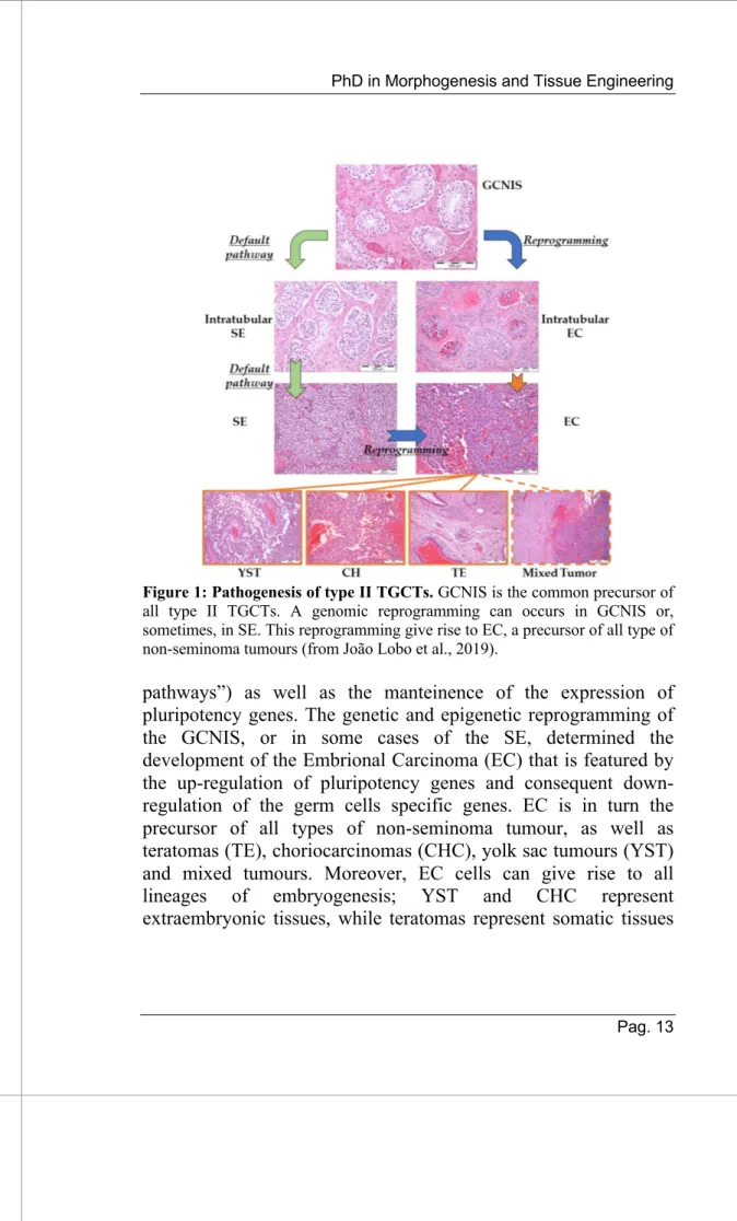
Figure 1: Pathogenesis of type II TGCTs. GCNIS is the common precursor of all type II TGCTs. A genomic reprogramming can occurs in GCNIS or, sometimes, in SE. This reprogramming give rise to EC, a precursor of all type of non-seminoma tumours (from João Lobo et al., 2019).
pathways”) as well as the manteinence of the expression of pluripotency genes. The genetic and epigenetic reprogramming of the GCNIS, or in some cases of the SE, determined the development of the Embrional Carcinoma (EC) that is featured by the up-regulation of pluripotency genes and consequent down-regulation of the germ cells specific genes. EC is in turn the precursor of all types of non-seminoma tumour, as well as teratomas (TE), choriocarcinomas (CHC), yolk sac tumours (YST) and mixed tumours. Moreover, EC cells can give rise to all lineages of embryogenesis; YST and CHC represent extraembryonic tissues, while teratomas represent somatic tissues
with expression features of all the three germ layers of the developing embryo (2).
The clinical interest about this type of tumours is enhanced in the last decades, due to an increase of their incidence especially in the Western countries, such as Europe, North America, South America, and Australia. In particular, it is worth mentioning that the highest incidence was recorded in Denmark, Norway and Switzerland (11-13). The global incidence is 1.5 affected individuals per 100.000 with difference of 20-times between area with the lowest and higher incidence (14). This non-randomic distribution of these pathologies has suggested that environmental factors, more available in high developed countries, can lead the development of these pathologies. However, the high variety of incidence among different ethnic groups in the same society demonstrate also the genetic relevance in the pathogenesis of these tumors. TGCTs is in fact a notable example of the close relationship between genetic and environmental factor which act in synergy with each other promoting the malignant transformation. The concept that fuses both factors has led to creating a new word called ‘genvironment”.
2.1.4 Risk Factor
2.1.4.1 Environmental Risk
Previously mentioned block of the gonocytes is due to an altered microenvironment, which lead to a multivariegate syndromes, as well as the Testicular Dysgenesis Syndrome (TDS), which increase approximately five times the risk to develop TGCTs (15). TDS is considered as a relatively mild disturbance of sex differentiation, due to hypovirilizing factors in utero (16-18). Features of TDS, which confer higher risk to develop TGCTs, include cryptorchidism, previous inguinal hernia, hypospadias, previous testicular cancer (TGCTs are bilateral in 3-4% of the cases), impaired spermatogenesis and familial history of testicular type II GCT.
Beside, other important risk factors in the onset of TGCTs are considered the low birthweight, short gestational age, twin, tall stature, first born child and disturbed hormonal conditions in utero (2).
Literature data demonstrated contrasting hypotheses but it seems that in the pathogenesis of type II TGCTs contribute also external environmental risk factor which act post-natally, such as diet (in particular the use of high in fat and dairy products), exposure to hormone disruptive organochlorine insecticides which influences the masculinization process, or a jobs like fire-fighting and aircraft- maintenance. Cannabis smoking is also considered a risk factor for the onset of TGCTs, and, in particular, it seems to be involved predominantly in the non-seminoma development. The last overview highlights that cannabis smoking can promote the reprogramming of seminoma tumours (19-25).
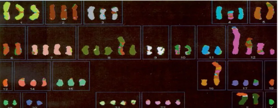
Figure 3: Spectral karyotype of type II GCT. Normally each chromosome have a specific color, but appears clear that chromosome 12 contains other chromosome fragments (from Oosterhuis H.W. and Looijenga L.H.J., Pathology and Biology of Human Germ Cell Tumors, Nogales, R.E. Jimenez (eds.), Springer-Verlag GmbH, Germany 2017).
2.1.4.2 Genetic Risk
A hallmark of type II TGCTs is their poliploidy. One of the peculiarity of their chromosome remodelling is the presence of duplication of the short arm of chromosome 12, frequently present
as isochromosome 12p (7, 26-28). On chromosome 12 are localized NANOG and STELLAR, genes that are involved in the maintenance of pluripotency, and cyclin D2 gene that regulates positively cell proliferation (29). Therefore, the gain of function of chromosome 12 promotes the block of differentiation of gonocytes as well as the increase of their proliferation. In addition, it has been reported that TGCTs are characterized also by other genomic aberrations that are the duplication of chromosomes 7, 8, 14, 15, 17 and X (30-32). Among them, the mentioned gain of 17q is frequent in non-seminoma tumours (33, 34).
In spite of all these chromosomal anomalies, there are few gene mutations identified as a risk factor for the onset and progression of TGCTs. However, it should be highlighted that mutations of KIT gene and the de-regulation of KIT/KITL pathway, important in the PGC/gonocyte survival and migration, represents one of the risk factor for the onset of TGCTs. This de-regulation is peculiar in particular in the seminoma tumors (35-37). Somatic mutations of KRAS and NRAS genes have been found both in seminoma and non-seminoma lesion, as observed in other solid tumours.
Moreover, very recently, a mutation of PDE11A
(phosphodiesterase 11A) was discovered in a familial form of this pathologies. Notably, in sporadic cases, the de-regulation of sex determination/germ cell specification, as DMRT1 and TPSY genes are involved in TGCTs pathogenesis (38, 39). Disorders of sex development (DSD, in its more aggressive form: 46,XY, characterize by atypical development of the gonads, sexual organs and/or secondary sex characteristics and considered as the most severe form of TDS), is also implicated in the onset of TGCTs. Essentially a defect in any gene involved in gonadal development and sex differentiation may cause DSD. These defects are considered to affect gonadal development and increasing the risk for GCNIS and TGCT. An example is the mutation in AR gene, related to DSD, that are associated with high risk of germ cell neoplasia (15, 18). Furthermore, it is known that a deletion in azoospermia factor c region of Y (AZF) represent a risk factor in
TGCTs oncogenesis (40, 41). Finally, Insulin-like growth factor receptor-1 (IGF1R) has an important role in the proliferation and survival of the PGCs and gonocytes. In a recent study phosphorylation of IGF1R was found in non-seminoma cell lines and was observed that it can promote acquired cisplatin resistance in non-seminoma cells (42, 43).
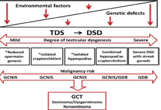
Figure 3: Schematic representation of the pathogenesis of germ cell tumours (GCT). The implication of testicular dysgenesis syndrome (TDS), predominantly caused by environmental factors, and DSD predominantly genetically determined, in GCT pathogenesis are shown. (from Jorgansen Anne et al.; 2015).
2.1.4.3 Epigenetics
With greater understanding on onset and progression of TGCTs, epigenetic acquired more and more importance. Study of type II TGCTs demonstrated that they are characterized by demethylation and erasure of parental imprinting. In fact, during the embryonic period, the Primordial Germ Cells (PGCs) are imprinted with female features. When testis develop, PGCs surronded by Sertoli cells become pre-spermatogoni and went to a de-methylation process. After that a "new primary metilation" is generated. This mechanism is sex-dependent, hence the genome of male and female gametes has different metylation patterns. In the de-methylation period the cell is more sensitive to mutational events.
Several studies demonstrated that serum of patients affected by type II TGCTs showed epigenetic alteration in correlation with healthy samples, and metilation of BRCA1 or RAD51C genes are only few examples of the implication of epigenetics in the TGCTs pathogenesis (2, 44-46).
2.1.4.4 Genvironmental hypotesis
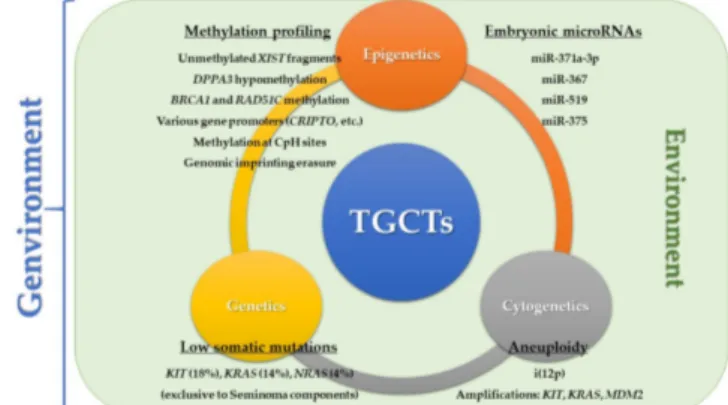
The interplay between environmental and genetic risk is called “Genvironmetal hypotesis”. These pathologies represent in an exemplary way the interchange between the environment and genetic and epigenetic factors in the development and progression of the disease.
Figure 4: Genvironmental Hypotesis. Representation of the genvironmental model focused on genetic, cytogenetic, and epigenetic factors, which are continuously modified and conditioned by environment (from João Lobo et al., 2019).
2.1.5 Chemio- and radio- resistance acquisition
Nowadays, testicular germ cells tumors are among the most curable solid tumors existent, with an average of 95% patients survival in the 10 years following the therapies (47, 48). The gold standard of the therapy in use for these tumors is based on a combination of radio- and chemo- therapy, with surgical removal. This protocol normally offers a good prognosis. Although, a little percentage of patients affected by TGCTs (approximately 5-10%)
developed resistance or long-term toxicity of the treatment previously described. Althoug these data seem positive, TGCTs represent the major cause of death in young men between 15-39 years old. . Because of the young age of the TGCTs onset, they have become cause of concern for the world cancer community. The most widely used chemotherapy protocols includes: BEP (bleomycin, etoposide, and cisplatin), EP (etoposide, cisplatin) and VIP (cisplatin, etoposide, ifosfamide) and the majority of the patients are treated with high doses of these drugs (49). As a consequence, patients can develop long terms toxicity due to at high cisplatin doses find in the plasma and urine also after years (50). The long-term toxicity can give origin to several diseases, as peripheral neuropathy, ototoxicity, secondary malignancies, cardiovascular toxicity, renal or pulmonary toxicity and infertility. On the other hand, other patients developed drug resistance with unclear mechanism. This acquired resistance kills about 5% of the patients. Even if the mechanism of resistance has been only partially elucidated, one of the hypotheses that are commonly considered is the decreasing of the cysplatin-dependant repair mechanism after DNA damage. TGCT patients show decreasing of these repair systems. Other supposed mechanisms of drug resistance are hypothesized, firstly the inactivation of p53 or de-regulation of PI3K/AKT pathway. In fact, normally p53 is not mutated in TGCTs patients even if they show high expression of MDM2, the principal physiologic antagonist of p53. Moreover, in EC it was observed high expression of OCT4 related with high expression of NOXA (Bcl-2 family member), suggest the involvement of OCT4 in the escape mechanism of apoptosis resistance-related drug (51).
Taken together, from these observations it appears clear that a better understanding of the onset and progression of these types of tumour are needed to make up new personalized therapies in order to improve quality of treatments and long-term status of the patients.
2.1.6 Cell lines and models
Type II TGCTs are probably unique in humans. In fact, no convincing examples of spontaneous or induced type II TGCTs have been reported in animals. The long time required for all the oncogenic steps may explain why there are no evidences of these pathologies in other animal models.
Regarding seminoma tumours, there is only one experimental model, called TCam-2 cell line, derived from a human primary TGCT with a seminoma component. This cell line is characterized by the expression of specific seminoma tumours markers, as well as NANOG and c-KIT, in combination with SOX17 (52, 53). Instead, there are some cell lines representative of non-seminoma tumours. The first established and well-characterized non-seminoma cell line is NT2D1. This cell line, used in my work, is a sub-clone (D1) derived from a human pluripotent embryonal carcinoma parent line NTERA-2. In turn, this cell line was established from a nude mouse xenograft tumour of TERA-2 cells, which were isolated from a lung metastasis of a 22 years old patient with primary embryonal carcinoma of the testis. NT2D1 cells express pluripotency genes, among which SOX2, OCT4 and NANOG (53). Noteworthy, they are negative for SOX17, specific for seminoma tumours. NT2D1 is widely used to study EC differentiation, in fact when this cell line is cultured at low density can differentiate, while at high density their pluripotency is maintained.
2.2 HGF/c-MET system
2.2.1 Hepatocyte Growth Factor (HGF)
The Hepatocyte Growth Factor (HGF), also known as Scatter Factor (SF), was described for the first time in 1989 (54).
It belongs to serin protease super-family, and it is normally secreted by cells of mesenchymal origins. Moreover,it acts as a pleitropic factor and cytokine promoting cell motility, survival, proliferation, differentiation and morphogenesis. HGF is secreted as a single-chain precursor and its activation is regulated at post-traslational level. In fact, the single chain precursor is converted in
an active form, a two-chain functional heterodimer extracellular protease, by an extracellular protease called HGF activator (HGFA) as well as the type II transmembrane enzymes metriptase and hepsin. It is also related to urokinase plasminogen activator (uPA) and tissue plasminogen activator (tPA) that are able to convert pro-HGF in two chain HGF. Active HGF form is composed by an a-chain of 69 kDa that binds a b-chain of 34 kDa through a disulfide bond. The a-chain is formed by an hairpin loop and four kringle domains, while b-chain is characterized by a serine protease homology domain which lacks proteolytic activity (55, 56).
Activation of HGF is finely regulated by the expression and secretion of two potent inhibitors of HGF activator knows as HGF activator inhibitor 1 (HAI1 also knows as SPINT1) and HGF activator inhibitor 2 (HAI2 or SPINT2).
2.2.2 Mesenchymal Epithelial Transition (c-MET)
c-MET is a member of receptor tyrosine kinases (RTKs) family, first discovered in 1984 (57) and normally expressed on the surface of epithelial cells. This receptor has a significant morphogenic and mitogenic role and it is essential during embryogenesis. It is also important in the adult life during wound healing,tissue repair, and homeostasis. In humans the proto-oncogene is located at 7q21q31 cromosome. The gene encoded for a single chain precursor of 150 kDa and, after translation, a glycosilation and a proteolitic cleavage are involved in the Golgi apparatus in the maturation of the receptor. The mature form of c-MET is a heterodimer of 190 kDa and it is composed by an a-chain of 50 kDa and a transmembrane b-chain of 145 kDa linked by disulfide bonds. The extracellular region is formed by three functional domains, which include: 1) the, Sema domain at the N-terminus site, that includes the whole a-chain and part of b-chain; 2) PSI (plexin, semaphorine and integrine cystein-rich) domain; 3) and four IPT (immunoglobulin plexin transcription) domains. The intracellular domain contains a catalitic region, a juxtamembrane sequence and a carboxy-terminal
multifunctional docking site. (58). The binding of HGF to the Sema domain of c-MET receptor triggers two different phosphorilations in the c-MET intacytoplasmic tail: the first is the trans-phosphorilation of Tyr1234 and Tyr1235 residues, responsible of the kinase activity, whereas the second is the phosphorilation in the docking site at Tyr1349 and Tyr1356, responsible for the recruitment of several molecular adaptors (59). In the juxtamembrane domain there are two residues, Ser 975 (that inhibits the c-MET receptor after its phosporilation) and Tyr1003 (that interact with cbl), both implicated in the downregulation of the receptor: c-MET is ubiquitinated through the action of cbl (E3 ubiquitin ligase casitas B lineage lymphoma). Cbl is able to attract ubiquitin-loaded E2 molecules, which are responsible for the tag of the receptor with ubiquitin. This way, it can be sorted into clathrin-coated areas on the plasma membrane. Subsequently, it undergoes lysosomal degradation. At the same time, cbl may determine membrane recycling (60). Receptor-mediated endocytosis is required for the full activation of c-MET signaling pathway and consequently for the various cellular responses (61-63). Moreover, it is known that c-MET degradation can occur through several mechanisms. One of this is the ubiquitination as previously mentioned. Moreoiver, it is also been demonstrated another mechanism wich involve ADAM metalloprotease, that determines the shedding of the c-MET ectodomain and the proteasome degradation of the cytoplasmic tail (59).
On the other hand, c-MET has several truncated isoforms generated by post-traslational processessing or alternative splicing. In particular, two isoforms are described, one is the isoform of 130 kDa, and the other is the isoform of 140 kDa, both without the tyrosin kinese domain. While the 130 KDa isoform were released from the cells, the other one is localized on the cell surface (64). Nowadays, the specific biological function of these c-MET truncated isoforms is unknown. One of the mechanism proposed described that these isoforms can prevent the full length c-MET activation blocking HGF. But also other mechanisms are proposed,
for example the loss of function in the Cbl-binding region which determine impared of the c-MET downregulation (59, 65, 66).
Figure 5: General Structure of c-MET Receptor (a) and its ligand HGF (b) described in detail in the text (Organ and Tsao, 2014)
2.2.3 HGF/c-MET signal transduction
The binding of HGF to its unique receptor, c-MET, starts a complex signal trasduction cascade. First of all, this event determines a conformational change of the receptor that in turn determines a dimerization of the receptor. Subsequently, two phosphorilation events occur in the receptor tail, as previously described. The last phosphorilations, which create a SH2 recognition motif unique to c-MET, are responsible for the recruitment of several adaptors responsible of different biological responses. Among these, there are various cytoplasmic effector proteins including phospholipase C (PLC), phosphatidylinositol 3-kinase (PI3K), Signal Trasducer and Activator of Trascription (STAT3) and the non-receptor tyrosine-kinase Src which are recruited by active c-MET. Frequently, these proteins are directly phosphorilated in tyrosine by the receptor. Moreover, there are several adaptor proteins, as well as Growth Factor Receptor-bound
2 (GRB2), Src homology-2-containing (SHC) and GRB2 Associated Binding Protein (GAB1), a multi-adaptor protein that creates binding sites for different downstream pathways.
c-MET downstream responses are similar to other RTKs. There are canonical pathways that transmit biochemical information from the cell surface, where c-MET resides, to the nucleus. The receptor through PI3K results in the formation of phosphatidylinosiltol 3,4,5-triphosphate (PIP3) which binds pleckstrin homology domain (PH domain) of AKT (Ser/Thr kinase) to the plasma membrane (in correspondence with PIP3) and this event determined several cell-dependant responses, such as anti-apoptotic signals, with the inactivation of Bcl-2 and the activation of MDM2 (promoting p53 degradation), or promoting survival responses activating the mammalian target of rapamycin mTOR, as well as the AKT-dependant inhactivation of GSK3b determined the downregulation of the cell cicle regulator, like cyclin D1 or Myc (59). c-MET can also activate MAPK cascade. Through moleular adaptors, previously described, it generates the activation of the sarcoma viral oncogene homology (RAS) that leads to the activation of RAF kinase that in turn can activate Mitogen Activated Protein Kinases (MAPKs) cascades (59) involved in proliferation, cell cycle progression or cell motility. c-Src is also activated by c-MET and this molecule normally determined the phosphorilation of Focal Adesion Kinases (FAK) implicated in migration and invation processes (58). The role of STAT-3 is widely described, and it is well known that c-MET active determined directly the phosphorilation, dimerization and traslocation in the nucleus resulting in invasiveness responses (67) or in tubulogenesis (68).
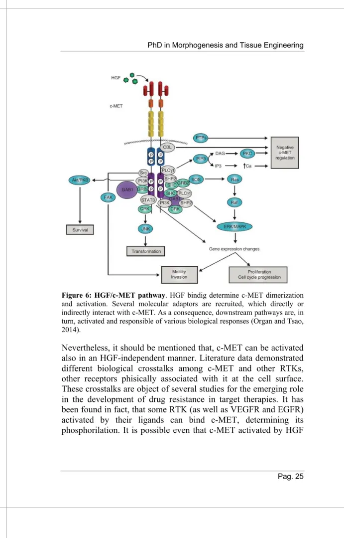
Figure 6: HGF/c-MET pathway. HGF bindig determine c-MET dimerization and activation. Several molecular adaptors are recruited, which directly or indirectly interact with c-MET. As a consequence, downstream pathways are, in turn, activated and responsible of various biological responses (Organ and Tsao, 2014).
Nevertheless, it should be mentioned that, c-MET can be activated also in an HGF-independent manner. Literature data demonstrated different biological crosstalks among c-MET and other RTKs, other receptors phisically associated with it at the cell surface. These crosstalks are object of several studies for the emerging role in the development of drug resistance in target therapies. It has been found in fact, that some RTK (as well as VEGFR and EGFR) activated by their ligands can bind c-MET, determining its phosphorilation. It is possible even that c-MET activated by HGF
can dimerize with other RTKs activating their signalling pathways. In both cases, signal transdution amplification occurs (58). It is also woth mentioning that, the hyaluronan receptor (CD44) can bind c-MET lead to the activation of RAS/ERK signaling through SOS reclutation and binds c-MET to the cytoskeleton (59, 69). The relationship between c-MET and the Epidermal Growth Factor receptor (EGFR) is widely provided, as well as its link with the Trasforming Growth Factor (TGF-a) (70, 71). Even the binding with laminin receptor-a6b4 integrin, or with semaphorin receptors of plexin B family or the linking with Vascular Endothelial Growth Factor Receptor (VEGFR-2) are proved (72). These events determined the synergistic activation of the downstream RTKs related pathways.
2.2.4 HGF/c-MET in testis
c-MET has an essential role in embryonic development, as demonstrated by the lethal phenotype observed in c-MET knock-out mice due to several placental defects. This observation led to deeper investigate HGF/c-MET system during prenatal phases, Notably, it has been found expressed and active during all phases of pre-natal and post-natal development of several organs whose morphogenesis appears regulated by a tight epithelium-mesenchyme cross-talk. Gonads can be numbered among these organs. Studying the mouse embryo, in fact, HGF/c-MET system has been detected in the urogenital ridge since 11.5 dies post coitum (dpc), and it probably collaborates with other growth factor, such as FGF9 and PDGF, in driving the sex-specific cells migration of mesonephric cells toward the developing testis (73-75). HGF/c-MET system is also involved in Fetal Leydig cell development starting from 15.5 dpc, regulating their endocrine function, differentiation and survival (76, 77).
In rat post-natal testis c-MET receptor has been found expressed in all the main cell lineages, including germ cells, even if at different levels and in different phases of testicular post-natal development (78). In this animal model HGF has been found to exert several
functions: it is involved in Sertoli polarization and differentiation; it promotes survival and endocrine function of Adult Leydig cells; it is involved in the regulation of cytoskeletal organization of myoid cells (79-82); it regulates sperm motility (78).
Few data are available on HGF/c-MET system expression and role in human testis. Among them, in 1996 the expression of the receptor was described in human testicular tissue, in particular in the germ cells (83). The same group after two years identified also the expression of its ligand HGF, and they observed altered levels of HGF in the seminal fluid of patients with andrological disease (84).
2.2.5 HGF/c-MET de-regulation in human cancer
HGF/c-MET system is very important for tissue development and homeostasis, and then it is not surprising that its de-regulation has been observed in several human cancers, being associated with aggressive pathologic features, poor prognosis and treatment resistance (85). Aberrant c-MET signaling in human malignancies can occur by a variety of mechanisms. Among these, a copy number increase of c-MET was found in several of neoplasias, associated with a gene amplification with higher c-MET gene level. An example of this mechanism is observable in the non-small cell lung cancer (NSCLC) and high level of c-MET protein has been associated with a decrease of patients survival. Mutation of the c-MET gene represents a not so frequent event in cancer, althoug it is well demonstrated that in some cases it represents a pivotal role in the carcinogenesis. There are several mechanisms of activating mutations described, in particular, for instance missense mutations in the catalitic domain of c-MET determines the constitutively activation of the receptor. This kind of mutation has been observed in hereditary and sporadic forms of papillary renal cell carcinoma (86, 87), in childhood hepatocellular carcinoma (88), and in rarely other cancer types. Moreover, somatic mutations were also observed in the juxtamembrane domain of c-MET, necessary for the recruitment of the E3-ubiquitin protein ligase Cbl, resulting in
the accumulation of the receptor. In 2002 it was described that somatic c-MET mutations that constitutivaly activate its signalling are frequent at metastatic sites. This observation strong indicates the tight relantionship between c-MET activation and cancerc progression (89). Another mechanism, which is involved in constitutivaly activation of c-MET, is the increase secretion of HGF by tumour cells or intringuingly by stroma. This event is observable in glioma, osteosarcoma, pancreatic breast and gastric cancers (90, 91).
Functional crosstalk between c-MET and other receptors results in an emerging important mechanism implicated in the cancer progression and drug resistance (66). The dimerization with c-MET and IGF1R, EGFR, ERBB2 or VEGFR and others has also been studied in several human cancers.
2.2.5.1 Cancer treatments and HGF/c-MET
As previously stated, de-regulation of HGF/c-MET system is frequently observed in many tumours and the activation of this signaling determined more aggressive clinical behaviour with higher invasion, metastasis and acquired drug resistance. It has become evident that tumour microenvironment acquired a very important role in the onset and progression. In fact, it is well known that important changes occur during carcinogenesis and tumours are able to create an enviroment suitable for tumour mainteinance. Cytokines, growth factors and chemokines secreted by stromal cells are foundamental in these processes (71).
RTKs has become very attractive drug targets in the development of novel treatment strategies. Moreover, as previously described, HGF/c-MET system is an intricate and highly regulated signalling in the cells. Within this view, c-MET is a promising specific target and it is used in different cancer therapies (92). Nowadays, a number of strategies targeting the HGF/c-MET axis are in development. These approaches use respectively HGF or c-MET as a target.
Among the first category, we distinguish HGF activation inhibitors (preventing the activation from pro-HGF to its active form) and HGF inhibitors (preventing the binding between HGF to c-MET receptor). The activation of HGF depends on the balance between the activators (HGFAs) and inhibitors (HAIs) and this may rapresent a new interesting target, but the development of HAIs is in the early stages. Regardig HGF inhibitors, several antibodies against this growth factor are tested and among these Ficlatuzumab and Rilotumumab, two monoclonal antibodies, are used in different clinical trials (85).
As for the second category, c-MET antagonists (which compete with HGF for receptor binding) or c-MET kinase inhibitor were identified (85, 93). MET antagonist compete with HGF for c-MET binding causing the degradation of the receptor and its inactivation. Onartuzumab is an example of these and it is in phase III in the treatment of NSCLC and phase II of breast cancer. Regarding c-MET kinase inhibitor, we can distinguish c-MET selective small molecules, such as Tivantinib in several clinical trials in phase I, II, and III or Savolitinib that displayed in vitro activity against c-MET. Among c-MET non selective drugs we found Foretinib, Cabozantinib and Crizotinib, all of these demonstrate an additional activity against other receptors. These drugs are in pre-clinical and clinical studies and patients are consistently recruited for different phases of anti-tumour terapies (http://www.clinicaltrials.gov;2013).
Nowadays, it is well known that c-MET is implicated in the resistance to target terapies, including approved EGFR inhibitors (94, 95). Treatment with monoclonal antibody against EGFR are used in several solid tumors and generally acquired resistance appears in 3-12 mounth after treament. Some of these mechanisms of resistance are dependant on the amplification of c-MET. Cetuximab resistance in colon cancer cells was observed and this cellular response is related to an up-regulation of p-c-MET. Na Song et al. described that in in-sensitive Caco-2 cells active c-MET was found and this activation is involved in the escape from the
inhibitory effect of cetuximab (95, 96). This effect was reverted using a c-MET inhibitor which is able to restore cetuximab responses. In the same work it is also demonstrated that p-c-Src is related to p-c-MET in Caco-2 cells and c-Src activation cooperate in the cetuximab resistance activating in turn PI3K/AKT or ERK pathways as effectors (96). Biologic agents targeting the VEGF pathway have also produced significant clinical benefits in some cancers but drug responses are usually short-lived. Inevitably, drug resistance emerges. In fact, HGF/c-MET axis appears important also in acquired VEGFR inhibitor resistance and vascular remodeling in non-small cell lung cancer (NSCLC) (97). In summary, tumour microenvironmet sustained cancer progression. HGF/c-MET axis results up-regulated after TKIs therapies and this event results implicated in drug resistance. Recently, it was observed that cancer associated fibroblasts (CAFs) produce HGF which sustaining adaptive resistance to targeted therapies (98). These evidences shed new light on the novel combined target therapies development and combinatorial TKIs and HGF/c-MET targeting may represent a reasonable approach to improve patient outcomes.
2.3 c-MET in TGCTs
The possible role of c-MET in TGCT pathogenesis is still a matter of debate, since literature data on this subject is very poor. To this regard, in 1996 Mostert and collaborators described a gain of chromosome 7 in primary tumours of patients affected by TGCTs (99). Moreover, another work published in 2004 mutation for c-MET have not been reported in TGCTs (100). Svetloska et al. analyzed several circulating cytokines, among which HGF, in the serum plasma of patients affected by TGCTs and an inverse correlation between higher HGF level and good prognosis was demonstrated (101).
2.3.2 My previous work
Recently, my group observed the differential expression of c-MET in cell lines derives from patients affected by seminoma and non-seminoma tumours (TCam-2, NCCIT and NT2D1 cells). In this work we demonstrated that non-seminoma cell lines (NT2D1) show the higher cellular responses to HGF administration, with respect to the other cell lines, in terms of proliferation, invasion and migration. In line with these results we also observed, in human biopsies, that non-seminoma cancer lesion exhibit c-MET at higher levels with respect to GCNIS and seminoma lesions (102).
Starting from these evidences, my group and I decided to analyze c-MET-activated pathways by HGF administration in NT2D1 cells. In particular, we focused on c-Src and PI3K dependent pathways triggered by c-MET activation. The results obtained on the analysis of c-Src activation, that I will report in this thesis, has been recently published. A graphical representation is shown in Figure 7 (103). The data focused on PI3K signalling belong to a manuscript in preparation.
Figure 7: Graphical abstract. HGF administration in NT2D1 cells determines activation of c-MET. It can activate c-Src protein, which changes its localization and translocates in the nucleous. This phenomenon is responsible for NT2D1
biological responses, such as proliferation, migration and invasion. In this system c-Src can also be activated by an unknown factor, without HGF/c-MET involvement. This results in higher collective migration and invasion (from Leonetti E. et al., 2019).
3. Aims
HGF/c-MET system has a very intricate signalling whose importance is well known during embryogenesis. c-MET de-regulation is widely described in several human cancers, and for this purpose, it is used as a target in several personalized therapies (www.vai.org/met). Though, few data are present in literature about this system in TGCTs. c-MET is encoded on chromosome 7, region 7q31. A duplication of this chromosome was described in TGCT patients. Noteworthy, high levels of HGF in serum plasma of patients affected by TGCT were found.
TGCTs are a group of pathologies that arise from altered testicular niche during embryogenesis. This alteration is responsible for a block of gonocyte differentiation, which can give rise to all type II TGCTs. TGCTs are mostly curable, but there is still a little percentage of patients who develop drug resistance, radio-resistance, or long-term toxicity due to the used chemo- and radio- therapy. For these reasons, as well as for the young age of the patients and the increasing incidence in recent years, it is a paramount to have a better understanding on the onset and progression of TGCTs.
In a previous work we demonstrated that c-MET is expressed by several cell lines derived from type II TGCTs and that HGF administration determines strong biological responses, especially in non-seminoma cell lines. In line with these results, we also observed that c-MET was present in human biopsies derived from patients affected by all type II TGCTs (102). In order to better characterize onset and progression of TGCTs, we decided to investigate:
1) The specific role of the c-MET receptor in chemotaxis and collective migration (continuing the previous work).
2) The signalling pathways triggered by HGF and mediated by c-MET activation, by identifying the main adaptor proteins related to it in NT2D1 cells and their role.
3) The synergic relationship among different activated adaptors in NT2D1 cells.
4) The differential HGF expression in biopsies derived from patients affected by type II TGCTs with specific concern on SE and EC.
4. Results
4.1 c-MET modulates the migration of NT2D1 cells induced by HGF
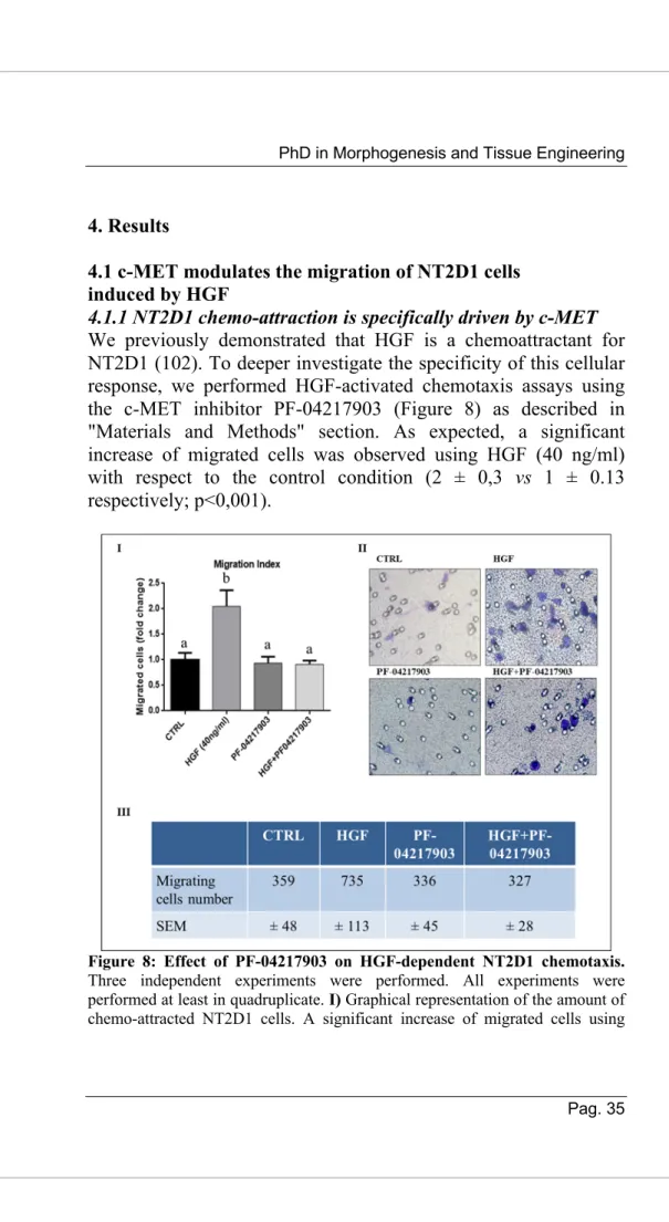
4.1.1 NT2D1 chemo-attraction is specifically driven by c-MET We previously demonstrated that HGF is a chemoattractant for NT2D1 (102). To deeper investigate the specificity of this cellular response, we performed HGF-activated chemotaxis assays using the c-MET inhibitor PF-04217903 (Figure 8) as described in "Materials and Methods" section. As expected, a significant increase of migrated cells was observed using HGF (40 ng/ml) with respect to the control condition (2 ± 0,3 vs 1 ± 0.13 respectively; p<0,001).
Figure 8: Effect of PF-04217903 on HGF-dependent NT2D1 chemotaxis. Three independent experiments were performed. All experiments were performed at least in quadruplicate. I) Graphical representation of the amount of chemo-attracted NT2D1 cells. A significant increase of migrated cells using
HGF was observed (b vs a; p <0.001). The use of PF-04217903 in combination with HGF, abrogates the effect induced by HGF. Control condition was arbitrarily considered as 1, and the values were calculated as “fold change” compared with control (± S.E.M.). II) Representative images of NT2D1 migration experiments obtained with Diff quick staining. The images were recorded at 40X magnification. III) Table illustrating the number of migrating cells/filter in all the experimental conditions reported in the respective “fold change”.
Notably, PF-04217903 alone does not modify the migratory capability of NT2D1 cells compared to the control (0.94 ± 0.12 vs 1 ± 0,13 respectively; p=n.s.), whereas the co-administration of HGF+PF-04217903 causes the reversion of the HGF-induced chemotactic effect (0.91 ± 0.08 vs 2 ± 0,31 respectively; p<0,001) (Figure 8, panel I). Taken together, these results indicate that HGF-induced chemotaxis is specifically activated by c-MET.
4.1.2a c-MET modulates the collective migration of NT2D1 cells induced by HGF
We analysed in detail the effects of HGF on the collective motility of NT2D1 through wound healing assays performed as described in "Materials and Methods" section. We calculated by Image J v 1.47h software the percentage of the open surface residual area of each experimental condition in correlation with baseline points T0, considered as 100% open area. We did not observe any reduction of open residual area in the wells treated with HGF with respect to control wells after 24 hours from insert removal. After 48h of culture the open area of HGF treated cells resulted significantly reduced compared to the open area of the control condition. This result demonstrates that HGF induces in NT2D1 cells an increase of migration capabilities.
To evaluate the specificity of HGF/c-MET response, we performed the experiments in presence of the c-MET inhibitor PF-04217903. The inhibitor alone results similar to the control condition after 24 and 48h the insert removal. After 24h of culture the open residual area of PF-04217903+HGF treated wells was not significantly different from the open residual area of all conditions, while after
48h of culture the open residual area of PF-04217903+HGF treated wells was similar to the control conditions and significantly higher than the open area of HGF treated wells (41,1% ± 9 vs 10,4% ± 6 respectively; p< 0,01) (Figure 9). These data indicate that c-MET inhibition with the ATP-competitor PF-04217903 significantly reduces (about 30%) the migratory effect induced by HGF.
Taken together our results reveal that HGF treatment is able to enhance the collective migration of NT2D1 cells and confirm that this response is specifically dependent on c-MET.
Figure 9: Role of HGF/c-MET system on NT2D1 cells collective motility. Wound healing assay after cells treatment with fresh 2% FBS DMEM (CTRL)
or containing HGF alone, PF-04217903 or HGF+ PF-04217903. Three independent experiments were performed at least in triplicate. I) Representative phase contrast images of wound healing assay recovered immediately after insert removal for baseline wound measurement (T0) and 24h and 48h after wounding. Images were photographed at 10X magnification (scale bar: 100 µm). II) Quantitative analysis of wound closure after 24h (A) and 48h (B). Data are expressed as the mean percentage of residual open area compared to the respective cell-free surface recovered at T0. After 24h the difference of open area was not statistically significant in all the considered experimental conditions. After 48h the decrease of open area in HGF treated cells was statistically significant with respect to the control (* p<0.05). PF-04217903 alone was similar to the control while PF-04217903+ HGF abrogated the HGF induced effect (# p<0.01).
4.1.2b Proliferation does not affect collective migration induced by HGF
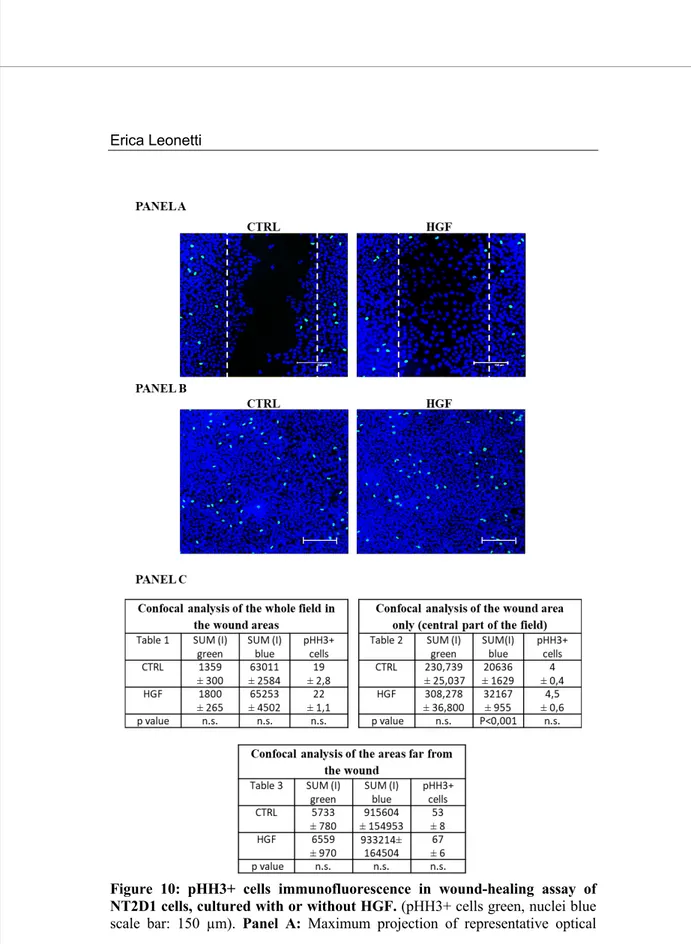
Since HGF is able to promote NT2D1 cell proliferation, as above reported, we wanted to check whether the observed HGF-mediated increase in the rate of wound closure were due to a trigger to cell proliferation rather than to an actual migratory effect. To this end we performed immunofluorescence experiments, using the phosphohistone H3 (pHH3) as mitotic marker, on wound-healing samples at the end of their culture time. To quantify the immunofluorescence results, samples were recovered using the Leica Confocal microscopy (Figure 10, Panel A and B) and analysed by Leica Confocal Software (Figure 10, Panel C). The Sum of Fluorescence Intensity (SUM (I)) of pHH3 signal (FITC/green signal) and nuclei staining (TO-PRO3/blue signal) was calculated in three different areas of each sample. The analysed areas were:
1) the whole photographic-field of each wound area (562,500 μm2
for each image), data reported in Table 1;
2) the presumptive wound area (263,486 μm2; inside the dotted
lines of the representative images of panel A) data reported in Table 2;
3) the confluent areas recovered far from the wound (562,500 μm2)
We also counted the pHH3+ cells in each area and even these results are reported in data reported in Tables in figure 10, panel C. The values reported in Table 1 clearly show that the SUM(I) analysis as well as the count of PHH3+ cells did not reveal any increase of pHH3+ cell in the whole photographic-field of wound area (562,500 μm2) both in control condition and in HGF treated
samples.
In Table 2, the values reported show that there is not a significant difference in the amount of pHH3+ cells between control and HGF treated cells in the areas of the wound. Notably, an increase of blue fluorescence (that represents nuclear staining) is observable between control condition and HGF treated cells. Altogether these observations indicate that HGF stimulates the NT2D1 cells collective migration and that, in these conditions, HGF-triggered cell proliferation does not contribute significantly to wound closure.
The reported values in Table 3 show that there is not a significant difference in the amount of pHH3+ cells between control and HGF treated cells, even in the confluent part of the culture dishes.
Therefore, these experiments demonstrate that the SUM(I) analysis as well as the count of pHH3+ cells does not reveal any increase of pHH3+ cell in the area of the wound in all samples considered. Furthermore, proliferation appears not responsible of NT2D1 cell collective migration. It is fair to consider that even in the areas far from the wound the number of pHH3+ cells as well as the SUM (I) of blue nuclear staining was not statistically significant. These data clearly indicate that the wound repair depends on a migratory effect of HGF on NT2D1, and that the cultural condition in which this experiment has been carried-out did not allow to underline the HGF- mediated NT2D1 cell proliferation, probably due to the over-confluence of the cells at the plating time.
Figure 10: pHH3+ cells immunofluorescence in wound-healing assay of NT2D1 cells, cultured with or without HGF. (pHH3+ cells green, nuclei blue scale bar: 150 µm). Panel A: Maximum projection of representative optical
spatial series with step size of 1 μm recovered at the area of the wound. The dotted lines indicate the presumptive area of the wound, calculated at the beginning of the wound-healing assay. Panel B. Maximum projection of representative optical spatial series of the same samples of Panel A recovered in areas far from the wound. Panel C: Confocal microscopy quantitative analysis by Leica Confocal Software, of the SUM(I) of pHH3+ cells and nuclei. pHH3+ cells per field were also counted. All experiments were performed in triplicate and reported as mean ± S.E.M.
4.2 c-Src is involved in HGF-dependent NT2D1 responses
4.2.1 Src inhibitor-1 does not affect cell viability
The c-MET/HGF system is a very intricate signalling, whose activation is responsible of a complex pathway activation cascade. Among the numerous proteins activated, c-Src represents one of the most intriguing ones. In light of this observation, we decided to start the investigation analysing possible c-Src activation after HGF treatment in NT2D1 cells. In this regard, we decided to use a pharmacological approach, using an inhibitor for this molecule, an ATP-competitor, called Src inhibitor-1. Dose response experiments were performed to highlight the better working dilution of Src inhibitor-1. Different concentrations of Src inhibitor-1 (1, 2.5, 5, 10 µM) were used for 48 hours. 10 µM of Src inhibitor-1 caused a significant increase in the percentage of cell death compared to the control condition (p<0,05). For this reason, we decided to use this inhibitor at 5 µM, a concentration that can be considered not toxic also after 72 hours of treatment (Figure 11, panel I). As an additional check, “trypan blue exclusion test” was performed, demonstrating that the concentration of 5 µM Src inhibitor-1 does not cause cell death in this cellular line (data not shown).
This concentration was tested also with immunofluorescence analysis using an antibody against cleaved caspase3, demonstrating that the number of cleaved caspase3 positive cells was not statistically different between treated cells and control condition (Figure 11, panel II).
Figure 11: I) Cell death FACS analysis on NT2D1 cells. On the left: graphical representation of the percentage of dead cells obtained culturing NT2D1 cells with different concentration of Src inhibitor-1 for 48h (*p<0,05). On the right: graphical representation of the percentage of dead cells obtained culturing for 72h NT2D1 cells with 5 µM Src inhibitor-1 (p=n.s.). II) on the left: representative images of cleaved caspase3 immunofluorescence on cells treated with 5 µM Src inhibitor-1 for 48h (scale bar 80 µM). On the right: quantitative analysis, by Leica Confocal analysis of the number/field of cleaved caspase3 positive cells (FITC/green signal) normalized versus and the number of nuclei (TOPRO-3/blue signal) (p=n.s.).
4.2.2 c-Src is involved in HGF-dependent NT2D1 proliferation As previously mentioned, HGF specifically activates its receptor c-MET on NT2D1 cells, resulting in an increase of proliferation rate after 48h of culture. To investigate the role of c-Src in HGF-dependent c-MET activated cell proliferation, we performed proliferation assay using Src inhibitor-1. On the basis of the mentioned previous work (102), NT2D1 cells were cultured for 48h in basal condition or with the following treatments: Src
inhibitor-1, HGF, or HGF+ Src inhibitor-1 (Figure 12). Then cells were detached and counted. As expected HGF administration induced a significant increase of cell number with respect to control samples (1,2 ± 0,06 vs 1 ± 0.04, respectively; p<0.001).
Figure 12: Effect of Src Inbhibitor-1 on NT2D1 cell proliferation induced by HGF. I) Graphical representation of the number of NT2D1 cell cultured for
48h in 2% FBS DMEM (CTRL), or with: HGF, Src inhibitor-1, or their combination. HGF treatment shows a significant increase of cell number (b vs a; p<0,001). Using the inhibitor, with or without HGF, we observed a significant reduction of cell proliferation compared to the HGF (c vs b; p<0,001), and to the control condition (c vs a; p<0,001). Four independent experiments were performed at least in triplicate. Results were expressed in fold change and the control was considered as 1 (± S.E.M.). II) Table illustrating the results of cell cycle analysis on NT2D1 cell cultured for 6, 24, 30 and 48h with or without Src inhibitor-1 (* vs the respective control p< 0,05).
Using HGF+ Src inhibitor-1, we observed that the treatment completely abrogates the HGF induced proliferation in NT2D1 cells (0,7 ± 0,04 vs 1 ± 0,04 respectively; p<0.001). Surprisingly, using Src inhibitor-1 alone determined a proliferation rate significantly different compared to the control (0,7 ± 0,04 vs 1 ± 0.04 respectively; p<0.001). To better characterize this phenomenon, we performed cell cycle analyses, which revealed that Src inhibitor-1 administered alone, causes a significant decrease of cells in G2 phase after 6 hours of culture, a significant increase of cells in G1 phase after 24 hours of culture and a subsequent significant increase of cells in S phase after 30 hours of culture (Figure 12; panel II). These results indicate that Src inhibitor-1 causes a slight cell cycle slowdown, when administered alone. Moreover, in light of these results, we can speculate that c-Src regulates NT2D1 cell proliferation both in HGF-dependent and HGF-independent way.
4.2.3 c-Src is specifically involved in HGF-dependent NT2D1 chemo-attraction
To deeper investigate the molecular effectors involved in a migration process, we decided to test if c-Src is required for HGF-mediated chemo-attraction of NT2D1 cells. We performed the above-mentioned chemotaxis assay, using Src inhibitor-1 (Figure 13). We observed that this inhibitor administered alone did not affect migration rate with respect to basal condition (1.4 ± 0.2 vs 1 ± 0.09 respectively; p=n.s.), while, as expected, HGF induced the chemotactic migration of NT2D1 cells (2 ± 0.16 vs 1 ± 0,09
respectively; p<0,001). But, in detail, the treatment of cells with Src inhibitor-1 in the upper chamber of the trans-well apparatus significantly reverted the migratory effect exerted by HGF added in the lower chamber (1,2 ± 0,13 vs 2 ± 0,16 respectively; p<0.01). Thanks to these results, we can state that c-Src is involved in the positive regulation of this phenomenon.
Figure 13: Effect of Src inhibitor-1 on NT2D1 chemo-attraction: I) Graphical representation of the amount of chemo-attracted NT2D1 cells. A significant increase of migrated cells using HGF was observed (b vs a; p<0.001). The use of Src inhibitor -1 in combination with HGF abrogates the effect induced by HGF. Control condition was arbitrarily considered as 1, and the values were calculated as “fold change” compared with control. II) Representative images of NT2D1 migration, obtained with Diff quick staining. Images were recorded at 40X magnification. III) Table illustrating the number of migrating cells/filter in all the experimental conditions reported in the respective “fold change” graph (b vs a; p<0.001).
4.2.4 c-Src is involved in HGF-induced collective migration It is well known that HGF binding activates c-MET, resulting into tyrosine residues phosphorylation. Active c-MET recruits signaling
effectors that activate protein kinases, such as c-Src, ultimately leading to cell migration (59). To investigate whether HGF-induced wound migration is dependent on c-Src recruitment,
Figure 14: Effect of Src Inhibitor-1 on NT2D1 cell collective migration. Three independent experiments were performed at least in quadruplicate. I) Representative phase contrast images of wound healing assay recovered immediately after insert removal (T0) and 24h and 48h after wounding. Images were photographed at 10X magnification (scale bar: 100 µm). II) Quantitative analysis of wound closure after 24h (A) and 48h (B). Data are expressed as the mean percentage of residual open area compared to the respective cell-free
surface recovered at T0. At 24h the decrease of open area in HGF treated cells was not statistically significant compared with control condition, but the closure was almost complete at 48h (*p<0.001). Src inhibitor-1 in combination with HGF both at 24h (# p<0.01) and 48 h (# p<0.001) abrogated the migratory effect induced by HGF. Src inhibitor-1 alone was also able to inhibit the collective migration of the cells cultured for 24 h ($ p<0.05), and 48 h ($ p<0.001). NT2D1 cells were incubated with medium alone or with HGF in the presence or not of Src inhibitor-1.
The results showed collective cell migration more evident in HGF-exposed NT2D1 cells after 48h treatment with respect to constitutive migration observed in the control condition (Figure 14).
More in detail, after 24h of culture the open residual area of samples treated with Src inhibitor-1 alone showed values similar to its own T0 (93.1% ± 1.5) and, therefore, significantly higher compared with control condition (64.4% ± 7.1) and HGF treated wells (55.2% ± 0.6). After 48h, these areas remained almost unchanged (83.5% ± 0.3) with respect to 24h, whereas the open residual area of control samples and HGF treated wells decreased (35.4% ± 1.5 and 12.2% ± 1 respectively). The inhibition of collective cell migration in the presence of Src inhibitor-1 alone clearly indicates the involvement of this adaptor-protein in constitutive collective migration of NT2D1 cells, even if further investigations are needed to clarify this point.
Src inhibitor-1 administration reduces also the enhanced collective migration induced by HGF. The values of open residual area of HGF+ Src inhibitor-1 treated wells were significantly higher, compared with HGF treated wells, already after 24h of culture (90.9% ± 0.9 vs 55.2% ± 0.6 respectively). This difference was even more significant after 48h of culture (77.6% ± 12 vs 12.2% ± 1 respectively) (Figure 14).
4.2.5 c-Src is involved in HGF-dependent cells invasion
Our previous work demonstrated that HGF is able to induce higher invasive capabilities in NT2D1 cell line (102). To better understand the mechanism involved in this biological response, we
performed invasion assays, using the Src inhibitor-1. As shown in Figure 15, we confirm that HGF treatment significantly modulates the invading capability of NT2D1 cells (2,2 ± 0,3 vs 1 ± 0,16 respectively). On the other hand, when Src inhibitor-1 was used in combination with HGF, the number of invading cells reverts to control condition (1 ± 0,21 vs 1 ± 0,16 respectively; p=n.s.). Notably, we also found that Src inhibitor-1 alone significantly increases NT2D1 cell invasiveness compared to the control condition (1,75 ± 0,4 vs 1 ± 0,16 respectively; p<0,05) and the value results similar to HGF administration (1,75 ± 0,4 vs 2,2 ± 0,3; p=n.s.). This result is relevant and deserves further investigation to fully characterize the mechanism underlying it. Taken together, these findings demonstrate that c-Src is involved in the regulation of NT2D1 cell invasiveness, using both HGF-dependent and HGF-inHGF-dependent pathways.
Figure 15: Effect of Src Inhibitor-1 on NT2D1 cell invading capability: Invasion assay was performed using filters coated with GFR Matrigel. The filters were analyzed by optical microscope and images were recovered at 10X magnification. Three independent experiments were performed at least in triplicate. Left panel: Quantitative analysis of invading cells. The results are
expressed as fold change (± S.E.M.) and the control condition is considered as 1. HGF and Src inhibitor-1 alone induced both a statistically significant increase in NT2D1 cell invasion with respect to the control (b vs a; p<0.05). The treatment of HGF+ Src inhibitor-1, abrogated this effect. Right panel: Representative phase contrast images of the different culture conditions. Images demonstrate a higher invasiveness behaviour of cells treated with HGF or Src inhibitor-1 alone compared with HGF+ Src inhibitor-1 condition. The table in the lower panel illustrates the number of invading cells/filter in all the experimental conditions reported in the respective “fold change” graph (b vs a; p<0.05).