Università degli Studi del Piemonte Orientale
“Amedeo Avogadro”
Dipartimento di Scienze del Farmaco
Dottorato di Ricerca in Biotecnologie Farmaceutiche ed
Alimentari
XXVIII ciclo A.A. 2012-2015
Neural Progenitor Cell-astroglia cross-talk:
involvement of the NF-
B p50 subunit.
Suzana Cvijetic
Supervised by Prof. Mariagrazia Grilli
TABLE OF CONTENTS
CHAPTER 1.
Introduction
1.1 Adult neurogenesis ... 7
1.1.1 Adult hippocampal neurogenesis ... 11
1.1.2 The neurogenic niche ... 17
1.2 Astroglia in the central nervous system ... 21
1.2.1 Astroglial phenotypes in health and disease ... 23
1.2.2 Astrocytes as modulators in adult neurogenesis ... 30
1.3 NF-κB signaling in adult neurogenesis ... 36
1.3.1 The NF-κB family of transcription factors ... 37
1.3.2 NF-κB signaling in the central nervous system: a role in synaptic plasticity, learning and memory ... 41
1.3.3 NF-κB signaling in adult neurogenesis: in vitro and in vivo supporting data ... 45
References ... 51
CHAPTER 2. Outline of the Thesis ... 71
References ... 74
CHAPTER 3. Astrocyte-mediated neuronal fate specification of adult hippocampal neural progenitors is regulated by NF-κB p50 ……….... 79
Introduction ………...…… 81
Materials and methods ……….……… 83
Results ………...… 89
Discussion ………... 102
References ……….. 108
CHAPTER 4
Modulatory events within the adult hippocampal neurogenic niche: novel insight into region-specific astrocyte-derived signaling cues and the role
played by NF-B p50 ... 125
Introduction ………...….. 126
Materials and methods ……….…..…. 128
Results ………... 130
CHAPTER 5. Discussion ... 143
Conclusions and future perspectives ……….. 147
References ... 151
CHAPTER 6. List of Pubblications and poster sessions ... 155
5
7 1.1 Adult neurogenesis
Adult neurogenesis is a process of structural plasticity characterized by de novo generation and integration of new neurons into the existing circuitry in the adult central nervous system (CNS) (Ming and Song 2005; Christian et al. 2014). Basal level of neurogenesis occurs in two discrete regions: the subventricular zone (SVZ) in the lateral wall of the lateral ventricle and the subgranular zone (SGZ) of the dentate gyrus (DG) in the hippocampal region (Lois and Alvarez-Buylla 1994; Kempermann and Gage 2000). These two areas, referred to as “canonical” sites of active adult neurogenesis, have been found in all mammalian species studied so far, including primates and humans (Eriksson et al. 1998; Gould et al. 1999; Knoth et al. 2010). The adult neurogenic process comprises a series of sequential developmental steps spanning from proliferation, survival, fate specification, neuronal migration, maturation and functional integration of neuronal progeny. The source of new neurons is the adult neural stem cell (NSC) and their progeny of neural progenitor cells (NPC), which were first isolated from the adult CNS of rodents by Reynolds and Weiss (1992) and Kilpatrick and Bartlett (1993). Their pioneering work on SVZ-derived NSC was pivotal, given that they reported methods for culturing and growing neural stem/progenitor cells (NSPC) in the neurosphere culture assay, where individual cells proliferate and generate clusters of cells in suspension. Additionally, these scientists obtained adhesive cultures of NSPC, grown as monolayers on coated substrates (such as laminin). In these proliferating conditions, basic fibroblast growth factor (bFGF) and epidermal growth factor (EGF) were defined as the primary mitogens used to propagate adult neural progenitors in vitro (Reynolds and Weiss 1992; Kilpatrick and Bartlett 1993). These studies established the basic protocols for keeping and maintaining NSPC self-renewal property among many passages, as well as their multipotentiality. Under these conditions the differentiation ability of NSPC is maintained over many passages and can be observed upon growth factor removal, when the molecular machinery drives the differentiation toward the
8
neural lineages, neurons, astrocytes and oligodendrocytes. Later on, similar protocols were also developed to expand and differentiate NSPC derived from the SGZ of the hippocampus (Gage et al. 1995).
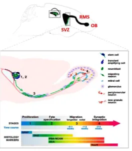
The adult rodent SVZ contains four distinct cell types defined by their morphological features, molecular markers and electrophysiological properties: (1) migrating neuroblasts, type A cells; two distinct populations of NSPC, type B (2) and type C (3) cells; and (4) ependymal ciliated (type E) cells, that separate the SVZ from the ventricular cavity (Lledo et al. 2006). Type B cells are slow cycling cells lining the border between the striatum and the lateral ventricle and expressing the astrocytic marker glial fibrillary acidic protein (GFAP). Type B cells give rise to rapidly dividing intermediate progenitors, type C cells, that lose GFAP expression, and acquire distal-less homeobox-2 (Dlx-2). From type C cells type A cells arise, expressing PSA-NCAM (poly-sialylated neural cell adhesion molecule) and the neuroblast marker doublecortin (DCX) (Alvarez-Buylla and Garcia-Verdugo 2002). Neuroblasts leave the SVZ and move along the rostral migratory stream (RMS) in chains formed by glial cells before terminating into the olfactory bulb (OB), where they differentiate into granule and periglomerular interneurons (Doetsch et al. 1999) (Figure 1). Herein integrated newborn interneurons make local contacts and modulate sensory information. The current hypothesys is that they are involved in changes in the neural representation of an odor, thereby supporting long-term olfactory memory (Sultan et al. 2010).
9
Figure 1. Adult neurogenesis in rodent SVZ/olfactory systems undergoes four developmental stages. A schematic
illustration of the adult mouse brain with an enlarged view of the olfactory system. Four developing stages define SVZ neurogenesis. Stage 1. Proliferation: stem cells in the SVZ of the lateral ventricles give rise to transient amplifying cells (type C cells). Stage 2. Fate specification: transient amplifying cells differentiate into immature neurons or neuroblasts (type A cells). Stage 3. Migration: immature neurons migrate in chains through the RMS to the OB. Stage 4. Synaptic integration: immature neurons differentiate into either granule neurons (orange) or periglomerular neurons (red), (Modified from Ming and Song 2005).
Similarly, in the SGZ, stem cells termed type 1 cells, display a radial glial morphology and give rise to intermediate progenitors (type 2a and type 2b cells) which express neuronal specific markers before moving to the dentate granule cell layers where they integrate into the trisynaptic hippocampal circuit (Seri et al. 2001, 2004; Bonaguidi et al. 2012). The detailed developing process of adult hippocampal neurogenesis is described in the following section.
In addition to neurogenesis occurring in the two forebrain regions, emerging evidence arose for a novel neurogenic site along the walls of the third ventricle, in the hypothalamic area. The hypothalamus is a small brain area that surrounds the third ventricle and contains distinct nuclei. It serves as a central homeostatic regulator of numerous physiological and behavioural functions, such as feeding, metabolism, body temperature, thirst, fatigue, aggression, sleep, circadian rhythms, and sexual behaviour. Several studies using 5-bromo-2'-deoxyuridine (BrdU) as well as genetic lineage tracing techniques reported constitutive neurogenesis in the adult hypothalamus of mammals, including rats and mice (Kokoeva et al. 2005; Pierce et al. 2010; Lee et al. 2014). Under unstimulated
10
conditions, the hypothalamus displays a low rate of neurogenesis and it is suggested that the de novo formation of newborn neurons might serve as a compensatory mechanism contributing to homeostatic control of dietary and energy balance (Yon et al. 2013).
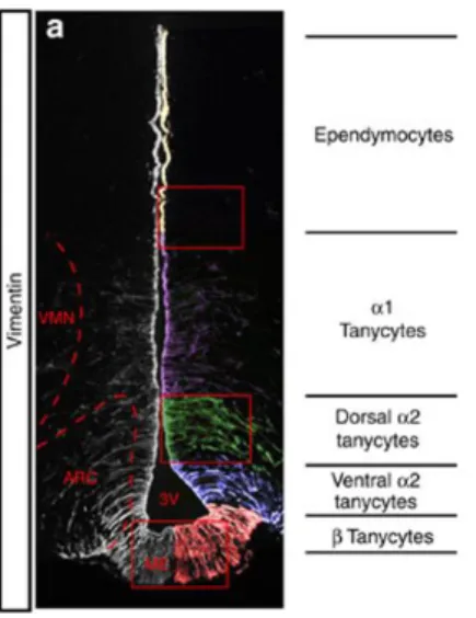
Within the adult hypothalamus, cells around the third ventricle form neurospheres, a hallmark of NSC. Cells like tanycytes, ependymocytes, subventricular astrocytes and parenchymal glial cells, all reside near the third ventricle and contribute to the neurogenic potential of the hypothalamus. Hypothalamic tanycytes constitute the main cell type lining the ventral third of the third ventricle, and resemble radial glia. Within the hypothalamus, tanycyte cell bodies are localised in the central and posterior hypothalamus, at the level of median eminence. Morphological studies have mapped and defined two subpopulations of tanycytes according to their position and process projection: i) β-tanycytes, lining the median eminence (ME); ii) α-tanycytes, extending into the arcuate nucleus (ARC) and ventromedial nucleus (VMN), lining the ventricular zone. Robins and colleagues (2013) have reported that α-tanycytes have NSC features and act as neural progenitors in the hypothalamus. It was demonstrated through a series of in vitro and in vivo experiments in adult mice (using a GLAST::CreERT2 mice), that GLAST+ (glutamate transporter) α-tanycytes are self-renewing cells, that constitutively give rise to new α-tanycytes, as well as astrocytes and neurons (Robins et al. 2013) (Figure 2).
11
Figure 2. Distinc tanycyte populations line the hypothalamic third ventricle. Left hand
panel: coronal section through third ventricle (3V), immunolabelled with Vimentin (intermediate filament marker, expressed by all tanycytes). The position and projection of the Vimentin+ process defines different tanycyte subsets: β-tanycytes line the median eminence; α2-tanycytes reside adjacent to the arcuate nucleus; α1-tanycytes extend from the level of the ventromedial nucleus to the dorso-medial nucleus. VMN, ventromedial nucleus; ARC, arcuate nucleus; ME, median eminence. (Modified from Robins et al. 2013)
1.1.1 Adult hippocampal neurogenesis
Large efforts have been made in order to understand the function of adult-born neurons both in physio- and pathological conditions. The enormous interest in hippocampal neurogenesis was mainly due to the involvement of this brain region in cognition, and in particular in learning and memory (Aimone et al. 2010). In the hippocampus, neurogenesis is localized within the SGZ in the dentate gyrus, where excitatory granule cells are continuously produced throughout life. The adult SGZ is a narrow band of cells made only of one to three nuclei. The SGZ contains the cell bodies of radial glia-like cells, with processes extending to the densely packed granular cell layer and to the molecular layer (Nicola et al. 2015). The DG has a complex local circuitry, with both inhibitory interneurons and excitatory feedback neurons (mossy cells) constituting the network. The sensory information from specific cortical areas converge to the entorhinal cortex and flows through medial and lateral perforant pathways to the dentate DG, then to CA3 pyramidal cells via mossy fiber axons of granule cells, then to CA1 pyramidal cells via Schaffer collateral projections
12
of CA3 and back to the entorhinal cortex and subiculum regions, closing the “hippocampal loop” (Aimone et al. 2014).
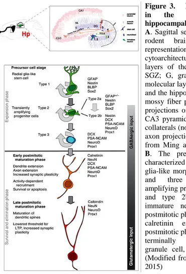
According to a recent publication by Kempermann and colleagues, adult hippocampal neurogenesis can be divided into four phases, defined as precursor cell phase, early survival phase, postmitotic phase and late survival phase (Kempermann et al. 2015) (Figure 3). The proposed model goes through different developmental stages, identified based on cell morphology and protein marker expression (Kempermann et al. 2004). In general, from a radial glia-like precursor cell, adult neurogenesis progresses throughout three identifiable progenitor stages to a postmitotic maturation phase ending with the formation of a mature granule cell.
In the adult SGZ, four developmental stages define the precursor cell phase of NSPC pool expansion. In vivo clonal studies have revealed that radial glia-like precursors are multipotent stem cells capable of repeated self-renewal and generation of both neurons and astrocytes, but not oligodendrocytes, in the adult hippocampus (Bonaguidi et al. 2011). As it is, adult neurogenesis starts with a precursor cell population with morphological and antigenic properties of radial-glia-like cells, named as type 1 cells. The type 1 cells express GFAP, astrocytic marker and nestin. The cell body of type 1 cells is found in the SGZ whereas the processes extends into the molecular layer. Type 1 cells show also electrophysiological properties of astrocytes (Filippov et al. 2003). One transcription factor specifically expressed in SGZ radial glia stem cells is the SRY-related HMG box family member Sox2, which plays a key role in stem cells self-renewal and is critical for the balance between proliferation and differentiation (Julian et al. 2013). The developmental potential of type 1 cells has been demonstrated with tracing experiments (Seri et al. 2004, 2001). Radial glial-like type 1 cells divide asymmetrically, with one daughter cell giving rise to the stem cell and another one giving rise to the intermediate progenitor, or type 2 cells.
13
Type 2 cells or transient amplifying progenitors, are highly proliferative cells with short processes, that are oriented tangentially to the DG. Type 2 cells come in two subtypes, both nestin positive. One subtype called type-2a cells still express the GFAP, but lack the radial glia morphology, the other cellular phenotype called type-2b, is positive for the immature neuronal marker DCX. Another recently added marker for type 2 cells, is the T-box brain protein 2 (Tbr2), a transcription factor that identifies basal progenitor cells. Tbr2 is required for neurogenesis in the dentate gyrus of developing and adult mice. Tbr2 is critical for the transition from stem cells to intermediate progenitors, as it regulates Sox2 suppression (Hodge et al. 2008). Type 2 cells receive first synaptic GABAergic inputs, and respond to physiological stimuli, such as voluntary wheel running (Kronenberg et al. 2003) or pharmacological stimulation via serotoninergic-dependent mechanisms (Encinas et al. 2006) and GABA sets the pace for this regulation (Ge et al. 2007). Kempermann and colleagues propose that the fate choice decision occurs at the level of type 2a cells, given that already in type-2b cells there is expression of transcription factors NeuroD1 and Prox1, involved in neuronal fate choice (Kempermann et al. 2015).
Type 3 cells are DCX positive, but nestin negative. DCX expression extends from a proliferative stage, through cell cycle exit, for a period of two to three weeks (Couillard-Despres et al. 2005). DCX also overlaps with PSA-NCAM expression.
Next phase is the early survival phase. DCX expression still persists until next stage of newly formed neurons expressing the postmitotic marker NeuN and the transient Ca2+ binding protein calretinin (Brandt et al. 2003). The majority of cells reaches this stage only three days after the initial division, then these immature neurons are subjected to a selection process, during which they can be eliminated or functionally recruited. Within few more days the number of NeuN+ new neurons decreases via apoptosis (Kuhn et al. 2005). The majority of
14
cells are eliminated before that the functional integration is established in the CA3 region and before receiving dendritic input from the enthorinal cortex, in the molecular layer. Cells that survive retain a vertical morphology with a rounded nucleus and the apical dendrite projecting toward the molecular layer. Then cells send out their axon to target CA3 region, in what is called the mossy fiber path. The main synaptic input to these cells is still GABAergic, and GABA remains excitatory (depolarizing), until sufficient glutamatergic contact has been made (Wang et al. 2000). GABA plays different roles in NPC and their progeny. The response to GABA is depolarizing in NPC and young neurons. In NPC this depolarizing effect is necessary for calcium influx, that in turn promotes the expression of NeuroD gene and leads to neuronal differentiation. The absence of a strong GABAergic inhibition in immature neurons represents an important property of these newborn cells. In acute brain slices newborn neurons exhibit enhanced synaptic plasticity, due to lower induction threshold and larger amplitude of long-term potentiation (LTP) of perforant path synaptic inputs compared with mature granule cells. It is suggested that regulation occurs at the survival phase, where only stimuli that are more specific to the hippocampus can affect it as it is for exposure to an enriched environment or to learning and memory hippocampus-dependent tasks, that are known to increase survival at this stage (Gould et al. 1999; Döbrössy et al. 2003).
During the maturation phase, the depolarizing GABA has been shown to be critical for the maturation of adult-born neurons allowing the formation of glutamatergic synapses (Chancey et al. 2013). GABA remains depolarizing in young neurons until two week of age, which corresponds to the time in which glutamatergic spines develop on immature neurons. By 2.5 week of age young neurons have dendrites with spines and receive glutamatergic inputs. Once glutamatergic synaptic connections have been made, the new neurons go through a phase of late survival where increased synaptic plasticity occurs. Inhibitory inputs to adult-born neurons gradually increase with maturation. This
15
increased inhibition, together with the onset of glutamatergic synapses, sets up a critical period for newborn neurons in memory encoding.
The period of calretinin expression lasts for three to four weeks. Then newly formed cells switch the expression of calretinin to calbindin, that is found in all mature granule neurons (Brandt et al. 2003). At this time point the new neurons still require several additional weeks to become electrophysiologically indistinguishable from older granule neurons.
Figure 3. Developmental stages in the course of adult hippocampal neurogenesis. Panel A. Sagittal section view of an adult
rodent brain and a detailed representation of the cytoarchitecture of the different layers of the hippocampus (hilus; SGZ; G, granular cell layer; ML molecular layer; area CA3 and CA1) and the hippocampal circuitry of the mossy fiber path connecting axonal projections of immature neurons to CA3 pyramidal cell layer; Schaffer collaterals (not shown) connect CA3 axon projections to CA1, (modified from Ming and Song 2005). Panel
B. The precursor cell stage is
characterized by cells with radial glia-like morphology (Type 1 cells) and three putative transient amplifying progenitor cells (Type 2a and type 2b cells and type 3 immature neuroblast). The early postmitotic phase is characterized by calretinin expression. The late postmitotic phase is characterized by terminally differentiated new granule cell, expressing calbindin. (Modified from Kempermann et al., 2015)
An intriguing question is about the functional importance of adult hippocampal neurogenesis. In other words, how newborn neurons contribute to adult brain functions. Many behavioural studies described the correlation between the
16
levels of adult hippocampal neurogenesis with performances in hippocampus-dependent tasks and thereby put a correlation link between neurogenesis and hippocampus-dependent processes of cognition and emotion. Adult hippocampal neurogenesis is a highly dynamic process, and many extrinsic positive modulators (i.e. enriched environment; physical activity; treatment with certain antidepressants; learning) have been reported to enhance performance in spatial navigation tasks (Morris water maze, MWM) and in tests of anxiety-related behaviour (Kim et al. 2012). On the other hand, physio- and pathological conditions like aging, stress and inflammation, have been reported as negative regulators, and result in decreased performance in tasks of spatial navigation and emotion-related behaviour. Methodological advances in conditional gene targeting improved temporal and tissue specific control of NSPC and neuronal progeny. The primary function that has been mainly associated with adult neurogenesis is pattern separation and hippocampal newborn granule neurons contribute to dentate gyrus pattern separation. Behaviourally, mice in spatial discrimination tasks (i.e. radial-arm maze; two choice discrimination task) must discriminate between two spatially proximate stimuli. Patter separation is the ability to distinguish similar stimuli, and it is linked to discrimination tasks. In has been demonstrated by several groups that by manipulating the rate of adult neurogenesis, new neurons are critical for making discrimination between close spatial location or similar environments in tests of working memory and long-term memory (Lacar et al. 2014; Vadodaria and Jessberger 2014). In this contest, Sahay et al. (2011) reported that by boosting adult hippocampal neurogenesis, utilizing transgenic mice where the proapoptotic gene BAX is conditionally deleted in nestin-expressing NSPC (iBAXnestin mice), enhances discrimination between similar contexts (Sahay et al. 2011). Thus, one of the current hypothesis is that adult hippocampal neurogenesis is critical for discriminating between highly dissimilar contexts, given that discrimination of proximal (in space) choices is selectively impaired without neurogenesis (two-choice discrimination task). Another possible role for adult hippocampal neurogenesis is in encoding time. Young neurons have
17
been shown to be suited to encode novel information better than the mature neurons (Aimone et al. 2006).
1.1.2 The neurogenic niche
The SGZ, as well the SVZ, form the proper microenvironment that is permissive for neurogenesis to occur. This microenvironment is called neurogenic “niche” (Morrens et al. 2012; Kempermann et al. 2015; Lin and Iacovitti 2015). Neurogenic niches can be defined as functional units within neurogenic areas, consisting of the stem cell pool, their progeny and the immature neurons, and other cellular components, like glial cells, microglia, and endothelial cells. The niche includes as well the extracellular matrix (ECM) and the vasculature and provides a milieu of non-cellular factors, such as secreted molecules and cell–to-cell contacts. All the components in the niche contributes to create a permissive microenvironment that tightly regulate neuronal development of the precursor cells. The local environment in the niche is pivotal in regulating the balance between self-renewal and multipotentiality maintenance for stem cells and for their progeny, and for controlling the different steps of lineage progression until the final commitment.
The architecture within the niche of the SGZ and SVZ differs because it reflects the location differences of the two neurogenic areas (Lin and Iacovitti 2015) (Figure 4). The SVZ is located in proximity to the cerebral ventricles and along the anterolateral ventricle multi-ciliated ependymal cells (E cells) are located. The cilium of B stem cells protrude the E cell wall and senses the cerebrospinal fluid (CSF) in order to gain access to its components, whereas the SGZ does not get in contact with the CSF.
18 Figure 4. Schematic representations of the cellular components that made up the canonical neurogenic niches of SVZ (panel B) and SGZ (panel D).
(modified from Lin and Iacovitti 2015)
A key factor of both neurogenic niches is the vasculature that surrounds the stem cells. For this reason, the niche is often referred also as “neurovascular niche” (Palmer et al. 2000). The vessels of the neurovascular niche lack the astrocytic end feet and the tight junctions that are found in endothelial cells of other tissues. These results in a highly permeable blood-brain barrier in the niche site (Shen et al. 2008). As a consequence, NSPC have access to both local endothelial derived factors and to systemic factors (i.e. vascular endothelial growth factor – VEGF; brain derived neurotrophic factor – BDNF; signaling components like cytokines and chemokines, etc.) (Lin and Iacovitti 2015).
Microglia are present in both SVZ and SGZ niches. They are considered important in regulating neurogenesis in both healthy and injured/diseased brain (Ekdahl 2012). The role of microglia in the niche includes phagocytosis. Sierra and colleagues (2010) showed that newborn cells undergoing death by apoptosis are rapidly cleared via phagocytosis by unchallenged microglia present in the adult SGZ niche (Sierra et al. 2010). In the healthy brain, microglia is also involved in synaptic elimination and pruning by phagocytic engulfment of synapses on mature neurons (Paolicelli et al. 2011) and by this way microglia keep the homeostasis in the presynaptic environment (Platel et
19
al. 2010). Altogether these functions contribute to the establishment of proper connections for newly formed neurons. Conversely, microglial activation and release of proinflammatory cytokines, upon brain inflammation, has been associated with decline in neurogenesis (Pluchino et al. 2008). Therefore, microglial activation state determines different, even opposite, effects on adult neurogenesis.
Astrocyte cells represent one of the major contributors of the neurogenic niches. Astrocytes have a broad diversity of subtypes, some behaving like stem cells (Seri et al. 2001) and some providing synaptogenic factors, that are critical for local dendritic spine maturation (Sultan et al. 2015). It has been shown that astrocytes also provide neurogenic signals, by instructing neural precursor cells to adopt a neuronal phenotype (Lim and Alvarez-Buylla 1999; Doetsch et al. 1999; Song et al. 2002). Astrocytes of the SGZ are coupled by gap junctions and create what is called the glial syncytium, by which they propagate signals and may regulate activation and differentiation of the stem cell pool. It has been shown that abolishment of gap junctions coupling, via inducible or constitutive depletion of connexin-30 and 43 in radial glia-like cells of the DG (Cx43fl/fl/hGFAP-Cre/Cx30–/– mice), results in a dramatic decrease in the proliferation of precursor cells and in the percentage of newly born granule neurons (Kunze et al. 2009).
ECM molecules (i.e. laminin; heparan sulfate proteoglycans - HSPG; etc.) are also important because they modulate the signals coming from the surrounding parenchyma and blood vessels. Stem cells have laminin receptors (i.e. integrins) on their surface that interact with components of the basal lamina and this interaction helps stem cells to orient in the niche (Erickson and Couchman 2000). Moreover, HSPG of the matrix bind to mitogens like FGF, thereby regulating neurogenesis via ECM/growth factors interactions in structures called fractones (Kerever et al. 2007).
20
Astrocytes also contributes to the production of the ECM. Thrombospondins (TSP) are a family of astrocyte-secreted proteins that are components of the ECM. Previous work suggested that TSP may play a role in hippocampal synaptogenesis. In particular, TSP-1 has been included in the key neurogenic factors released by astrocytes, given that TSP-1 is able to enhance precursor cell proliferation and neuronal differentiation (Hughes et al. 2010; Lu and Kipnis 2010). In the neurogenic niche, astrocytes are as well a source of other important justacrine/paracrine signals (Notch; sonic hedgehog – SHH; bone morphogenetic proteins - BMP), neurotrophic factors (FGF2; BDNF; VEGF) and cytokines (Interleukine-1β - IL-1β; Interleukine-6 - IL-6). An interesting molecule, secreted by astrocytes is the high-mobility group box 1 (HMGB1) protein. HMGB1 is a common proinflammatory cytokine mediating inflammation, neuronal apoptosis and tissue damage. However emerging data suggest that HMGB1 appears to have beneficial effects in brain development, as it is associated with neurogenesis, in NPC survival, proliferation and neuronal differentiation via NF-B signaling (Meneghini et al. 2013; Li et al. 2014). Altogether astrocytes may be considered as crucial contributors to neurogenic permissiveness and their contribution in CNS and neurogenesis is deeply discussed in the following section.
21 1.2 Astroglia in the central nervous system
Astrocytes are a heterogeneous cell population and represent the most abundant cells in the mammalian brain. Astrocytes exist in several morphologically different types all bearing multiple stellate processes. In the morphological classification the following phenotypes are included: i) the protoplasmic astrocytes (localized in brain grey matter), that provide para-arterial influx of nutrients, and para-venous clearance of toxic metabolites, with their perivascular endfeets; ii) the fibrous astrocytes (in brain white matter), involved mainly in the repair of damaged tissue; iii) the radial astrocytes that are the first developing cells from NSC during early embryogenesis and form a scaffold for neuronal migration and, after maturation, became stellate astrocytes. In the mammalian CNS astrocytes undergo differentiation from the NSC pool with the peak of astrogliogenesis occurring during late prenatal/early postnatal developmental (P1 to P10 in rodents), once the bulk of neurogenesis has occurred (Liddelow and Barres 2015). This switch from a largely neurogenic to a gliogenic potential of the NSC pool, reflects the temporal separation in the production of neurons versus astroglia. The increase in the number and complexity of glia during evolution suggests a possible function for astrocytes in human cognition (Molofsky et al. 2012). From an evolutionary point of view, both number and complexity of astrocytes increased. Invertebrates CNS contains only 20% of astrocytes, whereas human astrocytes constitute up to 50% of CNS cells, contacting up to 10-times more synapses than their rodent counterparts (Oberheim et al. 2009). Thus, human cortical astrocytes have a more complex branching profile than those of rodents. Given the astrocyte dimension in the human brain, Sloan and Barres (2014) raised the hypothesis that information processing and circuit organization modulated by astroglia may contribute to improved human cognitive abilities. Conversely, human neurodevelopmental disorders may represent an end-product of disrupted astrocyte development (Sloan and Barres 2014).
22
Originally astrocytes were considered mainly as passive bystanders, playing critical functions in energy metabolism, K+ buffering and neurotransmitter recycling finalized to keep brain homeostasis. Nowadays, astrocytes cannot be considered anymore only as sustaining cells in the CNS, providing only metabolic and trophic support and passively maintaining neurons (Banker 1980). Great interest grew for the role of astrocytes in regulating neuron-glia interaction, in order to control several processes of brain development, like neurogenesis and differentiation (Barkho et al. 2006; Lim and Alvarez-Buylla 1999; Song et al. 2002), synapses formation (Pfrieger and Barres 1997) neuronal migration/axon guidance (Anton et al. 1997) and neuronal signaling (Fróes et al. 1999), all aspects that provide metabolic and electrophysiological interconnections between neurons and astrocytes. A vast array of data proves the importance of astrocyte-derived soluble factors in several steps of neuronal morphogenesis, ranging from precursor cell proliferation to neuronal differentiation and circuit formation (Gomes et al. 2001). Recently, the metabolic coupling between astrocytes and neurons has been considered to be essential for memory formation. The high metabolic demand for long-term memory formation is possible because astrocytes provide to neurons lactate as a source of energy, that is essential for long-term but not short-term memory formation, for synaptic plasticity and LTP (Suzuki et al. 2011).
Emerging evidence support the concept of regional differences in glia/neuron ratio across the different mammalian brain areas (Herculano-Houzel 2014). This issue highlights how important for brain physiology and function must be the interaction between glial cells and neurons.
23
1.2.1 Astroglial phenotypes in health and disease
Astrocytes become reactive and undergo reactive astrocytosis/astrogliosis in response to a vast array of CNS insults and the related mediators (such as cytokines and chemokines released from damaged surrounding neurons and active microglia) or in chronic neurological CNS diseases associated with aging, like Parkinson’s, Huntington’s and Alzheimer’s diseases (Sofroniew 2015). In response to tissue damage or inflammation, the category of scar-forming astrocytes form scar borders that segregate the damaged tissue in the CNS. This scar-forming astrocytes demarcate the compartment where damage (trauma, stroke, or infection) has occurred, meanwhile preserving the adjacent tissue. Conversely, in response to mild or diffuse CNS insults, or in tissue that are adjacent to scar borders, astrocytes undergo hypertrophic reactive astrogliosis. The changes that are associated with hypertrophic astrogliosis vary with the nature of the triggering insult, the CNS region involved, the distance from the insult itself, and the presence of co-morbid peripheral infections (Khakh and Sofroniew 2015). Under these circumstances, reactive astrocytes display hypertrophic cell body and nucleus, and the expression of characteristic marker of astrocytes, like GFAP and S100β is upregulated. Reactive astrocytes are involved in neuroinflammatory processes through the release of proinflammatory molecules and reduced expression of genes involved in neuronal support.
24
I focused on two specific astrocytic players.
i) Lipocalin-2 (LCN-2)
LCN-2 is a 24-kDa secreted protein, described first as neutrophil gelatinase-associated lipocalin (NGAL) and also known as 24-kDa superinducible protein (24p3). LCN-2 is a member of the lipocalin family of proteins, known to bind and transport hydrophobic molecules (Flower et al., 2000). LCN-2 acts as a carrier/transporter and binds to two distinct cell-surface receptors: brain type organic cation transporter (BOCT/24p3R), that is expressed constitutively and interestingly is present at high levels in astrocytes; megalin receptor, that is expressed in a wide variety of tissues and cell types, such as ependymal cells; neural progenitors in mice spinal cord; oligodendrocytes, neurons and cultured astrocytes (Lee et al., 2015).
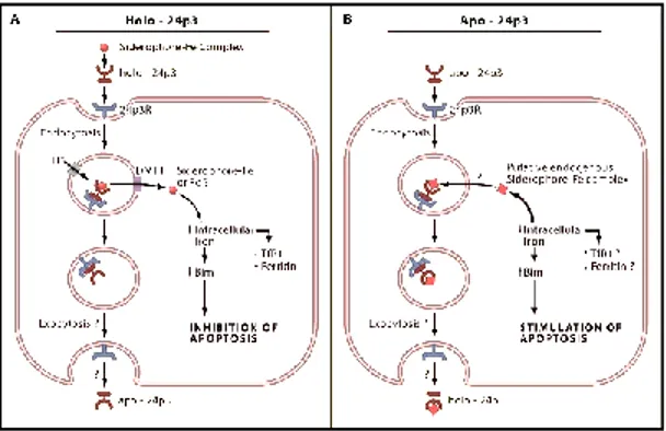
LCN-2 has been implicated in a vast array of cellular processed ranging from cell death/survival (Devireddy et al., 2005), cell migration/invasion in cancer (Yang and Moses, 2009), cell differentiation (Bolignano et al., 2010). In physiological conditions LCN-2 is reported to have high affinity to bind and transport iron, therefore it modulates iron content into cells and this impacts on cell homeostasis (regulating proliferation versus apoptosis). Iron-containing (holo-) LCN-2 binds to 24p3R and is internalized, thereby increasing intracellular iron concentration. Instead, when the iron-free (apo-) LCN-2 is internalized upon 24p3R binding, it chelates iron and transfers it in the extracellular milieu, thereby impacting on cellular apoptosis (Devireddy et al. 2005) (Figure 5). As well, LCN-2 can transport iron when complexed with siderophores. This binding confers the ability to LCN-2 to work as part of the acute-phase response in innate immunity.
25
Figure 5. Potential effects of LCN-2 iron binding activity on cellular homeostatsis. A. Holo-24p3/LCN-2 containing the iron complex of the bacterial siderophore, donates
iron to cells via the 24p3 receptor. Internalization of LCN-2 and its receptor leads to the uptake of iron from the siderophore-iron complex. Donation of iron to the cell leads to a decrease in TfR1 (Transferrin receptor protein 1) expression and an increase in ferritin levels. In addition, donation of iron to the cell prevents apoptosis by decreasing the expression of the proapoptotic protein Bim. B. Apo-24p3/LCN-2 binds to 24p3R and is internalized into the cell. Devireddy and colleagues (2005) suggest that a putative intracellular mammalian “siderophore iron complex”, called catechol, becomes bound to apo-24p3 to form holo-24p3, which is subsequently released from the cell by exocytosis. Depletion of iron from the cell results in the upregulation of the proapoptotic molecule, Bim, which leads to apoptosis (Richardson 2005).
Initially, LCN-2 was proposed as an acute phase protein also in glial cells, based its role in the regulation of astrocyte death, morphology and migration. LCN-2 can be secreted by reactive astrocytes under inflammatory brain conditions. In astrocytes, released LCN-2 acts as an autocrine mediator of reactive astrocytosis. Following exposure to LCN-2, it was reported that, in parallel to increased GFAP immunoreactivity, there are also phenotypic changes in the number and lenght of cellular processes of astrocytes (Lee et al., 2015). LCN-2 upregulation also promotes astrocytes migration via release of chemokine ligand-10 (CXCL10), chemokine that promotes the migration of other CNS cell types. Altogether LCN-2 upregulation is consistent with the hallmarks of reactive astrocytes.
26
In the CNS, LCN-2 expression is described to occur in response to injury or in inflammatory conditions. On the other hand, Chia and colleagues (2011) reported the distribution and the expression of LCN-2 in normal rat brain in physiological conditions, with low levels of expression in most brain regions, comprising the hippocampus, and highest levels of expression in olfactory bulbs, brainstream and cerebellum (Chia et al. 2011). They reported that LCN-2+ cells co-localize with GFAP+ astrocytes of these regions.
In addition, in the CNS, LCN-2 has been suggested to play a modulatory action in cognitive functions (Ferreira et al. 2013), neuronal excitability and anxiety (Mucha et al. 2011) and depression in human beings (Naudé et al. 2013).
Interestingly, LCN-2 null mice (LCN-2-/-) display anxious and depressive-like behaviors, cognitive impairment in spatial learning tasks and in parallel synaptic impairment in hippocampal long-term potentiation (Ferreira et al. 2013). Altogether behavioral studies, performed in order to assess animal anxiety-like behavior (i.e. elevated plus maze; light/dark box; open field test; Morris water maze), associated with hippocampal morphological analysis support the involvement of LCN-2 in emotion and cognition.
Recently, Mucha and collegues (2011) reported that both Lcn2 gene and the encoded protein are upregulated in the hippocampus of male mice upon psychological stress (6h/daily restraint for 3days) and that LCN-2 contributes to stress-induced neuronal plasticity (Mucha et al., 2011). On one hand, they demonstrated through in vitro experiments that treating hippocampal neurons with lipocalin-2 (100 ng/ml), as an attempt to mimick stress induced upregulation, caused a decrease of mushroom spines (known as “memory spines”) and a concomitant increase of the thin spines (known as “learning spines”) in neuronal cultures. On the other hand, they observed that disruption of the Lcn2 gene in mice, caused an increase in the percentage of mushroom spines and their density. These ex vivo data correlated also with higher
anxious-27
like behavior of LCN-2-/- mice upon stress. This scientific report suggested that lipocalin-2 might regulate brain allostasis, and act as a mechanism of cellular adaptation in response to harmful stimuli.
ii) Thrombospondins (TSPs)
TSPs are astrocyte-secreted glycoproteins, that participate in a vast array of biological functions as part of the ECM, and regulate cell-cell and cell-matrix interactions. The TSP family consist of two subgroups. Subgroup A includes TSP-1 and TSP-2, whereas subgroup B includes TSP-3/4/5 (Figure 6).
Figure 6. Thrombospondin isoforms and oligomerization states. TSP can be divided into two
subgroups. TSP-1 and -2 (subgroup A) form trimers, whereas TSP-3, -4, and -5 (subgroup B) are assembled as pentameric proteins. All TSPs contain a variable number of epidermal growth factor (EGF)-like repeats (blue) that are contiguous with calcium binding repeats (gray) and a globular conserved C-terminal region. A distinctive characteristic of group A TSP is the presence of a pro-collagen (von Willebrand factor, red) homology domain and the three properdin-like repeats (orange). In contrast, the pentameric thrombospondins (TSP-3, -4, and -5) lack the procollagen homology domain and the properdin-like repeats and contain four (instead of three) copies of the EGF-like repeats (Modified from Eroglu et al. 2009)
Recently, it was demonstrated that TSPs promote the formation of new excitatory/gabaergic synapses in the developing CNS. This process is called synaptogenesis and is stimulated by astrocyte-secreted prosynaptogenic signals, such as TSP, and not only controlled by intrinsic neuronal mechanism (Risher and Eroglu 2012). Excitatory synaptogenesis occurs in the mammalian brain after birth, and in rodents this period occurs during the second/third postnatal weeks. TSP-1 and TSP-2 were found to play active roles by promoting the formation of excitatory synapses, and their deficiency resulted in reduced synaptic density during development (Liauw et al. 2008). The increase in synaptogenesis is mediated by the interaction between the TSP EGF-like
28
domains (common to all TSPs) and the α2δ-1 neuronal receptor (Eroglu et al. 2009).
Considering the critical role of TSPs in developmental synaptogenesis, it was also discovered that these ECM proteins are upregulated/downregulated in response to CNS injuries and in several neuropathological disorders. The role of TSPs was investigated in stroke recovery. It was reported that TSP-1/2 double KO mice exhibited significant reduction in synaptic density, as well as significant deficits in their ability to recover functions. Instead in WT mice TSP-1 and TSP-2 mRNA and protein expression levels increased following stroke and the proteins were found to be co-localized primarily in astrocytes (Liauw et al., 2008). These studies suggest that deficiency in TSP-1 and TSP-2 leads to impaired recovery after stroke and that TSPs are beneficial in term of synaptic formation. Altered TSPs expression has been reported in many neurological disorders. Down's syndrome (DS) is the most common genetic form of mental retardation and characterized by a reduced number of dendritic spines. DS astrocytes were found directly involved in the development of spine malformations and reduced synaptic density. This DS neurological feature has been linked to TSP-1 decreased protein expression in astrocytes (Garcia et al. 2010).
Alzheimer’s disease (AD) is a progressive neurodegenerative disorder characterized by impaired cognitive function and memory loss due to deposition of senile plaques (Amyloid-β), neurofibrillary tangles, loss of neurons, microgliosis and astrogliosis. Along with these features the clinical stage is characterized by massive synaptic loss that correlates with cognitive decline in AD patients. It has been demonstrated that TSP-1 expression levels are significantly decreased in brain of AD patients and in two AD mouse models (Tg2576 mouse model, expressing Aβ precursor protein - APP, Swedish mutation K670N; and Tg6799/5xFAD mouse model, overexpressing mutant human APP695 with the Swedish, Florida I716V , and London V717I familial AD mutations, as well as human Presenillin 1 harboring two familial mutations M146L and L286V), (Son et al. 2015). This authors demonstrated that TSP-1
29
plays a protective role against Aβ toxicity on dendritic spines. Aβ1-42 was found able to regulate TSP-1 secretion levels in astrocyte and only TSP-1 was found reduced in astrocyte secretome. Furthermore, co-incubation of TSP-1 with Aβ 1-42 -treated hippocampal neurons, was shown to mitigate the dendritic spine loss
via interaction with the TSP-1 receptor α2δ-1 in vitro.
Lu and Kipnis (2010) proposed TSP-1 as key regulator of NPC self-renewal and neuronal differentiation in vivo and in vitro (Lu and Kipnis 2010). The authors showed that deficiency of TSP-1 impairs cell proliferation and neuronal differentiation, both in vivo and in vitro. First, by quantifying BrdU and Ki67 proliferation markers in the neurogenic SGZ and SVZ areas in WT and TSP-1 -/-mice (on a B6129SF1/J background), they reported a significant reduction in proliferating cells in mutant mice. In addition, in the same regions they observed a significant reduction in the number of DCX+/BrdU+ cells in TSP-1 -/-mice compared with the WT counterparts. In the in vitro study, the authors confirmed that TSP-1 deficiency impairs cell proliferation and neuronal differentiation of SVZ-derived NPC. For the first time they demonstrated that incubation with exogenous TSP-1 (2 µg/ml) promotes WT NPC neuronal differentiation and that TSP-1-/- astrocytes are unable to promote neuronal differentiation of NPC. Altogether these observations suggest that TSP-1 is one of the astrocytes-derived molecules required for astrocyte induced neuronal differentiation.
Our group demonstrated that adult hippocampal NPC derived from wild type animals do express the α2-δ subunit of the voltage-gated calcium channels, and this receptor mediates the proneurogenic effect of antiepileptic drugs, pregabalin and gabapentin (Valente et al., 2012).
30
1.2.2 Astrocytes as modulators in adult neurogenesis
Astrocytes, as influential components of the neurogenic niches, can contribute to regulation of new neurons formation. An active role for astrocytes and astrocyte-secreted factors in neurogenesis, has been demonstrated in the last two decades. The group of Gage was among the first ones to provide evidence for the active and instructive role of astrocytes in the adult brain (Song et al. 2002). These scientists showed that hippocampal-derived astrocytes from postnatal mice actively regulate neurogenesis, instructing the adult stem cells to a neuronal fate commitment. They examined the fate choice of adult hippocampi-derived neural stem cells in vitro, establishing a co-culture system where NSPC were plated onto a feeder layer of primary hippocampal astrocytes from neonatal rodent brain. Furthermore, they showed that even adult hippocampal astrocytes retain the ability to promote neurogenesis from adult NSPC but are less effective compared to neonatal astrocytes. By examining the effects of astrocytes derived from neonatal and adult spinal cord, an area where multipotent stem cells can be isolated, they demonstrated the lower magnitude of neuronal differentiation of adult NSPC in presence of neonatal astrocytes. Conversely, astrocyte derived from adult spinal cord were ineffective in promoting neurogenesis, while were effective in promoting glial over neurogenic fate, data confirmed also by others (Lie et al., 2002). These results suggested for the first time the possibility that astrocytes from different CNS regions may provide regionally specific cues and that astrocytes at different developmental stages may exhibit different effects on NSPC fate choice.
Previously, in a similar work, Lim and Alvarez-Buylla reported that the interaction between NPC derived from adult SVZ and astrocytes is necessary to stimulate neurogenesis (Lim and Alvarez-Buylla, 1999). Given the ultrastructure of SVZ, with astrocytes in close contact with type B cells (slow cycling stem cells, GFAP+) and with type C cells (rapidly dividing intermediate progenitors, Dlx-2+), these researchers explored the possibility that astrocytes
31
provide a neurogenic microenvironment for SVZ cells. In their publication they showed that direct contact with astrocytes deriving from cortices supports the proliferation of SVZ precursor cells and the differentiation of these into type A neuroblasts (DCX+). They suggested that it is necessary the direct contact between astrocytes and precursor cells for neurogenesis to occur. When they tested conditioned medium from astrocytes, they observed that the medium does not support neurogenesis, suggesting that the soluble factors produced by astrocytes may be necessary but not sufficient.
Later on the work by Song and colleagues was supported by the group of Zhao, who performed gene expression profiling studies on primary astrocytes isolated from different regions of the CNS. The gene expression analysis was performed on astrocytes isolated from regions with high plasticity, such as newborn and adult hippocampal astrocytes and newborn spinal cord astrocytes, shown to promote neuronal differentiation of adult NSPC, and on astrocytes isolated from the non-neurogenic area of adult spinal cord, known to inhibit neuronal differentiation (Barkho et al., 2006). Briefly, they demonstrated that astrocytes isolated from different brain areas have distinct gene expression profiles. They found out that neurogenesis-promoting astrocytes release interleukine-1β (IL-1β) and interleukine-6 (IL-6), which are proinflammatory citokines, that could promote NSPC neuronal differentiation in combination with other factors, such as vascular cell adhesion molecule-1 (VCAM-1) and interferon-induced protein 10 (IP-10). Instead, astrocytes isolated from the spinal cord express more insulin-like growth factor binding protein 6 (IGFBP6, a negative regulator of insulin-like growth factor II signaling) and decorin (proteoglycan that can inhibit transforming growth factor 2, TGFβ2) which negatively regulate neurogenesis. In fact, it was demonstrated that this factors inhibited neuronal differentiation in vitro when co-cultured NSPC and neurogenic astrocytes were treated with them. These authors proposed that the combo of factors secreted by astrocytes into the local microenvironment is affecting the neurogenic niche.
32
Recently, IL-6 was identified to promote neuronal, but not glial differentiation of adult hippocampal NPC when applied directly in culture, by increasing the fraction of class III β-tubulin (TUJ1+) cells (Oh et al., 2010). Furthermore, was observed that also the average length of TUJ1+ neurites was increased compared to vehicle-treated cells, and the effects observed were abolished when IL-6 was neutralized by anti-IL-6 blocking antibody. This recent study supported the work done by Barkho and colleagues (2006).
While proinflammatory cytokines generally inhibit neurogenesis, the fact that neurogenesis-promoting astrocytes express higher levels of inflammation related proteins than neurogenesis-inhibiting astrocytes, suggests that cytokines and chemokines may have context- and concentration-dependent effects. Astrocytes have been shown to secrete many positive, as well as negative modulators of hippocampal neurogenesis. Neurogenesin-1 (Ng1) has been identified as an astrocyte-derived soluble factor that positively modulate neuronal differentiation both in vitro and in vivo (Ueki et al., 2003). Ng1 has been found prominently expressed in neurogenic brain areas and co-localize mainly within astrocytes of the SGZ of the adult brain. The implication of Ng1 in adult neurogenesis was confirmed by in vitro experiments on hippocampus-derived NSC cultures. In these experiments, Ng1 neurogenic effect was antagonized with a neutralizing antibody against-Ng1 applied to NSC cultures prior to treatment with the supernatant of hippocampal neurogenic region. Thus this work supported the concept that astrocytes from the hippocampus contribute to the favorable environment for adult neurogenesis.
Among other positive modulators, components related to glial metabolism also appear to be key regulators of adult hippocampal neurogenesis. ATP released from astrocytic metabolism, has been identified as a proliferative factor required for astrocyte-mediated NSC proliferation in the adult hippocampus and
in vitro (Cao et al., 2013). Similarly, Sultan and colleagues reported that
D-serine a secreted astrocyte gliotransmitter abundant in hippocampal astrocytes, is able to increase the number of NSPC in vitro and when administered exogenously, it increases both the number of RGL cells and the number of
33
transient amplifying progenitors in the dentate gyrus of adult mice (Sultan et al., 2013). Additionally the reduction of glutamine levels with fluorocitrate, that selectively inhibits glial metabolism, results in decreased levels of NPC proliferation (Fonnum et al., 1997).
An interesting protein is the neurotrophin brain-derived neurotrophic factor (BDNF), that has been reported to regulate adult hippocampal neurogenesis (Waterhouse et al., 2012). Exogenous BDNF injection into the hippocampus boost NPC proliferation in vivo and promotes the differentiation and maturation of adult NPC in culture. This is consistent with evidence that neurogenesis decreases in BDNF knockout mice. SGZ-derived NPC have been demonstrated to express BDNF-TrkB receptor, that is required for the neurogenic and behavioral response to antidepressive treatments, supporting the concept that SGZ-derived NPC are a required component in the amelioration of depression (Li et al., 2008). Studies have demonstrated that hippocampal astrocytes minimally produce BDNF (Rudge et al., 1992). During aging the levels of the neurotrophin does not change, whereas those of the receptor TrkB decreases with increasing age, as reported in the rat hippocampus. This phenomen might be one of those correlated with aging-related decline in adult hippocampal neurogenesis.
Instead, among the factors that are secreted by astrocytes and promote astroglial differentiation, there are bone morphogenetic proteins (BMP), that alter the fate of neural stem cells from neurogenesis to astrocytogenesis. BMP are members of the transforming growth factor β (TGFβ) superfamily, known to mediate the growth, differentiation and survival of different cell types (Dobolyi et al., 2012). First studies reported that BMPs (BMP2/4/5/6) induce astroglial differentiation of SVZ-generated neural progenitors and the concurrent decrease of oligodendroglial and neuronal lineages, and this instructive effect is merely differentiative with no influences on both proliferation and survival of astroglial lineage cells (Gross et al., 1996). Other reports, demonstrated that NSC treatment with low-dose of BMP4 (0.1 ng/ml) or BMP7 (1 ng/ml) stimulates NSC to differentiate exclusively into neurons (TUJ-1+ cells), with other cells
34
remaining undifferentiated, suggesting a concentration-dependent effect of BMP (Chang et al., 2003). Astrocytes secrete ciliary neurotrophic factor (CNTF), that is also important for differentiation of NSC into astrocytic lineage. CNTF alone (20 ng/ml) has a dramatic effect on astrocytic differentiation, with GFAP+ cells > 90% of total cells. Interestingly, the combined treatment of the astrogenic/neurogenic BMP with CNTF results in a decrease in astrocytes and an increase in neurons. This demonstrates the context-dependent regulation of BMP, regardless its concentration (Chang et al., 2003).
Furthermore, Ng1 involvement in neurogenic activity can be traced back to its structure (cysteine-rich domains), characteristic of BMP antagonists. Ueki and colleagues (2003) reported that Ng1 antagonize BMP-4 and alters that fate commitment of NSC from gliogenesis to neurogenesis, and suggests that Ng1 may influence hippocampal plasticity by downregulating the activity of BMP (Ueki et al., 2003).
Astrocytes supplement the local microenvironment also with mitotic factors and most notably FGF-2 and VEGF contribute to neurogenesis by enhancing the proliferation of NSC and their progeny both in the SVZ and SGZ. Bernal and colleagues (2011) asked whether a decreased availability of astrocyte-derived growth factors may contribute to the age-related decline in neurogenesis (Bernal and Peterson, 2011). GFAP-positive astrocytes express both VEGF and FGF-2. The authors found out that the gene expression levels of VEGF are significantly reduced in aged DG of rats compared to young one. In DG aging resulted also in the emergence of a phenotype of GFAP-positive astrocytes that did not express FGF-2, whereas in the young brain all GFAP-positive astrocytes co-localize with FGF-2. Anyway the overall density of astrocytes immunopositive for GFAP did not change. The authors suggested that the local environment during aging becomes less supportive for neurogenesis. VEGF and its receptor Flk-1 were detected also in Type-1 NSC and the co-expression of the two proteins is prior to neuronal commitment. Once DCX+ cells are formed, VEGF expression is lost, but Flk-1-mediated sensitivity to VEGF signaling is still maintained indicating that immature neuroblasts may be influenced by VEGF
35
signals presented by neighboring astrocytes. Normal aging is accompanied by a decline in CNS function and reduced neurogenesis. Astrocytes might be considered as the major contributors in maintaining the neurogenic niche given that the altered profile of astrocytes in the aged hippocampus negatively affects neurogenesis.
In a recent publication a group of researchers investigated the neurogenic-inducing properties of astrocytes isolated from cortical, hippocampal and midbrain areas, by analyzing the secretome profile of astrocyte conditioned media (ACM) and the effect on neuronal differentiation of human NPC (Cordero-Llana et al., 2011). They cultured human NPC for one and four weeks in control media or in regional-specific ACM. They demonstrated that ACM derived from hippocampus and midbrain is able to promote neuronal differentiation of human NPC, whereas ACM from cortex does not. By using mass spectrometry (MS) they identified clusterin as a protein secreted only in hippocampal and midbrain and not in cortex ACM. Clusterin is an interesting secretory glycoprotein (75-80 kDa), with high expression in astrocytes. It has no effect on the proliferation of NPC, but it promotes both their neuronal differentiation and survival.
Generally, the control of NSPC proliferation and differentiation is influenced by several factors that are expressed by many cell types residing within the niche. The role of astrocytes on cell fate is different, with no clear knowledge of the instructive signals and the molecular pathways involved. Astrocytes are organizers of the neurogenic niche, and the signal they provide to the neurogenic process are a sum of complex microenvironmental cues, and influences of the surrounding cells. Astrocytes by releasing soluble factors, may play both instructive and permissive functions. On one hand astrocytes secreted molecules, may promote one fate at the expenses of another, on the other hand astrocytes cues may inhibit one fate, thereby allowing progenitors to differentiate in the other cell populations.
36 1.3 NF-κB signaling in adult neurogenesis
The term NF-κB (Nuclear Factor kappa-light chain enhancer of activated B cells) refers to a family of transcription factors, firstly discovered in immune B cells to bind to the intronic enhancer of the kappa light chain gene, the κB site. These proteins are ubiquitously expressed and responsible for regulating the expression of multiple genes whose products are involved in pleiotropic functions, including cell survival/death, cell proliferation and differentiation and immune and inflammatory responses (Grilli and Memo 1999; Kovacs et al., 2004; Vallabhapurapu and Karin 2009). Often NF-B signaling appears to play opposite functions, since the final outcome of its activation depends on the cell type involved, the type of disease or injuries and the target genes activated or repressed. NF-B signaling was initially studied mainly in the field of immunology, as major regulator of innate and adaptive immunity. In the CNS, NF-κB is expressed in both neurons and glia. In general, NF-κB in CNS glial cells, like microglia and astrocytes is mainly neurodestructive due to the overwhelming production of pro-inflammatory cytokines (Mattson and Meffert 2006). The role of NF-κB in the nervous system has gained interest because of its involvement in neuroprotection/neurodegeneration (Kaltschmidt and Kaltschmidt 2009), synaptic plasticity and neurite outgrowth (Gavalda et al. 2009) and formation of functional dendritic spines (Boersma et al. 2011). On one hand many in vitro studies have demonstrated that neurotoxic and pro-apoptotic stimuli including high concentrations of glutamate, amyloid-β or cytokines, cause NF-κB activation in neuronal cells, resulting in cell death (Uberti et al. 1999; Ryan et al. 2000; Zhang et al. 2005). On the other hand, it has been reported that neuron-restricted ablation of NF-B-driven gene expression promotes neurodegeneration (Fridmacher et al. 2003), suggesting a neuroprotective role for NF-κB. Recent finding, suggest that neuronal NF-B signaling is neuroprotective via maintaining neuronal survival, synaptogenesis, synapse to nucleus communication (Gutierrez and Davies 2011; Mattson and
37
Meffert 2006) and its role in regulating neural development and CNS functions such as learning and memory (Salles et al. 2014).
Herein, I will introduce NF-κB family members and their signaling pathways, focusing on the emerging evidence of a role for NF-κB in regulating adult hippocamapal neurogenesis.
1.3.1 The NF-κB family of transcription factors
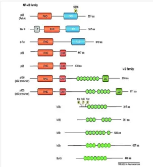
The NF-κB family of transcription factors is composed of five subunits including p50/p105 (NF-κB1), p52/p100 (NF-κB2), p65 (RelA), RelB and c-Rel, which all share a conserved and multifunctional N-terminal Rel homology domain (RHD), that is responsible for homo- and hetero-dimerization of the different subunits. The RHD also contains nuclear localization signal (NLS), masked by binding of inhibitory IκB protein. Upon IκB inhibitory protein phosphorylation and degradation, NLS unmasking allows nuclear translocation of dimeric complexes which can then bind regulatory sequences in target genes. According to their structure, NF-κB family members can be divided into two subfamilies. One includes RelA, RelB and c-Rel, which are generally considered as transcriptionally activators, since they possess a transactivational domain (TAD), while the other subfamily includes p50 and p52 subunits, which are commonly considered as repressors, since lacking the TAD. The IκB family of inhibitors includes p100, p105, IκBα, IκBβ, IκBγ and Bcl-3. These inhibitors proteins are carachterized by the presence of ankyrin-repeat motives that mediate their binding with NLS/RHD of NF-B subunits. The p105 and p100 C-terminal region, also containing ankyrin repeats, that acts like an auto-inhibitory domain, retaining the precursors of p50 and p52, respectively, into the cytoplasm. (Figure 7)
38
Figure 7. Schematic diagram illustrating the mammalian members of the NF-κB and IκB families. NF-κB family members
have in common the RHD, with RelA, RelB and c-Rel additionally possessing a TAD [RelB has a leucine zipper (LZ), while p52 and p50 and their precursors have glycine-rich regions-(GRR); additionally, precursor possess regions with homology to death domains (DD)]. The ankyrin repeats of IκB members (denoted by green ovals), mediates the RHD binding. Within the IκBα phosphorylation sites relevant for regulation of NF-κB activation have been identified (Ser32, Ser36, Tyr42). (Gutierrez and Davies, 2011)
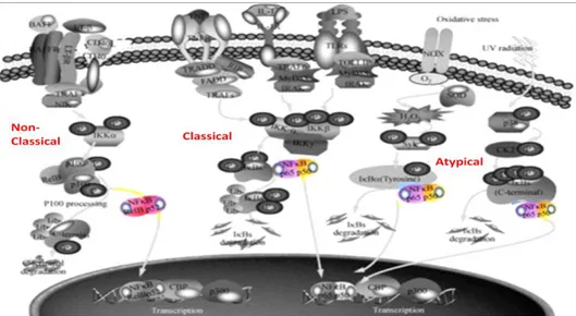
At the cellular level, under unstimulated conditions, NF-κB dimers are retained in the cytosol through the interaction with a member of the inhibitory IκB protein family. The IκB subunit masks NLS in the NF-κB complex, thereby maintaining it in the inactive state. There is a variety of potential combinations for the NF-κB complex. In the CNS, the most abundant forms are the p65/p50 heterodimer and the p50 homodimer, while IκBα is the most common inhibitory protein. A vast array of stimuli are able to trigger NF-κB activation by two main activating pathways, referred to as the canonical (or classic) and the non-canonical (or alternative) pathways. The non-canonical NF-κB signaling pathway is crucial for the activation of innate immunity and inflammation. In response to a several proinflammatory cytokines, including tumor necrosis factor α (TNFα), interleukin-1β (IL-1β), and agonists of the toll-like receptor, such as lipopolysaccharide (LPS), the IκB Kinase (IKK) complex is activated via phosphorylation of consensus motives in the IκB members (IκBα, IκBβ, IκBε and p105). More specifically, phosphorylation on Ser32 and Ser36 residues of
39
IκBα by the IκB Kinase-β (IKKβ) catalytic subunit of the IKK complex (composed of IKKβ and IKKα, the catalytic subunits and the IKKγ, the regulatory subunit) triggers ubiquitination and proteasome-mediated degradation of the inhibitory protein. Once activated, the NF-κB p65/p50 dimer translocates into the nucleus, where it binds to the DNA consensus sequence of target genes. The non-canonical NF-κB signaling pathway is critical for adaptative immunity and is activated by some particular members of the TNF family (i.e. CD40L; Lymphotoxin-β). The non-canonical pathway involves NF-κB inducing kinase (NIK)-induced phosphorylation of the IKKα subunit within the IKK complex, that in turn phosphorylates the p100 subunit of the heterodimeric complex p100/RelB. Following ubiquitination and proteasome-dependent processing of p100 into p52, the nuclear translocation of the dimer RelB/p52 occurs and regulates target genes expression. A third distinct NF-κB signaling pathway triggered by DNA damage and oxidative stress, is defined as atypical because it is independent of the IKK complex (Viatour et al. 2005). Although in this pathway proteasome-mediated degradation of IκB is involved, the upstream kinases and the phosphorylation sites are different and, as of today, not completely understood (Figure 8).
40
Figure 8. Signaling pathways involved in NF-κB activation. In the canonical
pathway, IKK complex activation leads to IκBs phosphorylation, followed by their ubiquitination and degradation by the proteasome, allowing the release of the heterodimer of p65/p50, which then translocates into the nucleus. The non-canonical pathway is dependent on NIK and IKKα, but not on the trimeric IKK complex, and mediates the activation of RelB/p52 complex. In the non-canonical pathway NIK-induced phosphorylation of IKKα leads to the phosphorylation of p100 and proteasome-mediated processing of p100 into p52, allowing the nuclear translocation of RelB/p52 to regulate target gene expression. The atypical or IKK-independent pathway of NF-κB is stimulated by DNA damage (Modified from Zhang and Hu 2012)