UNIVERSITÀ DI PISA
FACOLTÀ DI INGEGNERIA
THE NETHERLANDS
PHILIPS RESEARCH
Effects of polarization, plasma and thermal
initiation pathway on irradiance threshold of
laser induced optical breakdown
Corso di Laurea Magistrale in Ingegneria Biomedica
Master of Science Thesis
Valentina Bonito
~ Confidential ~
Care & Health Applications Department - Philips Research, Eindhoven, The Netherlands October 2012 – September 2013
Supervisors: Student:
Dr. Babu Varghese, Philips Research Prof. Luigi Landini, Università di Pisa
2 This work was carried out at Care & Health Application department at Philips Research Europe, Eindhoven, The Netherlands
Approved by:
Supervisors: Dr. Babu Varghese - Philips Research Europe
3
Declaration of the Master Thesis
I warrant that the thesis is my original work and that I have not received outside assistance. Only the sources cited have been used in this draft. Parts that are direct quotes or paraphrased are identified as such.
Eindhoven, ______________________________ ____________________________
4
All’Amore tra Mamma e Papá, che non è mutato quando ha trovato mutamento.
Ho visto entrare una donna con gli occhi bendati e nelle mani teneva tutti i miei desideri. Lei mi porta lontano la mente, nel gradino più stretto del cielo.
5
Contents
Acknowledgments ... 7 Abstract ... 8 Chapter 1. Introduction ... 10 1.1. Introduction ... 10 1.2. Research goal ... 10 1.3. Research method ... 111.4. Structure of the thesis ... 11
Chapter 2. Background Information ... 13
2.1 Introduction ... 13
2.2 Polarization of light ... 13
2.3 Skin rejuvenation ... 16
2.3.1 Skin anatomy ... 16
2.3.2 Skin aging and wrinkle formation ... 18
2.3.3 Light skin propagation ... 18
2.3.4 Light skin interactions ... 20
2.3.5 Laser based skin rejuvenation methods ... 23
2.4 Laser Induced Breakdown... 26
2.4.1 Laser induced optical breakdown (LIOB) ... 27
2.4.2 Laser induced thermal breakdown (LITB) ... 29
2.4.3 Comparison between LIOB and LITB ... 29
2.5 The fourth state of matter: plasma ... 30
2.5.1 Generation mechanisms of cold plasma ... 31
2.6 Biomedical applications ... 32
2.6.1 Laser plasma medicine ... 32
2.6.2 Plasma skin resurfacing ... 32
Chapter 3. Investigation of the focal spot and the irradiance threshold for linearly and radially polarized light ... 35
3.1. Introduction ... 35
3.2 Apodization ... 35
3.3 Materials and methods ... 36
6
3.3.2 Knife edge method ... 38
3.4 Results and analysis ... 39
3.4.1 Demonstration of feasibility in skin rejuvenation device: pulse energy measurements 39 3.4.2 Knife edge measurements ... 40
3.4.3 Demonstration of feasibility in skin rejuvenation device: irradiance thresholds calculations ... 42
3.4.4 Investigation of the electric field and the scrambling of polarization in the focus ... 43
3.5 Conclusions ... 47
Chapter 4. Experimental investigation of irradiance threshold for LIOB and LITB ... 48
4.1. Introduction ... 48
4.1.1 Laser induced optical and thermal breakdown ... 49
4.2. Materials and Methods... 50
4.2.1 Measurement of optical properties ... 50
4.2.2 Experimental demonstration of laser induced optical and thermal breakdown ... 51
4.3. Results and Analysis ... 52
4.3.1 Measurement of optical properties ... 52
4.3.2 Experimental investigation of irradiance thresholds for LIOB and LITB ... 53
4.4. Conclusions ... 56
Chapter 5. Reduction of irradiance threshold for laser induced optical breakdown in an external plasma environment ... 57
5.1 Introduction ... 57
5.1.1 Laser induced optical breakdown in an external plasma source ... 57
5.2 Materials and Methods... 58
5.2.1 LIOB based skin rejuvenation device ... 59
5.2.2 Atmospheric pressure cold plasma device ... 59
5.3 Results and analysis ... 61
5.4 Conclusions ... 64
Chapter 6. Summary and Outlook... 65
6.1 Conclusions ... 65
6.2 Outlook ... 67
Appendix ... 69
7
Acknowledgments
My first mention goes to my supervisor Dr. Babu Varghese, who gave me the opportunity to work for this stimulating and high level project. You always found the right words to support me during this long internship. Your sense of humor and your helpfulness contributed widely to create a pleasant working environment. Thanks for your endless patience: you gave me the time to learn from my mistakes and improve myself, helping me reaching professional results I am proud of.
Thanks to all the people from the Department of Care & Health Applications: being part of such a mind opening working place has been a honor for me. Thanks Rieko and Martin for supporting me during the laboratory activities and for all your precious suggestions. Un particolare ringraziamento va al mio personale pezzetto di Italianità in Philips Research: Calina. Grazie per il tuo costante incoraggiamento e per i bei momenti passati insieme, dentro e fuori dall’ufficio. Nella tua casa mi sono sentita a casa. I would also like to thank all my past and present friends from Eindhoven. We built together an extended family that never let me feel alone. A special mention goes to Aura: I’m glad to share one of the best days of my life here, today, with you.
Roomina mia, grazie. Quell’emblematico secondo giorno di università e quel posticino riservato a lezione sono stati soltanto l’inizio di una innumerevole serie di esperienze indimenticabili insieme.
Grazie Martina. Lontano da tutto e da tutti ci siamo costruite, mattone dopo mattone, la nostra piccola Casa. E abbiamo fatto un lavoro cosi perfetto che non c’è altro posto al mondo dove vorrei stare adesso. Antonella, Brigida, Chiara, Luca, Luigi, Domenico, Chiara, Francesco, Monica, Gabriele: vi adoro. Così diversi tra noi, cosi vicini. Avete reso gli anni universitari i più belli della mia vita. Tutti i momenti vissuti con voi son marchiati a fuoco nella mente e nel cuore. Dal profondo, grazie.
Virginia, 23 anni di amicizia sconfiggono qualsiasi distanza. È bello sapere che dovunque andremo e qualsiasi cosa faremo, potremo sempre contare una sull’altra. Grazie per ieri, oggi e domani.
Grazie alle mie due splendide donne: Mamma e Laura. La vostra tenacia e l’amore che ci lega abbattono qualsiasi muro. Insieme formiamo una squadra imbattibile.
E l’ultimo pensiero va a Te. Sei padre, amico, alleato. Sei intelligenza, caparbietà, coraggio, ironia, integrità. Sei quanto di piu grande una figlia possa desiderare. Oggi ci laureiamo insieme, Babbino mio.
8
Abstract
The development of laser based skin rejuvenation techniques has significantly increased in recent years. However, the presently available ablative and non-ablative methods for skin rejuvenation typically balance between efficacy, safety, social downtime and pain perception [1]. Highly effective ablative techniques are characterized by long recovery time and significant risk profile whereas the non-ablative methods, safer due to the ability to create dermis thermal damage without affecting epidermis, show limited efficient clinical results. To overcome the existing trade off, Philips Research has recently developed a novel minimally invasive laser technology for wrinkle reduction using a form of laser induced breakdown (LIB), i.e. laser induced optical breakdown (LIOB).
LIB is the partial or complete ionization of a solid, liquid or gas through absorption of thermal or electromagnetic energy generated by a laser source. Laser-induced breakdown (LIB) takes two different forms: laser-induced optical breakdown (LIOB) and laser induced thermal breakdown (LITB). LIB can occur by pure multiphoton ionization, by avalanche (cascade) ionization or a combination of the two. For both the mechanisms the initial phase is the creation of free electrons in the focal volume. Depending on medium purity two generation processes of seed electrons can be distinguished. In case of pure medium, the seed electrons come from ionization of a few molecules through multi-photon absorption. Several photons together containing enough energy to ionize the molecule must be simultaneously absorbed and the breakdown produced is LIOB. In case of impure medium these seed electrons come from ionization of impurities by thermal excitation. In other words, by heating of linear absorbing chromophores in the target these seed electrons are produced by thermionic emission and LITB occurs.
LIOB has attracted great interest in recent years due to its numerous applications in medicine and biology, while LITB has received less attention. The principles of LIOB have been exploited for applications regarding ophthalmic microsurgery, stone fragmentation, angioplasty, and, recently, skin rejuvenation. When LIOB is generated inside the skin, a grid of lesions appears within the dermis that induces new collagen formation without affecting the epidermis, resulting in reduction of wrinkles and fine lines [2]. Subsurface laser skin ablation through LIOB requires high irradiance in the order of 1013 W/cm2 [3]. Beside the safety related advantages, reducing the irradiance required to create breakdown may allow reaching deeper layer of the skin, hence improving the efficacy of the treatment. In this work of thesis, three possible solutions have been provided to achieve the lowering of the irradiance threshold: the use of a properly polarized input beam, the exploitation of the LITB action mechanisms for the initial generation of seed electrons, and the combined use of the laser source with an external plasma source.
9 First of all we demonstrated the dependence of the irradiance threshold on the polarization state of the laser beam used, in order to consequently establish the polarization state giving a larger focal spot and a lower irradiance. We investigated the effects of polarization and apodization on laser induced optical breakdown (LIOB) irradiance threshold on liquid media resembling the optical properties of biological tissues relevant for LIOB based skin rejuvenation applications. Using a skin rejuvenation device recently developed by Philips, we measured the breakdown threshold obtained for linearly and radially polarized light under different apodization conditions. We proved that the irradiance threshold required to create LIOB in liquid media of different optical properties is lower for radial polarization due to a larger focal spot provided by it.
We proved the possibility to lower the irradiance threshold when the thermal pathway for the generation of seed electrons typical of LITB is induced in absorbing media. We calculated LIOB and LITB irradiance thresholds on transparent and absorbing aqueous media treated with the home built prototype device developed by Philips. We demonstrated a transition from laser-induced optical breakdown to laser-induced thermal breakdown as the absorption coefficient of the medium is increased. We observed also that irradiance threshold for optical breakdown, after correction for the path length dependent absorption losses in the medium, is nearly unaffected by the variation in the absorption properties of the medium, whereas irradiance threshold for thermal breakdown decreases with the increase in the absorption properties of the medium. Moreover we demonstrated that the irradiance threshold for LITB is always lower than LIOB threshold. Results obtained are the experimental confirmation of two of the most significant benefits of employing LITB in skin rejuvenation applications: (i) the selectivity of LITB for the target chromophore and the surrounding tissue as the process is dependent on the absorption properties of the target; (ii) the less demanding laser pulse parameters required for the LITB process as a result of the lower intensity threshold.
Also, we demonstrated the feasibility of lowering the intensity threshold for LIOB by providing seed free electrons in the focal volume of the tightly focused ultra-short laser pulses using an additional plasma source. Experiments were performed to confirm the proof of principle using a skin rejuvenation prototype developed by Philips Research in combination with a commercially available plasma device, kINPen®09. We proved that with a partial or weak ionization providing at least one seed electron in the focal volume, a 70% reduction of the irradiance threshold can be achieved.
Lowering the irradiance threshold with the above described methods may allow creating deeper lesions inside the skin and can lead to precise and well-localized tissue effects with less risk of collateral damage, thereby improving safety and efficacy of treatment. Furthermore the laser pulse parameters required for the LIOB process become significantly less demanding and this will result in better availability of laser sources with less critical requirements. Any laser energy reduction will have an exponential benefit on the price of the laser source, which is the main driver for cost of the entire system for devices based on LIOB.
10
Chapter 1
Introduction
Beauty is only skin deep, but it is only the skin you see
- Fifteenth century proverb
1.1.
Introduction
The development of laser based skin rejuvenation techniques has significantly increased in recent years. In spite of the technological developments in the skin rejuvenation techniques throughout the years, no revolutionary approach has been introduced that is capable of accurately defining the balance between efficacy, safety, social downtime and pain perception [1]. The ablative techniques provide significant results but the post-treatment care required is substantial and the incidence of side effects is relatively high. On the other hand the non-ablative methods are safer and social downtime is shorter but the clinical efficacy is limited compared to ablative treatment.
Philips Research has recently developed a novel minimally invasive laser technology for wrinkle reduction using laser induced optical breakdown (LIOB). When LIOB is generated, a grid of lesions appears within the dermis that induces new collagen formation without affecting the epidermis, resulting in reduction of wrinkles and fine lines [2].
1.2.
Research goal
LIOB requires high intensities in the order of 1013 W/cm2 [3]. The initiation of optical breakdown through multi-photon ionization involves the generation of one or more seed electrons by absorption of multiple photons having same polarization in the focal volume [4]. Multiple scattering and tissue birefringence results in depolarization of light and this effect accumulates as a function of focusing depth inside the skin. Therefore skin needs to be exposed to high laser intensities to create sufficient number of photons with same polarization in order to exceed the breakdown threshold. The pulse parameters and the focusing conditions that are used for creating optical breakdown are very critical and tight focusing is required. Reducing the irradiance threshold required to create breakdown is very important, especially for aspects regarding laser safety and treatment efficacy. Lowering the irradiance threshold may let deeper layers of tissue to be targeted with less risks of collateral damage.
The aim of this work of thesis is to find an effective method to lower the irradiance threshold for laser induced optical breakdown. Here we investigated three potential solutions: (i) use of radially polarized light and influence of size of focal spot, (ii) thermionic emission of seed electrons (thermal
11 initiation pathway) for creating optical breakdown, and (iii) presence of free electrons in the focal volume provided by an external plasma source.
1.3.
Research method
To create laser induced optical breakdown the skin rejuvenation prototype developed by Philips Research was used. This device provides high power laser pulses at a wavelength of 1064 nm. Performances of radial and linear polarization were analyzed in terms of irradiance threshold, conducting experiments on distilled water and diffuse media. The beam waist for linear and radial polarization for different apodization conditions was experimentally measured through the knife edge technique and the results were compared with numerical simulations. We investigated also the scrambling of polarization in the focus by means of the numerical simulations.
We experimentally measured the irradiance threshold for another form of laser induced breakdown, laser induced thermal breakdown (LITB), and we analyzed its dependence on the properties of the medium by conducting experiments in transparent and absorbing media resembling the optical properties of skin.
To demonstrate the reduction of the irradiance threshold for LIOB in plasma environment we used the same skin rejuvenation prototype described above in combination with a commercially available plasma device, kINPen®09. The plasma source created partial ionization of the medium and provided the free electron environment in the focal volume to initiate the breakdown process.
1.4.
Structure of the thesis
The layout of the thesis is as follows.In Chapter 2 the theoretical background underlying the content of thesis is given. First, mathematical aspects of light polarization are described. Then, a short description of the skin anatomy and its interaction with light is presented. An overview of different skin rejuvenation treatments is provided. Then the skin rejuvenation prototype developed by Philips is introduced. Principles of laser induced optical (LIOB) and thermal breakdown (LITB) are consequently explained. Then the mechanisms of plasma formation and some examples of cold plasma sources are described. Examples of laser devices for biomedical applications conclude the chapter.
In Chapter 3 the effects of polarization and apodization for LIOB threshold are investigated. The experimental measurements of the focal spot for linearly and radially polarized light through the knife edge technique are described. Results of numerical simulations are compared with experimental results and available literature. Finally the study of the scrambling of polarization in the focus by means of numerical simulations is presented.
12 In Chapter 4 the investigation of the irradiance threshold for LIOB and LITB in transparent and absorbing aqueous media resembling the skin optical properties is performed.
In Chapter 5 the effective solution for the reduction of the irradiance threshold for subsurface laser skin ablation through LIOB in plasma environment is presented.
13
Chapter 2
Background Information
Boswell: Then, Sir, what is poetry? Johnson: Why, Sir, it is much easier to say what it is not. We all know what light is; but it is not easy to tell what it is - Boswell’s Life of Johnson
2.1
Introduction
In this chapter, the theoretical background of the thesis is provided. First, principles of polarization of light and a brief description of homogeneous and spatially variable states of polarization are given. Then general description of the anatomy of the skin and its optical properties is presented; the skin rejuvenation methods available nowadays are then described. Particular attention is given to a novel minimally invasive technique developed by Philips based on the principles of laser induced optical breakdown. Then basic principles of plasma formation are explained and an overview of the mechanism of generation of cold plasma is provided. Examples of laser and plasma based biomedical applications conclude the chapter.
2.2
Polarization of light
Light consists of spatio-temporal electric (E) and magnetic fields (B). These fields are vectors, and their directions are perpendicular to each other and to the direction of propagation of light. Because the fields propagate together and maintain a constant phase difference (90◦) with one another, it is usually sufficient to describe the wave with either the electric vector or the magnetic vector. Conventionally it is chosen the electric vector, largely because the interaction of matter with the electric field is stronger than that with the magnetic field ([5],[6]).
Polarization is a property of waves that describes the orientation of the electric field. When the vector direction is predictable light is called polarized; in any other case, light is defined partially polarized or unpolarized. To define the different states of polarization, the theory of the propagation of electromagnetic waves must be introduced. Briefly, any electromagnetic field may be resolved into an infinity of monochromatic fields of angular frequency , of which any can be split up into an infinity of plane waves of wave vector [7+. The Fourier’s integral theory translates this concept in the following equation for the electric induction field :
14 ∫ ∫ [ ] (2.1) is the most significant field in crystalline media optics. All the other components of the electromagnetic field can be deduced from thanks to the Maxwell’s equations. In order to study the polarization of light it is then sufficient to consider only one of the monochromatic plane waves mentioned above. The electric field, for a propagation along the z-axis, can be described by its main components and , defined as follows:
where and are the amplitudes and is the wave number (modulus of the wave vector ).
If and oscillate in phase, that is , with being a real constant,
formula (2.2) represents the electric field of a linearly polarized plane wave. The wave is called linearly polarized because the observer sees the oscillating vector tracing out a straight line in the plane. If
or are equal to 0, the wave is referred to as linearly polarized along the direction or
the direction respectively.
If there is a phase shift between and , that is where is the
imaginary unit, the wave is said to have circular polarization because the end of the vector E traces out a circle. Circularly polarized waves can be left or right-handed. If the electric field
vector is tracing out a circle in a clockwise direction and the light is referred to as left-handed circularly polarized. If the end of the electric field vector traces out a circle in a
counterclockwise direction and the light is right-handed circularly polarized.
FIG.2.1. Linearly and circularly polarized light
15 Linear and circular states of polarization are particular cases of the most general fully polarized homogeneous state of polarization that is elliptical polarization. In that case ,
with being a generic complex number, and for an observer facing the wave source the end of the vector E is tracing out an ellipse [7].
FIG.2.2. Homogeneous states of polarization: linearly, circularly and elliptical polarized light
For all the polarization states described above the direction of the electric field is the same in every position throughout the beam; therefore linear, circular are elliptical polarization are called spatially uniform polarization states. In recent years there has been growing attention towards spatially variable states of polarization. In particular laser beams with cylindrical symmetry polarization have been studied because of the unique properties of the electric field in the focal region. By decomposition of a generalized cylindrical vector beam two new states of polarization can be defined: radial polarization and azimuthal polarization. A clear and intuitive definition of the wave polarization states is based on the definition of the polarization orientation, calculated as follows:
(2.3) The integer P is the polarization order number (a topological charge) that represents the number of polarization rotations per roundtrip (θ=0…2π), and is the initial polarization for . For instance P=0 fields represents linearly polarized light with orientation ψ0. Radial and azimuthal
16 FIG.2.3. Spatially variable states of polarization: radially and azimuthally polarized light
2.3
Skin rejuvenation
2.3.1
Skin anatomy
The skin covers the entire external surface of the human body and it is the principal site of interaction with the surrounding world. It serves as a protective barrier that prevents internal tissues from exposure to trauma, ultraviolet (UV) radiation, temperature extremes, toxins, and bacteria. Other important functions include sensory perception, immunologic surveillance, thermoregulation, and control of insensible fluid loss. The skin is made up of the following layers: epidermis, dermis and subcutaneous fat layer or “subcutis” (Fig.2.4).
.
17 The epidermis is the outer layer of the skin and is average thickness is 100 μm. It is composed by a stratum corneum (or “horny layer”, mostly characterized by dead cells) and four underlying layers: stratum lucidum, granulosum, spinosum and basale (Fig.2.5).
FIG.2.5. Section of the epidermis
The second layer of the skin is the dermis, tightly connected to the epidermis through a thin sheet of fibers constituting the basement membrane. The dermis contains the structural elements of the skin, the connective tissue, and can be from 1 to 4 mm thick. Two main areas can be identified: a superficial area adjacent to the epidermis, called papillary region, and a deep ticker area known as the reticular region. The papillary dermis is characterized by a network of thin (0.3-3 μm diameter) collagen fibers and elastic fibers (10-12 μm diameter), embedded in lose connective tissue and a highly developed microcirculation composed of arterioles, capillaries and venules. The reticular dermis is composed predominantly of dense bundles of thick (10-40 μm diameter) collagen fibers arranged parallel to the skin’s surface, elastic fibers and fibroblasts surrounded by an amorphous mix of water, electrolytes, plasma proteins and polysaccharides. Collagen is made by fibroblast and it is the main protein of connective tissue. Collagen consists of inter-woven fibrils of globular tropocollagen sub-units that spontaneously arrange themselves by numerous hydrogen and covalent bonds. Thanks to its great tensile strength, collagen is responsible for skin strength. Other properties of the skin, such as turgor and elasticity are given by glycosaminoglycans and elastin fibers respectively.
The deepest layer of skin is the subcutis (or “hypodermis), which can be from 1 to 6 mm thick depending on the body site. The subcutis, consisting of a network of collagen, elastin and fat cells, helps conserve the body's heat and protects the body from injury by acting as a "shock absorber."
18
2.3.2
Skin aging and wrinkle formation
The more dramatic effects of skin aging are seen in the dermis, which is thought to be mainly responsible for skin mechanical properties. In young skin, fibrillar collagen is highly organized. It forms a spread meshwork of rope-like small and thin bundles of tightly packed fibers in the papillary dermis, whereas in the reticular dermis it is more spaced with thicker bundles. Fibroblasts attached to collagen fibers via integrins are highly aligned with the collagen network and stretch it; in return, they are maintained in a tensed state. This well-organized collagenous structure has extraordinary properties in terms of resiliency and stiffness, together with a characteristic tensed aspect.
During chronological aging, dermal content in collagen is diminished, bundle becomes thinner and collagen meshwork tends to disappear. Therefore interactions between fibroblasts and collagen are reduced leading to randomly collapsed fibroblasts (Fig.2.6). These changes in the scaffolding of skin cause skin to wrinkle and sag [8].
FIG.2.6. Histological feature of skin from young (left) and old (right) individuals. With aging fibroblasts oriented in the plane of thick fiber bundles are replaced by round and oblong interstitial cells randomly disposed between disorganized fibers.
2.3.3
Light skin propagation
In laser skin therapy the knowledge of absorbing and scattering properties of the treated area is essential for the purpose of predicting a successful treatment.
Absorption is mainly caused by water molecules (in the IR region) or proteins, hemoglobin and melanin (in the UV and visible range). These absorbing molecules are termed as chromophores. When an incident electromagnetic wave is absorbed by a medium there is a decrease in the intensity expressed through the Lambert-Beer’s Law:
(2.4)
where Io indicates the incident light intensity, the light intensity at a distance inside the medium and the absorption coefficient. The absorption spectrum of human skin is presented below (Fig.2.7).
19 FIG.2.7. Absorption coefficient of the main constituents of the skin
Scattering in skin starts when the light interacts with single fibrils and more complex structure made by interlacement of them, called scattering centers. This phenomenon can be caused primarily by variation in polarizability, which can be characterized by variations of the optical index of refraction. This mismatch may be largely a function of shape, size and distribution of skin constituents and, in particular, of dermis constituents such as collagen, lipids, water, cells and their organelles. The epidermal scattering, which is affected by melanin granules and keratinocyte and melanocyte nuclei, is indeed sufficiently close to that of dermis and sufficiently thin to be not critical. The optical scattering properties of the dermis can be characterized by the scattering coefficient (μS), or sometimes by the
reduced scattering coefficient (μS’), related to each other by the following formula:
(2.5) where g is the anisotropy factor defined as the mean of the cosine of the scattering angle Ө :
(2.6) Depending on the value of g, it is possible to identify the scattering direction: purely forward scattering for and purely backward scattering for . Among the two layers of the dermis – papillary and reticular – the former is highly backscattering because of the small size of the collagen
20 fibers (diameter of an order of magnitude less than visible light wavelength). Contrariwise within the latter, the large size of collagen fiber bundles causes highly forward-directed scattering. The average scattering properties of skin are defined by the scattering properties of the reticular dermis because of its relatively big thickness (up to 4 mm) and typical values of are in the range of 0.7 - 0.95 [9].
FIG.2.8. Reduced scattering coefficient of skin
The total attenuation coefficient for skin, that takes into account the absorption process and the scattering of light, can be expressed as:
(2.7) As can be seen in Fig.2.7 and Fig.2.8, in a wavelength range of 600-1300 nm both absorption and scattering are low. Therefore this region, refer to as optical or therapeutic window, is used for imaging and treatment of the skin.
2.3.4
Light skin interactions
The laser-tissue interactions depend on the opto-thermal properties of tissue in combination with the characteristics of the laser source and in general they can be grouped as follows: photochemical interactions, photothermal (vaporization and coagulation) interactions, plasma induced ablation, photoablation and photodisruption (Fig.2.9). The characteristic energy density (dashed lines of Fig.2.9) varies over 15 orders of magnitude, ranging from approximately 1 mJ/mm2 to 1 J/mm2and a single parameter distinguishes and primarily controls these processes: the duration of laser exposure, which is mainly identical with the interaction time itself [10]. The time scale can be roughly divided into five sections: continuous wave or exposure times > 1 s for photochemical interactions, 1 min down to 1 μs
21 for thermal interactions, 1 μs down to 1 ns for photoablation, and < 1 ns for plasma induced ablation and photodisruption. The difference between the latter two is attributed to different energy densities, besides the fact that one is based exclusively on ionization whereas the other is primarily a mechanical effect. Thermal effects for instance may play also an important role when ultrashort laser pulses (< 100 ps) at high repetition rates are applied (10-20 Hz), adding up to a measurable increase in the temperature of the tissue treated.
FIG.2.9. Laser tissue interactions showing influence of exposure time and power density (double logarithmic map).
In the following part physical principles and possible applications of the mechanisms interesting for the purpose of our study, i.e. photothermal interactions and plasma induced ablation, are briefly presented.
In photothermal interactions, absorbed photons are converted into heat. The local increase in temperature is the most significant influencing factor and the effects of heating can be seen as denaturation and coagulation (T > 60◦C), which can proceed to necrosis and vaporization (T>100◦C), resulting in tissue ablation and carbonization (Fig.2.10).
22 FIG.2.10. Thermal effects of an ablative laser beam.
The heat generated within the target can be confined to that target by the appropriate selection of pulse duration. This is related to the thermal relaxation time of the target, defined as the intrinsic cooling time taken for the target to dissipate half of the incident thermal energy [10], [11]. If the laser pulse duration is equal to or less than the thermal relaxation time of the target, then thermal diffusion to adjacent tissue will be reduced. The thermal relaxation time can be calculated as follows:
(2.8)
The thermal diffusivity of the tissue is approximately , which is very similar to that of water ( ) of which most of the tissue is composed. is the
effective attenuation coefficient (equivalent to the molar extinction coefficient) and is the depth of
penetration at which the intensity has fallen by 1/e, equal to . The thermal relaxation time is
primarily related to the physical size of the target: the larger the target, the longer the thermal relaxation time, as it possible to see in Tab.2.1, where the thermal relaxation times of the main skin chromophores are presented.
Chromophore Size (μm) Thermal relaxation time
Melanosome 0.5-1 1 μs Erythyrocyte 7 1 μs Blood vessel 15 100 μs 50 1 ms 100 1 ms 200 20 ms
23 Plasma induced ablation is obtained when the power densities exceed 1011 W/cm2 in solids and fluids, or 1014 W/cm2 in air [10]. Once the critical irradiance threshold is surpassed a phenomenon called breakdown occurs. The critical threshold can be defined also in terms of electric field strength E, related to the local power density I by the basic electrodynamic equation:
(2.9) For power density of 1011 W/cm2, the corresponding electric field amounts to approximately 107 V/cm. This value is comparable to the average atomic or intramolecular Coulomb electric fields, thus providing the necessary condition for plasma ionization. The physical principles of breakdown and plasma formation have been extensively investigated in the past [12],[13],[14]. Further details will be explained in sec. 2.4 and 2.5.
2.3.5
Laser based skin rejuvenation methods
Laser and light based skin rejuvenation techniques has rapidly evolved over the last few decades from ablative to non ablative and more recently towards fractional resurfacing techniques (Fig.2.11).
FIG.2.11 : Laser based skin rejuvenation methods
The golden standard for laser resurfacing is based on ablative lasers such as CO2 and Er:YAG. The
ablation of the superficial layers of the skin is followed by the stimulation of new collagen growth in the dermis. New collagen replaces the disorganized collagen/elastin connective tissue matrix associated with wrinkled or photodamaged skin in the upper dermis. Since CO2 and Er:YAG laser wavelengths target
water as chromophore, these methods ablate the full epidermis before reaching the target papillary dermis. As a consequence a thin layer of heat-denatured dermal tissue remains. During the repair processes this heat damaged layer releases mediators which induce formation of the new and improved collagen matrix with an improved aesthetic appearance [1]. Despite the high efficacy of these ablative techniques, the significant risk of side effects and the prolonged postoperative recovery period associated with them prompted the development of alternative solutions.
24 Non ablative techniques exploit the principles of selective photothermolysis (SP) to injure the dermis leaving intact the epidermis. SP has become a widely used approach for the selective photocoagulation of blood vessels and pigmented cells [15]. The goal is indeed to deliver heat to the target in a time (pulse duration of the laser pulses) shorter than the time required for the heat to diffuse (thermal relaxation time) from the target to surrounding tissues such that heat remains confined to the target. Moreover superficial skin cooling is applied to avoid thermal damage in the epidermis.
Non ablative lasers can be classified into 3 main groups: infrared lasers (1064-nm neodymium yttrium aluminium garnet, Nd:YAG); visible pulsed lasers (or vascular lasers KTP and PDL), alone or in conjunction with the Nd:YAG laser; and intense pulsed light (IPL) sources. Among them, the Nd:YAG laser (at 1320 nm with a pulse duration of 200 μs) has the highest efficacy in terms of rhytides and scars improvement and induction of neocollagenesis.
More recently a new skin rejuvenation technique has been developed, called fractional photothermolysis (FP), based on linear absorption of optical energy by the skin constituents. In order to increase the ratio of damage between the dermis and epidermis, the optical energy is focused into the dermis at the desired depth and arrays of microscopic thermal wounds, called microscopic treatment zones (MTZs), are created.
FIG.2.12 : Effectiveness versus safety for each of the skin rejuvenation techniques
To overcome the limitations of the skin resurfacing techniques described above, Philips developed non-invasive laser technologies for skin rejuvenation based on laser induced optical breakdown (LIOB) and laser induced thermal breakdown (LITB). The physical principles of these methods are fundamentally different from the methods previously discussed. LIOB relies on a non-linear absorption process while the above mentioned techniques are based on linear absorption processes. By tightly focusing near-infrared laser pulses, a grid of intradermal lesions is created and the epidermis is preserved (Fig.2.13).
25 FIG.2.13 : (a) Working principle of skin rejuvenation technology using LIOB for skin rejuvenation. (b) Ex-vivo skin specimen
treated with the new prototype device, stained with hematoxylin and eosin (H&E) demonstrating the creation of lesion inside dermis of human skin
LITB starts with a linear absorption followed by a non linear absorption process. Isolated lesions are created only at the selected skin chromophores having high absorption coefficients at the specific laser wavelength (Fig2.14). For instance targeting melanin, that in the epidermis can causes a mottled or uneven skin tone, re-epithelialization is induced and a better skin tone is achieved. Tissue surrounding the damaged zone is virtually unaffected due to the high selectivity of the treatment, leading to a shorter healing time and downtime associated with this technique [18].
FIG. 2.14: Working principle of skin rejuvenation technology using LITB for skin rejuvenation: selective targeting of skin chromophores.
26
2.4
Laser Induced Breakdown
Dielectric breakdown is the partial or complete ionization of a solid, liquid or gas through absorption of thermal or electromagnetic energy. The gas of charged particles resulting after the ionization process is called “plasma”, which constitutes the fourth state of matter. Plasmas could occur in nature through thermal breakdown or electrostatic breakdown. In thermal breakdown, high temperatures cause the vaporization and ionization of the ordinary matter. In electrostatic or dc breakdown the production of plasma is due to electron cascade ionization in a strong static field. If the intense electromagnetic field is generated by a laser, the breakdown is called laser induced breakdown. Laser-induced breakdown (LIB), like naturally produced plasmas, takes two different forms: laser-induced optical breakdown and laser induced thermal breakdown. These mechanisms are discussed in details in the following sections.
In breakdown by cascade ionization seed electrons come from ionization of impurities by thermal excitation (thermionic emission by heating of linear absorbing chromophores in the target) in case of impure medium or by ionization of a few molecules through multi-photon absorption in case of pure medium.
In multi-photon absorption, free electrons are created by simultaneous absorption of several photons by a molecule, together containing enough energy to ionize the molecule. A variable number of photons with same polarization state and with total energy (Nhυ) exceeding the ionization potential of the investigated medium is required to produce an energetic electron.
FIG.2.15 : Initiation of ionization with subsequent electron avalanche [10].
Once a free electron exists in the medium, it can absorbs photons in a non-resonant process called “inverse Bremsstrahlung absorption” in the course of collisions with heavy charged particles, i.e. ions or atomic nuclei. During absorption, energy and momentum must be conserved so a third particle
27 (ion/atom) is involved in the process as well. The free electron gains kinetic energy during the absorption of the photon and after a sequence of K inverse Bremsstrahlung events, the kinetic energy exceeds the band gap energy ∆E. From that point on, the electron can produce another free electron through impact ionization and form two free electrons with low kinetic energies. The recurring sequence of inverse bremsstrahlung absorption events and impact ionization leads to an avalanche growth in the number of free electrons if the irradiance is high enough to overcome the losses of free electrons through diffusion out of the focal volume and through recombination [12]. During each collision a fraction of the kinetic electron energy proportional to the ratio of the electron and ion masses is transferred to the ion. Therefore the energy gain through inverse bremsstrahlung must be more rapid than the energy loss through collisions with heavy particles to sustain the electron avalanche growth.
2.4.1
Laser induced optical breakdown (LIOB)
Laser induced optical breakdown (LIOB) is a non-linear absorption process leading to plasma formation at locations where the threshold irradiance for breakdown is surpassed [12]. There are different mechanisms that can lead to LIOB: multiphoton absorption, cascade ionization (or avalanche ionization) or a combination of the two. The initial phase, for all of them, is the creation of free electrons in the focal volume and the minimum amount required is the medium nonspecific value of 1018-1020 cm-3.
Pulse duration and impurity presence are two of the main factors that influences the breakdown process. We can identify in particular three regimes, as shown in Tab.2.2.
Pulsewidth Pure Media Impure media Long 1. Ionization of impurities
2. Cascade Ionization
Intermediate 1. Multiphoton absorption 2. Cascade Ionization
Short Multiphoton absorption
TAB.2.2. Mechanisms of LIOB for different pulsewidths
For long pulsewidth, cascade ionization is the dominating mechanism. In pure media, however, photon ionization is needed for the creation of seed electrons. For intermediate pulsewidth multi-photon initiation dominates any contribution of seed electrons from impurities. Breakdown in both pure and impure media is by multi-photon initiated avalanche ionization. For short pulsewidth multi-photon ionization is the dominant mechanism, because the field is not present in the focal volume long enough to achieve an electron cascade. Each atom is independently ionized by the field, so neither
particle-28 particle interactions nor seed electrons are necessary. To summarize, decreasing the pulse duration (towards picoseconds and femtoseconds pulses) the multiphoton process becomes more important. This aspect can be justified also in terms of intensity dependence of cascade and multiphoton ionization rates. When electron losses are neglected, the cascade ionization rate is proportional to the light laser intensity . The multiphoton ionization rate is proportional to , where is the intensity
of the laser beam and is the number of photons required for the ionization. With decreasing pulse duration, must increase for breakdown to occur and multiphoton ionization, which has the strongest intensity dependence, becomes ever more important.
2.4.1.1 LIOB in aqueous media
The occurrence of electric breakdown in liquids has become of interest only in recent years in connection with medical applications of lasers such as for ophthalmic microsurgery, stone fragmentation and angioplasty [12]. This has raised an interest in gaining a better understanding of plasma formation in liquids which for many years received less attention than plasma formation in solids and gases.
Whereas the optical breakdown in gases leads to the generation of free electrons and ions, in condensed matter electrons are either bound to a particular molecule or they are “quasi free” if they have sufficient kinetic energy to be able to move without being captured by local potential energy barriers. Transitions between bound and quasi free states are equivalent of ionization of molecules in gases. From now on, for simplicity, we will use even in case of LIOB in aqueous media the terms “free electrons” and “ionization”, as abbreviations for quasi free electrons and excitation into the conduction band respectively.
FIG.2.16. LIOB in aqueous media. Formation of quasi free electrons by multiphoton ionization and consequent avalanche ionization
29 Plasma absorbs optical radiation much more strongly than ordinary matter. The dense plasma created can thus be rapidly heated by inverse bremsstrahlung absorption while the laser pulse remains in the focal volume. Bremsstrahlung emission from free electrons and emission from electron-ion recombination combine to produce a visible “flash”, i.e. broadband plasma emission from the UV to the IR. The combined effect of high temperatures and pressures can lead to plasma expansion at supersonic velocities and to the creation of a shock wave together with a characteristic acoustic signature [12].
When nanosecond and picosecond pulses are employed, the plasma luminescence serves as experimental breakdown criterion. The standard theoretical definition of breakdown is in this case a free electron density of 1019-20 cm-3. With shorter laser pulses (<10 ps), there is no plasma luminescence in the visible region of the spectrum and breakdown is experimentally detected by observing the cavitation bubbles formed in the liquid [12],[14].
2.4.2
Laser induced thermal breakdown (LITB)
Laser induced thermal breakdown refers to the damage inflicted in an absorbing medium by an intense laser field. The seed electrons are generated by the linearly absorbing chromophore (melanin, blood) in skin. The chromophore absorbs energy from the laser beam and quickly converts it into heat, increasing the probability of thermally liberating an electron. These free electrons gain energy from the laser field to ionize further atoms in collision (impact ionization) and repetition of this process leads to rapid multiplication of free electrons (avalanche ionization) with the resultant formation of a plasma and thereby surpassing the threshold for breakdown. In short, LITB starts with linear absorption of energy (linear absorption) and later proceeds to non-linear absorption via avalanche ionization [3],[18].
2.4.3
Comparison between LIOB and LITB
LITB occurs for long exposures to continuous wave (cw) or repetitively pulsed laser sources at high average power. It is encountered primarily in materials which have a fairly high linear absorption coefficient; i.e. those which are opaque at the laser wavelength. The (relatively) slow absorption of energy from the laser produces heating of the medium, followed by melting, vaporization, and collisional ionization. Contrariwise LIOB occurs primarily for short pulse exposures in the microsecond to femtosecond time regime, where short pulse interaction times do not allow breakdown by linear absorption or direct heating. In this regime, the high peak powers and irradiances characteristic of short pulses produce plasma formation through processes such as formation of an electron cascade and direct ionization of the medium by multiphoton absorption [3].
Optical breakdown does not differentiate between the target chromophore and the surrounding tissue as the process is independent of the absorption properties of the target. The process is therefore non-selective for the target chromophore and the surrounding tissue. In the case of thermal breakdown the effect will occur only at the locations where chromophores are present. The main differences between LIOB and LITB in terms of type of absorption, mechanisms involved, criteria to choose the laser
30 operating wavelength, and penetration depth are listed in Tab2.3. Benefits of both the processes are also presented [18].
LIOB
LITB
Type
Non-linear absorption 1. 2. Linear absorption of light Generation of seed electrons3. Non-linear absorption
Wavelength
Minimal absorption Peak absorptionSteps
1. Plasma
2. Photon absorption by plasma
3. Breakdown
1. Temperature rise
2. Plasma
3. Photon absorption by plasma
4. Breakdown
Ionization
a. Multi-photon ionization b. Avalanche ionization c. Multi-photon initiated avalanche ionization 1. Thermal ionization 2. Impact/Avalanche ionizationPenetration
depth
High MediumBenefits
Independent of wavelength Single wavelength
Localized lesions and also for homogenous media
Feedback required
Wavelength dependent Multiple wavelengths for
different targets Localized lesions Automated feedback
TAB.2.3. Comparison of laser induced optical breakdown and laser induced thermal breakdown.
2.5
The fourth state of matter: plasma
The plasma state occurs when matter is in the form of a charged mixture of positive ions and negative electrons after an ionization process. In all plasmas supported by electric field, electrons receive the external energy faster than the much heavier ions.
In non-thermal plasma, cooling of ions and uncharged molecules is more effective than energy transfer from electrons and the gas remains at low temperature. For this reason non-thermal plasma is also called non equilibrium plasma or “cold plasma”. In a thermal plasma (or “hot plasma”), on the other hand, energy flux from electrons to heavy particles equilibrates the energy flux from heavy particles to the environment only when temperature of heavy particles becomes almost equal to the electron
31 temperature [18]. Moreover, a difference in terms of ionization exists between thermal and non thermal plasma. In general hot plasma is nearly fully ionized, while in cold plasma only a small fraction (for example 1%) of the gas molecules is ionized.
2.5.1
Generation mechanisms of cold plasma
To generate artificial cold plasmas operating at atmospheric pressure for both direct and indirect treatments, three types of plasma sources are applicable: barrier discharges (BDs), plasma jets and corona discharges. So far, activities were focused on the first two types of plasma sources.
BDs are characterized by the presence of at least one isolating layer in the discharge gap. The classical configuration is the so called volume BD (VBD), where one or two electrodes with an isolating layer form the discharge gap. The VBD enables mainly direct treatment of the object to be treated, i.e., the object with stray capacity is the second electrode. Since the local current is limited by the capacity of the discharge configuration, a painless treatment is possible.
Special configurations of the BD are the surface discharge and the coplanar discharge. In a surface barrier discharge (SBD), both electrodes are in direct contact with the isolator and the plasma is formed around the electrodes on the isolator surface. In the case of coplanar discharge (CBD), both electrodes are embedded in the dielectric and the plasma is generated at the isolator surface. All the discharge types described enable also indirect treatment of wounds, skin area, or other objects because they are not a distinct part of the discharge configuration.
Plasma sources that use a barrier-like approach produce plasmas that are either geometrically confined to the area between the electrodes or contained within a chamber or containment enclosure. Although this is very useful in several applications, there are cases where it is more desirable if the plasma is launched outside an area not bound by anything. Plasma jets or plumes fill exactly such a niche [21]. For this reason, together with aspects related to practical manageability, atmospheric pressure plasma jets are the ones of highest interest for skin treatment applications. Its tool-like, small size and light weight plasma generation unit indeed allows fast and almost arbitrary 3D movements. The contracted and comparably cold plasmas allow focused small-spot treatments, even of small size objects, as well as large-scale treatments by moving the jet over a selected area by applying blower nozzles [23].
Briefly, plasma jets consist of a gas nozzle applied with one or two electrodes. The plasma is generated inside the nozzle and transported to the object to be treated by a gas flow. There are numerous plasma jets available and described in literature [20],[21],[22],[23]. They mainly differ in electrode configuration, type of gas, and frequency of applied voltage. In general, one must distinguish between remote plasma jets (i.e., the plasma is potential free and consists of relaxing and recombining active species from inside the nozzle) and active plasma jets (i.e., the expanding plasma contains free and high energetic electrons). In the latter case, the substrate must be considered as a second or third electrode, i.e., the plasma is not potential free [23].
Possible mechanisms responsible for the production of atmospheric plasma jets remain largely unknown, even if the fundamental principle of the mechanism could be found in the so called streamer theory. The conditions necessary for streamer propagation are (a) that sufficient high energy photons
32 must be created in the initial electron avalanche to ionize some of the gas atoms or molecules present, (b) that these photons must be absorbed to produce ions in adequate proximity to the streamer tip, and (c) that the space-charge field at the rear of the avalanche tip shall be great enough to give adequate secondary avalanches in the enhanced field [22].
2.6
Biomedical applications
2.6.1
Laser plasma medicine
In the last decades laser induced breakdown has become a prime area of scientific and technological interest for researchers in the fields of biomedical engineering and medicine. High peak powers delivered with nanosecond and sub-nanosecond laser pulses, combined with the strong focusing of the eye, can produce irradiances at the retina and in the vitreous humor which are sufficient to cause LIB [12]. Nowadays, ophthalmic procedures based on laser plasmas produced by LIB are: (i) holes puncturing in capsulotomies and iridotomies, and (ii) cutting of collagen strands which form within the vitreous humor [24]. Other areas of laser medicine in which plasma plays a central role are urology and gastroenterology, cardiology and vascular surgery, dermatology and esthetic surgery.
Laser lithotripsy uses the shock waves provoked by optical breakdown to fragment large stones that form in the biliary apparatus and the urogenital system. The plasma is initiated by absorption of optical energy and subsequent conversion to heat by the stone, until vaporization or desorption creates enough free electrons for cascade ionization to be initiated. Once the plasma is formed, laser energy continues to be coupled into the plasma. The expansion and the confinement of the plasma, together with the high temperatures and pressures developed in the surrounding medium, causes a shock wave intense enough to fragment the stone [24].
Laser angioplasty is another plasma mediated approach for the treatment of atherosclerosis. Atherosclerosis is a pathological condition in which, due to the accumulation of cholesterol and triglycerides in the arterial blood vessels, atherosclerotic plaques develop and compromise the blood flow, causing death by ischemia or infarction [25]. By means of microsecond-long pulses of visible radiation, the laser source can preferentially ablate calcified plaque in comparison with normal artery. Even in this ablative process, formation of plasma is involved, with mechanical fracture and removal of particulate material. Other examples of biomedical application that makes use of optical breakdown (optical and thermal) are the novel breakthrough methods developed by Philips for skin rejuvenation which have already been presented in par. 2.3.5.
2.6.2
Plasma skin resurfacing
Some of the earlier applications of plasma in skin treatments relied mainly on the thermal effects of plasma. Heat and high temperature, together with the capability of hot plasma to penetrate into the
33 skin, have been exploited in the past for the treatment of wrinkles, superficial skin lesions common on sun-exposed areas, and actinic keratosis.
The non-thermal plasma treatments in living tissues can be fit into two major categories: direct and indirect. In direct plasma treatment, living tissues or organs are considered as one of the plasma electrodes and some current may flow in the form of either a small conduction current, displacement current, or both. Therefore a flux of active uncharged atoms and molecules ((O3), NO, OH radicals, etc.) as well as ultraviolet (UV) radiation could reach the surface of the living tissue. To avoid any thermal effects or electrical stimulation of the tissue, conduction current should be limited. In contrast, in indirect plasma treatments uncharged atoms and molecules generated in plasma are typically delivered to the surface via flow of gas through a plasma region; as a consequence, small, if any, flux of charges reaches the tissue surface [27],[28].
Plasma skin regeneration (PSR) is a novel method of resurfacing that uses plasma energy to create a thermal effect on the skin. PSR is not chromophore dependent and does not vaporize the tissue, but leaves a layer of intact, desiccated epidermis that acts as a natural biologic dressing and promotes wound healing and rapid recovery [28].
34 The portrait PSR system from Rhytec is currently the only commercially available gas plasma resurfacing system to date. An ultrahigh frequency radiofrequency generator excites a tuned resonator and imparts energy to nitrogen gas molecules flowing through the hand-piece resulting in their ionization (Fig.2.17). Nitrogen is chosen to produce the plasma because of its unique properties as an inert diatomic molecule, which locks up energy in a predictable manner when excited. The Nitrogen source purges oxygen from the skin surface, thereby minimizing the risk of unpredictable hot spots, charring and scar formation. The plasma is directed through a quartz nozzle out of the tip of the hand-piece and onto the skin with no RF energy being transferred to the patient [27],[28]. The effect of PSR plasma on the skin architecture is characterized by two zones of treatment: a zone of thermal damage (ZTD), wherein the cells are rendered non-viable, and a zone of thermal modification (ZTM), wherein the cells are thermally modified, yet remain viable (Fig.2.18).
FIG.2.18. The two zones of effect of Portrait plasma skin regeneration (Rhytech) device on the skin: the ZTD (purple) and the ZTM (red)
This process exhibits a very low thermal time constant so it is virtually instantaneous. Each pulse of plasma energy is released onto the target in a Gaussian distribution and produces uniform tissue heating. Adjustment of the RF power and RF pulse width by the generator enables control of tissue effects by altering the amount of energy contained in the plasma pulse. At high energy settings, temperatures averaging 60°C are achieved in the dermis to a depth of approximately 500 μm. This leads to an immediate tissue contraction accomplished via thermal denaturation of dermal collagen, followed by neo-collagenesis and, consequently, skin rejuvenation.
Notwithstanding the deep thermal effects of PSR act in a unique fashion and the balance between efficacy and safety of this technique reaches satisfying levels, some disadvantages emerge during the use of this device on patients, i.e. an immediate and energy-dependent erythema, edema, and de-epithelialization with minimal charring. Moreover, several contraindications to PSR must be considered, i.e. pregnancy, predisposition to developing keloids, darker skin types (Fitzpatrick types V and VI), patients with skin barrier defects, inflammatory skin conditions, and active infections.
35
Chapter 3
Investigation of the focal spot and the irradiance
threshold for linearly and radially polarized light
Measure a thousand times and cut once
- Old Turkish proverb
* This chapter has been published as: Babu Varghese, Simona Turco, Valentina Bonito, Rieko Verhagen, “Effects of polarization and apodization on laser induced optical breakdown threshold”, Optics Express, 21 (15), 18304-18310 (2013).
3.1.
Introduction
When laser induced optical breakdown is created in-vivo in human tissue, lowering the incident irradiance required to create breakdown is very important, especially for reducing the risk profile and for increasing treatment efficacy. The irradiance threshold required for LIOB is a function of both medium characteristics (ionization energy, impurity level) and beam characteristics (wavelength, pulsewidth, beam diameter at focus).
In this chapter we investigate the influence of polarization and apodization on laser induced optical breakdown threshold in transparent and diffuse media using linearly and radially polarized light. We measure the focal spot size for linearly and radially polarized light and for different apodization conditions using the knife-edge technique. Results obtained are compared with numerical simulations.
The layout of the chapter is as follows. First the apodization function is explained and the effects of employing an annular illumination are described; the home built prototype for the generation of LIOB is presented; then the knife edge set up for the measurements of the beam waist is described; the results of the pulse energies for LIOB, the focal spots for linear and radial polarization and the irradiance thresholds are shown and discussed; finally the scrambling of polarization in the focus is analyzed by means of numerical simulations; a discussion about the benefits of radial polarization over linear polarization and vice versa concludes the chapter.
3.2
Apodization
An apodization function is used in general to change the input intensity profile of an optical system, with the intent to tailor the system to certain properties. One of the most common apodization function used in optic is annular illumination, created thanks to the blocking of the central part of the beam. In this way only of the rays at the periphery of the beam waist are able to pass. Since the outermost rays
36 are the ones that experience the highest bending especially when they propagates through high NA lens, the net effects of employing annular illumination is to enhance the longitudinal component in the focus and to yield a longer focal depth and a lower focal spot, on the expense of decreasing efficiency. In Fig.3.1 it is shown the effect of clear and annular illumination when the input beam is radially polarized.
FIG.3.1. Schematic sketch of thew focusing of a radially polarized beam in case of clear (left) and annular (right) aperture. The red and blue arrows indicate the longitudinal and the transverse electric fields, respectively. The sum of the transverse and the longitudinal components gives the total intensity profile (black line).
3.3 Materials and methods
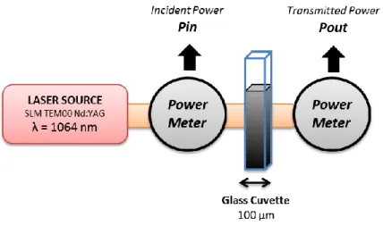
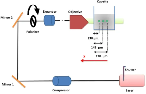
3.3.1 Demonstration of feasibility in skin rejuvenation device
The experimental setup used for the optical breakdown threshold measurements comprises a pulsed laser source, beam shaping optics and mirrors to guide the laser beam via an articulated arm to an aspheric focusing lens (NA = 0.67, f = 2.84 mm, AR: 1050-1620 nm) (Fig.3.2). The laser source is a flash lamp pumped SLM TEM00 Nd:YAG laser which delivers 200 ps laser pulses of 1064 nm. The pulse energy is less than 1 mJ and it was controlled using a polarization beam splitter and a half lambda wave-plate [29]. To create annular illumination, we introduced in the beam path glass slides with a dark spot of diameter 1 and 2 mm. The size of apodization masks is expressed in terms of ratio as it follows:
(3.1)
where is defined as the of the inner annulus of the apodization mask, while is the
of the objective lens. ratio=0 corresponds to the condition of clear aperture, without the glass slides. NA ratio=0.3 and 0.6 correspond to mask diameter of 1 mm and 2 mm respectively.
37 FIG.3.2. Schematic set up of the optical system
Radially polarized light was created using a commercially available radial polarization converter (ARCOptix, Switzerland) well described in ref [30]. Briefly, the main components of the radial polarizer are: (i) a retarder cell, (ii) a polarization rotator and (iii) a nematic liquid crystal cell (Ө-cell) (Fig.3.3).
FIG.3.3. Schematic of the radial polarization converter elements.
The laser power was initially set below threshold and was then increased until a visible flash and an audible sound were detected, which testify that LIOB had occurred. Optical breakdown was created in distilled water (sample 1) and scattering phantoms made by water suspensions of Polystyrene