Clinical and Functional Characterization of a
Novel Mutation in Lamin A/C Gene in a
Multigenerational Family with
Arrhythmogenic Cardiac Laminopathy
Cinzia Forleo1☯‡*, Monica Carmosino2☯‡*, Nicoletta Resta3, Alessandra Rampazzo4, Rosanna Valecce1, Sandro Sorrentino1, Massimo Iacoviello1, Francesco Pisani2,
Giuseppe Procino2, Andrea Gerbino2, Arnaldo Scardapane5, Cristiano Simone3, Martina Calore4, Silvia Torretta2, Maria Svelto2‡, Stefano Favale1‡
1 Cardiology Unit, Department of Emergency and Organ Transplantation, University of Bari, Bari, Italy, 2 Department of Biosciences, Biotechnology and Biopharmaceutics, University of Bari, Bari, Italy, 3 Section of Medical Genetics, Department of Biomedical Sciences and Human Oncology, University of Bari, Bari, Italy, 4 Department of Biology, University of Padua, Padua, Italy, 5 Section of Radiology, Interdisciplinary Department of Medicine, University of Bari, Bari, Italy
☯ These authors contributed equally to this work.
‡ C. Forleo and M. Carmosino are co-first authors on this work. M. Svelto and S. Favale contributed equally to this work as joint senior authors.
*[email protected](CF);[email protected](MC)
Abstract
Mutations in the lamin A/C gene (LMNA) were associated with dilated cardiomyopathy (DCM) and, recently, were related to severe forms of arrhythmogenic right ventricular car-diomyopathy (ARVC). Both genetic and phenotypic overlap between DCM and ARVC was observed; molecular pathomechanisms leading to the cardiac phenotypes caused by LMNA mutations are not yet fully elucidated. This study involved a large Italian family, span-ning 4 generations, with arrhythmogenic cardiomyopathy of different phenotypes, including ARVC, DCM, system conduction defects, ventricular arrhythmias, and sudden cardiac death. Mutation screening ofLMNA and ARVC-related genes PKP2, DSP, DSG2, DSC2, JUP, and CTNNA3 was performed. We identified a novel heterozygous mutation
(c.418_438dup) inLMNA gene exon 2, occurring in a highly conserved protein domain across several species. This newly identified variant was not found in 250 ethnically-matched control subjects. Genotype-phenotype correlation studies suggested a co-segre-gation of theLMNA mutation with the disease phenotype and an incomplete and age-relat-ed penetrance. Basage-relat-ed on clinical, page-relat-edigree, and molecular genetic data, this mutation was considered likely disease-causing. To clarify its potential pathophysiologic impact, function-al characterization of thisLMNA mutant was performed in cultured cardiomyocytes express-ing EGFP-tagged wild-type and mutated LMNA constructs, and indicated an increased nuclear envelope fragility, leading to stress-induced apoptosis as the main pathogenetic mechanism. This study further expands the role of theLMNA gene in the pathogenesis of cardiac laminopathies, suggesting thatLMNA should be included in mutation screening of OPEN ACCESS
Citation: Forleo C, Carmosino M, Resta N, Rampazzo A, Valecce R, Sorrentino S, et al. (2015) Clinical and Functional Characterization of a Novel Mutation in Lamin A/C Gene in a Multigenerational Family with Arrhythmogenic Cardiac Laminopathy. PLoS ONE 10(4): e0121723. doi:10.1371/journal. pone.0121723
Academic Editor: Akinori Kimura, Tokyo Medical and Dental University, JAPAN
Received: October 28, 2014 Accepted: February 3, 2015 Published: April 2, 2015
Copyright: © 2015 Forleo et al. This is an open access article distributed under the terms of the
Creative Commons Attribution License, which permits unrestricted use, distribution, and reproduction in any medium, provided the original author and source are credited.
Data Availability Statement: All relevant data are within the paper and its Supporting Information files. Funding: This work was supported by the Italian Ministry of University, Scientific and Technical Research (grant number ORBA1085M0 to SF), and by Fondo per gli Investimenti della Ricerca di Base-Rete Nazionale di Proteomica (grant number RBRN07BMCT_009 to MS). The funders had no role in study design, data collection and analysis, decision to publish, or preparation of the manuscript.
patients with suspected arrhythmogenic cardiomyopathy, particularly when they have ECG evidence for conduction defects. The combination of clinical, genetic, and functional data contribute insights into the pathogenesis of this form of life-threatening arrhythmogenic cardiac laminopathy.
Introduction
Lamins A and C, encoded by the lamin A/C gene (LMNA), are major structural components of the nuclear lamina, a protein meshwork supporting the inner nuclear membrane [1]. In addi-tion to sustaining the structural integrity and mechanical stability of the nuclear envelope, lamins are involved in multiple cellular processes, such as chromatin organization, DNA repli-cation, gene regulation, and nucleo-cytoskeletal coupling [2].LMNA gene mutations are impli-cated in a wide spectrum of laminopathies, inherited diseases characterized by phenotypic heterogeneity, including cardiac and skeletal myopathies, lipodystrophy, peripheral neuropa-thy, and premature aging syndromes [1,3].
The cardiac phenotype of laminopathies is characterized by conduction system disorders (CD), arrhythmias, and dilated cardiomyopathy (DCM) [4]. ManyLMNA mutation carriers have a poor prognosis [5], due to a high rate of major cardiac events, such as sudden cardiac death (SCD), life-threatening ventricular arrhythmias, extreme bradycardia due to high-degree atrioventricular block, and progression to end-stage heart failure [4]. In addition toLMNA DCM-CD, some atypical forms ofLMNA-related cardiac diseases were reported [6,7]. Recent-ly, severe forms of arrhythmogenic right ventricular cardiomyopathy (ARVC) have been linked to lamin A/C gene mutations [8], and both genetic and phenotypic overlap between DCM and ARVC was observed [8–11]. Although the role of lamins in cell functions has been widely in-vestigated, the pathophysiological mechanisms leading to cardiac phenotypes caused by LMNA mutations are not yet fully understood [1–3].
In this study, we detected a novelLMNA gene mutation in a large family with arrhythmo-genic cardiomyopathy of different phenotypes, including ARVC, DCM, conduction distur-bances, arrhythmias, and sudden cardiac death (SCD). We investigated the involvement of the LMNA gene in the pathogenesis of this arrhythmogenic, familial cardiac laminopathy and functionally characterized the newly-identifiedLMNA mutant.
Materials and Methods
Ethics Statement
All participants provided written informed consent. The Ethics Committee of University Hos-pital Consortium, Policlinico of Bari, Italy approved the study. This study conforms to the principles outlined in the Declaration of Helsinki (World Medical Association Declaration of Helsinki).
Clinical Evaluation
The study involved 20 individuals from 4 generations of a large Italian family, referred to our Unit dedicated to Cardiomyopathies. The 54-year-old male index patient was diagnosed in 2001 with ARVC, based on the original Task Force criteria (TFC) of 1994 [12].
Family cascade screening was performed [13]. All subjects underwent clinical workup, in-cluding medical history, physical examination, measurement of serum creatine phosphokinase,
Competing Interests: The authors have declared that no competing interests exist.
12-lead electrocardiogram (ECG), transthoracic echocardiography, 24-hour ECG recording, and exercise testing. When appropriate, coronary angiography was performed. Subjects with-out contraindication to magnetic resonance imaging, such as pacemaker, defibrillator, or severe claustrophobia underwent cardiac magnetic resonance (CMR) (for details, seeS1 Methods).
Genetic Analysis and Mutation Detection
Genomic DNA was obtained from peripheral blood samples using the Wizard Genomic DNA Purification kit (Promega Corporation, Madison, Wisconsin, USA), as recommended by the manufacturer. Mutation screening of plakophilin-2(PKP2), desmoplakin (DSP), desmoglein-2 (DSG2), desmocollin-2 (DSC2), plakoglobin (JUP), and αT-catenin (CTNNA3) genes was per-formed as previously reported [14,15]. All coding exons ofLMNA gene were amplified by PCR and analyzed by High Resolution Melting, as reported in G. Millat et al [16]. The PCR products of exon 2 and 10 were analyzed by direct sequencing on a 310 ABI sequencer. Num-bering of theLMNA nucleotides refers to GenBank accession number NM_170707.2. A control group of 250 healthy and unrelated Italian subjects (500 alleles) was used to exclude the possi-bility that any identified variation could be due to DNA polymorphism. All controls were unre-lated healthy volunteers matched to the index patient by ancestry from the general Italian population. Moreover, all identified variants were systematically searched for in dbSNP (http:// www.ncbi.nlm.nih.gov/projects/SNP/), in the 1000 Genomes Project database (http://www. 1000genomes.org), or in the Exome Variant Server (http://evs.gs.washington.edu/EVS/).
Functional Studies in HL-1 Cardiomyocytes
The functional characterization of this mutated lamin A protein (LMNA) was performed in cultured HL-1 cardiomyocytes expressing EGFP-N-terminal-tagged wild-type (WT) and mu-tated LMNA. Live-imaging experiments were carried out in a BioStation IM device (Nikon). The acquisition timing was set to every 5 minutes for 16–32 hours, and up to 10 cell fields were captured at each time point. Hyperosmotic stress was induced by incubating HL-1 cells in cul-ture medium with 300 mM mannitol for 2h. Hypoxic stress was performed in a Hypoxia Mod-ular Incubator Chamber (Billups-Rothenberg Inc) and a flow rate of 4 liters/minute of 100% N2was applied for 15 min. Cells, in the hypoxia chamber saturated with N2, were placed at
37°C for 8h. Oxidative stress was induced by incubating HL-1 cells in culture medium plus 300μM H2O2for 4 h.
For further details, seeS1 Methods.
Statistical Analysis
Continuous variables were expressed as mean values ± standard deviation, and frequencies as the number and percentage of patients. Between-group comparisons were made using the non-parametric Wilcoxon rank-sum test. Frequencies were compared using the Fisher’s exact test. The analyses were performed using STATA software, version 12 (StataCorp, College Station, TX, USA). A P-value of<0.05 was considered statistically significant.
Results
Genetic Analysis
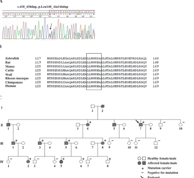
We identified in the index patient the novel heterozygous lamin A/C variant, c.418_438dup, p. Leu140_Ala146dup, consisting of a duplication of 21 nucleotides (CTGCTGAACTCCAAG-GAGGCC) in exon 2 of theLMNA gene (Fig. 1A), located in the coil 1B of the centralα-helical rod domain of the lamin A/C protein. TheLMNA variant is predicted to result in the duplication
of seven amino acids (LLNSKEA), from position 140 to position 146, in the lamin A/C protein, without a frame shift in the open reading frame and affects a highly conserved amino acid region across several species (Fig. 1B), suggesting a possible pathogenetic role.
ThisLMNA variant was subsequently detected in 10 of 20 family members who underwent family cascade genetic screening (Fig. 1C). The index patient (subject II-7 inFig. 1C) was also screened for mutations in the ARVC-related genesPKP2, DSP, DSG2, DSC2, JUP, and CTNNA3 without positive findings.
The novelLMNA variant was not present in 250 healthy control individuals nor found in the above- listed GenBank databases.
Fig 1. Family pedigree and mutation identification. (A) Electropherogram of theLMNA gene variant. (B) The 7 duplicated amino acids (LLNSKEA) are highly conserved amongLMNA gene homologs in vertebrates. (C) Family pedigree of the index patient with the novel LMNA gene mutation.
Clinical Findings
Pedigree structure (Fig. 1C) and clinical characteristics of all evaluated subjects (Table 1) are presented. ECG, Holter, and cardiac structural abnormalities of family members carrying the LMNA mutated variant are summarized inTable 2. Pedigree was consistent with autosomal dominant transmission (Fig. 1C). The index patient (II-7) in 2001 presented with palpitation. In the ECG, first-degree atrioventricular (AV) block and premature ventricular complexes (PVCs) of left bundle-branch block (LBBB) morphology were detected (Fig. 2A). Frequent (up to 35000 per day) multifocal PVCs and runs of non-sustained ventricular tachycardia (NSVT) were recorded using 24-hour Holter monitoring. An echocardiogram showed normal size and preserved global function of both ventricles. Coronary angiography was normal, and right ven-tricle (RV) angiography showed dyskinetic areas with bulging at the RV free wall. On the basis of clinical and instrumental features fulfilling the presence of 1 major plus 2 minor criteria, the index patient was diagnosed with ARVC, based on the original TFC [12], and received a pro-phylactic implantable cardioverter-defibrillator (ICD). He had a positive family history for DCM and SCD; his mother (I-2) died suddenly at rest at the age of 39 years and his brother (II-1) died from SCD at the age of 43 years while playing soccer. No autopsy data were available.
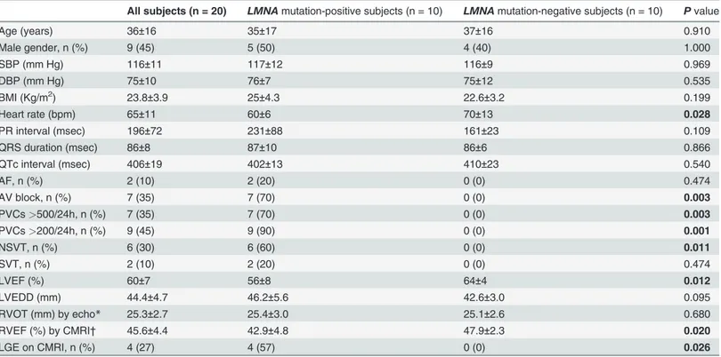
Table 1. Clinical characteristics of family members according toLMNA mutation carrier status.
All subjects (n = 20) LMNA mutation-positive subjects (n = 10) LMNA mutation-negative subjects (n = 10) P value
Age (years) 36±16 35±17 37±16 0.910 Male gender, n (%) 9 (45) 5 (50) 4 (40) 1.000 SBP (mm Hg) 116±11 117±12 116±9 0.969 DBP (mm Hg) 75±10 76±7 75±12 0.535 BMI (Kg/m2) 23.8±3.9 25±4.3 22.6±3.2 0.199 Heart rate (bpm) 65±11 60±6 70±13 0.028 PR interval (msec) 196±72 231±88 161±23 0.109 QRS duration (msec) 86±8 87±10 86±6 0.866 QTc interval (msec) 406±19 402±13 410±23 0.540 AF, n (%) 2 (10) 2 (20) 0 (0) 0.474 AV block, n (%) 7 (35) 7 (70) 0 (0) 0.003 PVCs>500/24h, n (%) 7 (35) 7 (70) 0 (0) 0.003 PVCs>200/24h, n (%) 9 (45) 9 (90) 0 (0) 0.001 NSVT, n (%) 6 (30) 6 (60) 0 (0) 0.011 SVT, n (%) 2 (10) 2 (20) 0 (0) 0.474 LVEF (%) 60±7 56±8 64±4 0.012 LVEDD (mm) 44.4±4.7 46.2±5.6 42.6±3.0 0.095 RVOT (mm) by echo* 25.3±2.7 25.4±3.0 25.1±2.6 0.680 RVEF (%) by CMRI† 45.6±4.4 42.9±4.8 47.9±2.3 0.020 LGE on CMRI, n (%) 4 (27) 4 (57) 0 (0) 0.026
Mean values± standard deviation or absolute frequencies and percentage of patients.
AF = atrialfibrillation; AVB = atrioventricular block; BMI = body mass index; CMRI = cardiac magnetic resonance imaging; DBP = diastolic blood pressure; Echo = echocardiography; LGE = late gadolinium enhancement; LVEDD = left ventricular end-diastolic diameter; LVEF = left ventricular ejection fraction; NSVT = non-sustained ventricular tachycardia; PVCs = premature ventricular complexes; RVEF = right ventricular ejection fraction; RVOT = right ventricular outflow tract; SBP = systolic blood pressure; SVT = sustained ventricular tachycardia.
*RVOT value by echocardiography was available in 19 of 20 patients, 9 of whom were LMNA mutation-positive subjects. †CMRI was performed in 15 subjects, 7 of whom were LMNA mutation carriers.
Another brother (II-5), suffering from DCM, underwent cardiac transplantation at the age of 48 years, and died 14 years later due to refractory heart failure.
Among the 10LMNA mutated variant carriers, subject II-4 in 2002, at the age of 58 years, received a pacemaker due to atrial fibrillation (AF) with a slow ventricular response alternating with sinus bradycardia. Subject III-1 developed DCM. Subject III-5, who fulfilled modified TFC for borderline ARVC diagnosis [17], showed left ventricle (LV) systolic dysfunction
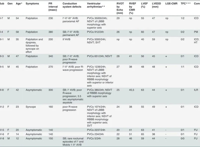
Table 2. Clinical characteristics ofLMNA mutation-positive family members.
Sub Gen Age* Symptoms PR
interval (msec) Conduction system defects Ventricular arrhythmias** RVOT by Echo (mm) RVEF by CMR (%) LVEF (%) LVEDD (mm) LGE-CMR TFC*** Com
II-7 M 54 Palpitation 230 I°-II°-III° AVB; paroxismal AF PVCs 35000/24h; NSVT of LBBB morphology with superior axis 29 np 55 47 np 1/2 ICD
II-4 F 58 Palpitation 380 SB; I°-II° AVB;
permanent AF
PVCs 912/24h 26 np 60 47 np 0/2 PM
III-1 M 35 Palpitation and dyspnea, followed by syncope on effort 200 II°AVB PVCs 5000/24h; NSVT; SVT np np 40 59 np 0/2 ICD; HT
III-3 M 47 Palpitation 340 SB; I°-II° AVB; poor R-wave progression
PVCs 651/24h; NSVT 28 41 56 45 + 0/1 ICD
III-5 M 45 Palpitation 270 I°-II° AVB; poor R-wave progression PVCs 1239/24h; NSVT of LBBB morphology with inferior axis; NSVT of RBBB morphology with superior or inferior axis
27 38 48 48 + 1/1 ICD
III-8 F 42 Asymptomatic 300 SB; I° AVB; poor R-wave progression; 3.5 sec asymptomatic asystole PVCs 380/24h; NSVT of RBBB morphology with superior axis
25 45,5 63 44 + 0/1 ILR
IV-2 F 23 Syncope 160 poor R-wave
progression PVCs 1074/24h; NSVT of LBBB morphology with inferior axis; NSVT of RBBB morphology with superior axis; SVT
24 38 55 49 + 0/1 ICD
IV-5 F 20 Asymptomatic 140 PVCs 2237/24h 20 41 63 41 - 0/1 FU
IV-6 F 14 Asymptomatic 140 PVCs 234/24h 22 51 65 38 - 0/1 FU
IV-8 M 12 Asymptomatic 150 SB; rare nocturnal episodes of I° and Mobitz 1 II° AVB
PVCs 5/24h 28 46 59 44 - 0/0 FU
AF = atrialfibrillation; ARVC = arrhythmogenic right ventricular cardiomyopathy; AVB = atrioventricular block; CMR = cardiac magnetic resonance imaging; Com = comments; Echo = echocardiography; EF = ejection fraction; F = female; FU = Follow-up; Gen = Gender; HT = heart transplantation; ICD = implantable cardioverter-defibrillator; ILR = implantable loop recorder; LBBB = left bundle-branch block; LGE = late gadolinium enhancement; LV = left ventricle; LVEDD = left ventricular end-diastolic diameter; M = male; m = minor; Mj = major; np = not performed; NSVT = non-sustained
ventricular tachycardia; PM = pacemaker; PVCs = premature ventricular complexes; RBBB = right bundle-branch block; RV = right ventricle; RVOT = right ventricular outflow tract; SB = sinus bradycardia; Sub = Subject; SVT = sustained ventricular tachycardia; TFC = Task Force criteria.
*at diagnosis/clinical presentation (years);
**Morphology of NSVT was determinable in 4 of 6 LMNA mutation-positive family members having NSVT; ***for ARVC diagnosis Mj/m.
without dilatation (Table 2). The distribution of major and minor criteria, according to the modified TFC [17], is reported inTable 2.
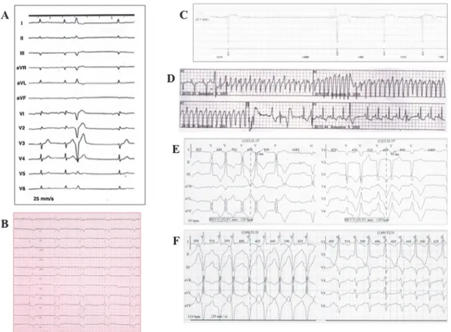
Abnormal ECG findings were present only in family members carrying the mutatedLMNA variant, seven (70%) of whom had, at clinical presentation or went on to develop, conduction disturbances (sinus bradycardia and/or first-, second-, or third-degree AV block) (Table 2;
Fig. 2A, B, and C); 2 subjects developed AF.
Ventricular arrhythmias were documented in 9 of 10 (90%)LMNA mutation-positive sub-jects, and ranged from sustained VT (n = 2), detected on telemetry (Fig. 2D) or ICD memory, to monomorphic NSVT with LBBB (n = 3) (Fig. 2E) and/or right bundle-branch block (RBBB) morphology (n = 3) (Fig. 2F), and polymorphic PVCs (n = 9) (Table 2), recorded on Holter monitoring. The total number of PVCs ranged from 234 to 35000/24 h. Considering the modi-fied criteria for familial ARVC [17], 9 (90%) individuals had>200 PVCs in 24 hours, and 7 (70%) had>500 PVCs in 24 hours (Tables1and2). One of the 2 patients with sustained VT, and 5 of 6 (83%) patients with NSVT, had concomitant AV block (Table 2).
Echocardiographic findings revealed LV involvement in 2LMNA mutation carriers (Table 2). Patient III-1, who had LV borderline systolic function (LVEF = 55%) at clinical pre-sentation at the age of 35, developed global LV hypokinesia (LVEF = 40%) and enlargement 6 years later (Table 2). Individual III-5, who had normal LV systolic function (LVEF = 60%) on
Fig 2. Clinical characteristics of index patient andLMNA mutation-positive family members. Index patient’s electrocardiogram (ECG) showing, at clinical presentation, (A) sinus rhythm, first-degree AV block, and PVC of LBBB morphology and, 13 years later, (B) complete AV block. (C) Asystole documented on implantable loop recorder memory (subject III-8). (D) Sustained VT detected on telemetry monitoring, effectively terminated by internal ICD shock (subject III-1). (E) Episodes of non-sustained VT with LBBB morphology and inferior axis (subject III-5) on 12-lead Holter monitoring. (E) Episodes of non-sustained VT with RBBB morphology and superior axis (subject IV-2) on 12-lead Holter monitoring.
echocardiography during familial screening at the age of 36, presented LV systolic dysfunction (LVEF = 48%) without LV enlargement 9 years later (Table 2). Taken together, mean LVEF values were relatively preserved (56% ± 8%) inLMNA mutation carriers, though significantly lower than those recorded inLMNA mutation-negative subjects (Table 1).
CMR imaging was performed in 15 subjects, 7 of whom carried the mutatedLMNA variant. Four of these 7 (57%) had RV involvement with a reduced RVEF (Table 2), and one (subject III-5) had dyskinetic areas with bulging at the RV free wall (Fig. 3Aa; for video file, seeS1 Movie). Taken together, mean RVEF values inLMNA mutation carriers were significantly reduced in comparison with those assessed inLMNA mutation-negative subjects (Table 1). Myocardial fibrosis by late gadolinium enhancement (LGE) imaging was detected in 4 of 7 (57%) of theLMNA mutation-positive patients (Table 2) and none of the 8LMNA mutation-negative subjects (Table 1). LGE-positive andLMNA mutation-positive subjects were characterized by older age (39 ± 11 vs. 15 ± 4 years, p = 0.034), and longer PR interval (268 ± 77 vs. 143 ± 6 msec, p = 0.032), compared with LGE-negative andLMNA mutation-positive subjects, suggesting an age-related phenotype expres-sion. Furthermore, all LGE-positive subjects had NSVT, and one developed sustained VT. LGE was located in the basal interventricular septum and LV inferior wall, and the pattern was linear and localized in the midwall myocardium (Fig. 3) in all subjects.
Fig 3. CMR imaging ofLMNA mutation-positive family members. (A) Short-axis cine (a) and LGE sequences in the Short-axis (b) and 2-chambers long-axis views (c). Bulging (arrow in a) and LGE of the RV free wall (arrowheads in b). Linear midwall LGE is localized at the interventricular septum and LV inferior wall (arrowheads in c) (subject III-5). (B) and (C) LGE sequences in the short axis (a) and 4-chambers long axis views (b). LGE with linear midwall pattern is shown on the LV inferior wall and basal interventricular septum (arrowheads) (subjects III-8, and IV-2).
Patients receiving ICD, before device implantation, underwent coronary angiography show-ing normal coronary arteries.
During a median follow-up of 122 (range: 12 to 162) months, five patients (II-7, III-1, III-3, III-5, and IV-2) received an ICD in primary prevention, 4 of whom had AV conduction de-fects. Subject III-1 underwent ICD implantation, unsuccessful VT transcatheter ablation and, 7 years later, at the age of 45, heart transplantation for both sustained VT recurrences in storm (Fig. 2D) and subsequent LV function deterioration (LVEF 30%). Subject IV-2, at the age of 24 years, received a prophylactic ICD that, 4 months after device implantation, discontinued sustained VT (mean cycle length 281 msec) by antitachycardia pacing. Patient III-8 refused prophylactic ICD implantation and has an implantable loop recorder that detected up to 3.5 sec asymptomatic asystoles (Fig. 2C). During follow-up, the index patient continued to have episodes of NSVT, showed paroxysmal AF documented on ICD memory and, after 13 years from diagnosis, he developed complete AV block (Fig. 2B).
Two of 10 (20%) of theLMNA mutation-positive family members (subjects IV-6 and IV-8, respectively aged 14 and 12 years) were asymptomatic, free of significant arrhythmias, and re-vealed normal cardiac function. AllLMNA mutation-negative family members were clinically asymptomatic, and phenotype negative after cardiac evaluation (Table 1). We did not observe any overlap with other known laminopathies in this family.
Characterization of LMNA Mutant in HL-1 Cardiomyocytes
To functionally characterize the newly identifiedLMNA mutation, two constructs were gener-ated to express both WT and mutgener-ated LMNA as N-terminally tagged EGFP-fusion proteins. For brevity, the (p.Leu140_Ala146dup) LMNA mutation will be termed“LMNA DUP” in the following results and figures.
To analyze LMNA subcellular localization, as well as the overall organization of the nuclear envelope (NE) upon ectopic LMNA expression, HL-1 cardiomyocytes were transiently trans-fected with the LMNA constructs then subjected to fluorescent confocal microscopy analysis and co-localization experiments with other nuclear components.
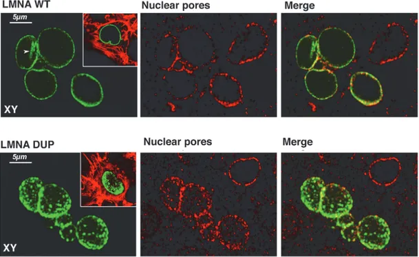
As expected, LMNA WT was uniformly distributed along the NE rim and typically in intra-nuclear invaginations of intra-nuclear membrane (Fig. 4, LMNA WT, arrow). Cardiomyocytes ex-pressing LMNA WT showed nuclei with regular rounded shapes. Co-localization experiments using antibodies against the nuclear pore complex showed that LMNA WT is tightly associated with nuclear pores (Fig. 4, LMNA WT, Nuclear Pores), which, in turn, showed the regular ex-pected distribution along the whole nuclear periphery.
In contrast, the localization of LMNA DUP appeared profoundly impaired, clearly express-ed in aggregates of different sizes, not uniformly distributexpress-ed along the NE, and notably absent from the intranuclear invagination of the NE. In addition to LMNA disorganization, nuclear pores were also altered in the LMNA DUP-expressing cells, resulting in LMNA organization in clusters (Fig. 4, LMNA DUP, Nuclear Pores).
Interestingly, as assessed by live imaging, both lamin A proteins have the same rate of syn-thesis and stability in cultured cardiomyocytes. Moreover, western blotting analysis on lysates from the same cells clearly showed that the expression levels of both WT and DUP LMNA con-structs were comparable, regardless of the antibody used in the analysis (S1 Fig).
In addition, LMNA DUP-expressing cells were able to normally cycle and divide like LMNA WT-expressing cells (S2 Fig).
Analysis of Nuclear Envelope Integrity upon Cell Stresses in WT and
Mutant LMNA-Expressing HL-1 Cells
In order to assess functional consequences of the altered nuclear lamina structure, the integrity of the NE of LMNA DUP-expressing cells, and its resistance to cellular stress, were checked.
Interestingly, cardiac myocytes, differently than other cell types, do not exhibit the volume regulatory response after exposure to hypertonic conditions [18]. Indeed, an osmotically-in-duced and uncompensated cell shrinkage may strain the nucleus.
To examine NE integrity in this experimental condition, we monitored the subcellular loca-tion of the nuclear marker CellLight Nucleus-Red Fluorescent Protein (NRF, Invitrogen) ex-pressed simultaneously to lamin A proteins in HL-1 cells by transient transfection.
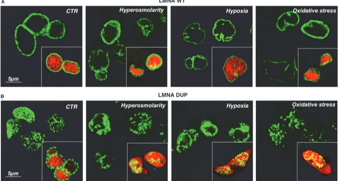
As shown inFig. 5, the red nuclear marker was confined to the nucleus in both LMNA WT and DUP-expressing cells in control conditions, indicating that nuclear integrity was not sig-nificantly impaired in LMNA DUP-expressing cells under resting conditions (Fig. 5, LMNA WT, LMNA DUP, CTR). After 2 h in 300 mM mannitol, added to the culture medium, the nu-clear morphology of the LMNA WT-expressing cells, as well as the LMNA WT labelling, was slightly compromised (Fig. 5A, LMNA WT, Hyper), but the nuclear marker was still contained into the nucleus (Fig. 5A, LMNA WT, Hyper, inset); this suggested that NE was not leaky under this challenging condition.
In contrast, in the same condition, extensive nuclear deformations appeared in LMNA DUP-expressing cells and the red nuclear marker escaped from the nucleus into the cytoplasm, suggesting that NE integrity was impaired under this stressing condition (Fig. 5B, LMNA DUP, Hyper, inset).
Interestingly, similar results were obtained when cardiomyocytes were subjected to either hypoxic (5% O2 for 8 h) or H2O2-induced oxidative stress (300μM H2O2for 4 h), suggesting
Fig 4. Immunofluorescence confocal analysis of LMNA transfected HL-1 cells. Cells transfected with both LMNA WT and DUP are depicted. LMNA is visualized in green, Nuclear Pores in red, and colocalization in yellow in the merge panels. In the insets, a merged image of LMNA and Phalloidin-TRITC is shown. Planar XY projections were depicted in each experimental condition.
that the NE of the LMNA DUP-expressing cardiomyocytes was more fragile under different cellular stresses (Fig. 5).
The expected final consequence of the increased NE fragility under different cellular stress-ors is apoptosis. We performed the apoptosis assay by using Ethidium homodimer-1 (EthD-1), a membrane-impermeable fluorescent dye, which only enters dying cells with leaky plasma membranes and binds to DNA in the nucleus, emitting red fluorescence (Fig. 6A).
As shown inFig. 6B, under all the stressing conditions tested, apoptosis increased by about 4 times in cells expressing LMNA DUP, compared to WT LMNA-expressing cells.
We then analysed whether the canonical Wnt/β-catenin signalling was altered in cells ex-pressing LMNA DUP to tentatively identify the pathomechanism underlying this form of cardiomyopathy.
When we analysedβ-catenin localization and phosphorylation levels in LMNA-expressing HL-1 cells, we found thatβ-catenin was localized to the cell-to-cell contacts in HL-1 cardio-myocytes expressing either WT or mutated lamin A (S3A Fig). Moreover, the amount of phos-pho-β-Catenin was unchanged upon LMNA DUP expression in HL-1 cells even under hypoxic conditions, suggesting that the canonical Wnt signaling pathway was not suppressed in LMNA DUP-expressing cardiomyocytes (S3B Fig).
Discussion
In this study, we identified a novel lamin A/C gene mutation associated with a familial form of arrhythmogenic cardiac laminopathy and characterized it, defining a possible pathogenic mechanism leading to disease development.
Mutations in theLMNA gene account for approximately 6–8% of all DCMs and 33% of DCM cases in association with cardiac conduction defects [4,19–21]. In recent years, muta-tions of the lamin A/C gene associated with an ARVC-related phenotype were found [8,22].
Fig 5. Analysis of nuclear envelope integrity under stressing condition in LMNA transfected HL-1 cells. Nuclear WT LMNA (A) and DUP LMNA (B) signals in control (CTR) and stressing conditions (Hyperosmolarity, Hypoxia, Oxidative stress). The merged signals of LMNA proteins and the RFP nuclear marker are shown in the insets. Confocal XY planar projections are depicted in each experimental condition.
Moreover, a combination of morphofunctional phenotypes between DCM and ARVC were highlighted, suggesting a new classification of cardiomyopathies [23].
The newly identified mutatedLMNA variant can be convincingly considered causative of the clinical features observed in this family for several reasons. First,LMNA is a disease gene for both cardiac laminopathies and ARVC [4,8,22]. Additionally, a co-segregation of the novel lamin A/C mutation with the disease phenotype was observed within the family. The subjects carrying theLMNA variant displayed arrhythmogenic cardiomyopathy of different phenotypes, including ARVC, DCM, LV systolic dysfunction without LV enlargement, system conduction defects, and arrhythmias, showing intra-familial variability of the cardiac pheno-type [4,13]. Importantly, we documented NSVT in 60%, and conduction system disturbances in 70%, ofLMNA mutation-positive family members, emphasizing the value of family genetic screening to identify silent mutation carriers [13] and the need for tailored clinical monitoring aimed to undertake early treatment strategies and prevent sudden death [24]. In agreement with previous studies [4,13,20], twoLMNA mutation-positive family members under the age of 20 years have no evidence of cardiac structural abnormalities, thus suggesting incomplete and age-related penetrance of the mutation. Moreover, absence of the mutation was associated with normal clinical status in all evaluated relatives of the index patient.
The newly detectedLMNA variant is in an amino acid region localized to coil 1B of the cen-tralα-helical rod domain of the lamin A/C proteins, highly conserved among several species, and was not found in 500 control chromosomes or in the aforementioned databases of genetic variants (seeMethods).
In our study, the genetic and clinical data forLMNA mutation in this family were strength-ened with functional studies. Thein vitro characterization of this new LMNA variant showed that mutated LMNA loses the uniform expression along the nuclear rime and perturbs nuclear shape and nuclear pore complex organization in cultured cardiomyocytes in resting conditions.
Fig 6. Apoptosis assay in LMNA transfected HL-1 cells. (A) Representative XY confocal planar projections of LMNA transfected cells (green) labelled with EthD-1 (red) in control conditions are depicted. (B) Quantitative analysis of apoptotic cells in control and under hyperosmotic (Hyper), hypoxic (Hypoxia), and oxidative (H2O2) conditions. Data are reported as % of apoptotic cells (double-labelled cells) in overall LMNA-expressing cells (green labelled cells).
Statistical analysis was performed on 3 independent experiments and significance calculated by Student’s T-test for unpaired samples. *P< 0.0002 is relative to CTR vs. stressing conditions in WT LMNA expressing cells and**P < 0.0001 is relative to CTR vs. stressing conditions in DUP LMNA expressing cells.
The loss of the higher-order assembly of lamin mutated polymers probably leads to a loss of nuclear stability and enhanced sensitivity to mechanical strain [25,26]; this LMNA mutant sig-nificantly increases nuclear envelope fragility upon different cellular stresses, such as hyperton-ic, hypoxhyperton-ic, and oxidative stresses. The leakage of NE in mutated lamin A-expressing
cardiomyocytes under hypertonic conditions suggested a decreased mechanic resistance of the NE. A similar nuclear fragility was observed under hypoxic and oxidative stresses. It is indeed possible that this newly identified LMNA mutation drastically decreases both the tolerance and the adaptation of myocardium to stressing conditions, making cardiomyocytes more suscepti-ble to nuclear breakage and cell death during mechanical stress [26].
It is recognized that lamin A is involved into physical and functional connections between the nucleus and cytoskeleton required for effective mechanotransduction in cells (for review, see [2]). It is indeed possible that the mutated lamin A causes not only a decreased mechanic resistance of the NE but also altered nuclear-cytoskeletal coupling with impairment of the mechanotransduction machinery. In hypoxic conditions, in which beating frequency and cellu-lar work increase in cardiomyocytes [27], continuously underwent to mechanical strain due to contraction cycles, the impairment of the nuclear- cytoskeletal connection may result in inap-propriate constraints onto the NE, which can, in turn, lose its integrity due to expression of the mutated lamin A. Moreover, it was reported that the nuclear-cytoplasmic compartmentaliza-tion can be profoundly affected by ROS, including H2O2, since nuclear transport factors are the
primary cellular targets for oxidants [28]. This effect, together with the nuclear pore clustering induced by the expression of mutated lamin A, may further affect the selective permeability of the NE, ultimately inducing massive nucleoplasm leakage, as observed in our functional studies.
Importantly, the impairment of the nuclear-cytoskeletal connection, due to the expression of the mutated lamin A, may increase the energy cost of contraction and oxygen demand, thus mimicking hypoxic stress even in the absence of physical exercise. Moreover, it has been re-ported that, in cardiomyocytes, hypoxic conditions increase ROS species production [29], sug-gesting that both hypoxic and oxidative stresses in mutated lamin A-expressing
cardiomyocytes can be continuously induced, even in resting conditions.
In addition to the decrease in mechanic resistance to stressing conditions, it is possible that the newly-identified LMNA makes cardiomyocytes more prone to pro-apoptotic pathways, speeding up the cardiomyocyte apoptotic process once initiated by a stressing condition.
One of the intracellular pathways altered in forms of ARVC due to desmoplakin mutations is the canonical Wnt/β-catenin signalling [30]. Suppression of this pathway induces adipogen-esis, fibrogenadipogen-esis, and apoptosis, the histological hallmark of the disease [31,32].
However, we found that the canonical Wnt signaling pathway was not suppressed in LMNA DUP-expressing cardiomyocytes. Further experiments will be necessary to identify the intra-cellular pathway/s involved in the pathogenesis of the cardiolaminopathy described in this study.
Regardless of the pathways, we showed that the final fatal consequence of thisLMNA muta-tion is cell death under cell-stressing condimuta-tions.
Cardiomyocyte apoptosis may lead to the development of arrhythmias, potentially resulting in sudden cardiac death [33]. An arrhythmogenic effect of apoptosis may be mediated in at least two ways. First, in the progress of dying, a cardiomyocyte passes through phases of in-creased excitability or becomes automatic, at least until it is dead. Second, from a random grouping of several such dead cardiomyocytes, the process of normal activation in that area of heart muscle must be deranged and redirected in a way that would provide a suitable anatomi-cal substrate for re-entrant arrhythmias (for review, see [34]). Sudden cardiac death in patients withLMNA mutations may occur due to ventricular arrhythmias, bradyarrhythmias, or
asystole [4,35]. Previous studies suggested that apoptosis in system conduction cardiomyo-cytes could cause either tachyarrhythmias or bradyarrhythmias, including complete AV block, as observed in our patients, probably playing an important role in the pathogenesis of sudden cardiac death [33].
Pathophysiological mechanisms leading to the cardiac phenotypes caused byLMNA muta-tions are not yet fully understood [25,26]. Our experimental data shed light on the clinical findings we collected. In this study, the majority ofLMNA mutation-positive subjects had ven-tricular arrhythmias and/or conduction system defects, including severe arrhythmic pheno-types, such as sustained VT and complete AV block, while cardiac function was variable. These findings are in line with previous observations showing that cardiac laminopathies carry high arrhythmogenic risk, even if left ventricular ejection fraction is preserved [4,24,35–38].
In our study, myocardial fibrosis by LGE-CMR was found in fourLMNA mutation carriers who had documented NSVT, one of whom developed sustained VT, and 3 who showed con-duction system disturbances. These findings agree with a recent study that included 41 lamin A/C mutation-positive subjects and showed association of myocardial septal fibrosis with ven-tricular arrhythmias and a prolonged PR-interval [39]. Furthermore, the typical pattern of LGE detected by CMR imaging in our patients was linear and midwall, predominantly located in the basal interventricular septum, consistent with the distribution of myocardial fibrosis pre-viously described in lamin A/C mutation-positive patients [39,40].
Taken together, our clinical, genetic, and functional data allow us to hypothesize a possible disease mechanism by which the mutatedLMNA variant causes decreased nuclear stability and impaired nuclear-cytoskeletal coupling, resulting in a higher susceptibility/sensitivity to nucle-ar rupture and cnucle-ardiomyocyte apoptosis in tissue subjected to mechanical stress, like the henucle-art [21,31,32,36,41]. Apoptosis may lead to heterogeneity of cardiac conduction and dispersion of refractoriness [42], providing a basis for the arrhythmias we observed in our patients. In ad-dition, throughout the disease course, cardiomyocyte loss, likely repaired by replacement fibro-sis, may provide a possible substrate for conduction block and re-entrant arrhythmias [42,43]; this hypothesis is in line with previous studies showing thatLMNA mutation carriers with con-duction defects and arrhythmias have myocardial fibrosis involving cardiac concon-duction system, documented by histopathological examinations [21,36,41].
Our study suggests that myocardial fibrosis detected by LGE-CMR may be considered a marker of higher arrhythmic risk in patients withLMNA mutations, contributing to identify those that would benefit from ICD implantation, in agreement with recent clinical findings [39,44].
However, there is growing evidence that life-threatening arrhythmias or sudden cardiac death may occur without myocardial fibrosis in arrhythmogenic cardiomyopathy [42,45]. In these cases, apoptosis-related enhanced excitability may play a role, which must be proved in further studies.
Some limitations of this study need to be mentioned. The findings of myocardial fibrosis by CMR imaging could be only documented in our patients without pacemaker or ICD. More-over, LGE-negative andLMNA mutation-positive family members were significantly younger than LGE-positive subjects, suggesting an age-related cardiac phenotype expression. Future larger studies, including CMR evaluations and long-term follow-up of healthy mutated sub-jects, should help to elucidate the timing of expression of the phenotypic traits.
Conclusions
In conclusions, our functional data, combined with clinical and genetic findings, indicate LMNA p.Leu140_Ala146dup as a disease-causing mutation, and suggest cardiomyocyte
apoptosis as a possible molecular mechanism leading to the clinical features observed in this family, thus confirming the emergent role of theLMNA gene in the pathogenesis of a wide spectrum of cardiac laminopathies. The current family is a striking example of the possibility of shared cardiac phenotypes between laminopathies and arrhythmogenic cardiomyopathy. The major clinical implication of our findings is that theLMNA gene should be included in mutational screening of patients with suspected arrhythmogenic cardiomyopathy, particularly when they have ECG evidence for conduction defects and/or myocardial septal fibrosis on CMR. These results, by integrating clinical, genetic, and functional data, could contribute to fu-ture studies aimed at improving risk stratification algorithms and testing possible tailored ther-apeutic approaches in patients withLMNA mutations.
Supporting Information
S1 Fig. LMNA protein biosynthesis dynamics.(A) To verify whether the mutated LMNA shows impaired biosynthetic pathways compared to the wild-type (WT) lamin protein, HL-1 cells were transiently transfected with plasmids containing the WT or alternatively the mutated variant of lamin A and then monitored by live imaging for 16 h in the Biostation device. Both proteins appeared within the cells after about 5 h post-transfection and were trackable until the end of the experimental time course, suggesting that both lamin A proteins have, at least in range of time of 16 h, the same rate of synthesis and stability in cultured cells (two representa-tive movie frames were shown). (B) Western blotting analysis on lysates from 24 h transiently transfected HL-1 cells, clearly showed that the expression levels of both WT and DUP LMNA constructs, were comparable regardless of whether the antibody used in the analysis (anti LMNA antibodies, anti EGFP antibodies; anti-GAPDH antibodies; GAPDH was used as load-ing control).
(TIF)
S2 Fig. Cell division visualization.The fate of single nuclei of HL-1 cells transfected with ei-ther WT or DUP LMNA construct were depicted as the most representative movie frames at 24, 28, 30 and 32 h of live imaging analysis. The red markers frame LMNA signals to facilitate the nuclei division visualization. The cell was able to normally divide and to survive at the cell division regardless of whether the type of LMNA expressed.
(TIF)
S3 Fig. Analysis ofβ-catenin pathway in either LMNA WT or LMNA DUP-expressing HL-1 cells.A)β-catenin (red) and LMNA constructs (green) co-localization in HL-1 cells. β-cate-nin was localized to the cell-to-cell contacts in HL-1 cardiomyocytes expressing either WT or mutated lamin A. B) Detection and semi-quantification of phosphoβ-catenin by western blot-ting. The amount of phosphoβ-catenin was unchanged upon LMNA DUP expression in HL-1 cells even under hypoxic conditions, suggesting that the canonical Wnt signaling pathway was not suppressed in LMNA DUP-expressing cardiomyocytes.
(TIF)
S1 Methods. This section describes: Cardiac Magnetic Resonance Imaging Protocol, LMNA cloning and vector construction, Cell culture and transient transfection, Western Blotting and Immunofluorescence Confocal analysis, Live imaging analysis, and Stressing assays. (DOC)
S1 Movie. CMR imaging movie.The short axis cine sequence of subject III-5 demonstrates bulging of the right ventricle free wall consistent with dyskinesia.
Acknowledgments
We thank family members for their participation. We are grateful to Dr. Pietro Guida for statis-tical analysis. Furthermore, we thank Mrs. Giovanna Petragallo and Mrs. Rosanna Bagnulo for their technical support. In addition, we thank Prof. William Claycomb (LSU Health New Orle-ans, USA) for the generous gift of the HL-1 cardiomyocyte cell line.
Author Contributions
Conceived and designed the experiments: CF MC MS SF AR. Performed the experiments: RV MI FP GP AG AS MC ST NR. Analyzed the data: CF MC SS CS SF. Wrote the paper: CF MC.
References
1. Worman HJ. Nuclear lamins and laminopathies. J Pathol. 2012; 226: 316–325. doi:10.1002/path.2999
PMID:21953297
2. Carmosino M, Torretta S, Procino G, Gerbino A, Forleo C, Favale S, et al. Role of nuclear Lamin A/C in cardiomyocyte functions. Biol Cell. 2014; 106: 346–58. doi:10.1111/boc.201400033PMID:25055884
3. Bertrand AT, Chikhaoui K, Yaou RB, Bonne G. Clinical and genetic heterogeneity in laminopathies. Bio-chem Soc Trans. 2011; 39: 1687–1692. doi:10.1042/BST20110670PMID:22103508
4. Cattin ME, Muchir A, Bonne G. 'State-of-the-heart' of cardiac laminopathies. Curr Opin Cardiol. 2013; 28: 297–304. doi:10.1097/HCO.0b013e32835f0c79PMID:23455585
5. Taylor MR, Fain PR, Sinagra G, Robinson ML, Robertson AD, Carniel E, et al. Natural history of dilated cardiomyopathy due to lamin A/C gene mutations. J Am Coll Cardiol. 2003; 41: 771–780. PMID:
12628721
6. Forissier JF, Bonne G, Bouchier C, Duboscq-Bidot L, Richard P, Wisnewski C, et al. Apical left ventricu-lar aneurysm without atrio-ventricuventricu-lar block due to a lamin A/C gene mutation. Eur J Heart Fail. 2003; 5: 821–825. PMID:14675861
7. Hermida-Prieto M, Monserrat L, Castro-Beiras A, Laredo R, Soler R, Peteiro J, et al. Familial dilated cardiomyopathy and isolated left ventricular noncompaction associated with lamin A/C gene mutations. Am J Cardiol. 2004; 94: 50–54. PMID:15219508
8. Quarta G, Syrris P, Ashworth M, Jenkins S, Zuborne Alapi K, Morgan J, et al. Mutations in the lamin A/ C gene mimic arrhythmogenic right ventricular cardiomyopathy. Eur Heart J. 2012; 33: 1128–1136. doi:
10.1093/eurheartj/ehr451PMID:22199124
9. Basso C, Corrado D, Bauce B, Thiene G. Arrhythmogenic right ventricular cardiomyopathy. Circ Arrhythm Electrophysiol. 2012; 5: 1233–1246. doi:10.1161/CIRCEP.111.962035PMID:23022706
10. Taylor M, Graw S, Sinagra G, Barnes C, Slavov D, Brun F, et al. Genetic variation in titin in arrhythmo-genic right ventricular cardiomyopathy-overlap syndromes. Circulation. 2011; 124: 876–885. doi:10. 1161/CIRCULATIONAHA.110.005405PMID:21810661
11. van der Zwaag PA, van Rijsingen IA, Asimaki A, Jongbloed JD, van Veldhuisen DJ, Wiesfeld AC, et al. Phospholamban R14del mutation in patients diagnosed with dilated cardiomyopathy or arrhythmogenic right ventricular cardiomyopathy: evidence supporting the concept of arrhythmogenic cardiomyopathy. Eur J Heart Fail. 2012; 14: 1199–1207. doi:10.1093/eurjhf/hfs119PMID:22820313
12. McKenna WJ, Thiene G, Nava A, Fontaliran F, Blomstrom-Lundqvist C, Fontaine G, et al. Diagnosis of arrhythmogenic right ventricular dysplasia/cardiomyopathy. Task Force of the Working Group Myocar-dial and PericarMyocar-dial Disease of the European Society of Cardiology and of the Scientific Council on Car-diomyopathies of the International Society and Federation of Cardiology. Br Heart J. 1994; 71: 215– 218. PMID:8142187
13. Charron P, Arad M, Arbustini E, Basso C, Bilinska Z, Elliott P, et al. Genetic counselling and testing in cardiomyopathies: a position statement of the European Society of Cardiology Working Group on Myo-cardial and PeriMyo-cardial Diseases. Eur Heart J. 2010; 31: 2715–2726. doi:10.1093/eurheartj/ehq271
PMID:20823110
14. Bauce B, Nava A, Beffagna G, Basso C, Lorenzon A, Smaniotto G, et al. Multiple mutations in desmo-somal proteins encoding genes in arrhythmogenic right ventricular cardiomyopathy/dysplasia. Heart Rhythm. 2010; 7: 22–29. doi:10.1016/j.hrthm.2009.09.070PMID:20129281
15. van Hengel J, Calore M, Bauce B, Dazzo E, Mazzotti E, De Bortoli M, et al. Mutations in the area com-posita proteinαT-catenin are associated with arrhythmogenic right ventricular cardiomyopathy. Eur Heart J. 2013; 34: 201–210. doi:10.1093/eurheartj/ehs373PMID:23136403
16. Millat G, Chanavat V, Julia S, Crehalet H, Bouvagnet P, Rousson R. Validation of high-resolution DNA melting analysis for mutation scanning of the LMNA gene. Clin Biochem. 2009; 42: 892–898. doi:10. 1016/j.clinbiochem.2009.01.016PMID:19318026
17. Marcus FI, McKenna WJ, Sherrill D, Basso C, Bauce B, Bluemke DA, et al. Diagnosis of arrhythmo-genic right ventricular cardiomyopathy/dysplasia: proposed modification of the task force criteria. Circu-lation. 2010; 121: 1533–1541. doi:10.1161/CIRCULATIONAHA.108.840827PMID:20172911
18. Wright AR, Rees SA. Cardiac cell volume: crystal clear or murky waters? A comparison with other cell types. Pharmacol Ther. 1998; 80: 89–121. PMID:9804055
19. Millat G, Bouvagnet P, Chevalier P, Sebbag L, Dulac A, Dauphin C, et al. Clinical and mutational spec-trum in a cohort of 105 unrelated patients with dilated cardiomyopathy. Eur J Med Genet. 2011; 54: e570–575. doi:10.1016/j.ejmg.2011.07.005PMID:21846512
20. Tesson F, Saj M, Uvaize MM, Nicolas H, Płoski R, Bilińska Z. Lamin A/C mutations in dilated cardiomy-opathy. Cardiol J. 2014; 21: 331–342. doi:10.5603/CJ.a2014.0037PMID:24846508
21. Arbustini E, Pilotto A, Repetto A, Grasso M, Negri A, Diegoli M, et al. Autosomal dominant dilated car-diomyopathy with atrioventricular block: a lamin A/C defect-related disease. J Am Coll Cardiol. 2002; 39: 981–990. PMID:11897440
22. Valtuille L, Paterson I, Kim DH, Mullen J, Sergi C, Oudit GY. A case of lamin A/C mutation cardiomyopa-thy with overlap features of ARVC: A critical role of genetic testing. Int J Cardiol. 2013; 168: 4325–4327. doi:10.1016/j.ijcard.2013.04.177PMID:23684604
23. Arbustini E, Narula N, Dec GW, Reddy KS, Greenberg B, Kushwaha S, et al. The MOGE(S) classifica-tion for a phenotype-genotype nomenclature of cardiomyopathy: endorsed by the World Heart Federa-tion. J Am Coll Cardiol. 2013; 62: 2046–2072. doi:10.1016/j.jacc.2013.08.1644PMID:24263073
24. Meune C, Van Berlo JH, Anselme F, Bonne G, Pinto YM, Duboc D. Primary prevention of sudden death in patients with lamin A/C gene mutations. N Engl J Med. 2006; 354: 209–210. PMID:16407523
25. Zwerger M, Jaalouk DE, Lombardi ML, Isermann P, Mauermann M, Dialynas G, et al. Myopathic lamin mutations impair nuclear stability in cells and tissue and disrupt nucleo-cytoskeletal coupling. Hum Mol Genet. 2013; 22: 2335–2349. doi:10.1093/hmg/ddt079PMID:23427149
26. Davidson PM, Lammerding J. Broken nuclei—lamins, nuclear mechanics, and disease. Trends Cell Biol. 2014; 24: 247–256. doi:10.1016/j.tcb.2013.11.004PMID:24309562
27. Sofija Jovanovic´, Nenad Jovanovic´, Aleksandar Jovanovic´. High glucose protects single beating adult cardiomyocytes against hypoxia. Biochemical and Biophysical Research Communications. 2006; 341: 57–66. PMID:16412383
28. Marinho HS, Real C, Cyrne L, Soares H, Antunes F. Hydrogen peroxide sensing, signaling and regula-tion of transcripregula-tion factors. Redox Biol. 2014; 2: 535–562. doi:10.1016/j.redox.2014.02.006PMID:
24634836
29. Duranteau J, Chandel NS, Kulisz A, Shao Z, Schumacker PT. Intracellular signaling by reactive oxygen species during hypoxia in cardiomyocytes. J Biol Chem. 1998; 273: 11619–11624. PMID:9565580
30. Tilgner K, Wojciechowicz K, Jahoda C, Hutchison C, Markiewicz E. Dynamic complexes of A-type lamins and emerin influence adipogenic capacity of the cell via nucleocytoplasmic distribution of beta-catenin. J Cell Sci. 2009; 122: 401–413. doi:10.1242/jcs.026179PMID:19126678
31. Mallat Z, Tedgui A, Fontaliran F, Frank R, Durigon M, Fontaine G. Evidence of apoptosis in arrhythmo-genic right ventricular dysplasia. N Engl J Med. 1996; 335: 1190–1196. PMID:8815941
32. Valente M, Calabrese F, Thiene G, Angelini A, Basso C, Nava A, et al. In vivo evidence of apoptosis in arrhythmogenic right ventricular cardiomyopathy. Am J Pathol. 1998; 152: 479–484. PMID:9466574
33. James TN. Normal and abnormal consequences of apoptosis in the human heart. From postnatal mor-phogenesis to paroxysmal arrhythmias. Circulation. 1994; 90: 556–573. PMID:8026044
34. Nerheim P, Krishnan SC, Olshansky B, Shivkumar K. Apoptosis in the genesis of cardiac rhythm disor-ders. Cardiol Clin. 2001; 19: 155–163. PMID:11787809
35. van Berlo JH, de Voogt WG, van der Kooi AJ, van Tintelen JP, Bonne G, Yaou RB, et al. Meta-analysis of clinical characteristics of 299 carriers of LMNA gene mutations: do lamin A/C mutations portend a high risk of sudden death? J Mol Med. 2005; 83: 79–83. PMID:15551023
36. van Tintelen JP, Tio RA, Kerstjens-Frederikse WS, van Berlo JH, Boven LG, Suurmeijer AJ, et al. Se-vere myocardial fibrosis caused by a deletion of the 5' end of the lamin A/C gene. J Am Coll Cardiol. 2007; 49:2430–2439. PMID:17599607
37. Fernández X, Dumont CA, Monserrat L, Hermida-Prieto M, Castro-Beiras A. Sudden death in a patient with lamin A/C gene mutation and near normal left ventricular systolic function. Int J Cardiol. 2008; 126: 136–137. PMID:17442430
38. De Backer J, Van Beeumen K, Loeys B, Duytschaever M. Expanding the phenotype of sudden cardiac death-An unusual presentation of a family with a Lamin A/C mutation. Int J Cardiol. 2010; 138: 97–99. doi:10.1016/j.ijcard.2008.06.008PMID:18691775
39. Hasselberg NE, Edvardsen T, Petri H, Berge KE, Leren TP, Bundgaard H, et al. Risk prediction of ven-tricular arrhythmias and myocardial function in Lamin A/C mutation positive subjects. Europace. 2014; 16: 563–571. doi:10.1093/europace/eut291PMID:24058181
40. Holmström M, Kivistö S, Heliö T, Jurkko R, Kaartinen M, Antila M, et al. Late gadolinium enhanced car-diovascular magnetic resonance of lamin A/C gene mutation related dilated cardiomyopathy. J Cardio-vasc Magn Reson. 2011; 13: 30. doi:10.1186/1532-429X-13-30PMID:21689390
41. Fatkin D, MacRae C, Sasaki T, Wolff MR, Porcu M, Frenneaux M, et al. Missense mutations in the rod domain of the lamin A/C gene as causes of dilated cardiomyopathy and conduction-system disease. N Engl J Med. 1999; 341: 1715–1724. PMID:10580070
42. Sen-Chowdhry S, McKenna WJ. Sudden death from genetic and acquired cardiomyopathies. Circula-tion. 2012; 125: 1563–1576. doi:10.1161/CIRCULATIONAHA.111.025528PMID:22451606
43. Basso C, Bauce B, Corrado D, Thiene G. Pathophysiology of arrhythmogenic cardiomyopathy. Nat Rev Cardiol. 2012; 9: 223–233. doi:10.1038/nrcardio.2011.173PMID:22124316
44. Anselme F, Moubarak G, Savouré A, Godin B, Borz B, Drouin-Garraud V, et al. Implantable cardiover-ter-defibrillators in lamin A/C mutation carriers with cardiac conduction disorders. Heart Rhythm. 2013; 10: 1492–1498. doi:10.1016/j.hrthm.2013.06.020PMID:23811080
45. Cerrone M, Noorman M, Lin X, Chkourko H, Liang FX, van der Nagel R, et al. Sodium current deficit and arrhythmogenesis in a murine model of plakophilin-2 haploinsufficiency. Cardiovasc Res. 2012; 95: 460–468. doi:10.1093/cvr/cvs218PMID:22764151