CLINICAL SCIENCE
EULAR recommendations for the reporting of
ultrasound studies in rheumatic and musculoskeletal
diseases (RMDs)
Félicie Costantino ,
1,2Loreto Carmona ,
3Maarten Boers,
4Marina Backhaus,
5Peter V Balint,
6George A Bruyn ,
7,8Robin Christensen,
9Philip G Conaghan ,
10Ricardo J O Ferreira ,
11,12Juan Luis Garrido- Castro ,
13Francis Guillemin,
14Hilde Berner Hammer ,
15Désirée van der Heijde ,
16Annamaria Iagnocco,
17Marion C Kortekaas ,
16Robert BM Landewé ,
18,19Peter Mandl ,
20Esperanza Naredo,
21,22Wolfgang A Schmidt,
23Lene Terslev ,
24Caroline B Terwee,
4Ralf Thiele,
25Maria- Antonietta D’Agostino
1,2,26To cite: Costantino F, Carmona L, Boers M, et al. Ann Rheum Dis Epub ahead of print: [please include Day Month Year]. doi:10.1136/ annrheumdis-2020-219816 Handling editor Josef S Smolen
►Additional material is published online only. To view, please visit the journal online (http:// dx. doi. org/ 10. 1136/ annrheumdis- 2020- 219816). For numbered affiliations see end of article.
Correspondence to Professor Maria- Antonietta D’Agostino, Rheumatology Department, Università Cattolica del Sacro Cuore, Policlinico Universitario Agostino Gemelli IRCSS, 00187 Roma, Italy; mariaantonietta. dagostino@ unicatt. it
MB, PVB, GAB, RC, PGC, RJOF, JLG- C, FG, HBH, DvdH, AI, MCK, RBL, PM, EN, WAS, LT, CBT and RT contributed equally. Received 29 December 2020 Revised 11 January 2021 Accepted 12 January 2021
© Author(s) (or their employer(s)) 2021. No commercial re- use. See rights and permissions. Published by BMJ.
ABSTRACT
Objective To produce European League Against
Rheumatism (EULAR) recommendations for the reporting of ultrasound studies in rheumatic and musculoskeletal diseases (RMDs).
Methods Based on the literature reviews and
expert opinion (through Delphi surveys), a taskforce of 23 members (12 experts in ultrasound in RMDs, 9 in methodology and biostatistics together with a patient research partner and a health professional in rheumatology) developed a checklist of items to be reported in every RMD study using ultrasound. This checklist was further refined by involving a panel of 79 external experts (musculoskeletal imaging experts, methodologists, journal editors), who evaluated its comprehensibility, feasibility and comprehensiveness. Agreement on each proposed item was assessed with an 11- point Likert scale, grading from 0 (total disagreement) to 10 (full agreement).
Results Two face- to- face meetings, as well as two
Delphi rounds of voting, resulted in a final checklist of 23 items, including a glossary of terminology. Twenty- one of these were considered ’mandatory’ items to be reported in every study (such as blinding, development of scoring systems, definition of target pathologies) and 2 ’optional’ to be reported only if applicable, such as possible confounding factors (ie, ambient conditions) or experience of the sonographers.
Conclusion An EULAR taskforce developed a checklist
to ensure transparent and comprehensive reporting of aspects concerning research and procedures that need to be presented in studies using ultrasound in RMDs. This checklist, if widely adopted by authors and editors, will greatly improve the interpretability of study development and results, including the assessment of validity, generalisability and applicability.
Ultrasound is an imaging technique widely used in patients with rheumatic and musculoskeletal diseases (RMDs) to detect signs of inflammation and destructive changes.1 Despite an increased use in clinical practice facilitated by major technical advances in the resolution of soft tissue contrast
(B- mode or grey scale (GS)) and of vascular perfu-sion (Doppler techniques), a relatively long learning curve2 and, until recently, the absence of agreed scoring systems have hampered its utilisation for research.3 4
The European League Against Rheumatism (EULAR) and the Outcome Measures in Rheuma-tology (OMERACT) Ultrasound Working Group have actively worked towards the standardisa-tion of the technique by developing educastandardisa-tional programmes and by performing several studies evaluating its reliability, validity and feasibility.5–8 These initiatives have underlined that factors such as nomenclature, definitions of ultrasound- detected pathologies, scoring systems and technical issues
Key messages
What is already known about this subject? ► Nomenclature, definitions of ultrasound-
detected pathologies, scoring systems and technical issues may affect the validity and generalisability of results of ultrasound studies in rheumatic and musculoskeletal diseases.
► These aspects, along with critical design characteristics, are often suboptimally reported in current ultrasound studies.
What does this study add?
► A 23- item recommendation checklist was developed by a European League Against Rheumatism taskforce to ensure transparent and comprehensive reporting of ultrasound research.
► This is the first reporting checklist focused on how to report characteristics of imaging measurement tools.
How might this impact on clinical practice or future developments?
► The use of this checklist may improve the interpretability, reproducibility and generalisability of study results.
copyright.
on May 18, 2021 at IRCCS Gemelli Roma. Protected by
with the ultrasound equipment may affect the validity and generalisability of these results. These aspects, along with critical design characteristics, such as reproducibility, blinding, patient selection and clearly defined purposes of the ultrasound evalua-tion, are often suboptimally reported in the current ultrasound studies.5 6 9 10
Complete and accurate reporting is necessary to detect poten-tial biases in the study (internal validity) and to assess the gener-alisability and applicability of the results (external validity). Over the last 20 years, many guidelines have been developed to improve the quality of reporting of research articles, including those for randomised controlled trials (RCT) (Consolidated Standards of Reporting Trials)11 and diagnostic accuracy studies (Standards for Reporting Diagnostic accuracy studies).12 13 EULAR has also contributed by developing recommendations for reporting registers and clinical trial extension studies.14 15 We are not aware of recommendations focused on how to report characteristics of imaging measurement tools such as the equip-ment characteristics, procedures or scoring, which can influence the validity and generalisability of study results. Therefore, an EULAR taskforce was convened to propose recommendations for the reporting of such aspects in ultrasound studies in RMDs. METHODS
The convenor (MADA), EULAR methodologist (LC) and project fellow (FC) led a multidisciplinary taskforce in accordance with the EULAR Standardised Operating Procedures (SOPs).16 The taskforce included 23 members from 11 European countries and from the USA and was composed as follows: 11 experts in ultra-sound in RMDs, 7 in methodology, 1 in both ultraultra-sound and methodology, 2 in biostatistics, 1 patient research partner and 1 health professional in rheumatology. Three of the 23 members were members of EMEUNET and 13 of them were also part of an editorial board.
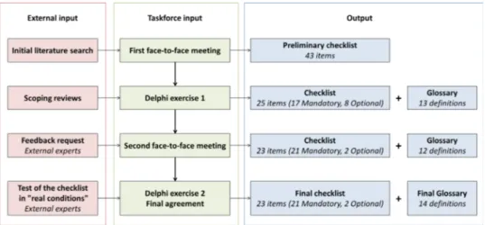
The taskforce employed a stepwise process summarised in
figure 1, including two face- to- face meetings and several Delphi rounds. First the EULAR methodologist, convenor and fellow searched for evidence of quality of reporting of ultrasound studies in RMDs. The choice was made to focus on an extensively studied topic, that is, ultrasound assessment of synovitis in rheu-matoid arthritis. In PubMed Clinical Queries, a broad search was performed; 80 studies were randomly selected and divided in four categories: diagnosis, aetiology, prognosis and therapy. The articles were summarised in table format to highlight objective, design, technical data, measures and outcomes (online supple-mental file 1). These tables were sent to each member of the
taskforce prior to the first face- to- face meeting, with the request to identify possible sources of bias and error and the absence of information considered important for the generalisability of the results. During the first face- to- face meeting, the members of the taskforce discussed the results and the unmet requirements in the selected literature, agreed on the format of presentation of the project (checklist or statement document) and elabo-rated a first list of items to be included. Other objectives of this meeting were the definition of a target audience and the need for systematic reviews. After the meeting, a number of focused literature reviews addressed specific issues; a summary of their results, along with the total list of items, were subsequently sent to the taskforce members. Relevance and comprehensibility of each proposed item were tested in a Delphi exercise, first by the taskforce members (excluding the convenor, EULAR methodol-ogist and fellow), then by a panel of external experts chosen from the fields of musculoskeletal imaging, epidemiology and methodology, including journal editors. External experts were also asked if no key aspects were missed (comprehensiveness). During the second face- to- face meeting, the optimal format of the checklist document was established. Inclusion of each item was either supported by empirical evidence, when available, or by consensus within the task force, that the information requested by the item was methodologically important to assess in a study, as recommended by the Enhancing the QUAlity and Transpar-ency Of health Research (EQUATOR) ‘guidance for developers of health research reporting guidelines’.17 In the same way, it was agreed not to include a level of evidence for each proposed item. The external experts were then invited to apply the checklist to a selection of ultrasound articles and to comment on its feasibility and comprehensibility; this resulted in minor modifications to the items. Finally, an online Delphi survey was performed among the taskforce experts to obtain their level of agreement with each final item, including each term of an accompanying glossary, included to define the checklist terminology. Agreement was assessed with a Likert scale, grading from 0 (total disagreement) to 10 (full agreement). Consensus was defined as a mean agree-ment ≥7 and with at least eight responders (2/3 of participants) having an agreement ≥7.
RESULTS
Figure 1 shows the flowchart of the project. During the first face- to- face meeting, a preliminary checklist of 43 items was established, and three scoping reviews were requested on factors potentially influencing the ultrasound evaluation and therefore the generalisability of the results: (a) contextual factors (eg,
Figure 1 Process flowchart of the project.
copyright.
on May 18, 2021 at IRCCS Gemelli Roma. Protected by
smoking, temperature), (b) machine quality (eg, device, settings) and acquisition methods (eg, joint or transducer position) (online supplemental file 2).
A first Delphi exercise helped decide which of the 43 items should be considered as ‘mandatory’ (always reported in every ultrasound study) or ‘optional’ (reported only according to specific study designs). After two voting rounds, several items were rephrased, deleted or combined, resulting in a checklist of 17 ‘mandatory’ and 8 ‘optional’ items (figure 1).
This new checklist was distributed among 218 external panel-lists (external Delphi exercise): 123 experts in muscoloskeletal imaging, 67 in epidemiology, 7 in methodology as well as 21 journal editors. Seventy- nine of them (36%) were participated. The external experts rated the initiative as very important (96%), the checklist comprehensive (95%), and all items were considered clear by the majority of them (median: 96%, range: 86%–96%). Additional suggestions were made to clarify some terminology. The results were discussed during the second face- to- face meeting, where the format of the checklist was agreed. Each item was verified to ensure comprehensibility, and a preliminary glossary including 12 terms was prepared. After this process, the checklist included 23 items (21 ‘mandatory’ and 2 ‘optional’) organised into 13 categories. This version and the glossary were distributed to the 79 external experts who had participated in the previous evaluation. Twenty- nine (37%) of them agreed to test the new checklist on additional selected articles and to comment on the comprehensibility and comprehensiveness of the items and the glossary as well as on the feasibility of applying the checklist. The median time needed to assess the articles for reporting the checklist items was 30 min (range 10–240). Comprehensibility was assessed as good with a median of 8 (range 1–10); and additional sugges-tions on the glossary terminology were made. The convenor and fellow incorporated all suggestions, and the final product (checklist with accompanying recommendation guidelines and glossary) was submitted to the taskforce members. Two rounds of an additional Delphi exercise (the second) were needed to obtain a final agreement on each item of the checklist and each term of the glossary.
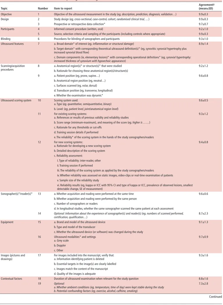
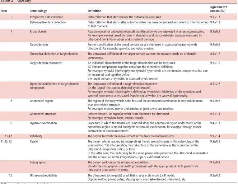
The final checklist, composed of 23 items (21 ‘mandatory’ and 2 ‘optional’), organised into 13 categories, along with the level of agreement for each item, is reported in table 1; the accompanying glossary and the recommendation guidelines on how to use and interpret each item in the following paragraphs are presented in table 2.
Target audience and when to apply the recommendations The target audience comprises health or scientific researchers reporting or assessing observational and interventional studies using ultrasound in RMDs: that is, rheumatologists, radiolo-gists and healthcare professionals using ultrasound, manuscript reviewers, grant applicants and reviewers, journal editors. Each mandatory item of the checklist should be considered as essen-tial to be reported in every ultrasound study regardless of the purpose of the study. Such a report will allow proper appraisal of the validity and applicability of the results. The checklist is meant to be applied whenever ultrasound is used (investigation of measurement properties, diagnostic or prognostic accuracy and therapeutic studies). It is focused specifically on ultrasound issues and is neither intended to be totally comprehensive for all study design issues, nor intended to replace other existing reporting guidelines (eg, RCT, observational diagnostic studies, etc).
General items
The first six items on ‘objective’, ‘design’, ‘participants’ and ‘blinding’ as well as items 20 (‘statistical analysis’) and 21 (‘disclosures’) are not specifically related to ultrasound and some of them have already been included in other reporting checklists. However, the taskforce members felt it was essential to include them in this checklist to emphasise their importance. For example, the objective of the ultrasound measurement in a study might be different from the main objective of the study, and then such difference should be clearly described. Another example is the blinding of the ultrasound evaluation. Blinding is of utmost importance, especially in diagnostic or therapeutic studies, since the lack of patient or operator blinding can influence the results.18 19 Item 20 (‘statistical analysis’) refers mainly to the way in which the analysis of ultrasound variables should be performed, for example, the importance to clearly state whether analyses are performed at patient or at joint/site level.
Ultrasound features
Item 7 refers to the ultrasound definition of the pathological lesions under study. It is crucial to be able to check for consistency between what authors say they want to measure (eg, erosions (target domain) as a measure of structural damage (broad domain of interest)) and what was really measured with ultrasound. The concepts of broad and target domains are explained in table 2. In such cases, reporting the domain components (ie, elementary lesions) really measured by ultrasound will help evaluate consis-tency. A precise definition of ultrasound elementary lesions used in the study helps to check whether ultrasound is able to measure what it is supposed to measure (domain match). Here we used the terminology proposed by OMERACT for the development of imaging outcome measurement instruments,20 and in partic-ular of ultrasound.5 In recent years, the OMERACT ultrasound working group, frequently in collaboration with EULAR, has undertaken considerable efforts to develop and improve defini-tions of ultrasound elementary lesions for a defined pathology (eg, synovitis, enthesitis, bone erosion).5 8
Scanning/acquisition procedures
Several sources of variability may affect the reliability of ultra-sound measurements and generalisability of the study results. These include the quality of the equipment, positioning of patient and transducer and training of the examiner. One of the scoping reviews (online supplemental file 2) studied whether acquisition methods (ie, joint or transducer position and dynamic acquisitions) affected the reliability and accuracy of ultrasound. All retrieved studies confirmed the importance of a standardised joint position for the reliability and general-isability of the results; this applies to all anatomical sites (eg, knee, wrist, Achilles tendon, etc) and all target pathologies under study (eg, synovitis, joint effusion, etc),.21–26 In addi-tion, appropriate transducer manipulation is needed to avoid artefacts.23–25 For example, transducer pressure may cause the synovial hypertrophy or Doppler signal to disappear or be reduced.27 The 2001 EULAR guidelines for performing ultra-sound in rheumatology (updated in 2017) addresses both joint (and patient) positioning as well as transducer use.28 29 Authors are invited to refer to these latest guidelines in their studies. Items 8 and 9 describe what details should be provided about the scanning and acquisition procedures to assess compliance with these guidelines.
copyright.
on May 18, 2021 at IRCCS Gemelli Roma. Protected by
Table 1 Recommendations checklist for reporting studies using ultrasound in rheumatic and musculoskeletal diseases
Topic Number Item to report Agreement† (mean±SD)
Objective 1 Objective of the ultrasound measurement in the study (eg, description, prediction, diagnosis, validation…) 9.9±0.3
Design 2 Study design (eg, cross- sectional, case- control, cohort, randomised clinical trial, …) 9.9±0.3
3 Prospective or retrospective data collection* 9.7±0.7
Participants 4 Informed consent procedure (written, oral) 9.2±1.0
5 Source, selection criteria and sampling of the participants (including controls where appropriate) 9.9±0.3
Blinding 6 Procedures for blinding of sonographers and participants 9.3±1.0
Ultrasound features 7 a. Broad domain* of interest (eg, inflammation or structural damage) 8.9±1.4
b. Target domain* with corresponding theoretical ultrasound definition(s)* (eg, synovitis: synovial hypertrophy plus increased synovial blood flow)
c. Domain components (ie, elementary lesions)* with corresponding operational definitions* (eg, synovial hypertrophy:
increased thickness of synovium with hypoechoic appearance)
Scanning/acquisition procedures
8 a. Anatomical region(s)* or structure(s)* that were studied 9.2±1.2
b. Rationale for choosing these anatomical region(s)/structure(s)
9 a. Patient position (eg, prone, supine…) 9.6±0.8
b. Anatomical region position (eg, neutral…) c. Surfaces scanned (eg, volar, dorsal)
d. Transducer position (eg, transverse, longitudinal) e. Whether the examination was dynamic* Ultrasound scoring system 10 Scoring system used:
a. Type (eg, quantitative, semiquantitative, binary) 9.6±0.5
b. Level: (eg, patient level, joint/anatomical region level) 11 For existing scoring systems:
a. References or results of previous validity and reliability studies 9.3±1.2
b. Score range (minimum- maximum), and meaning of the score (eg, higher is ……) c. Rationale for any thresholds or cut- offs
d. Training session details if performed
e. The reliability* of the scoring system in the hands of the study sonographers/readers 12 For new scoring systems:
a. Rationale for developing a new scoring system 9.4±0.8
b. Detailed description of the scoring system c. Reliability assessment:
I. Type of reliability: inter- reader, other ii. Training session if performed
Iii. The reliability of the scoring system as applied by the study sonographers/readers
iv. Whether reliability was assessed on static images, video- clips or real- time examination of patients v. Sample size of the reliability study
vi. Reliability results (eg, kappa or ICC with 95% CI and type of kappa or ICC, prevalence of observed lesions, smallest detectable change, SE of measurement)
Sonographer(s)*/reader(s)* 13 a. Whether acquisition and reading were performed at the same time 9.6±0.6
b. Whether acquisition and reading were performed by the same person c. Number of sonographers or readers
d. In longitudinal studies, whether the same sonographer scanned the same patient at each assessment
14 Optional: Information about the experience of sonographer(s) and reader(s) (eg, numbers of scanned performed, certification, qualification…)
8.7±2.3
Equipment 15 a. Brand and model of the ultrasound device 9.1±1.3
b. Type and model of the transducer
c. Whether the ultrasound device (or software) was changed during the study 16 Ultrasound modalities* and settings
a. Grey scale
9.7±0.9 b. Doppler
c. Other Images (pictures and
drawings) 17 For images included into the manuscript, verify that:a. Information identifying patient is deleted 9.3±1.6 b. Essential targets in the image(s) are clearly labelled
c. Images match the content of the manuscript d. Quality of the images is adequate
Contextual factors 18 Duration of ultrasound examination when relevant for the study question 8.8±1.6
19 Optional:
a. Whether ambient conditions (eg, temperature, time of day) were kept stable during the study b. Potential confounding factors (eg, exercise, alcohol, caffeine, smoking)
7.3±2.8
Continued
copyright.
on May 18, 2021 at IRCCS Gemelli Roma. Protected by
Ultrasound scoring systems
Items 10, 11 and 12 focus on a clear description of ultrasound scoring systems, especially if a newly developed scoring system is used. Special attention should be paid to the documentation of the development of the scoring system. As ultrasound is frequently considered the most operator- dependent imaging technique, intra- rater and inter- rater reliability is an important concern and a strong argument for standardisation. For new scoring systems, results of intra- sonographer and inter- sonographer/reader reli-ability studies should be reported. For existing scoring systems, reference to previous reliability studies should be given as well
as the results of reliability assessments among the sonographers/ readers in the context of the study.
Sonographers/readers
Depending on the setting, the person who performs the ultra-sound acquisition of the images (sonographer) can be a healthcare professional or a medical doctor (radiologist or rheumatologist). The images can be interpreted at the time of acquisition or later, by the same or another person. Choices made here affect ultra-sound scores and generalisability, so details on who performs
Topic Number Item to report
Agreement† (mean±SD)
Statistical analysis 20 a. Existence of a pre- specified statistical analysis plan and specification of post- hoc analyses 9.3±1.2 b. Analyses performed
c. Whether the analyses were performed at patient or at joint/region level d. Extent of missing data
e. Handling of missing data
Disclosures 21 Potential conflicts of interest including those related to ultrasound 9.2±1.4
*Items are explained in detail in the glossary (table 2). ICC, intra class correlation; SE, standard error.
Table 1 Continued
Table 2 Glossary
Item Terminology Definition
Agreement† (mean±SD)
3 Prospective data collection Data collection that starts before the outcome has occurred. 9.2±1.7
Retrospective data collection Data collection that starts after outcome status has been determined and refers to information up to that moment.
9.4±1.2 7 Broad domain A pathological (or pathophysiological) manifestation we are interested in assessing/measuring.
For example, current broad domains in rheumatic and musculoskeletal diseases measured by ultrasound are ‘inflammation’ and ‘structural damage’.
9.7±0.8
Target domain Further specification of the broad domain we are interested in assessing/measuring with ultrasound. For example, synovitis, enthesitis, erosion.
9.7±0.6 Theoretical definition of target domain The ultrasound definition of the target domain we want to measure, made up of domain
components.
8.9±1.7 Target domain component An individual characteristic of the target domain that can be measured.
All domain components together constitute the theoretical definition.
For example, synovial hypertrophy and synovial hyperaemia are the domain components that can be measured, and together define
the target domain of synovitis as assessed by ultrasound.
9.1±1.1
Operational definition of target domain component
The ultrasound definition of a target domain component (ie, the ‘signal’ that can be detected by ultrasound).
For example, synovial hypertrophy is defined as hypoechoic thickening of the synovium; and synovial hyperaemia as increased Doppler signal within the synovial hypertrophy.
8.9±2.2
8 Anatomical region The region of the body which is the focus of the ultrasound examination; it may include more than one related structure.
For example, muscles, nerves and arteries, or joint cavity and tendons.
9.9±0.2
Anatomical structure Isolated tissue(s) or organ(s) which is/are examined by ultrasound. For example, synovium, bone, tendon, muscle.
9.6±1.3 9 Dynamic examination Procedure in which the transducer is moved along the anatomical region under study; or the
anatomical region is moved during the ultrasound examination, for example through muscle contraction or tendon movement.
9.4±1.3
11,12 Reliability The degree to which the measurement is free from measurement error. 9.1±1.4
11,12,13 Reader The person who is reading (ie, interpreting) the ultrasound images or the video- clips of the examination. This interpretation may take place at the same time as the acquisition of the ultrasound images/video- clips, or later.
In the latter case, the reader may be the same person who performed the ultrasound examination and the acquisition of the images/video- clips or a different person.
9.9±0.3
Sonographer The person performing the ultrasound evaluation.
Usually the sonographer is a health professional with the appropriate skills to perform an ultrasound examination in RMDs.
9.7±0.9
16 Ultrasound modalities The ultrasound technique(s) used, that is, grey scale mode (or B mode),
Doppler (colour, power, pulse), elastography, contrast- enhanced ultrasound, etc. 9.9±0.2 RMD, rheumatic and musculoskeletal disease.
copyright.
on May 18, 2021 at IRCCS Gemelli Roma. Protected by
the acquisition and the interpretation are a mandatory reporting requirement. Item 14 on the experience of sonographer(s) and/ or reader(s) is optional, mainly because no consensus exists on how to report such experience. EULAR and American College of Rheumatology suggest a competency assessment in ultrasound to improve the quality of the examination.30 31
Equipment
Technical characteristics of the imaging device (item 15), ultra-sound modalities and settings used (item 16) may affect the intrasonographer and intersonographer/reader reliability and generalisability of the results. A second scoping review (online supplemental file 2) addressed this question, that is, whether the ultrasound device (model, age, acquisition software, transducers and settings) affect the reliability or accuracy of the ultrasound examination. We found no study investigating the influence of device age or software on ultrasound results. However, seven studies assessed the influence of the machine (eg, ultrasound device, transducer frequency, settings) on ultrasound results whatever the anatomical site studied, the ultrasound modality used (ie, Doppler, GS) or the target pathology under study (eg, joint effusion, synovitis, erosion).32–38 Three studies used a phantom to compare the ultrasound devices.34 36 37 Five of the seven studies showed differences in the performance between machines and therefore an influence on the study results.35 36 However, in these studies, the relevance and the magnitude of such differences were reported, but no sensitivity analysis was conducted.
Images, pictures and figures
Since ultrasound is a tomographic imaging modality, the appear-ance of the structures may change following the orientation and position of the transducer. Standardised images should always be presented (item 17) so that the reader can easily recognise the anatomical structures as well as the target pathology and elementary lesions described in the study. The use of drawings can facilitate the interpretation of the images for readers not experienced in ultrasound. Images should never contain patient information and should be accompanied by clear legends and points of reference.
Contextual factors
Item 18 deals with the feasibility of ultrasound, in particular, the time necessary for the evaluation, which depends on the number of sites (or joints) examined and the number of ultrasound exam-inations performed over the duration of the study. Although these aspects are highly important for the acceptability of the technique, the taskforce members felt that time spent should be reported only if relevant to the study question.
Item 19 refers to additional potential sources of variability: ambient and patient conditions. This item was made optional because the third scoping review (online supplemental file 2) failed to find strong evidence of the influence of these factors on the ultrasound results. It reviewed the effect of three ambient conditions (room temperature, atmospheric pressure and time of the day) and five patient conditions (exercise, skin tempera-ture, smoking, alcohol consumption and caffeine) on ultrasound measurements. There was a potential influence of time of the day on Doppler signal evaluation (with contradictory effects)39 40 and on GS results41; and potential influences on Doppler signal following the application of cold (ice and cold water)42–44 and after physical exercise.45–47
DISCUSSION
This EULAR taskforce developed a recommendation checklist to ensure transparent and comprehensive reporting of ultrasound research and procedures aspects, which may affect the interpre-tation and generalisability of the results. The checklist consists of 23 items (21 ‘mandatory’ and 2 ‘optional’), organised into 13 categories. Its organisation allows authors to choose the order and format for presenting information, depending on their pref-erences and on journal style. Content validity of the recommen-dations checklist was confirmed by a panel of external experts, who considered each item of the checklist an essential reporting point, crucial to make an informed judgement on the quality of the scientific report. Moreover, all items were considered comprehensible and the checklist as a whole was considered to comprehensively cover all relevant reporting issues.
Along with sufficient content validity of this checklist, addi-tional strengths include the development process that followed EULAR SOPs for a stepwise consensual approach16 and the
guid-ance from the EQUATOR network,17 also, the panel members
reflected a wide range of expertise and stakeholders. In addition, agreement on comprehensiveness and comprehensibility of the checklist was obtained in the first round of voting for all items of the checklist and all definitions of the glossary.
A possible limitation of this project is the fact that the face- to- face meetings comprised mostly Europeans, with only one colleague from USA, and only one patient. We partially over-came this in the external Delphi panels, including more interna-tional experts, including several radiologists.
The checklist was purposefully focused and is complementary to other existing guidelines, depending on the study design. It has not been developed as a tool to assess the quality of published research; however, it can certainly serve as a basis to develop such a tool, and its use may improve the quality of studies, as seen with other reporting recommendations.48 49 We hope that this reporting checklist will be widely adopted by authors and editors, which, in turn, will greatly improve the interpretability, reproducibility and generalisability of the study results.
Author affiliations
1UVSQ, Inserm U1173, Infection et inflammation, Laboratory of Excellence INFLAMEX, Université Paris- Saclay, Montigny- le- Bretonneux, France
2Rheumatology Department, Ambroise Paré Hospital, AP- HP, Boulogne- Billancourt, Île- de- France, France
3Instituto de Salud Musculoesquelética (INMUSC), Madrid, Madrid, Spain 4Epidemiology and Biostatistics, Vrije Universiteit Amsterdam, Amsterdam, Noord- Holland, The Netherlands
5Department of Internal Medicine—Rheumatology and Clinical Immunology, Park- Klinik Weissensee, Berlin, Berlin, Germany
63rd Department of Rheumatology, National Institute of Rheumatology and Physiotherapy, Budapest, Hungary
7Rheumatology Department, MC Group Hospitals, Lelystad, The Netherlands 8Rheumatology Department, Reumakliniek Flevoland, Lelystad, The Netherlands 9Department of Clinical Research, Odense University Hospital, Odense, Denmark 10Rheumatology, Leeds Teaching Hospitals NHS Trust, Leeds, UK
11Rheumatology, Centro Hospitalar e Universitário de Coimbra, Coimbra, Portugal 12Health Sciences Research Unit: Nursing (UICiSA:E), Coimbra, Portugal 13Spanish Federation of Spondyloarthritis Associations (CEADE), Madrid, Spain 14Medecine, School of Public Health, Vandoeuvre, France
15Rheumatology, Diakonhjemmet Sykehus, Oslo, Norway
16Rheumatology, Leiden University Medical Center, Leiden, The Netherlands 17Scienze Cliniche e Biologiche, Università degli Studi di Torino, Torino, Italy 18Rheumatology Department, Amsterdam Rheumatology Center, AMC, Amsterdam, The Netherlands
19Rheumatology, Zuyderland MC, Heerlen, The Netherlands
20Internal Medicine 3, Division of Rheumatology, Medical University Vienna, Vienna, Austria
21Department of Rheumatology, Bone and Joint Research Unit, Hospital Universitario Fundación Jiménez Díaz, Madrid, Spain
22IIS Fundación Jiménez Díaz, Universidad Autónoma de Madrid, Madrid, Spain
copyright.
on May 18, 2021 at IRCCS Gemelli Roma. Protected by
23Rheumatology Department, Medical Centre for Rheumatology Berlin Buch, Berlin, Germany
24Center for Rheumatology and Spine Diseases, Rigshospitalet, Kobenhavn, Denmark 25Department of Medicine, Division of Allergy, Immunology and Rheumatology, University of Rochester School of Medicine and Dentistry, Rochester, New York, USA 26Rheumatology Department, Università Cattolica del Sacro Cuore, Policlinico Universitario Agostino Gemelli IRCSS, Roma, Italy
Twitter Loreto Carmona @carmona_loreto and Ricardo J O Ferreira @FerreiraRJO Acknowledgements The authors want to thank all contributing experts who participated in the online survey (see online supplemental file 3 for the full list). Contributors Full- text review, data abstraction and Delphi assessments were performed by FC, supervised by MADA and independently double- checked by LC. MADA and LC supervised the methodology of the scoping literature review and FC prepared the evidence report. FC and MADA prepared the first draft of recommendations, and all authors participated in the discussion and formulation of recommendations. MADA supervised the project and FC, MADA, LC, MB and PGC drafted the manuscript. All authors reviewed the manuscript and approved its final version.
Funding This project was funded by the European League Against Rheumatism (EULAR). PGC is supported in part by the UK National Institute for Health Research (NIHR) Leeds Biomedical Research Centre. RC is supported by a core grant from the Oak Foundation (OCAY-18–774- OFIL).
Disclaimer The views expressed are those of the authors and not necessarily those of the NHS, the NIHR or the Department of Health.
Competing interests FC reports personal fees from Lilly and Novartis France. MB reports personal fees from Novartis, BMS and Pfizer. RC is a founding member of the Technical Advisory Group of OMERACT, an organization that develops outcome measures in rheumatology and receives arms- length funding from 12 companies. DvdH reports personal fees from AbbVie, Amgen, Astellas, AstraZeneca, Bayer, BMS, Boehringer Ingelheim, Celgene, Cyxone, Daiichi, Eisai, Eli- Lilly, Galapagos, Gilead, Glaxo- Smith- Kline, Janssen, Merck, Novartis, Pfizer, Regeneron, Roche, Sanofi, Takeda, UCB Pharma and is Director of Imaging Rheumatology bv. AI reports grants from Abbvie, MSD, and Alfasigma and personal fees from AbbVie, Abiogen, Alfasigma, Biogen, BMS, Celgene, Eli- Lilly, Janssen, MSD, Novartis, Sanofi, Sanofi Genzyme. RBL reports personal fees from AbbVie, Galapagos, Gilead, Jansen, Eli- Lilly, Novartis, Pfizer, UCB. PM reports grants and personal fees from AbbVie, Novartis, Janssen, personal fees from Celgene, grants from Merck Sharp & Dohme, UCB, Roche. RT reports personal fees from Amgen, AbbVie, Novartis. MADA reports personal fees from Abbvie, BMS, Novartis, Celgene, Janssen and grants from Pfizer.
Patient consent for publication Not required.
Provenance and peer review Not commissioned; internally peer reviewed. Data availability statement All data relevant to the study are included in the article or uploaded as supplementary information.
Supplemental material This content has been supplied by the author(s). It has not been vetted by BMJ Publishing Group Limited (BMJ) and may not have been peer- reviewed. Any opinions or recommendations discussed are solely those of the author(s) and are not endorsed by BMJ. BMJ disclaims all liability and responsibility arising from any reliance placed on the content. Where the content includes any translated material, BMJ does not warrant the accuracy and reliability of the translations (including but not limited to local regulations, clinical guidelines, terminology, drug names and drug dosages), and is not responsible for any error and/or omissions arising from translation and adaptation or otherwise.
ORCID iDs
Félicie Costantino http:// orcid. org/ 0000- 0002- 1449- 959X
Loreto Carmona http:// orcid. org/ 0000- 0002- 4401- 2551
George A Bruyn http:// orcid. org/ 0000- 0001- 7020- 5798
Philip G Conaghan http:// orcid. org/ 0000- 0002- 3478- 5665
Ricardo J O Ferreira http:// orcid. org/ 0000- 0002- 2517- 0247
Juan Luis Garrido- Castro http:// orcid. org/ 0000- 0002- 0871- 3780
Hilde Berner Hammer http:// orcid. org/ 0000- 0001- 7317- 8991
Désirée van der Heijde http:// orcid. org/ 0000- 0002- 5781- 158X
Marion C Kortekaas http:// orcid. org/ 0000- 0003- 4334- 552X
Robert BM Landewé http:// orcid. org/ 0000- 0002- 0577- 6620
Peter Mandl http:// orcid. org/ 0000- 0003- 1526- 4052
Lene Terslev http:// orcid. org/ 0000- 0003- 3690- 467X REFERENCES
1 Mandl P, Ciechomska A, Terslev L, et al. Implementation and role of modern musculoskeletal imaging in rheumatological practice in member countries of EULAR.
RMD Open 2019;5:e000950.
2 Scheel AK, Schmidt WA, Hermann K- GA, et al. Interobserver reliability of rheumatologists performing musculoskeletal ultrasonography: results from a EULAR "Train the trainers" course. Ann Rheum Dis 2005;64:1043–9.
3 Joshua F. Ultrasound applications for the practicing rheumatologist. Best Pract Res Clin Rheumatol 2012;26:853–67.
4 van Holsbeeck M, van HM. Fury over sound. Arthritis Rheum 2004;51:877–80. 5 Terslev L, Naredo E, Keen HI, et al. The OMERACT stepwise approach to select and
develop imaging outcome measurement instruments: the musculoskeletal ultrasound example. J Rheumatol 2019;46:1394–400.
6 Joshua F, Lassere M, Bruyn GA, et al. Summary findings of a systematic review of the ultrasound assessment of synovitis. J Rheumatol 2007;34:839–47.
7 Wakefield RJ, Balint PV, Szkudlarek M, et al. Musculoskeletal ultrasound including definitions for ultrasonographic pathology. J Rheumatol 2005;32:2485–7. 8 Bruyn GA, Iagnocco A, Naredo E, et al. OMERACT definitions for ultrasonographic
pathologies and elementary lesions of rheumatic disorders 15 years on. J Rheumatol
2019;46:1388–93.
9 Mandl P, Naredo E, Wakefield RJ, et al. A systematic literature review analysis of ultrasound joint count and scoring systems to assess synovitis in rheumatoid arthritis according to the OMERACT filter. J Rheumatol 2011;38:2055–62.
10 Lazarou I, D’Agostino M- A, Naredo E, et al. Ultrasound- guided synovial biopsy: a systematic review according to the OMERACT filter and recommendations for minimal reporting standards in clinical studies. Rheumatology 2015;54:1867–75.
11 Schulz KF, Altman DG, Moher D, et al. Consort 2010 statement: updated guidelines for reporting parallel group randomized trials. Ann Intern Med 2010;152:726–32. 12 Bossuyt PM, Reitsma JB, Bruns DE, et al. Towards complete and accurate reporting of
studies of diagnostic accuracy: the STARD initiative. Ann Intern Med 2003;138:40–4. 13 Bossuyt PM, Reitsma JB, Bruns DE, et al. Stard 2015: an updated list of essential
items for reporting diagnostic accuracy studies. BMJ 2015;351:h5527.
14 Dixon WG, Carmona L, Finckh A, et al. EULAR points to consider when establishing, analysing and reporting safety data of biologics registers in rheumatology. Ann Rheum Dis 2010;69:1596–602.
15 Buch MH, Silva- Fernandez L, Carmona L, et al. Development of EULAR
recommendations for the reporting of clinical trial extension studies in rheumatology.
Ann Rheum Dis 2015;74:963–9.
16 van der Heijde D, Aletaha D, Carmona L, et al. 2014 update of the EULAR standardised operating procedures for EULAR- endorsed recommendations. Ann Rheum Dis 2015;74:8–13.
17 Moher D, Schulz KF, Simera I, et al. Guidance for developers of health research reporting guidelines. PLoS Med 2010;7:e1000217.
18 Hróbjartsson A, Emanuelsson F, Skou Thomsen AS, et al. Bias due to lack of patient blinding in clinical trials. A systematic review of trials randomizing patients to blind and nonblind sub- studies. Int J Epidemiol 2014;43:1272–83.
19 Hróbjartsson A, Thomsen ASS, Emanuelsson F, et al. Observer bias in randomized clinical trials with measurement scale outcomes: a systematic review of trials with both blinded and nonblinded assessors. CMAJ 2013;185:E201–11.
20 Boers M, Beaton DE, Shea BJ, et al. OMERACT filter 2.1: elaboration of the conceptual framework for outcome measurement in health intervention studies. J Rheumatol
2019;46:1021–7.
21 Zappia M, Cuomo G, Martino MT, et al. The effect of foot position on power Doppler ultrasound grading of Achilles enthesitis. Rheumatol Int 2016;36:871–4. 22 Zayat AS, Freeston JE, Conaghan PG, et al. Does joint position affect us findings in
inflammatory arthritis? Rheumatology 2012;51:921–5.
23 Terslev L, D’Agostino MA, Brossard M, et al. Which knee and probe position determines the final diagnosis of knee inflammation by ultrasound? results from a European multicenter study. Ultraschall Med 2012;33:e374–8.
24 Mandl P, Brossard M, Aegerter P, et al. Ultrasound evaluation of fluid in knee recesses at varying degrees of flexion. Arthritis Care Res 2012;64:773–9.
25 Hong BY, Lim SH, Cho YR, et al. Detection of knee effusion by ultrasonography. Am J Phys Med Rehabil 2010;89:715–21.
26 Koenig MJ, Torp- Pedersen ST, Christensen R, et al. Effect of knee position on ultrasound Doppler findings in patients with Patellar tendon hyperaemia (jumper’s knee). Ultraschall Med 2007;28:479–83.
27 Joshua F, de Carle R, Rayment M, et al. Power Doppler ’blanching’ after the application of transducer pressure. Australas Radiol 2005;49:218–21.
28 Möller I, Janta I, Backhaus M, et al. The 2017 EULAR standardised procedures for ultrasound imaging in rheumatology. Ann Rheum Dis 2017;76:1974–9.
29 Backhaus M, Burmester GR, Gerber T, et al. Guidelines for musculoskeletal ultrasound in rheumatology. Ann Rheum Dis 2001;60:641–9.
30 Eular. US-2022. Available: https:// esor. eular. org/ enrol/ index. php? id= 159 [Accessed 3 Apr 2020].
31 American College of Rheumatology. RhMSUS™ certification. Available: https://www. rheumatology. org/ Learning- Center/ RhMSUS- Certification [Accessed 3 Apr 2020]. 32 Torp- Pedersen S, Christensen R, Szkudlarek M, et al. Power and color Doppler ultrasound
settings for inflammatory flow: impact on scoring of disease activity in patients with rheumatoid arthritis. Arthritis Rheumatol 2015;67:386–95.
33 Brulhart L, Ziswiler H- R, Tamborrini G, et al. The importance of sonographer experience and machine quality with regards to the role of musculoskeletal ultrasound in routine care of rheumatoid arthritis patients. Clin Exp Rheumatol 2015;33:98–101.
copyright.
on May 18, 2021 at IRCCS Gemelli Roma. Protected by
34 Ten Cate DF, Luime JJ, Swen N, et al. Role of ultrasonography in diagnosing early rheumatoid arthritis and remission of rheumatoid arthritis--a systematic review of the literature. Arthritis Res Ther 2013;15:R4.
35 Peterlein C- D, Fuchs- Winkelmann S, Schüttler K- F, et al. Does probe frequency influence diagnostic accuracy in newborn hip ultrasound? Ultrasound Med Biol 2012;38:1116–20. 36 Koski JM, Alasaarela E, Soini I, et al. Ability of ultrasound imaging to detect erosions in a
bone phantom model. Ann Rheum Dis 2010;69:1618–22.
37 Koski JM, Saarakkala S, Helle M, et al. Assessing the intra- and inter- reader reliability of dynamic ultrasound images in power Doppler ultrasonography. Ann Rheum Dis
2006;65:1658–60.
38 Albrecht K, Grob K, Lange U, et al. Reliability of different Doppler ultrasound quantification methods and devices in the assessment of therapeutic response in arthritis. Rheumatology 2008;47:1521–6.
39 Lazaar H, Lhoste- Trouilloud A, Pereira B, et al. Does rheumatoid synovitis activity vary during the day? evaluation with color Doppler sonography. BMC Musculoskelet Disord 2017;18:98.
40 Semerano L, Gutierrez M, Falgarone G, et al. Diurnal variation of power Doppler in metacarpophalangeal joints of patients with rheumatoid arthritis: a preliminary study.
Ann Rheum Dis 2011;70:1699–700.
41 Kilic G, Kilic E, Akgul O, et al. Ultrasonographic assessment of diurnal variation in the femoral condylar cartilage thickness in healthy young adults. Am J Phys Med Rehabil
2015;94:297–303.
42 Guillot X, Tordi N, Prati C, et al. Cryotherapy decreases synovial Doppler activity and pain in knee arthritis: a randomized- controlled trial. Joint Bone Spine 2017;84:477–83. 43 Ellegaard K, Torp- Pedersen S, Henriksen M, et al. Influence of recent exercise and skin
temperature on ultrasound Doppler measurements in patients with rheumatoid arthritis--an intervention study. Rheumatology 2009;48:1520–3.
44 Strunk J, Strube K, Müller- Ladner U, et al. Three dimensional power Doppler ultrasonography confirms early reduction of synovial perfusion after intra- articular steroid injection. Ann Rheum Dis 2006;65:411–2.
45 Malliaras P, Chan O, Simran G, et al. Doppler ultrasound signal in Achilles tendinopathy reduces immediately after activity. Int J Sports Med 2012;33:480–4.
46 Boesen MI, Koenig MJ, Torp- Pedersen S, et al. Tendinopathy and Doppler activity: the vascular response of the Achilles tendon to exercise. Scand J Med Sci Sports
2006;16:463–9.
47 Cook JL, Kiss ZS, Ptasznik R, et al. Is vascularity more evident after exercise? implications for tendon imaging. AJR Am J Roentgenol 2005;185:1138–40. 48 Plint AC, Moher D, Morrison A, et al. Does the CONSORT checklist improve the
quality of reports of randomised controlled trials? A systematic review. Med J Aust
2006;185:263–7.
49 Smidt N, Rutjes AWS, van der Windt DAWM, WM vanderWDa, et al. The quality of diagnostic accuracy studies since the STARD statement: has it improved? Neurology
2006;67:792–7.
copyright.
on May 18, 2021 at IRCCS Gemelli Roma. Protected by