UNIVERSITÀ DEGLI STUDI DELLA TUSCIA DI VITERBO
FACOLTÀ DI SCIENZE MATEMATICHE FISICHE E NATURALI
DIPARTIMENTO DI SCIENZE AMBIENTALI
DOTTORATO DI RICERCA IN EVOLUZIONE BIOLOGICA E
BIOCHIMICA - XIX CICLO
ATTIVITÀ CELLULARI E MOLECOLARI
NEI LEUOCITI DEI TELESOTEI
(Settore scientifico-disciplinare: BIO 05)
Tutor: Prof. Massimo Mazzini Correlatore: Prof. Giuseppe Scapigliati
Firma: Firma:
Coordinatore: Prof. Lucia Mastrolia Dottoranda: Stefania Benedetti
Firma: Firma:
Ai miei genitori
per il loro sostegno
a Marco
“Ich sah dich, und die milde Freude
Floß aus dem süßen Blick auf mich.
Ganz war mein Herz an deiner Seite,
Und jeder Atemzug für dich“
Io credo nel mio Dio Onnipotente.
Nella Mente che tutto comprende.
Nel cuore misericordioso
e nel braccio vittorioso.
Io credo nella Spada che non fallisce.
Nella Vita che non finisce.
Nella Giustizia mai assente.
La Libertà che non si svende.
Nella Dignità perigliosa.
Nella Carità meravigliosa
Nello Scudo per l'indifeso.
Nel Guanto per chi è offeso.
Io credo nella Fede implacabile.
Nella Forza incrollabile.
Nell'Onore per mantello.
La Verità per suggello.
Nel Sogno incandescente.
Nell'Amore sorprendente.
Io credo nel coraggio di chi osa.
Nella certezza che mai riposa.
Negli antichi padri e la loro Gloria.
In chi ero, sono e ancor sarò. Nella mia storia.
Io credo nella Volontà che non vacilla.
Nella Conoscenza che scintilla.
Nello Spirito non cadente.
La pietra e la stella che tocca a me far più splendente.
Nella Magia del Mondo, il Fato e nel mio cammino.
La candidata Benedetti Stefania, ha frequentato il triennio del corso di Dottorato in “Evoluzione Biologica e Biochimica” (XIX ciclo), svolgendo una tesi dal titolo “Attività
cellulari e molecolari nei leucociti dei Teleostei”. In particolare, si è dedicata allo studio della
trasduzione del segnale via calcio, indotta da: IL-1β ricombinante, terreno condizionato,
Photobacterium damselae e nodavirus, ed ha investigato gli effetti della stimolazione sia a
livello cellulare che a livello molecolare.
Durante gli anni del corso di Dottorato ha avuto modo di apprendere numerose tecniche, in particolare: produzione e purificazione di citochine ricombinati, stimolazione cellulare, misurazione del calcio intracellulare, purificazione per sorting magnetico, cross-linking, SDS-PAGE, western blotting, 2D-gel, marcature metaboliche, proteomica, reazioni “MLR”. Durante il suo soggiorno presso l’Istituto Regina Elena di Roma ha, inoltre, imparato a manipolare sostanze radioattive.
Nel trienno ha sempre svolto autonomamente e con perizia il suo lavoro presso il Laboratorio di Biotecnologie Animali e Zoologia Applicata dell’Università degli studi della Tuscia (prof. Giuseppe Scapigliati), ed ha partecipato alla vita del laboratorio partecipando anche ad altri progetti di ricerca in studio nel suddetto laboratorio. Il suo impegno è stato sempre continuo e i risultati del suo lavoro sono stati pubblicati su diverse riviste scientifiche di buon livello internazionale e discusso in numerosi congressi sia nazionali che internazionali. Ha, inoltre, partecipato a diversi corsi per approfondire le sue conoscenze in ambito biochimico e biotecnologico, ed è risultata vincitrice di premi di ricerca e borse di specializzazione, come mostrato nel curriculum allegato.
Per i motivi esposti, ritengo che la candidata abbia raggiunto una buona maturità scientifica, che risulta del tutto idonea per il titolo di Dottore di Ricerca che si accinge a conseguire. Visto che il prof. Mazzini è fuori per motivi logistici, la dottoranda è stata valutata, dopo aver sentito il professore, dalla sottoscritta prof. Anna Maria Fausto, docente nel settore zoologico e facente parte del consiglio di questo Dottorato. Pertanto tale giudizio scaturisce da collaborazione tra il prof. Mazzini e me.
CURRICULUM VITAE
PARTECIPAZIONI A CONGRESSI SCIENTIFICI
2004
XVII Meeting of the Protein Workgroup
E. Randelli, M. Forlenza, S. Meloni, S. Benedetti, C.J. Secombes, J. Zou, G. Scapigliati, S. Costantini, A. Facchiano, F. Buonocore POTENTIAL APPLICATION OF SEA BASS INTERLEUKIN-1β IN FISH VACCINATION. Viterbo, 20-22 maggio.
65° Congresso Nazionale dell’Unione Zoologica Italiana
F. Buonocore, E. Ovidi, D. Casani, E. Randelli, S. Benedetti, S. Meloni, G. Scapigliati, M. Mazzini PRODUZIONE DI IMMUNOGLOBULINE NELLA SPIGOLA (Dicentrarchus
labrax) dopo vaccinazione contro Vibrio anguillarum tramite marcatori molecolari e cellulari.
Taormina-Giardini-Naxos (ME), 21-25 settembre.
2005
7° Incontro scientifico della Società Italiana di Immunobiologia Comparata e dello Sviluppo
S. Benedetti, M. Tiberi, E. Randelli, F. Buonocore, M. Mazzini and G. Scapigliati
RECOMBINANT IL-1β DIRECTLY AFFECTS INTRACELLULAR Ca2+
CONCENTRATION ON LEUCOCYTES OF THE SEA BASS (Dicentrarchus labrax). Trapani, 10-12 febbraio.
7° Incontro scientifico della Società Italiana di Immunobiologia Comparata e dello Sviluppo
S. Meloni, G. Zarletti, S. Benedetti, E. Randelli, F. Buonocore, G. Scapigliati LYMPHOCYTE ACTIVITIES “IN VITRO” OF THE SEA BASS DICENTRARCHUS
LABRAX. Trapani, 10-12 febbraio.
International Marine Biotechnology Conference
S. Meloni, F. Buonocore, E. Randelli, S. Benedetti, G. Zarletti, M. Mazzini and G. Scapigliati
IMMUNE SYSTEM AND BIOTECHNOLOGY: “IN VITRO” LYMPHOCYTE
ACTIVITIES OF THE SEA BASS DICENTRARCHUS LABRAX. Wageningen, The Nederland, 15-16 April.
66° congresso dell’Unione Zoologica Italiana
S. Benedetti, M. Tiberi, E. Randelli, F. Buonocore, M. Mazzini, J. Zou, C. J. Secombes, G.
Scapigliati EVOLUZIONE DELLE RISPOSTE DI SEGNALAZIONE ORMONALE:
INTERLEUCHINA-1β RICOMBINANTE DI TELEOSTEI E CONCENTRAZIONE
INTRACELLULARE DI Ca2+. Roma, 19-22 settembre.
2006
10° congress of International Society of Developmental and Comparative Immunology
G. Scapigliati, S. Benedetti, E. Randelli, F. Buonocore, J. Zou, C.J. Secombes EVOLUTION OF CYTOKINE RESPONSES: IL-1β DIRECTLY AFFECTS INTRACELLULAR Ca2+ CONCENTRATION OF TELEOST FISH LEUKOCYTES THROUGH A RECEPTOR-MEDIATED MECHANISM. Charleston, South Carolina (USA) 1-6 luglio.
2007
8°Incontro scientifico della Società Italiana di Immunobiologia Comparata e dello Sviluppo
F. Buonocore, E.Randelli, S. Bird, CJ., Secombes, S. Costantini, A., Facchiano, S. Benedetti, G. Scapigliati MOLECULAR CLONING, STRUCTURAL ANALYSIS AND
ANTIGEN-INDUCED “IN VIVO” EXPRESSION OF INTERLEUKIN-10 IN SEA BASS (DICENTRARCHUS LABRAX L.). Napoli, 1-2 marzo.
Vth SIICA National Conference
S. Bendetti, E. Randelli, F. Buonocore, J. Zou, C.J. Secombes, G. Scapigliati EVOLUTION
OF CYTOKINE RESPONSES: IL-1β DIRECTLY AFFECTS INTRACELLULAR Ca2+ CONCENTRATION OF TELEOST FISH LEUKOCYTES THROUGH A RECEPTOR-MEDIATED MECHANISM. Trieste, 6-9 giugno.
PUBBLICAZIONI
2004
E. Randelli, M. Forlenza, S. Meloni, S. Benedetti, C.J. Secombes, J. Zou, G. Scapigliati, S. Costantini, A. Facchiano, F. Buonocore POTENTIAL APPLICATION OF SEA BASS
INTERLEUKIN-1β IN FISH VACCINATION. Italian Journal of Biochemistry Vol. 53 (Suppl.1) p. 68.
2005
F. Buonocore, M. Forlenza, E. Randelli, S. Benedetti, P. Bossù, S. Meloni, C.J. Secombes,
M. Mazzini and G. Scapigliati BIOLOGICAL ACTIVITY OF SEA BASS
(DICENTRARCHUS LABRAX L.) RECOMBINANT INTERLEUKIN-1β. Marine
Biotechnology 7, 609-17.
S. Meloni, G. Zarletti, S. Benedetti, E. Randelli, F. Buonocore, G. Scapigliati THE MIXED LEUKOCYTE REACTION IN THE TELEOST (DICENTRARCHUS LABRAX). Fish and
Shellfish Immunology 20, 739-49
S. Benedetti, E. Randelli, F. Buonocore, J. Zou, C.J. Secombes, and G. Scapigliati
EVOLUTION OF CYTOKINE RESPONSES: IL-1β DIRECTLY AFFECTS
INTRACELLULAR Ca2+ CONCENTRATION ON TELEOST FISH LEUKOCYTES
THROUGH A RECEPTOR-MEDIATED MECHANISM. Cytokine 34(1-2):9-16.
ALTRE INFORMAZIONI
2004
Abilitazione all’esercizio della professione di biologo conseguita presso la Facoltà di Scienze Matematiche Fisiche e Naturali dell’Università degli Studi della Tuscia di Viterbo
Partecipazione al workshop “RNA-2Day” presso l’università La Sapienza (Roma), 10-11 Giugno. Coordinazione: Consorzio Interuniversitario per le Biotecnologie, Consiglio Nazionale delle Ricerche
Partecipazione al corso “TUTELA DEL TERRITORIO E QUALITÀ DELL’AMBIENTE NELLA PROVINCIA DI VITERBO” presso l’università degli Studi della Tuscia (Viterbo), 29 settembre. Coordinazione: Ministero dell’ambiente e della tutela del territorio; ARPA-Lazio, Regione Lazio.
Vincitrice di un contratto di prestazione occasionale presso il Dipartimento di Scienze Ambientali dell’Università degli studi della Tuscia-Viterbo riguardante il progetto UE QLK2-CT-2000-01076 “STIMULATION OF FISH LARVAL DEFENCE MECHANISMS AGAINST INFECTIOUS DISEASES”, consistente nell’analisi immunoenzimatica di campioni di larve di spigola-Responsabile scientifico prof. G. Scapigliati.
2005
Partecipazione al corso: “LA REAL-TIME PCR NELLA VALUTAZIONE
QUANTITATIVA DELL’ESPRESSIONE GENICA: APPLICAZIONI IN
IMMUNOBIOLOGIA” presso il Polo Universitario di Trapani, 12 febbraio. Coordinazione del corso: Società italiana di immunobiologia comparata e dello sviluppo (SIICS) e Applied Biosystems.
Vincitrice di un premio per giovani ricercatori indetto dal Consorzio Interuniversatario per le Biotecnologie (con sede amministrativa a Padriciano-Trieste).
Vincitrice di una borsa di specializzazione annuale della Fondazione Ca.Ri.Vit. (con sede amministrativa a Viterbo) inerente il progetto di ricerca “EVOLUZIONE DELLE RISPOSTE IMMUNITARIE INNATE ED ACQUISITE”.
2006
Vincitrice di una borsa di studio del Dipartimento di Scienze Ambientali dell’Università della Tuscia (Viterbo), inerente il progetto di ricerca “IMPIEGO DI SAGGI IMMUNOENZIMATICI PER LO STUDIO DELLA RISPOSTA IMMUNITARIA DI PESCI TELESOTEI”.
Biological
Activity
of
Sea
Bass
(Dicentrarchus labrax L.) Recombinant
Interleukin-1
b
Francesco Buonocore,1 Maria Forlenza,1 Elisa Randelli,1 Stefania Benedetti,1 Paola Bossu`,1 Sabrina Meloni,1 Christopher J. Secombes,2 Massimo Mazzini,1 Giuseppe Scapigliati1
1 Dipartimento di Scienze Ambientali, Universita` della Tuscia, I-01100 Viterbo, Italy
2 Scottish Fish Immunology Research Centre, University of Aberdeen, AB24 2TZ Aberdeen, Scotland
Abstract
Biological activities of a putative mature sea bass interleukin-1b peptide, produced as a recombinant protein (rIL-1b) in Escherichia coli, have been investigated. The rIL-1b contains a 6-histidine tag at the N-terminus, and protein purification has been achieved through this tag by affinity chromatogra- phy. Biological activities have been investigated both at the cellular and gene expression levels. In in vitro assays sea bass rIL-1b induced the prolifer- ation of murine D10.G4.1 cells and increased yeast phagocytosis by sea bass head kidney leukocytes. The purified cytokine was also tested in a lympho- cyte-activation factor assay, where it induced the proliferation of sea bass thymocytes. Finally, in an in vivo assay, rIL-1b administered intraperitoneally increased expression levels of the IL-1b gene and activated macrophages to produce a cyclooxygenase 2 homologue (COX-2) gene in the head kidney.
Key words: European sea bass — cytokine — interleukin 1b — proliferation — phagocytosis — cyclooxygenase 2
Introduction
Interleukin-1 (IL-1) is a common name for a family of diverse cytokine proteins of which IL-1a (IL-1F1), IL- 1b (IL-1F2), and IL-1 receptor antagonist (IL-1ra, IL- 1F3) are the most studied. Other members of the IL-1 family (IL-1F) have been discovered recently in mammals and include IL-1F to IL-1F10 (D.E. Smith
Correspondence to: Francesco Buonocore; E-mail: [email protected]
et al., 2000; Debets et al., 2001). In mammals IL-1a and IL-1b are produced as 31-kDa precursor mole- cules that have about 23% homology in their peptide sequence. In common with the fibroblast growth factor (FGF) family, IL-1F members have a b-trefoil structure composed of 12 b sheets (Nicola 1994). IL- 1b has pleiotropic activity (Dinarello 1997) affecting nearly every cell type (Auron 1998). In vivo, IL-1 plays a pivotal role in the regulation of acute inflammatory events (Dinarello 1997). Other in vivo effects include the activation of lymphocytes (K.A. Smith et al., 1980) and natural killer cells (Kullberg and Vander Meer 1995), and the stimulation of macrophages to secrete proinflammatory mediators. IL-1 activity occurs as a consequence of binding to its receptor complex (IL-1R) on the cell surface of target cells (Sims and Dower 1994; Dinarello 1996), with the type I receptor transducing the signal, whereas a second, type II, receptor acts as a decoy receptor and lacks the necessary intracellular do- mains for signal transduction.
In recent years there has been clear evidence that fish produce IL-1b during immune responses (Secombes et al., 1999), and this cytokine has been cloned in a number of different fish species (Zou et al., 1999a; Fujiki et al., 2000; Pelegrin et al., 2001; Bird et al., 2002). In addition, 3 genes homologous to the IL-1RI and IL-1RII have been cloned in salmonids
(Holland et al., 2000; Sangrador-Vegas et al., 2000; Subramaniam et al., 2002). The production of fish recombinant IL-1b (rIL-1b) molecules has allowed the characterization of in vitro and in vivo biological activities of IL-1b (Hong et al., 2001; Hong et al., 2003) and the production of a monoclonal antibody to carp rIL-1b (Mathew et al., 2002). These studies have shown that rIL-1b affects cell proliferation, phagocytosis, and immune gene expression and can act as a chemoattractant for phagocytes (Peddie
610
et al., 2001). Carp rIL-1b has also been shown to act as an adjuvant for bacterial vaccines (Yin and Kwang, 2000), and it has been demonstrated that trout rIL-1b can affect the hypothalamic-pituitary-interrenal axis (Holland et al., 2002).
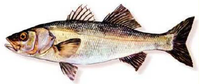
Sea bass is one of the most important cultured fish species in the Mediterranean (Scapigliati et al., 2003), and the IL-1b gene has been cloned from this species (Scapigliati et al., 2001). As for other nonmammalian IL-1b molecules (Zou et al., 1999a; Engelsma et al., 2001), analysis of the sea bass IL-1b sequence reveals the absence of a putative cut site for the interleukin 1 converting enzyme (ICE) that in mammals cleaves the precursor peptide to give the biologically active mature protein. The sequence alignment, together with the gene organization (Buonocore et al., 2003) and predicted 3-dimensional molecular structure (Scapigliati et al., 2004), sug- gests that a possible mature peptide would start at Ala86, giving a protein of 176 amino acids (with a predicted molecular weight of 19.7 kDa), larger than that in mammals. This putative mature peptide has been expressed in E. coli, purified, and tested as an immunoadjuvant (Buonocore et al., 2004). In this paper, we describe the bioactivity (Vega-Rubin de Celis et al., 2003) of the recombinant protein in a range of in vitro and in vivo immune assays.
Materials and Methods
Production and Purification of Recombinant Sea Bass IL-1b. Recombinant sea bass IL-1b was pro- duced and purified as described previously (Buonocore et al., 2004). Briefly, rIL-1b was expressed in E. coli after cloning into the pQE-30 vector (Invitrogen) and purified by affinity chromatography through a 6-his- tidine tag present at the N-terminus of the protein. The rIL-1b purity was determined by sodium dode- cylsulfate polyacrylamide gel electrophoresis (SDS- PAGE), and the endotoxin levels in the protein sample were determined with a Limulus Amebocyte Lysate Kit (LAL Test, Bio Whittaker) as previously docu- mented (Hong et al., 2001; Buonocore et al., 2004) to ensure no lipopolysaccharide (LPS) contamination.
Proliferation of D10.G4.1 Cells. The rIL-1b biological activity was first tested in vitro using the murine D10.G4.1 cells (Th2 clone) proliferation as- say (Horowitz et al., 1986). Initially the concentra- tion of concanavalin A (Con A, from Canavalia ensiformis) that just caused proliferation of the murine cells was determined.
Cells were plated in triplicate at 4 · 104 per well in 50 ll of RPMI medium (Gibco) plus 10% fetal bovine serum (FBS, Hyclone) and antibiotics. To
FRANCESCO BUONCORE ET AL.: BIOACTIVITY OF SEA BASS rIL-1b
such wells 50 ll of serially diluted sea bass (from 0.1 to 30 ng/ml) or human (from 0.1 to 30 pg/ml) rIL-1b was added in the presence of 0.3 lg/ml Con A (pre- viously determined concentration) as a costimula- tory factor. To the control wells 50 ll of medium only was added. After 48 hours at 37_C in a 5% CO2
atmosphere, the cells were pulsed with 10 ll per well of medium containing 0.5 lCi3 H-thymidine (Pharmacia Amersham). Subsequent to an overnight incubation the cells were harvested, and the incor- poration of radioactivity was determined with a scintillation counter. Two independent experiments were performed, and the results were presented as the mean ± SE of the combined data. Data were analyzed for statistical significance by one-way analysis of variance (ANOVA).
Proliferation of Sea Bass Thymocytes. Sea bass thymocytes were isolated from juvenile fish (20-30 g), grown, and maintained in a local fish farm (Nuova Azzurro, Civitavecchia, Italy), as described previously (Scapigliati et al., 2001). As for the D10.G4.1 cell test, a suboptimal concentration of Con A that was still capable of inducing an appre- ciable proliferation of thymocytes was first tested. Cells were plated in triplicate at 5 · 105 per well in 50 ll of RPMI medium plus 5% FBS and antibiotics. Then 50 ll of serially diluted sea bass (from 0.5 to 50 ng/ml) rIL-1b was added to the wells in the presence of the suboptimal concentration of Con A (0.1 ng/ml) as a costimulatory factor. After incuba- tion for 48 hours at 18_C, cell proliferation was measured by an ATP bioluminescence method using a CytoLux Kit (Wallac, PerkinElmer) accord- ing to the manufacturer s instructions. Lumines- cence emission was monitored as relative light units (RLU) over a 10-second period using a lumi- nometer (Victor II, Wallac, PerkinElmer). Two independent experiments were performed, and the results were presented as the mean ± SE of the combined data. Data were analyzed for statistical significance by one-way ANOVA followed by Tu- key s pairwise comparisons.
Phagocytosis of Sea Bass Head Kidney Cells. Head kidney leukocytes were purified from sea bass juveniles (300-500 g) as described previously (Scapigliati et al., 2001), and the phagocytosis assay was performed in a similar manner to that previ- ously reported (Crampe et al., 1997; Hong et al., 2001). Briefly, the cells were adjusted to 1.6 · 107 viable leukocytes per milliliter of L-15 (Gibco) medium. Glass-culture chamber slides (0.5 cm2, Becton Dickinson), each containing 250 ll of head kidney cells (4 · 106 cells), were incubated with
FRANCESCO BUONCORE ET AL.: BIOACTIVITY OF SEA BASS rIL-1b
various concentrations (from 0 to 100 ng/ml) of rIL- 1b for 4 hours at 18_C. Each experimental point was in triplicate. The slides were then washed twice with Hanks balanced salt solution (HBSS, Gibco) to re- move nonadherent cells, and 250 ll of a heat-killed (15 minutes in boiling water) FITC-labeled Saccharomyces cerevisiae yeast suspension was ad- ded at a concentration of 4.8 · 105 cells/ml in L-15. Phagocytosis was allowed to occur for 1 hour at 18_C in the chamber slide before excess yeast was washed off with HBSS and the slides air dried. Following methanol fixation the preparations were stained sequentially with May-Grunwald and Giemsa stains. Slides were viewed at · 40 magnification on a fluorescence microscope. Approximately 100 cells were counted in random fields of view for each experimental point, and the number of phagocytosed yeast cells and the number of yeast particles en- gulfed by each cell were recorded. Data were ex- pressed as the mean percentage ± SE of phagocytosis and phagocytic index and analyzed for statistical significance by one-way ANOVA followed by Tu- key s pairwise comparisons.
In Vivo Induction of Immune Gene Expres- sion. The in vivo biological activity of rIL-1b was studied through its effects on the induction of IL-1b and cyclooxygenase 2 (COX-2) homologue gene expression in head kidney leukocytes. All the solu- tions used were adjusted to sea bass osmolarity (360 mOsm/kg). Three different groups of 5 fish (50 g in weight) were used: one was injected intraperitone- ally with 100 ll of PBS; the second was injected intraperitoneally with 100 ll of PBS containing 5 lg/ ml LPS (E. coli 055:B5, Sigma); the third was injected intraperitoneally with 100 ll of PBS containing 500 ng of rIL-1b. After 24 hours, head kidney leukocytes were isolated and total RNA was purified from cells using Tripure (Roche) following the manufacturer s instructions. Reverse transeriptase polymerase chain reaction (RT-PCR) was performed using Ready-To- Go RT-PCR Beads (Amersham Pharmacia). For complementarsDNA synthesis, 1 lg of total RNA and 0.5 lg of random primers (pd(N)6) were used for
each reverse transcription reaction in a 50 ll total volume. To check the RNA quality and quantity, PCRs were performed initially using primers that amplified a 543-bp product from sea bass b-actin, a gene constitutively expressed. The primers were 5 ¢-
ATCGTGGGGCGCCCCAGGCACC (forward) and 5 ¢-
CTCCTTAATGTCACGCACGATTTC (reverse), and the cycling protocol was one cycle of 94_C for 5 minutes, 17 cycles of 94_C for 45 seconds, 55_C for 45 sec- onds, and 72_C for 45 seconds, followed by one cycle of 72_C for 10 minutes. To exclude DNA contami-
611
nation, a PCR was also performed using the RNA as template with the same protocol and primers that were designed to span an intron.
Subsequently, RT-PCR was performed using specific primers designed for the amplification of a 240-bp product of sea bass IL-1b. The primers were 5 ¢-CACAAGGATGGAGAGGAGCC (forward) and 5 ¢
-GAT-
GTTGAAGGCTCGGTGG (reverse), and the cycling pro- tocol was one cycle of 94_C for 5 minutes, 17 cycles of 94_C for 45 seconds, 60_C for 45 seconds, 72_C for 45 seconds, followed by one cycle at 72_C for 10 minuets. Finally, a RT-PCR was performed using specific primers designed for the amplification of a 180-bp product of a sea bass COX-2 gene (Buonocore et al., 2005). The primers were 5 ¢-GGTGGACCTCAGC- CACATT (forward) and 5 ¢-ACAGCGAAGCGG-TGAGCTT
(reverse), and the cycling protocol was one cycle of 94_C for 5 minuets, 17 cycles of 94_C for 45 seconds, 50_C for 45 seconds, and 72_C for 45 seconds, fol- lowed by one cycle of 72_C for 10 minuets. A low cycle number was used to ensure the linearity of the assay and to allow semiquantitative analysis of the IL-1b and COX-2 expression by comparison to the b- actin product, as described below.
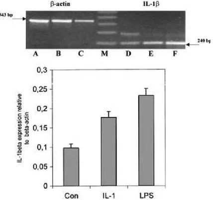
All PCR reactions were conducted using the MiniCycler model PTC-150-16 (MJ Research) and were overlaid with mineral oil. PCR products (25 ll) were visualized on 1% (w/v) agarose gel containing ethidium bromide (10 ng/ll) using a 123-bp ladder (Sigma) as size marker. PCR fragments were purified from agarose gels using a QIAquick Gel Extraction Kit (Qiagen) and directly sequenced on an ABI 377 automated sequencer (Applied Biosystems, U.K.). The messenger RNA IL-1b and COX-2 levels were expressed as a ratio relative to b-actin level after densitometric scanning of the agarose gels using GeneImage IR software from Li-COR, and data were expressed as the mean ± SE of the values for 5 fish used per group.
Results
Proliferation Assays. The in vitro biological activity of purified sea bass rIL-1b was first investigated for its effects on the D10.G4.1 murine cell lines. The results, (Figure 1) showed a clear enhancement of proliferation depending on the concentration of rIL- 1b used (P < 0.05 by one-way ANOVA). The max- imum amount of sea bass rIL-1b used (30 ng/ml) induced the highest proliferation of the murine cells. The positive control employed in this assay, human rIL-1b, gave the same proliferative effect at a con- centration of 30 pg/ml (data not shown).
The results obtained using sea bass rIL-1b in the thymocytes assay (Figure 2) showed a dose-depen-
612
dent increase of proliferation with increasing con- centrations of the recombinant cytokine from 0.5 to 5 ng/ml (P < 0.05 by one-way ANOVA). The highest quantity of rIL-1b employed (50 ng/ml) showed less stimulation, and the degree of proliferation was not significantly different from that with the control. Tukey s pairwise comparison test showed that cells treated with 0.5 and 5 ng/ml sea bass IL-1b had sig- nificantly higher values than those for the control, and that these two groups were significantly differ- ent from each other.
Phagocytosis Assay. The effect of rIL-1b on sea bass head kidney cell phagocytosis was next studied (Figure. 3). Macrophages were stimulated with rIL-1b diluted in L-15 medium at concentrations ranging from 1 to 100 ng/ml. Percentage of phagocytosis in cells treated with 1 ng/ml rIL-1b showed little vari- ation in comparison with the controls and was not statistically significant. In contrast, stimulation was clearly increased in samples treated with 10 and 30 ng/ml rIL-1b (P < 0.05 by one-way ANOVA). How- ever, treatment with 100 ng/ml rIL-1b showed no such effect (P < 0.05 by one-way ANOVA), and the values were not significantly higher than those in untreated and elution buffer controls. Similar results were obtained for the phagocytic index (Figure. 3). Values from cells treated with 1 ng/ml rIL-1b were
FRANCESCO BUONCORE ET AL.: BIOACTIVITY OF SEA BASS rIL-1b
Fig. 1. Proliferation of murine D10.G4.1 cells stimulated with varying concentrations of sea bass rIL-1b (0.1, 0.3, 1, 3, and 30 ng/ml) in the presence of a suboptimal dose of Con A (0.3 ng/ ml). Cells were stimulated without rIL-1b and Con A (Con) as a negative control and with Con A (ConA) alone as a positive control. Data are presented as means ± SE of triplicate samples, with each experiment performed twice. *P < 0.05.
not statistically different from those for the two controls. A clear stimulation was observed in cul- tures treated with 10 and 30 ng/ml rIL-1b (P < 0.001
Fig. 2. Proliferation of sea bass thymocyte cells stimulated with varying concentrations of sea bass rIL-1b (0.5, 5, and 50 ng/ml) in the presence of a suboptimal dose of Con A (0.1 ng/ml). Cells were stimulated without rIL-1b and Con A (Con) as a negative control and only with Con A (ConA) as a positive control. Data are presented as means ± SE of triplicate samples, with each experiment performed twice. *P < 0.05. Means with no letters in common are sig- nificantly different (thus ‘‘a’’ is significantly different from ‘‘b’’ and ‘‘c’’).
FRANCESCO BUONCORE ET AL.: BIOACTIVITY OF SEA BASS rIL-1b
by one-way ANOVA), whereas treatment with 100 ng/ml rIL-1b showed no such effect (P < 0.001 by one-way ANOVA). In both cases Tukey s pairwise comparison test showed that cells treated with 10 and 30 ng/ml sea bass IL-1b had significantly higher values than those in untreated and buffer controls, and that these two groups were significantly differ- ent from each other. Values from cells treated with 1 ng/ml and 100 ng/ml showed no significant effect for either percentage of phagocytosis or phagocytic in- dex.
In Vivo Immune Gene Expression Assay. The biological activity of rIL-1b was tested in vivo through its effects on the induction of IL-1b and COX-2 homologue gene expression in head kidney cells purified from sea bass (Figures. 4 and 5). LPS
613
Fig. 3. Yeast phagocytosis of sea bass head kidney leukocytes stimulated with varying
concentrations of sea bass rIL-1b (1, 10, 30 and 100 ng/ml). Cells were stimulated without rIL-1b (Con) and with the elution buffer (Buffer) used in rIL-1b production as negative controls. Data are presented as means ± SE of triplicate samples, with each experiment performed twice. *P < 0.05. Means with no letters in common are
significantly different (thus ‘‘a’’ is significantly different from ‘‘b’’ and ‘‘c’’).
was used as a positive control because it is well know that this molecule can upregulate IL-1b and COX-2 gene expression (Zou et al., 1999a; 1999b; Scapigliati et al., 2001). After 24 hours of induction, total RNA was extracted from all samples and its quality and quantity were checked by RT-PCR with b-actin primers. The absence of DNA contamination was determined by performing a PCR using the RNA sample as template for PCR with b-actin primers that crossed an intron. In no case was a b-actin product larger than 543 bp obtained.
Similar results were obtained for each fish of the 3 tested experimental groups. Therefore the agarose gel data for only one fish of each group are presented. RT-PCR analyses of the cDNA samples for b-actin generated a product of the expected size (excluding the presence of an intron) in each case (upper panels,
Fig. 4. IL-1b gene expression in sea bass head kidney leukocytes. Upper panel: 1% (w/v) agarose gel loaded with the RT-PCR products obtained using primers for b-actin (left side) and IL-1b (right side) are shown for a representative fish of the 5 examined. Sea bass were injected
intraperitoneally with PBS (Con) (lanes A and D), with rIL-1b (500 ng) (lanes B and E) or with LPS (5 lg/ml) (lanes C and F). Lower panel: IL-1b mRNA levels expressed as a ratio relative to b- actin levels in the same samples, after
densitometric scanning of the gels with the Gene Image IR software (Li-COR). The DNA molecular weight markers were loaded in lane M.
614
Fig. 5. COX-2 gene expression in sea bass head kidney leucocytes. Upper panel: 1% (w/v) agarose gels loaded with the RT-PCR products obtained using primers for b-actin and COX-2 are shown for a representative fish of the 5 examined. Sea bass were injected intraperitoneally with PBS (Con) (lanes A and D), with LPS (5 lg/ml) (lane B and E), or with rIL-1b (500 ng) (lane C and F). Lower panel: COX-2 mRNA levels expressed as a ratio relative to b- actin levels in the same samples, after densitometric scanning of the gels with the Gene Image IR software (LI- COR). The DNA molecular weight markers were loaded in lane M.
Figures 4 and 5). IL-1b expression was next examined using the specific sea bass primers. An amplified product of the expected size (240 bp) was seen in all samples (upper panel, Figure. 4), with lower levels of transcript seen in cells from fish injected with PBS relative to those from fish injected with LPS or rIL- 1b. A larger amplification product at approximately 346 bp was also clearly observed in samples from fish injected with PBS and, to a lesser extent, in the stimulated samples, and this was due to the presence of an incompletely spliced transcript containing an intron of 108 bp as reported previously (Scapigliati et al., 2001).
COX-2 expression was studied (Figure. 5) using the specific sea bass primers obtained from a COX-2 sequence (Buonocore et al., 2005). An amplified product of the expected size (180 bp) for the COX-2 gene was seen in samples from LPS-and IL-1b stim- ulated fish (Figure. 5), whereas in the control a very faint band was visible because this gene is usually not constitutively expressed, as already reported in rainbow trout (Zou et al., 1999b).
When IL-1b mRNA levels were expressed as a ratio relative to the b-actin products obtained after
FRANCESCO BUONCORE ET AL.: BIOACTIVITY OF SEA BASS rIL-1b
densitometric scanning of the agarose gels (lower panel, Figure 4), it was clear that under the condi- tions of these experiments IL-1b mRNA induction was higher with LPS stimulation (approx. 2.5 times more than the control), than with rIL-1b stimulation (approx. 2 times more than the control). Similar re- sults were obtained for COX-2 mRNA levels: stim- ulation with LPS gave higher induction (approx. 2 times more than the control) than stimulation with rIL-1b (approx. 1.8 times more than the control).
Whether these results would be true for other timings and doses of LPS and rIL-1b remains to be determined.
Discussion
IL-1b is a key modulator of immune responses in vertebrates, and the aim of the present work was to study the biological activity of a sea bass re- combinant IL-1b peptide expressed in E. coli. As al- ready stated, mammalian IL-1b is produced as a precursor molecule that must be cleaved by the en- zyme ICE to give the biologically active mature protein (Thornberry et al., 1992). While fish IL-1b precursors lack signal peptide sequences and there is no clear ICE cut site (Zou et al., 1999a; Engelsma et al., 2001; Scapigliati et al., 2001), it has been re- cently demonstrated that IL-1b is processed in trout macrophages in a way analogous to the situation in mammals (Hong et al., 2004).
In sea bass it has been possible to predict a putative mature peptide starting at Ala86, identified by IL-1b sequence alignment, resulting in a putative mature protein of 176 amino acids (Scapigliati et al., 2001). The sequence coding for this peptide was cloned in a commercial expression vector, and de- tails of the rIL-1b production in E. coli and purifi- cation have been described previously (Buonocore et al., 2004). In addition, the bacterial endotoxin con- tamination of the rIL-1b preparation was deter- mined, as it could be a contributor to the effects induced by the sea bass recombinant peptide. Lim- ulus amebocyte lysate (LAL) test data showed that the LPS concentration was low, typically 0.02 to 10 ng/ml over the concentration range used in this study, and this is less than the amount considered to be the minimum dose able to induce IL-1b expres- sion in trout head kidney leukocytes (Zou et al., 2000) and found in mammalian rIL-1b preparations (Blecha et al., 1995).
One of the most widely employed assays to de- tect IL-1b bioactivity is the in vitro IL-1-induced proliferation of the mouse T-helper cell clone D10.G4.1. These Th2-type cells, specifically require exogenous IL-1 for proliferation in the presence of
FRANCESCO BUONCORE ET AL.: BIOACTIVITY OF SEA BASS rIL-1b
Con A as a co-stimulator (Tartakovsky et al., 1986), which stimulates IL-4 production that is an essential growth factor for the cells (Kupper et al., 1987). With this assay, using human recombinant IL-1b as a po- sitive control, sea bass rIL-1b induced a dose- dependent proliferation response of D10.G4.1 cells in the range of 1 to 30 ng/ml. It is interesting that, as for rainbow trout rIL-1b (Hong et al., 2001), the amount of sea bass rIL-1b needed to induce levels of proliferation comparable to those induced by human rIL-1b was approximately 1000 times higher. This is likely a consequence of differences between mouse and sea bass IL-1b 3-dimensional structures. In a recent modeling study (Scapigliati et al., 2004), it was shown that sea bass IL-1b is predicted to interact with the mouse IL-1R with considerably less energy than mouse IL-1b and for this reason more sea bass IL-1b may be needed relative to human IL-1b, which has a 3-dimensional structure similar to mouse IL- 1b. Nevertheless, despite this drawback, the D10 assay appears a sensitive and reliable test for the bioactivity of fish rIL-1b.
We also investigated the ability of sea bass rIL-1b to induce the proliferation of sea bass thymocytes, in an assay that has been employed extensively in mammalian studies of IL-1 action (Dalmau et al., 1993). Sea bass rIL-1b induced the proliferation of thymocytes from juveniles in a dose-dependent manner, although the highest dose used (50 ng/ml) was significantly less stimulatory, potentially attributable to rIL-1b toxicity for cells at this con- centration or to receptor saturation.
Phagocytosis of foreign particles by leukocytes and its modulation by various immunoregulatory molecules is a well studied process in fish (Secombes and Fletcher 1992; Benanni et al., 1995; Solem et al., 1995). IL-1b has been shown to upregulate phagocy- tosis by rainbow trout leukocytes (Hong et al., 2001). To better study in vitro bioactivity of sea bass rIL-1b, we performed phagocytosis experiments with adherent head kidney leukocytes and fluorescently labeled yeast particles. The results from these experiments clearly showed that both the percentage of phagocytosis and the phagocytic index were af- fected by rIL-1b. As for the thymocyte proliferation test, the highest dose tested (100 ng/ml) was less stimulatory, and again one can speculate that this IL-1 concentration is toxic for cells or that receptor saturation has occurred.
Finally, we investigated the in vivo biological activity of rIL-1b on immune gene expression, through the induction of the expression of two proinflammatory molecules (i.e., IL-1b itself and COX-2) after intraperitoneal injection of rIL-1b into sea bass juveniles. Head kidney leukocyte gene
615
expression was examined because these cells are known to be responsive to IL-1b. LPS was used as a positive control for both IL-1b and COX-2 produc- tion by these cells. Results obtained from these experiments showed that IL-1b and COX-2 tran- scription was increased in head kidney leukocytes after rIL-1b injection, to a similar extent to that obtained by LPS induction. Other studies have shown similar increases of IL-1b and COX-2 levels induced by comparable quantities of rIL-1b, as seen in humans (Warner et al., 1987) and in trout (Hong et al., 2001,2003). A second IL-1b transcript was observed in the RT-PCR study, most obvious in the control (PBS-injected) fish. This transcript has been detected in other RT-PCR experiments and is attributable to the presence of an unspliced intron (Scapigliati et al., 2001). This incompletely spliced form may have a role in posttranslational regulation of the mature, fully spliced transcript (Jarrous and Kaempfer 1994).
In conclusion, in this study both in vitro and in vivo approaches have been used to examine the bioactivity of a recombinant fish cytokine. The re- sults obtained suggest that sea bass rIL-1b is likely to be an important cytokine in fish, as in mammals, with a wide range of effects. In addition, rIL-1b could be a useful molecule for the modulation of the im- mune system of farmed sea bass, although such po- tential applications will require much further work to establish.
References
1. Auron PE (1998) The interleukin 1 receptor: ligand interactions and signal transduction. Cytokine Growth Factor Rev 9, 221-237
2. Benanni N, Schmid-Alliana A, Lafaurie M (1995) Evaluation of phagocytic activity in a teleost fish, Dicentrarchus labrax. Fish Shellfish Immunol 5, 237-246
3. Bird S, Wang T, Zou J, Cunningham C, Secombes CJ (2002) The first cytokine sequence within cartilagi- nous fish: IL-1b in the small spotted catshark (Scyliorhinus canicula). J Immunol 168, 3329-3340 4. Blecha F, Reddy DN, Chitko-Mckown CG, Mcvey
DS, Chengappa MM, Goodband RD, Nelssen J.L (1995) Influence of recombinant bovine interleukin- 1b and interleukin2 in pigs vaccinated and challenged with Streptococcus suis. Vet Immunol Immunopa- thol 44, 329-346
5. Buonocore F, Frugnoli D, Falasca C, Secombes CJ, Scapigliati G (2003) Peculiar gene organisation and incomplete splicing of sea bass (Dicentrarchus labrax L.) interleukin 1b. Cytokine 21, 257-264
6. Buonocore F, Mazzini M, Forlenza M, Randelli E, Secombes CJ, Zou J, Scapigliati G (2004) Expression in E. coli and purification of sea bass (Dicentrarchus
616
labrax) interleukin-1b, a possible immuno-adjuvant in aquaculture. Mar Biotechnol 6, 53-59
7. Buonocore F, Randelli E, Casani D, Mazzini M, Cappuccio I, Secombes CJ, Scapigliati G (2005) cDNA cloning and expression analysis of a cyclooxygenase2 from sea bass (Dicentrarchus labrax L.) after vacci- nation. Aquaculture 245, 301-310
8. Crampe M, Farley SR, Langston A, Pulsford AL. (1997) Measurement of phagocytosis: a comparative evaluation of microscopic and microplate methods. In: Methodology in Fish Diseases Research. Aber- deen: Fisheries Research Services, pp. 81-89
9. Dalmau SR, Freitas CS, Savino W (1993) A modified assay for measuring thymocyte co-stimulatory activ- ity. Mem Inst Oswaldo Cruz 88, 419-425
10. Debets R, Timans JC, Homey B, Zurawski S, Sana TR, Lo S, Wagner J, Edwards G, Clifford T, Menon S, Bazan JF, Kastelein RA (2001) Two novel IL-1 family members, IL-1d¢ and IL-1 , function as an antagonist and agonist of NF-jB activation through the orphan IL-1 receptor-related protein 2. J Immunol 167, 1440- 1446
11. Dinarello CA (1996) Biologic basis for interleukin 1 in disease. Blood 87, 2095-2147
12. Dinarello CA (1997) Interleukin 1. Cytokine Growth Factor Rev 8, 253-265
13. Engelsma MY, Stet RJM, Schipper H, Verburg-van Kememade BML (2001) Regulation of interleukin-1 beta RNA expression in the common carp, Cyprinus carpio L. Dev Comp Immunol 25, 195-203
14. Fujiki K, Shin DH, Nakao M, Yano T (2000) Molec- ular cloning and expression analysis of carp (Cyprinus carpio) interleukin-1b high affinity immunoglobulin E Fc receptor (b subunit and serum amyloid A. Fish Shellfish Immunol 10, 229-242
15. Holland JW, Pottinger TG, Cunningham C, Secombes CJ (2000) Sequencing and expression anal- ysis of the type 1 interleukin-1 receptor gene. EMBL accession number AJ295296
16. Holland JW, Pottinger TG, Secombes CJ (2002) Re- combinant interleukin1 b activates the hypotha- lamic-pituitary-interrenal axis in rainbow trout, Oncorhynchus mykiss. J Endocrinol 175, 261-267 17. Hong S, Zou J, Crampe M, Peddie S, Scapigliati G,
Bols N, Cunningham C, Secombes CJ (2001) The production and bioactivity of rainbow trout (Oncorhynchus mykiss) recombinant IL-1b. Vet Immunol Immunopathol 81, 1-14
18. Hong S, Peddie S, Campos-Perez J, Zou J, Secombes CJ (2003) The effect of intraperitoneally administered recombinant IL-1b on immune parameters and resis- tance to Aeromonas salmonicida in the rainbow trout (Oncorhynchus mykiss). Dev Comp Immun 27, 801-812
19. Hong S, Zou J, Collet B, Bols NC, Secombes CJ (2004) Analysis and characterisation of IL-1b processing in rainbow trout, Oncorhynchus mykiss. Fish Shellfish Immunol 16, 453-459
20. Horowitz JB, Kaye J, Conrad PJ, Katz ME, Janeway Jr CA (1986) Autocrine growth inhibition of a cloned
FRANCESCO BUONCORE ET AL.: BIOACTIVITY OF SEA BASS rIL-1b
line of helper T cells. Proc Natl Acad Sci USA 83, 1886-1890
21. Jarrous N, Kaempfer R (1994) Induction of human interleukin 1 gene expression by retinoic acid and its regulation at processing of precursor transcripts. Journ Biol Chem 269, 23141-23149
22. Kullberg BJ, Van der Meer JWM (1995) Cytokines in the Treatment of Infectious Diseases: Options for the Modulation of the Host Defense. (Dordrecht, Neth- erlands: Kluwer Academic Publishers)
23. Kupper T, Flood P, Coleman D, Horowitz M (1987) Growth of an interleukin 2/interleukin 4-dependent T cell line induced by granulocyte-macrophage col- ony-stimulating factor (GM-CSF). J Immunol 138, 4288-4292
24. Mathew JA, Guo YX, Goh KP, Chan J, Verburg-Van Kemenade BML, Kwang J (2002) Characterisation of a monoclonal antibody to carp IL-1b and the develop- ment of a sensitive capture ELISA. Fish Shellfish Immunol 13, 85-95
25. Nicola NA (1994) Guidebook to Cytokines and Their Receptors. (Oxford, U.K: Sambrook and Tooze) 26. Peddie S, Zou J, Cunningham C, Secombes CJ (2001)
Rainbow trout (Oncorhynchus mykiss) recombinant IL-1b and derived peptide induce migration of head- kidney leucocytes in vitro. Fish Shellfish Immunol 11,697-709
27. Pelegrin P, Garcia-Castillo J, Mulero V, Meseguer J (2001) Interleukin1b isolated from a marine fish re- veals up-regulated expression in macrophages fol- lowing activation with lipopolysaccharide and lymphokines. Cytokine 16, 67-72
28. Scapigliati G, Buonocore F, Bird S, Zou J, Pelegrin P, Falasco C, Frugnoli D, Secombes C.J (2001) Phylog- eny of cytokines: molecular cloning and expression analysis of sea bass Dicentrarchus labrax interleu- kin1 b. Fish Shellfish Immunol 11, 711-726 29. Scapigliati G, Meloni S, Buonocore F, Bossu` P,
Prugnoli D, Secombes CJ (2003) Immunopurification of B lymphocytes from sea bass Dicentrarchus labrax (L.) Mar Biotechnol 5, 214-221
30. Scapigliati G, Costantini S, Colonna G, Facchiano A, Buonocore F, Bossu` P, Cunningham C, Holland JW, Secombes CJ (2004) Modelling of fish interleukin1 and its receptor. Dev Comp Immun 28, 429-441 31. Sangrador-Vegas A, Martin SAM, O Dea PG, Smith TJ
(2000) Cloning and characterisation of the rainbow trout (Oncorhynchus mikyss) type II interleukin1 receptor cDNA. Eur J Biochem 267, 7031-7037 32. Secombes CJ, Fletcher TC (1992) The role of phago-
cytes in the protective mechanism of fish. Ann Rev Fish Des 2, 53-71
33. Secombes CJ, Bird S, Cunningham C, Zou J (1999) Interleukin1 in fish. Fish Shellfish Immunol 9, 335- 343
34. Sims JE, Dower SK (1994) Interleukin1 receptors. Eur Cytokine Netw 5, 517-531
35. Smith DE, Renshaw BR, Ketchem RR, Kubin M, Garka KE, Sims JE (2000) Four new members expand the IL-1 superfamily. J Biol Chem 275, 1169-1175
FRANCESCO BUONCORE ET AL.: BIOACTIVITY OF SEA BASS rIL-1b
36. Smith KA, Lachman LB, Oppenheim JJ, Favata MM (1980) The functional relationship of the interleu- kins. J Exp Med 151, 1551-1556
37. Solem ST, Jo¨rgensen JB, Robertsen B (1995) Stimula- tion of respiratory burst and phagocytic activity in Atlantic salmon (Salmo salar L.) macrophages by lipopolysaccharide. Fish Shellfish Immunol 5, 475- 491
38. Subramaniam S, Stansberg C, Olsen L, Zou J, Secombes CJ, Cunningham C (2002) Cloning of a Salmo salar interleukin-1 receptor-like cDNA. Dev Comp Immunol 26, 415-431
39. Tartakovsky B, Kovacs EJ, Takacs L, Durum SK (1986) T cell clone producing an IL1-like activity after stimulation by antigen-presenting B cells. J Immunol 137,160-166
40. Thornberry NA, Bull HG, Calaycay JR, Chapman KT, Howard AD, Kostura MJ, Miller DK, Molineaux SM, Weidner JR, Aunins J (1992) A novel heterodimeric cysteine protease is required for interleukin1b pro- cessing in monocytes. Nature 356, 768-774
41. Vega-Rubin Celis de S, Gomez P, Calduch-Giner JA, Medale F, Perez-Sanchez J (2003) Expression and
617
characterization of European sea bass (Dicentrarchus labrax) somatolactin: assessment of in vivo meta-
bolic effects. Mar Biotechnol 5, 92-101
42. Warner SJC, Auger KR, Libby P (1987) Interleukin 1 induces interleukin 1, II: recombinant human inter- leukin 1 induces interleukin 1 production by adult human vascular endothelial cells. J Immun 139, 1911-1917
43. Yin Z, Kwang J (2000) Carp interleukin 1b in the role of an immuno-adjuvant. Fish Shellfish Immunol 10, 375-378
44. Zou J, Grabowski PS, Cunningham C, Secombes CJ
(1999) Molecular cloning of interleukin 1b from rainbow trout Oncorhynchus mykiss reveals no evi- dence of an ICE cut site. Cytokine 11, 552-560 45. Zou J, Neumann NF, Holland JW, Belosevic M,
Cunningham C, Secombes C.J, Rowley F (1999a) Fish macrophages express a cyclo-oxygenase-2 homologue after activation. Biochem J 340, 153-159
46. Zou J, Holland J, Pleguezuelos O, Cunningham C, Secombes CJ (2000) Factors influencing the expres- sion of interleukin1b in rainbow trout. Dev Comp Immunol 24, 575-582
Fish & Shellfish Immunology 20 (2006) 739e749
www.elsevie r.com/locate/ fsi
Cellular activities during a mixed leucocyte
reaction
in
the
teleost sea bass
Dicentrarchus labrax
Sabrina Meloni, Gianpaolo Zarletti, Stefania Benedetti,
Elisa Randelli,
Francesco Buonocore, Giuseppe
Scapigliati
*
Dipartimento di Scienze Ambientali, Universita` della Tuscia, Largo dell’Universita`, I-01100 Viterbo, Italy
Received 19 July 2005; revised 20 September 2005; accepted 4 October 2005 Available online 15 November 2005
Abstract
In this investigation a number of ‘‘in vitro’’ activities of sea bass peripheral blood leucocytes (PBL) against allogeneic PBL inactivated by irradiation were studied. Stimulator PBL were cultured with
inactivated allogeneic PBL, and direct counting of lym-
phocytes was done after 2 weeks by immunofluorescence and flow cytometry using mAbs DLT15 and DLIg3 specific for T-cells and B-cells, respectively. In a one-way mixed leucocyte reaction (MLR), results showed an increase of T lymphocytes, whereas B lymphocytes had values similar to those in control PBL. The increase of T-cells in MLR cultures was also confirmed using RT-PCR by analyzing the expression of the T-cell receptor (b-subunit) mRNA. The addition of 5 mg/ml of cyclosporin A (CsA) to the MLR caused a significant decrease in T-cell proliferation. Leucocytes from MLR cultures displayed an enhanced cytotoxic activity against xenogeneic target cells with respect to control PBL, raising the possibility of the presence of cytotoxic-like T lymphocytes. Cellular activation of PBL was confirmed in 2 weeks MLR by measuring antibody-induced intracellular Caþþ mobilization with Fura-2 AM. This work represents the first direct quantitative determination of an ‘‘in vitro’’ T-cell activity in a teleost species.
2005 Elsevier Ltd. All rights reserved.
Keywords: Sea bass Dicentrarchus labrax; T-cell; Leucocyte; Teleost; Allogeneic recognition; Cytotoxicity; In vitro
1. Introduction
In recent years, many fundamental molecules produced by leucocytes of teleosts have been cloned, which include immunoglobulins (Ig) [1e3], class I and class II major histocompatibility complex (MHC) antigens [4,5], T-cell receptors (TcR) [6e8], CD8 and CD4 coreceptors [9,10], and a growing number of cytokines [11]. Despite this impressive progress, the biology of teleost lymphocytes is poorly known, and this is mainly due to the lack of good specific cell surface markers, especially for T-cells [12].
The response to non-self recognition MHC molecules by T lymphocytes is a common
feature in all jawed verte-
brates [13], and is characterized by an intense primary immune reaction, due to a very high frequency of alloreactive precursor T-cells. This behaviour may be reproduced ‘‘in vitro’’ using the mixed leucocyte reaction (MLR), and ‘‘in
* Corresponding author. Tel.: þ39 0761 357137; fax: þ39 0761 357029.
E-mail address: [email protected] (G. Scapigliati).
1050-4648/$ - see front matter 2005 Elsevier Ltd. All rights reserved. doi:10.1016/j.fsi.2005.10.001
740 S. Meloni et al. / Fish & Shellfish Immunology 20 (2006) 739e749
vivo’’ leads to allograft rejection and to graft versus host reaction (GVRH). Both these ‘‘in vivo’’ activities have been shown to take place in teleost species such as damselfish [14], sea bass [15], and carp [16]. The ‘‘in vitro’’ activities of teleost T-cells have been known for many years, when peripheral blood leucocytes (PBL) from rainbow trout were found to exhibit an MLR against an allogeneic target [17]. A two-ways and one-way MLR were shown in carp
[18], and this work showed that a weak leucocyte recognition response could be strongly increased by mutual ‘‘in vivo’’ priming of the reacting donor fish. Allorecognition mechanisms are known in teleosts to generate natural killer (NK) cells bearing the receptor for the Fc of immunoglobulins (FcR) [19]. A good indirect indication of the involve- ment of T lymphocytes in the MLR came from work in catfish [20], which showed their putative proliferation after depletion of B-cells and macrophages from PBL. It has recently been suggested that the activation of teleost T-cells can very likely be influenced by cytokines [21], since the ‘‘in vitro’’ proliferation of catfish PBL stimulated by Con A was inhibited by cyclosporin A (CsA), a substance known in mammals to inhibit T-cell activities through the depres- sion of T-cell growth factor production [22,23]. However, despite the clear evidence of the involvement of T lympho- cytes described in these previous works, their participation in fish MLR was only supposed, since no specific markers were available for direct analysis. We previously investigated the involvement of T-cells and B-cells during ‘‘in vivo’’ allograft rejection in sea bass by quantitative immunocytochemistry [15], and provided data on the participation of T lymphocytes during non-self recognition processes in fish.
In this study we show for the first time in a teleost species the direct involvement of T-cells during ‘‘in vitro’’ allorecognition. We also investigated the effects induced by the presence of CsA, the antibody-induced cellular activation of leucocytes, and the possible presence of cytotoxic T lymphocyte-like cells (CTL-like).
2. Materials and methods
2.1. Fish and leucocyte cultures
Sea bass were bred and reared in seawater at a local fish farm (Nuova Azzurro, Civitavecchia, Italy), and geneti- cally outbred, 2-year-old fish of ca. 300 g in weight were used in the experiments. Fish were vaccinated by immersion against vibriosis 1 year before the experiments and did not display any sign of pathology at sampling. All buffers and solutions used in handling fish cells were brought to 355 mOsm kg_1 with 2 M NaCl. Cell viability of leucocytes was
determined by counting in a haemocytometer with trypan blue. Fish were lethally anaesthetised with 1 g l_1 of ben-
zocaine (Sigma), and 5 ml of blood per fish was drawn from the caudal vein using HBSSeheparin. Whole blood from individual fish was washed twice in HBSSeheparin, resuspended in 8 ml of the same solution and loaded over 1.04 and 1.07 g cm_3 Percoll gradients as previously described [7]. After centrifugation (30 min at 840 _ g) at 4 _C, cells at the interface between the two densities were collected, washed twice with HBSS (10 min at 680 _ g) at 4 _C, re- suspended in flasks at 2 _ 106 cells/ml in 5 ml of L15 medium (Gibco) containing 5% heat-inactivated FCS (Gibco) and antibiotics, and incubated at 18 _C. PBL from one stimulator fish (1 _ 108 cells) in 5 ml of L15 in a 25-cm2 flask were treated at 20 _C with 22 Gy using an X-ray machine (Gilardoni, Italy) and cultured with PBL from other indi- vidual responders. Typically, 1 _ 107 irradiated cells were added to 5 _ 107 PBL in a T-25 flask, and five samples were run in each experiment. Controls were done by omitting stimulatory cells, and where necessary 5 mg/ml of cyclosporin A (Sigma) was added to cultures.
Cell proliferation was determined by assaying intracellular ATP levels of leucocyte cultures using a luminometric assay (ATP-lite, Perkin Elmer) [24]. Briefly, 100 ml of culture medium containing 5 _ 105 PBL was transferred into each well of a 96-well luminometric plate (Isoplate TC Perkin Elmer), cells were lysed with 50 ml of the solution contained in the kit following the manufacturer’s instructions and ATP read with a luminometer (Victor II, Wallac) by the light emitted when adding the kit reagent.
2.2. Immunofluorescence and flow cytometry
Indirect immunofluorescence (IIF) was carried out as previously described [7] at 4 _C in solutions containing 0.1% sodium azide. Briefly, cells were washed by centrifugation with phosphate buffered saline (PBS) and incubated with 250 ml of DLT15 (specific for sea bass thymocytes and T-cells, IgG3 subclass) or DLIg3 (specific for Ig and Ig-bearing cells, IgG1 subclass) mAb as undiluted culture supernatants for 45 min. These mAbs have been previously described and characterized (for review see [12]). Control staining was performed by incubating cells with myeloma culture
S. Meloni et al. / Fish & Shellfish Immunology 20 (2006) 739e749 741
supernatant or with an irrelevant mAb (anti-human CD4, BD-Pharmingen cat no. 555344). After washing with PBS, a 1:300 dilution of fluorescein-labelled goat anti-mouse Ig serum (Sigma) was added for 30 min. The cells were then washed and monitored for their morphology and fluorescence with a FACScalibur (Becton-Dikinson, USA). Analysis was done using the Cellquest program on 10,000 events acquired without gating.
To test for the presence of NK cells bearing receptors for Ig, leucocyte cultures were incubated for 45 min with 20 mg/ml of sea bass Ig purified by affinity chromatography from serum as previously described [25], and then processed for IIF using mAb DLIg3 as above.
2.3. Caþþ determinations
PBL cultures in flasks were washed twice in serum-free L15 by centrifugation (480 _ g, 10 min), cells resuspended in serum-free L15 medium and incubated for 45 min at 18 _C with the cell permeant Fura-2 AM (Sigma) at a final concentration of 4 mM. Cells were then washed by centrifugation (480 _ g, 10 min) with DMEM without FCS and phenol red and resuspended at 107 ml_1 in the same medium (control) or in undiluted mAb culture supernatant and dialyzed
overnight against PBS to remove phenol red. Cells were immediately plated at 1 _ 106 cells/well in triplicates in 96-well plates, and emitted fluorescence from each well automatically read for 5 min at 1-min intervals with a Victor II fluorimeter (WallacePerkin Elmer, USA).
2.4. RT-PCR
Total RNAwas isolated from 1 _ 107 viable cells of control and MLR cultures using Tripure
(Roche) following the
manufacturer’s instructions and suspended in diethylpyrocarbonate (DEPC)-treated water. For
cDNA synthesis, 1 mg
of total RNA and 0.5 mg of random primers (pd(N)6) were used for each reverse transcription
reaction in a 50-ml total
volume. To check the RNA quality and quantity, a PCR was performed using primers for the
amplification of a 543-bp
product from sea bass b-actin. To exclude DNA contamination, PCR was performed using the
RNA as template with
the same protocol. Subsequently, RT-PCR was performed using primers designed for the
amplification of a 503-bp
product from the sea bass TcRb constant region [7], and for a 224-bp product of sea bass Ig light
chain constant region
[26]. TcRb primers were as follows: 5#-AGATTACCGGACCATCAGTGAAAG-3# (forward) and
5#-TCAGTAGTTC
TGCTTTCCCTTTGA-3# (reverse). The cycling protocol was one cycle of 94 _C for 5 min, and
16 or 35 cycles of
94 _C for 45 s, 60 _C for 45 s and 72 _C for 45 s, followed by one cycle of 72 _C for 10 min. Ig
primers were 5#-
GAGCTGCAGAAGGACAGTG-3# (forward) and 5#-TCAGACTGGCCTCACAGCT-3#
(reverse). The cycling pro-
tocol was one cycle of 94 _C for 5 min, 16 or 35 cycles of 94 _C for 45 s, 50 _C for 45 s and 72 _C
for 45 s, followed
by one cycle of 72 _C for 10 min.
All PCR reactions were conducted using a MiniCycler , model PTC-150-16 (MJ Research). PCR products (15 ml) were visualised on a 1% (w/v) agarose gel containing ethidium bromide (10 ng ml_1) using 123-bp ladder (Sigma) or 1-kb ladder (Gibco) as a size marker. To confirm PCR sequences, fragments were purified from agarose gels using a QIAquick Gel Extraction Kit (Qiagen) and were directly sequenced on an ABI 377 automated sequencer.
2.5. Cytotoxicity assay
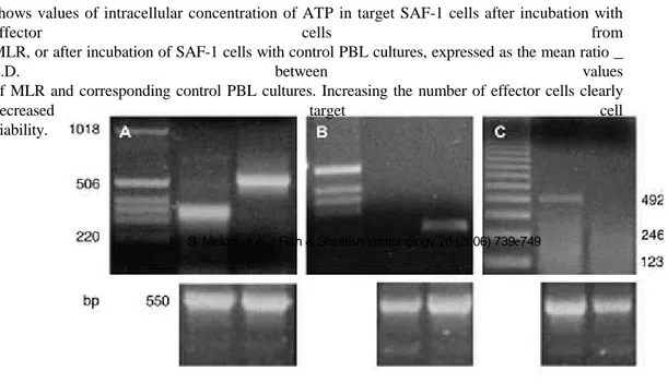
PBL cultures (control and MLR) were used as effector cells (E), and after 2 weeks of incubation cells were washed by centrifugation (680 _ g) with L15 medium, resuspended at the desired concentration in 100 ml of complete medium and added to microwells of a luminometric plate containing target cells. Target cells (T) were the fibroblast adherent cell line SAF-1 obtained from sea bream [27] plated 1 day before at 15,000 cells/well in complete L15 medium. Experimental wells (E þ T) were filled in triplicates with varying numbers of effector PBL to achieve the different effector to target cell (E/T) ratios of 20:1, 10:1, 5:1, and 1:1. Control wells were with T alone, and E alone at the same concentrations. Cultures were incubated for 72 h at 18 _C, then the effector cells from each well were removed, the wells washed twice with 100 ml of PBS and 100 ml of DMEM without phenol red (Gibco) were added. The killing activity was determined as previously described [24] by measuring intracellular ATP concentration in the remaining adherent SAF-1 cells using a luminometric assay and following the manufacturer’s instructions. The percentage of specific cytotoxicity was calculated using the formula: % target cell viability ¼ (E þ T) _ E/T _ 100.
742 S. Meloni et al. / Fish & Shellfish Immunology 20 (2006) 739e749
2.6. Statistical analysis
Data are represented as mean values _ standard deviation (S.D.), or mean values _
standard error of the mean (SEM). Statistical analysis of all experiments was performed by comparing groups using one-way ANOVA and a significant difference was assumed for p
values less than 0.05, unless indicated.
3. Results
3.1. Lymphocyte counting in the MLR
Irradiation of PBL with X-rays (22 Gy) completely blocked their proliferation since in two-way MLR with irradiated PBL no signs of cell proliferation were detected after 3 weeks (not shown).
The content of T-cells and B-cells during one-way MLR, analyzed by immunofluorescence and flow cytometry with mAb DLT15 and DLIg3, is shown in Fig. 1. Reported mean values are
the percent of positive cells after subtract-
ing the background staining. The mean content of B-cells (Fig. 1) decreased at 2 weeks in control PBL cultures from
21.1 _ 3.8% to 13.3 _ 7.2%. In the MLR, each sample was analyzed with respect to its own control, and the results showed that T-cells increased from 3.1 _ 0.8% in controls to 8.8 _
4.7%. In the MLR, B-cell percentages were
21.1 _ 3.8% in controls and became 6.71 _ 3.2% after 2 weeks. The number of total viable cells decreased from 1 _ 107 to 4 _ 1.6 _ 106 in control unstimulated PBL, whereas total viable cells decreased from 1.2 _ 107 (including irradiated cells that were present and counted) to 6 _ 0.8 _ 106 in MLR.
Statistical analysis by ANOVA showed that the decrease of B-cells and the increase of T-cells in the MLR were significant with respect to initial values.
Fig. 1. Lymphocytes in controls and MLR. IIF monitored by flow cytometry was performed in control PBL (a, b) and after 2 weeks in MLR cultures (c, d) with mAbs DLIg3 (B-cells, black bars) and DLT15 (T-cells, grey bars). Values are the mean percent _ S.D. of positive cells in cell cultures subtracted of background staining. Statistical analysis was performed by ANOVA, with significant differences taken at p < 0.05. Asterisks (* for B-cells, ** for T-cells) indicate significant differences between samples at 2 weeks with respect to 0 weeks.
S. Meloni et al. / Fish & Shellfish Immunology 20 (2006) 739e749 743 3.2. Effects of CsA
The effects of 5 mg/ml CsA on the MLR are shown in Fig. 2A. In the MLR, the percentage of T-cells increased from 3.8 _ 3.2% to 8.3 _ 3.8%, whereas the presence of CsA depressed the number of T-cells to a value of 3.8 _ 0.9%. Statistical analysis showed a significant difference between MLR and control data (indicated by an asterisk), but no difference between CsA and controls.
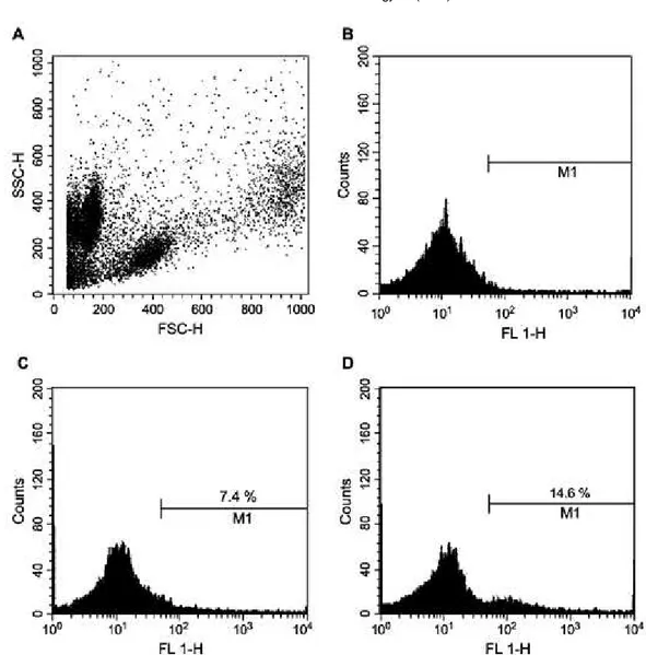
Fig. 2B shows the cellular activation of leucocytes tested by stimulating PBL cell cultures with mAbs DLT15 and DLIg3, and by measuring the concentration of intracellular Caþþ using Fura-2 AM. The Caþþ concentration is reported as the relative mean values of fluorescence units (RFU), which is the fluorimeter readout measured in all experiments after 5 min of incubation with mAb, with the background value subtracted. Using DLT15, control leucocytes of blood gave a mean RFU value of 1.250 _ 348 (SEM), cells from a 2-week MLR had a value of 2.500 _ 442 (SEM), and cells from MLR incubated for 2 weeks with CsA had a value of 1.500 _ 109 (SEM). Using DLIg3, control PBL gave a mean RFU value of 770 _ 260 (SEM), cells from a 2-week MLR had a value of 380 _ 180 (SEM), and cells from MLR with CsA added had a value of 900 _ 290 (SEM). Comparison of data between MLR and MLR þ CsA by statistical analysis (ANOVA) gave a p value of 0.001 for DLT15, and a p value of 0.009 for DLIg3. As a further control, the addition of CsA to cell cultures together with mAbs did not affect results (not shown).
The flow cytometric pattern of a selected MLR experiment is shown in Fig. 3. In Fig. 3A the cytometric morphology of PBL is shown, where three main populations can be recognized. No gating was used when analyzing these popu- lations for fluorescence intensity, and the background staining (3.2%) obtained when incubating PBL with an irrele- vant mAb is shown in Fig. 3B. DLT15 staining of control PBL (7.4%) is shown in Fig. 3C with a broad distribution of positive cells having a scarcely identifiable single peak. The DLT15 staining of an MLR experiment (14.6%) is shown in
Fig. 3D and in this case a clear peak of DLT15-positive cells at a higher fluorescence value can be appreciated.
3.3. RT-PCR
Fig. 4 shows the RT-PCR pattern of PBL cultures analyzed for the expression of sea bass Ig light chain gene (for B lymphocytes) and for sea bass TcRb (for T lymphocytes). In control unstimulated PBL there is an evident expression of
the Ig (224 bp) and the TcRb (503 bp) genes when performing 35 cycles of the PCR reaction (Fig. 4a). When the
number of PCR cycles was reduced to 16, only the expression for Ig could be detected in control PBL (Fig. 4b),
Fig. 2. Effects of cyclosporin A. IIF with mAb DLT15 monitored by flow cytometry was performed in control PBL, in a 2-week MLR, and in a 2week MLR added with 5 mg/ml of CsA (A). Values are the mean percent _ S.D. of positive cells subtracted of background values. (B) shows mean _ S.D. content of intracellular concentration of Caþþ measured with Fura-2 AM at 5 min. Cells from indicated samples were stimulated with mAb DLT15 (grey bars) and DLIg3 (black bars) as undiluted culture supernatants, and values are expressed as relative fluorescence units (RFU) subtracted of background staining. Statistical analysis was performed by ANOVA, with significant differences at p < 0.05. Asterisks indicate significant difference between MLR and control PBL values (*for B-cells, **for T-cells).