626
C L I N I C A L R E V I E W
European Advisory Board on Cat Diseases www.abcdcatsvets.org
www.abcd-vets.org
Corresponding author: Maria Grazia Pennisi Email: [email protected]
LUNGWORM DISEASE IN CATS
ABCD guidelines on prevention
and management
Maria Grazia Pennisi, Katrin Hartmann, Diane D Addie, Corine Boucraut-Baralon, Herman Egberink, Tadeusz Frymus, Tim Gruffydd-Jones, Marian C Horzinek, Margaret J Hosie, Albert Lloret, Hans Lutz, Fulvio Marsilio, Alan D Radford, Etienne Thiry, Uwe Truyen and Karin Möstl
Overview: Cardiopulmonary nematodes are emerging parasites of cats in Europe. A number of helminth parasites may be involved. The most prevalent lungworm in domestic cats is
Aelurostrongylus abstrusus. Oslerus rostratus and Troglostrongylus species are found mainly in wild
cats. The trichurid Capillaria aerophila has a low host specificity and is not uncommon in cats. Additionally the lung flukes Paragonimus species are reported in many species outside of Europe, including cats.
Clinical signs: Lungworm infections may be asymptomatic, or cause mild to severe respiratory signs, dependent on the worm species and burden; mixed infections are observed. Kittens can be vertically infected and may develop a more severe disease. Affected cats show a productive cough, mucopurulent nasal discharge, tachypnoea, dyspnoea and, in severe cases, respiratory failure and death.
Management: Early diagnosis and treatment greatly improves the prognosis. First-stage larvae can be easily detected in fresh faecal samples; the Baermann migration method is the enrichment technique of choice, but takes 24 h. Lungworm larvae can be found in tracheal swabs and bronchoalveolar lavage fluid, but with less sensitivity than in faeces. Molecular methods have been developed that exhibit high specificity and sensitivity, and allow diagnosis in the prepatent phase. Treatment options include fenbendazole paste, milbemycin oxime/praziquantel and various spot-on formulations. Severe cases should receive prompt medical care in an intensive care unit.
Prevention: Avoiding predation is at present the only preventive measure for pulmonary worms with indirect life cycles.
Zoonotic risk: C aerophila has zoonotic potential,
causing severe pulmonary disease in humans. Some Paragonimus species are also of zoonotic concern.
Introduction
Cardiopulmonary nematodes are emerging parasites of cats and dogs in Europe and have received growing attention from researchers in recent years.1–4Significant progress has been made, mainly in the diag-nosis and treatment of infection.
Disease agents
Infection of the lower respiratory tract can be caused by a number of parasitic nematodes. Certain metastrongyloid worms are commonly defined as lungworms because the adult stage is located in the lungs of their hosts, but actually some trichurids and flukes also live in the respiratory system.1–3,5 Aelurostrongylus abstrusus (Strongylida, Angiostrongylidae) is the most well known feline lungworm and is regarded as the most prevalent in domestic cats. It is small (5–10 mm) and very narrow (less than 100 μm) and capable of colonising the res-piratory bronchioles and alveolar ducts of domestic cats and other felids worldwide.2,6Other respiratory mollusc-borne metastrongylids are commonly reported at necropsy in wild felids but are considered very rare in domestic cats. Oslerus rostratus (Strongylida, Filaroididae) exceeds 30–40 mm in length and infects the bronchial submucosa mainly in wild cats such as bobcats or in feral cats.5,7 –10 Troglostrongylus species (Strongylida, Crenosomatidae) is reported in a wide variety of wild cats and occasionally in domestic cats;4,9,11–14these worms vary in length, according to the species, from about 10–25 mm and are up to 0.5 mm in width. They are located in the trachea and bronchi or even the bronchioles for the smallest species (T brevior).12,15
The trichurid Capillaria aerophila (syn Eucoleus aerophilus) has a low host specificity and is not uncommon in cats and dogs as well as wild carnivores.16It is also a zoonotic parasite, causing a potentially severe pulmonary disease in humans.17C aerophila is found in the submucosa of the trachea, bronchi and bronchioles.2,16
Mixed infections by respiratory nematodes are sometimes report-ed7,9,14,18–20and both Troglostrongylus species and O rostratus may be more prevalent than presumed in domestic cats since there is a risk
that these infections are being misdiagnosed as A abstrusus because of morphometric simi-larities of their first-stage larvae (L1) in faeces.3,4
Paragonimus species are lung flukes report-ed in many animals, including cats and humans, and some species are of zoonotic concern. Many species are found in cats, including P kellicotti, and between one and 10 adults measuring 8–18 mm x 4–8 mm live in subpleural cysts or bullae.1
Life cycle and transmission
A abstrusus, O rostratus and Troglostrongylus species all have an indirect life cycle involving terrestrial molluscs. Eggs of A abstrusus laid by female worms hatch in the respiratory tract and L1 larvae are coughed up, swallowed and eliminated in the environment with the faeces. They can actively enter slugs or snails where they moult into the infectious L3 stage.21–24 The biological cycle in the intermediate host is influenced by environmental temperature: a higher rate of larval development is observed at warmer temperatures.23 The L3 larvae are also found in a wide range of paratenic hosts (rat, mouse, lizard, frog, bird) commonly predated by cats.1,5,22The ingestion of L3 lar-vae by the cat is the best recognised means of transmission of lungworms, but vertical trans-mission via the placenta or milk cannot be excluded, as adult egg-laying worms have been found in kittens as young as 8 weeks of age.14 Experimentally, it has been demonstrat-ed that egg production starts 4–6 weeks after infection and may last for months, although it can be irregular.6,25–28
Vertical transmission of T brevior was recent-ly observed in a queen and patent infection was detected in 1-month-old kittens.13,14,29 T brevior and A abstrusus larvae may develop simultaneously in the same mollusc host (Helix aspersa) and overwinter for at least 120 days.24 Very recently environmental contamination has been suggested as an alternative means of transmission for both A abstrusus and T brevior L1 on the basis of an experimental study;30live larvae were found in the pedal mucus excreted by H aspersa and in the water where the snails died.
C aerophila has a direct life cycle and eggs laid by female worms in the respiratory tract are swallowed and reach the environment in the faeces. After 30–45 days, embryonated eggs become infective when ingested by cats. Earthworms are facultative paratenic hosts.16 When cats ingest infective eggs or earthworms carrying larvae, the larvae migrate to the lung and develop into the adult stage in 3–6 weeks.31
The life cycle of Paragonimus species is
asso-ciated with freshwater environments and is complex as it involves two intermediate hosts. Motile miracidia are released from eggs when swallowed and then passed in faeces from infected cats and penetrate aquatic snails; cercarial stages developed in snails will move from them, actively entering the second inter-mediate host (crab or crayfish). Cats are infect-ed after eating the second interminfect-ediate host where metacercariae finally develop. Young flukes develop from metacercariae in the cat intestine, and cross the intestinal wall and the diaphragm to the pleural cavity where they penetrate the lung parenchyma and become reproducing adults in about 6 weeks.1
Epidemiology
Feline lungworm infection is receiving increasing attention.2,6 A abstrusus is a well recognised agent of lower respiratory tract disease in cats.1,2Epidemiological studies and case reports have confirmed the presence of the parasite in the Americas, Europe, Asia and Australia.1,14,32–40 Prevalence rates vary and endemicity is linked to climatic and ecological factors that may influence: (a) the vitality and developmental capacity of L1; (b) the presence of suitable intermediate hosts in the environ-ment; and (c) the number of days needed for development of the infective stage (L3). The diagnostic method used in epidemiological studies and the characteristics of the popula-tion investigated heavily influence the results.2,37,41,42Feral and free-roaming cats are at higher risk because of their predator acti -vity, as are cats with respiratory signs and young cats.43,44 In Tirana (Albania), post-mortem examination of the lungs of 18 feral cats revealed that nine (50%) were positive for A abstrusus.45 Use of a lowsensitivity diag -nostic method, such as the standard faecal flotation technique, showed a prevalence rate of 1–25% in a general cat population (see Table 1).14,46–49
O rostratus is considered an uncommon parasite in domestic cats, but the prevalence in feral cats was found to be 24% in Majorca (Spain). It was also reported in a cat in north-ern Spain.7,8 Very recently, the incidental occurrence of a few adult O rostratus worms was reported in Sicily (Italy) at the necropsy of an adult cat that had died following a road traffic accident.10
C aerophila has a sporadic occurrence in cats, dogs and humans in Europe. In central Italy, a prevalence of 3–14% was found in the feline population.2,16,36
Single cases of Troglostrongylus species infec-tion were recently reported in cats from Ibiza (Spain), central and southern Italy and Crete (Greece).9,12,13,15,19,20,29,50The first
epidemiologi-Difficulties with
morphometric
differentiation
of L1 larvae
have meant that
many cases
of (co)infection
by other
metastrongylids
may have been
erroneously
attributed
to the
better-known
A abstrusus
lungworm.
European Advisory Board on Cat DiseasesThe European Advisory Board on Cat Diseases (ABCD) is a body of experts in immunology, vaccinology and clinical feline medicine that issues guidelines on prevention
and management of
feline infectious diseases in Europe, for the benefit of the health and welfare of cats. The guidelines are based on current scientific knowledge of the diseases and available vaccines concerned.
The latest version of the lungworm disease in cats guidelines
is available at www.abcdcatsvets.org and www.abcd-vets.org
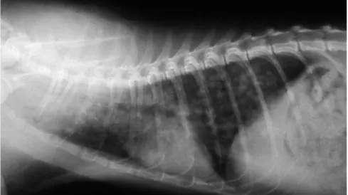
Figure 1 Right lateral thoracic radiograph of a kitten with severe aelurostrongylosis, showing a diffuse focal alveolar pattern. Courtesy of Maria Grazia Pennisi, Department of Veterinary Sciences, University of Messina, Italy
Figure 2 Alveolitis with larval accumulation, bronchiolitis and
bronchiectasis in the lung of a cat with aelurostrongylosis (haematoxylin and eosin stain). Courtesy of Maria Grazia Pennisi, Department of Veterinary Sciences, University of Messina, Italy
Figure 3 Multifocal subpleural nodules and haemorrhages in a severe case of aelurostrongylosis. Courtesy of Maria Grazia Pennisi, Department of Veterinary Sciences, University of Messina, Italy
cal data on T brevior in domestic cats were recently provided in Sardinia (Italy) where 6.5% of a sample of 107 cats tested positive compared with 25.2% that tested positive for A abstrusus,14confirming that Troglostrongylus is not a negligible lungworm of domestic cats.
The recent development of molecular assays specific for mollusc-borne
feline lungworms sharing the same ecological niches, as well as for C aerophila, is likely to be of great value for epidemiolog-ical investigations, overcoming the difficulties of copromicros -copy for differentiating the metastrongylid L1 larvae.3,9,50–52 Paragonimus species infec-tions are reported in cats from the Americas, Africa and Asia.1,53,54 Paragonimiasis is most prevalent in cats and dogs in some parts of Asia.55
Prevalence rates and/or
doc-umented cases of A abstrusus, C aerophila, O rostratus and Troglostrongylus species in some European countries are shown in Table 1.
Pathogenesis
The severity of lesions depends on the worm species and burden. Kittens also seem to develop a more severe disease.18,26,27,67 This may be explained by the smaller lung volume and small diameter of the trachea and bronchi, which are more easily blocked by worms, in particular by the larger Troglo -strongylus species. The immature immune system also seems to facilitate infection: experimental reinfection of kittens with A abstrusus L3 larvae about 1 year after the initial symptomatic infection failed to induce respiratory signs or lung lesions.25In cats with natural aelurostrongylosis, the more severe
radiological abnormalities and the higher larval burdens were found in younger ani-mals (Figure 1).68
An infective dose of <100 L3 A abstrusus lar-vae does not induce clinical signs but infective doses of 800–3200 larvae severely affect the lung and may even be lethal.69,70 However, at normal infective doses, the individual immune response significantly disrupts the parasite life cycle.28 Cats repeatedly infected with a low number of larvae do not develop clinical dis-ease when challenged with a high dose.71
The role of immunity is confirmed also by the protective effect of passive immunisation in experimentally infected kittens. In some cases it can halt the parasite life cycle and the patent phase of infection does not occur.28,72
It has been recognised for a long time that eosinophilia is evident 2–6 weeks after the ingestion of L3 larvae of A abstrusus and that immune-mediated reactions of types I, III and IV are associated with alveolar, interstitial, peribronchial and vascular lesions and may lead to the death of parasites several months later.1,73A more recent experimental study pro-vides more detailed information on the clinical signs, haematology, biochemistry, coagulation analysis, computed tomography, coprology and post-mortem findings in young adult cats.28,70Infected cats had moderate, non-specific clinical signs (fever, lethargy, weight loss, lymph node enlargement) and respiratory signs (dyspnoea, respiratory sounds, cough). Leucocytosis, massive and persist-ent eosinophilia and, in some cases, severe lymphocytosis were the most frequently observed abnormalities but no changes were detected on serum biochemistry. Various coag-ulation abnormalities were found, with a
frequent occurrence of low fibrinogen values suggesting an increased consumption of coag-ulation factors. Imaging changes in the thorax were related to the dose and consisted of pul-monary nodules, bronchial pattern and lymphadeno megaly and were found even in a cat that did not develop a patent infection.70
A abstrusus eggs accumulate in alveoli and bronchioles, inducing an inflammatory reac-tion in the lung (Figure 2). Multiple subpleur-al nodules (Figure 3) are caused by the granulomatous reaction surrounding clusters of eggs and adult worms, and emphysema is due to parasitic accumulation in the alveolar spaces. Bronchitis is severe and diffuse, usual-ly manifested by bronchial and peribronchial lymphoid hyperplasia, hypertrophy of the smooth muscle layer and mucosal hyperplasia with increased mucous cell secretion in the bronchi. Vascular and perivascular changes are also seen, with hypertrophy and hyper -plasia of pulmonary arteriolar smooth muscle, subendothelial fibrosis associated with eosinophilic infiltrates, endothelial and perivascular hyperplasia. Pulmonary hyper-tension may be the consequence of lung
dis-ease and arteriolar and bronchial changes may persist after the parasite dies, mimicking the changes found in feline asthma.27,73–75 Bacterial complication is frequent and can be associated with pleural effusion.26 Salmonella typhimurium, Pseudomonas species and Escherichia coli have been isolated in some cases and infection with enteric bacteria prob-ably results from larvae migrating from the intestine.54,76
In a kitten with severe pulmonary aelurostrongylosis, enteritis and mild diar-rhoea were associated with the presence of a high number of L1 larvae invading the small intestinal mucosa.40
Lethal T brevior infection was associated in three kittens with catarrhal bronchitis occlud-ing the lumen together with the adult worms, and multifocal pulmonary haemorrhages, consolidation and emphysematous foci.12,15
O rostratus does not seem to be associated with severe pathological changes in domestic cats, as few adult worms are found embedded in bronchial or peribronchial tissues inside pseudocysts.7,10 C aerophila usually induces chronic bronchitis.16,77
Kittens appear
to develop
more severe
lungworm
disease.
Country References A abstrusus O rostratus Troglostrongylus
species
C aerophila
Italy Brianti et al (2008)47
Traversa et al (2008)42
Iorio and Traversa (2008)43
Mugnaini et al (2012)56 Riggio et al (2013)57 Spada et al (2013)58 Brianti et al (2014)10 Tamponi et al (2014)14 Giannelli et al (2014)15 Varcasia et al (2015)20 1.2–25.2% (CR) (CR) 6.5% (CR) 1.2–14.3% (CR) Spain Miró et al (2004)46 Jefferies et al (2010)9 1% (CR) 24% (CR) (CR) 1.3% Greece Diakou et al (2014)29 – – (CR) – Portugal Payo-Puente et al (2008)59 17.4% – – 0.3–0.6% Netherlands Robben et al (2004)60 2.6% (CR) – – – Germany Taubert et al (2009)61 Becker et al (2012)62
Barutzki and Schaper (2013)44
0.7–6.5% (CR) – – 0.2% Croatia Grabarević et al (1999)63 22% – – – Albania Knaus et al (2011)45 50% – – – Romania Mircean et al (2010)48 5.6% – – 3.1%
Hungary András and Péter (2002)64
Capari et al (2013)65 14.5% (CR) – – 3.8% Bulgaria Stoichev et al (1982)66 33.3% – – – CR = case report
Prevalence rate (%) and documented cases of A abstrusus, C aerophila, O rostratus and Troglostrongylus species infection in various European countries. Case reports of A abstrusus respiratory disease exist also from the UK, Ireland, France, Switzerland, Belgium, Denmark, Poland and Greece2,6
Mixed
lungworm
infections are
increasingly
reported but
they do not
necessarily
have a more
severe clinical
picture or
poorer
outcome.
The penetration of Paragonimus species in the lung is associated with haemorrhagic foci, usually in the diaphragmatic lobe. Fluke cysts enter the bronchi and may evolve into bullae, with a consequent risk of pneumothorax.
Clinical signs
Although the majority of the publications in the literature concern A abstrusus, it has been suggested that many cases of infection or co-infection by other metastrongylids may have been erroneously attributed to the better-known A abstrusus because of difficulties with the morphometric differentiation of L1 larvae.3,4,9 Genetic characterisation of larvae now offers new insights and is likely to permit more accurate diagnosis.
Lungworm infections may be asymptomatic, or cause mild to severe respiratory signs due to bronchopneumonia, sometimes complicated by pleural effusion or pneumo thorax.26,67,78,79A productive cough is, therefore, the main clini-cal sign, together with mucopurulent nasal dis-charge, tachypnoea, dyspnoea with laboured, abdominal breathing and end-inspiratory crackles upon auscultation. In more severe cases, respiratory failure causes cyanotic mucosae and respiratory acidosis.9,18,27,42,80
diagnostic imaging (eg, thoracic radi -ography or computed tom-ography) reveals bronchial thickening and poorly defined, small nodules during the patent phase. These findings may persist after clearing the infec-tion and should be differentiated from other chronic bronchial disease such as asthma.59,81 Imaging changes may be evident even before the patent phase of disease.28,70
Right-sided cardiomegaly associated with eccentric hypertrophy and secondary to pul-monary hypertension has been described in two kittens affected by a severe bronchopneu-monia caused by A abstrusus.27 Both kittens presented with heart murmurs with maximum intensity on the right hemithorax due to tricus-pid and pulmonary regurgitation. One of the kittens died but, in the surviving kitten, the heart murmur disappeared several months after parasitological and clinical cure. Echo-doppler examination confirmed the resolution of pulmonary hypertension.27 It is, therefore, advisable to investigate for the presence of lungworm infection in cases of right heart disease associated with signs of pulmonary hypertension in outdoor cats. In a study of 54 cats that died during anaesthesia in spay– neutering programs in the USA, 9% of post-mortem investigations revealed the presence of A abstrusus.82Stray outdoor cats, such as those included in trap–neuter–release programs, are at higher risk of lungworm infection.
Eosinophilia is a frequent abnormality but is not found consistently in cell blood counts or in bronchoalveolar lavage (BAL) cytology.18,67,76,83 Troglostrongylus species was considered the cause of death of parasitised kittens present-ing with a cough and severe respiratory fail-ure at diagnosis, but cases of asymptomatic infection have also been reported.12,13,15,19,84
Capillaria infection may induce coughing (mostly dry cough), sneezing and wheezing in cats but asymptomatic carriers have also been reported.16,85
Mixed infections are increasingly reported but they do not necessarily have a more severe clinical picture or poorer outcome.19,84,85
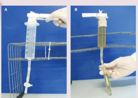
B a e r m a n n m e t h o d
< Fill a large (60 ml) syringe with tap water
< Connect the cone of the syringe to a rubber tube, which is clamped at the end
< Orientate the syringe vertically
< Fill a cheesecloth pouch with approximately 5–10 g of faeces
< Clamp the pouch and dip it in the water-filled syringe
< After 24 h any live larvae will have passed into the water and sedimented at the bottom of the system
< Collect a few millilitres of the water in a tube and centrifuge (400 g x 2 mins). Discharge the supernatant and put one drop of the sediment fluid on a
microscope slide. Cover with a coverslip and examine under a microscope at x 100 magnification
The Baermann method separates live larvae from a faecal sample as they are attracted by humidity (hydrotropism). It can be performed using an in-house system (Figure 4).
Figure 4(A,B) Baermann apparatus. Courtesy of Emanuele Brianti, Department of Veterinary Sciences, University of Messina, Italy
Diagnosis
L1 larvae are very active in the fae-ces and are readily detected in fresh faecal samples. Care should be taken to prevent soil contamination of samples, as the presence of free-liv-ing nematodes may lead to misdiag-nosis. L1 larvae can be observed in direct faecal smears or by the flota-tion technique. Note that, in the latter method, high specific gravity concentrated salt or sugar solutions may induce osmotic damage to the larvae, making identification diffi-cult.1 The Baermann migration method is considered the enrichment technique of choice for meta -strongyloid lungworms, and is based on the positive hydrotropism observed in live nematode larvae (see box and Figure 4, page 630).41,42,86 It can provide quantita-tive information on the number of larvae found in each gram of faeces, which correlates well with the sever-ity of disease.47,68 Unfortunately, 24 h are necessary to obtain the result
and the test should be repeated three times in the event of negative results, for optimum sensitivity.
A newer parasitological device for multi -valent quantitative estimation of eggs, larvae and oocysts, named FLOTAC, was evaluated for suitability in the diagnosis of A abstrusus infection. The authors reported that it was more sensitive than the Baermann method.87 However, the major limitation of copro -microscopy in general is the impossibility of making a diagnosis in the prepatent period, which lasts about 1–2 months, or when egg shedding has stopped but parasites persist and clinical signs are manifest. A well-trained observer is required to distinguish between the different strongylid L1 forms on the basis of their morphometric and morphological characteristics (Figures 5 and 6).3,12
Lungworm larvae can be found in tracheal swabs or wash and BAL cytology but with less sensitivity than in faeces, so there is no benefit in using these more invasive proce-dures that risk severe respiratory disease.41 Antibodies to A abstrusus can be detected as early as 3 weeks postinfection using an immunofluorescence antibody test, but past and currently active infections cannot be differentiated by serology.88
Significant progress has been made diag-nostically with the advent of molecular methods. A nested-PCR assay specific for A abstrusus has been validated on different biological samples (faeces, flotation supernatant, Baermann sediment and pharyngeal swabs) collected from cats with natural infections. A specificity of 100% and a sensitivity of up to 96.6% were recorded and the best results were obtained using pharyngeal swabs.51 This method allows early diagnosis in the prepatent phase, with a potential positive impact on prognosis. Molecular techniques are expected to significantly improve the under-standing of lungworm infections. A new multiplex PCR has also been developed for the simulta -neous detection of A abstrusus and T brevior.50
Capillariosis is diagnosed by standard faecal flotation but molecular techniques are also avail-able for screening and for human cases.2,52
Paragonimiasis is diagnosed using a formalin-ether sedimenta-tion technique.53Molecular methods are available for epidemiological purposes in cats and are used for human cases.89,90
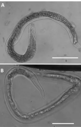
Figure 5 First-stage (L1) larvae of Aelurostrongylus
abstrusus (A and C) and Troglostrongylus brevior
(B and D) viewed by light microscopy (scale bars = 25 mm). (A) Anterior extremity of A abstrusus, lateral view. Note the terminal oral opening (arrowhead). (B) Anterior extremity of T brevior, lateral view. Note the pointed head and the subterminal oral opening (arrowhead). Morphology of the tail of
A abstrusus (C) and T brevior
(D) showing a dorsal spine at the end of the tail. Courtesy of Emanuele Brianti, Department of Veterinary Sciences, University of Messina, Italy
Figure 6 First-stage (L1) larvae of Oslerus rostratus viewed by light microscopy (scale bars = 100 µm). Note the morphology of the cephalic (A) and caudal (B) regions. Courtesy of Emanuele Brianti, Department of Veterinary Sciences, University of Messina, Italy
Treatment
Information on the efficacy of various drugs in the treatment of feline lungworm infection is available from controlled studies and clini-cal case reports (Table 2). Oral administration of fenbendazole has been suggested, with different doses and durations of therapy (from 20 mg/kg for 5 days to 50 mg/kg for 15 days), but an oral paste is licensed in the UK for treating aelurostrongylosis in cats at 50 mg/kg q24h for 3 days [EBM grade III].2
Off-label use of ivermectin has been report-ed, with inconclusive results, and should not be considered because of the risk of toxicity, principally in kittens [EBM grade III].67
Two spot-on formulations administered at the recommended dosage were compared with a 3 day course of fenbendazole therapy and were found to be effective and safe in the treatment of 12 naturally infected cats each: one formulation contained imidacloprid 10% and moxidectin 1% (Advocate; Bayer), the other emodepside 2.1% and praziquantel 8.6% (Profender; Bayer) [EBM grade I].91,92 The moxidectin formulation proved the most efficacious of the three protocols, with 100% efficacy after 30 days [EBM grade I].92 In an additional controlled study, the imidacloprid 10% and moxidectin 1% spot-on formulation was significantly effective also against C aerophila infection [EBM grade I].85 A new spot-on combination of fipronil 8.3%, (S)-methoprene 10%, eprinomectin 0.4% and praziquantel 8.3% (Broadline; Merial) was evaluated under experimental conditions and found to be highly effective for both the pre-vention and treatment of A abstrusus infection
[EBM grade II].93 Very recently the same prod-uct was found effective for treating cats with natural disease caused by A abstrusus (18 cats), as well as T brevior (three cats) or both lung-worms (two cats) [EBM grade III].94
In a case series study, cats with natural infection treated with the combination of imidacloprid 10% and moxidectin 1% were rechecked at day 14, and those still found pos-itive (4/7) were retreated and checked 1 week later. At that stage, one cat remained positive and was treated for a third time. At the end of the study (day 50), two negative faecal tests had been obtained for all treated cats, con-firming the efficacy of the treatment with this combination [EBM grade III].47
A combination of milbemycin oxime (4 mg) and praziquantel (10 mg) (Milbemax; Novartis) was administered as a single oral dose (half a tablet per kg) three times, 15 days apart, to a kitten with A abstrusus broncho -pneumonia and pulmonary hypertension, achieving parasitological and clinical cure [EBM grade IV].27Efficacy of standard topical administration of a selamectin spot-on formu-lation (6 mg/kg) (Stronghold; Zoetis) was reported in a case report and in two case series.67,83,95In one case series, selamectin was effective in one of four cats at day 30 and in two of the three cats retreated and followed up at day 60 [EBM grade III].67In the second case series, treatment was effective in nine of 10 cats [EBM grade III].95 Capillariosis was successfully treated in a cat with two injec-tions of abamectin (14 days apart) at a dose of 0.3 mg/kg [EBM grade IV].96
Information on the treatment of Troglo -strongylus, as well as on mixed infections, is
Drug Formulation Dosage Efficacy
Fenbendazole Oral paste 50 mg/kg q24h for 3 days A abstrusus (CS)
Imidacloprid 10% + moxidectin 1%
Spot on Licensed dosage
1 administration for C aerophila and
A abstrusus (CS)
1–3 administrations for A abstrusus (CR)
A abstrusus (CS, CR) C aerophila (CS)
Emodepside 2.1% + praziquantel 8.6%
Spot on Licensed dosage
Repeated after 15 days (CR)
A abstrusus (CS, CR) T brevior (CR) C aerophila (CR) Fipronil 8.3 % + (S)-methoprene 10% + eprinomectin 0.4% + praziquantel 8.3%
Spot on Licensed dosage A abstrusus (EI, CR)
T brevior (CR)
Milbemycin oxime (4 mg) + praziquantel (10 mg)
Tablet 1 tablet/2 kg every 15 days for three doses A abstrusus (CR)
Selamectin Spot on Licensed dosage for two to three doses A abstrusus (CR)
CS = controlled study, CR = case report, EI = experimental infection
Drugs used for the treatment of pulmonary nematode infections in cats Table 2
EBM grades
The ranking system for grading the level of evidence of various statements within the treatment and prevention sections of this article is described on page 574 of this Special Issue.
Bacterial
secondary
infections may
contribute to
the severity
of lungworm
disease and
require
broad-spectrum
antibiotic and
corticosteroid
therapy.
< Aelurostrongylus abstrusus (Strongylida, Angiostrongylidae) is the most well known feline lungworm and
is regarded as the most prevalent worldwide in domestic cats.
< Other lungworms in the cat include Oslerus rostratus, Troglostrongylus species, Capillaria aerophila and
Paragonimus species.
< A abstrusus, O rostratus and Troglostrongylus species may cause mixed infections as they share the same
intermediate and paratenic hosts.
< Lungworm infections may be asymptomatic, or cause mild to severe respiratory signs due to bronchopneumonia,
sometimes complicated by pleural effusion or pneumothorax (nasal discharge, tachypnoea, dyspnoea, coughing). The disease can be fatal.
< Kittens may be vertically infected and develop a more severe disease at an early stage, due to the smaller diameter
of the respiratory tract and their immature immune system.
< It is advisable to investigate for the presence of lungworm infection in outdoor cats with right-sided heart disease
associated with signs of pulmonary hypertension.
< Stray outdoor cats are at higher risk of lungworm infection.
< The Baermann migration method is considered the enrichment technique of choice, but takes 24 h to produce
results and false negatives may occur.
< The major limitation of copromicroscopy is that it is not diagnostic in the prepatent period, which lasts about
1–2 months.
< A nested-PCR assay specific for A abstrusus has been validated.
< Treatment options include fenbendazole paste, milbemycin oxime/praziquantel and various spot-on formulations
(imidacloprid 10 %/moxidectin 1%; emodepside 2.1%/praziquantel 8.6%; fipronil 8.3%/(S)-methoprene 10%/ eprinomectin 0.4%/praziquantel 8.3%; or selamectin).
< In severe cases, broad-spectrum antibiotics should be given, together with corticosteroids.
< C aerophila has zoonotic potential and sporadic cases of human capillariosis, manifesting with
a productive cough, haemoptysis and lung lesions, have been described .
KEY
pOINTS
derived from case reports only. Cases of severe respiratory disease associated with Troglo -strongylus infection were not cured by imida-cloprid 10% and moxidectin 1% or febendazole treatment [EBM grade IV].12A combination of mil bemycin oxime (4 mg) and praziquantel (10 mg) was administered as a single oral dose (half a tablet per kg) in two kittens with mixed infections caused by A abstrusus and T brevior. The asymptomatic kitten was cured but the sibling with severe respiratory disease died 2 days later [EBM grade IV].19Mixed T brevior/ A abstrusus and T brevior/C aerophila infections were cured in two kittens using the emodep-side 2.1% and praziquantel 8.6% spot-on com-bination; in one case, two administrations were required to clear Troglostrongylus larval shed-ding [EBM grade IV].84
Bacterial secondary infections may con-tribute to the severity of lungworm disease and broad-spectrum antibiotics should always be given together with corticosteroids at anti-inflammatory doses in cases with signs of bronchopneumonia. Pleural effusion and pneumothorax require immediate resolution by thoracocentesis, and medical care in an intensive care unit (oxygen administration) is required for all cats with respiratory failure.
Prognosis
In cases of A abstrusus infection, a delay in diagnosis and treatment may lead to fatal car-diopulmonary lesions, while early diagnosis and treatment greatly improves the prognosis. The level of larval burden determined by the Baermann test is usually related to the severi-ty of the disease but the prognosis should be based mainly on physical examination (sever-ity of dyspnoea and occurrence of cyanosis) and radiographic findings (severity of diffuse bronchial, alveolar and interstitial disease).
Prevention
Stray and free-roaming cats have a higher risk of becoming infected with lungworms in endemic areas.43 Avoiding predation is at present the only preventive measure for metastrongyloid or trematode pulmonary
Molecular techniques are expected to
significantly improve the understanding of
Z o o n o t i c r i s k
C aerophila has zoonotic potential and sporadic cases of human capillariosis
have been described worldwide. The disease manifests as bronchitis with a productive cough, but the presence of haemoptysis and nodular infiltrative lesions in the lung means that pulmonary neoplasia needs to be considered as a differential diagnosis.17
Paragonimiasis is a food-borne zoonosis acquired by people eating raw crustaceans. Infected cats do not present a risk to people.55,97
Funding
The authors received no specific grant from any funding agency in the public, commercial or not-for-profit sectors for the preparation of this article. The ABCd is supported by Merial, but is a scientifically independent body and its members receive no stipends from Merial.
Conflict of interest
The authors do not have any potential conflicts of interest to declare.
References
1 Conboy G. Helminth parasites of the canine and feline respira-tory tract. Vet Clin North Am Small Anim Pract 2009; 39: 1109–1126. 2 Traversa d, di Cesare A and Conboy G. Canine and feline
cardiopulmonary parasitic nematodes in Europe: emerging and underestimated. Parasit Vectors 2010; 3: 62.
3 Traversa d and di Cesare A. Feline lungworms: what a dilem-ma. Trends Parasitol 2013; 29: 423–430.
4 Brianti E, Giannetto S, dantas-Torres F, et al. Lungworms of the genus Troglostrongylus (Strongylida: Crenosomatidae): neg-lected parasites of domestic cats. Vet Parasitol 2014; 202: 104–112. 5 Bowman dd. Respiratory system parasites of the dog and cat (part ii): trachea and bronchi, and pulmonary vessels. In: Bowman dd (ed). Companion and exotic animal parasitology. International Veterinary Information Service IVIS, 2000; www.ivis.org.
6 Pennisi MG, Niutta PP and Giannetto S. Longwormziekte bij katten veroorzaakt door Aelurostrongylus abstrusus. Tijdschr
Diergeneesk 1995; 120: 262–266.
7 Juste RA, Garcia AL and Mencía L. Mixed infestation of a domestic cat by Aelurostrongylus abstrusus and Oslerus
ros-tratus. Angew Parasitol 1992; 33: 56–60.
8 Millan J and Casanova JC. High prevalence of helminth par-asite in feral cats in Majorca Island (Spain). Parasitol Res 2009; 106: 183–188.
9 Jefferies R, Vrhovec MG, Wallner N, et al. Aelurostrongylus
abstrusus and Troglostrongylus sp. (Nematosa: Meta
-strongyloidea) infections in cats inhabiting Ibiza, Spain.
Vet Parasitol 2010; 173: 344–348.
10 Brianti E, Gaglio G, Napoli E, et al. Feline lungworm
Oslerus rostratus (Strongylida: Filaridae) in Italy: first case
report and histopathological findings. Parasitol Res 2014; 113: 3853–3857.
11 Fitzsimmons WM. Bronchostrongylus subcrenatus (Raillet & Henry, 1913) a new parasite recorded from the domestic cat.
Vet Rec 1961; 73: 101–102.
12 Brianti E, Gaglio G, Giannetto S, et al. Troglostrongylus
brevior and Troglostrongylus subcrenatus (Strongylida:
Crenosomatidae) as agents of broncho-pulmonary infesta-tion in domestic cats. Parasit Vectors 2012; 5: 178.
13 Brianti E, Gaglio G, Napoli E, et al. Evidence for direct trans-mission of the cat lungworm Troglostrongylus brevior (Strongylida: Crenosomatidae). Parasitology 2013; 140: 821–824.
14 T amponi C, Varcasia A, Brianti E, et al. New insights on metastrongyloid lungworms infecting cats of Sardinia, Italy.
Vet Parasitol 2014; 16: 203: 222–226.
15 Giannelli A, Passantino G, Nascimento Ramos RA, et al. Pathological and histological findings associated with the feline lungworm Troglostrongylus brevior. Vet Parasitol 2014; 204: 416–419.
16 Traversa d, di Cesare A, Milillo P, et al. Infection by Eucoleus
aerophilus in dogs and cats: is another extra-intestinal
para-sitic nematode of pets emerging in Italy? Res Vet Sci 2009; 87: 270–272.
17 Lalosević d, Lalosević V, Klem I, et al. Pulmonary capillariosis miming bronchial carcinoma. Am J Trop Med Hyg 2008; 78: 14–16.
18 Risitano AL, Brianti E, Pennisi MG, et al. Aspetti clinici e anatomopatologici di aelurostrongilosi in un gattino. Proceedings of the LXII annual meeting of Società Italiana di Scienze Veterinarie (SISVet), 2008; 62: 169–170.
19 di Cesare A, Frangipane di Regalbono A, Tessarin C, et al. Mixed infection by Aelurostrongylus abstrusus and
Troglostrongylus brevior in kittens from the same litter in
Italy. Parasitol Res 2014; 113: 613–618.
20 Varcasia A, Brianti E, Tamponi C, et al. Simultaneous infec-tion by four feline lungworm species and implicainfec-tions for the diagnosis. Parasitol Res 2015; 114: 317–321.
21 Lòpez C, Panadero R, Paz A, et al. Larval development of
Aelurostrongylus abstrusus (Nematoda, Angiostrongylidae)
in experimentally infected Cernuella (Cernuella) virgata (Mollusca, Helicidae). Parasitol Res 2005; 95: 13–16.
22 Jezewski W, Buńkowska-Gawlik K, Hildebrand J, et al. Intermediate and paratenic hosts in the life cycle of Aelurostrongylus abstrusus in natural environment. Vet
Parasitol 2013; 198: 401–405.
23 di Cesare A, Crisi PE, di Giulio E, et al. Larval development of the feline lungworm Aelurostrongylus abstrusus in Helix
aspersa. Parasitol Res 2013; 112: 3101–3108.
24 Giannelli A, Ramos RA, Annoscia G, et al. Development of the feline lungworms Aelurostrongylus abstrusus and
Troglostrongylus brevior in Helix aspersa snails. Parasitol
2014; 141: 563–569.
25 Hamilton JM. Studies on re-infestation of the cat with
Aelurostrongylus abstrusus. J Comp Pathol 1968; 78: 69–72.
worms with indirect life cycles. The prophy-lactic activity of some molecules used to treat nematode respiratory infections – as for A vasorum infection in dogs – is currently unknown; but the spot-on combination of fipronil 8.3%, (S)-methoprene 10%, epri-nomectin 0.4% and praziquantel 8.3% (Broadline) was found effective as a preventa-tive treatment for aelurostrongylosis in an experimental setting [EBM grade II].93
26 Barrs VR, Swinney GR, Martin P, et al. Concurrent
Aelurosrongylus abstrusus infection and salmonellosis in a
kitten. Aust Vet J 1999; 77: 229–232.
27 dirven M, Szatmári V, van den Ingh T, et al. Reversible pul-monary hypertension associated with lungworm infection in a young cat. J Vet Cardiol 2012; 14: 465–474.
28 Schnyder M, di Cesare A, Basso W, et al. Clinical, laboratory and pathological findings in cats experimentally infected with Aelurostrongylus abstrusus. Parasitol Res 2014; 113: 1425–1433.
29 diakou A, di Cesare A, Aeriniotaki T, et al. First report of
Troglostrongylus brevior in a kitten in Greece. Parasitol Res
2014; 113: 3895–3898.
30 Giannelli A, Colella V, Abramo F, et al. Release of lungworm larvae from snails in the environment: potential for alterna-tive transmission pathways. PLoS Negl Trop Dis 2015; 9(4): e0003722.
31 Anderson RC. Nematode parasites of vertebrates. Their development and transmission. 2nd ed. CABI Publishing, Guildford, UK, 2000.
32 Gregory GG and Munday BL. Internal parasites of feral cats from the Tasmanian Midlands and King Island. Aust Vet J 1976; 52: 317–320.
33 Coman BJ, Jones EH and driesen MA. Helminth parasites and arthropods of feral cats. Aust Vet J 1981; 57: 324–327.
34 Mundim TCd, Oliveira Jr Sd, Rodrigues dC, et al. Freqüência de helmintos em gatos de Uberlândia, Minas Gerais. Arq Bras
Med Vet Zoo 2004; 56: 562–563.
35 Abu-Madi MA, Al-Ahbabi dA, Al-Mashhadani MM, et al. Patterns of parasitic infections in faecal samples from stray cat populations in Qatar. J Helminthol 2007; 81: 281–286. 36 di Cesare A, Castagna C, Meloni S, et al. Canine and feline
infections by cardiopulmonary nematodes in Central and Southern Italy. Parasitol Res 2011; 109: S87–S96.
37 Lucio-Forster A and Bowman dd. Prevalence of fecal-borne parasites detected by centrifugal flotation in feline samples from two shelters in upstate New York. J Feline Med Surg 2011; 13: 300–303.
38 Echeverry dM, Giraldo MI and Castaño JC. Prevalencia de helmintos intestinales en gatos domésticos del departamen-to del Quindío, Colombia. Biomedica 2012; 32: 430–436. 39 Kohart NA, Boes KM, Sponenberg dP, et al. What is your
diagnosis? Lung impression smear from a stray kitten.
Vet Clin Pathol 2014; 43: 113–114.
40 Philbey AW, Krause S and Jefferies R. Verminous pneumonia and enteritis due to hyperinfection with Aelurostrongylus
abstrusus in a kitten. J Comp Pathol 2014; 150: 357–360.
41 Lacorcia L, Gasser RB, Anderson GA, et al. Comparison of bronchoalveolar lavage fluid examination and other diag-nostic techniques with the Baermann technique for detec-tion of naturally occurring Aelurostrongylus abstrusus infection in cats. J Am Vet Med Assoc 2009; 35: 43–49.
42 Traversa d, Lia RP, Iorio R, et al. Diagnosis and risk factors of
Aelurostrongylus abstrusus (Nematoda, Strongylida)
infec-tion in cats from Italy. Vet Parasitol 2008; 153: 182–186. 43 Iorio R and Traversa d. New epidemiological and molecular
insights into feline lungworm infection. Ann NY Acad Sci 2008; 1149: 174–176.
44 Barutzki d and Schaper R. Occurrence and regional distribu-tion of Aelurostrongylus abstrusus in cats in Germany.
Parasitol Res 2013; 112: 855–861.
45 Knaus M, Kusi I, Rapti d, et al. Endoparasites of cats from
the Tirana area and the first report on Aelurostrongylus
abstrusus (Railliet, 1898) in Albania. Wien Klin Wochenschr
2011; 123 Suppl 1: 31–35.
46 Miró G, Montoya A, Jiménez S, et al. Prevalence of antibodies to Toxoplasma gondii and intestinal parasites in stray, farm and household cats in Spain. Vet Parasitol 2004; 126: 249–255.
47 Brianti E, Pennisi MG, Risitano AL, et al. Aelurostrongilosi
felina: epidemiologia e valuazione di un protocollo
terapeu-tico alternativo. Proceedings of the 59th SCIVAC International Congress, Rimini (Italy); 2008, May 30 – June 1.
48 Mircean V, Titilincu A and Vasile C. Prevalence of endopara-sites in household cat (Felis catus) populations from Transylvania (Romania) and association with risk factors.
Vet Parasitol 2010; 171: 163–166.
49 Barutzky d and Schaper R. Results of parasitological exami-nations of faecal samples from cats and dogs in Germany between 2003 and 2010. Parasitol Res 2011; 109: S45–S60. 50 Annoscia G, Latrofa MS, Campbell BE, et al. Simultaneous
detection of the feline lungworms Troglostrongylus brevior and Aelurostrongylus abstrusus by a newly developed duplex-PCR. Vet Parasitol 2014; 199: 172–178.
51 Traversa d, Iorio R and Otranto d. Diagnostic and clinical implications of a nested PCR specific for ribosomal DNA of the feline lungworm Aelurostrongylus abstrusus (Nematoda, Strongylida). J Clin Microbiol 2008; 46: 1811–1817.
52 di Cesare A, Castagna C, Otranto d, et al. Molecular detection of Capillaria aerophila, an agent of canine and feline pulmonary capillariosis. J Clin Microbiol 2012; 50: 1958–1963. 53 Sohn WM and Chai JY. Infection status with helminthes in
feral cats purchased from a market in Busan, Republic of Korea. Korean J Parasitol 2005; 43: 93–100.
54 Foster S and Martin P. Lower respiratory tract infections in cats. Reaching beyond empirical therapy. J Feline Med Surg 2011; 13: 313–332.
55 Liu Q, Wei F, Liu W, et al. Paragonimiasis: an important food-borne zoonosis in China. Trends Parasitol 2008; 24: 318–323. 56 Mugnaini L, Papini R, Gorini G, et al. Pattern and predictive
factors of endoparasitism in cats in central Italy. Rev Med Vet 2012; 163: 85–88.
57 Riggio F, Mannella R, Ariti G, et al. Intestinal and lung parasites in owned dogs and cats from central Italy.
Vet Parasitol 2013; 193: 78–84.
58 Spada E, Proverbio d, della Pepa A, et al. Prevalence of faecal-borne parasites in colony stray cats in northern Italy.
J Feline Med Surg 2013; 15: 672–677.
59 Payo-Puente P, Botelho-dinis M, Carvaja Urueña AM, et al. Prevalence study of the lungworm Aelurostrongylus
abstrusus in stray cats of Portugal. J Feline Med Surg 2008; 10:
242–246.
60 Robben SR, le Nobel WE, döpfer d, et al. Infections with helminths and/or protozoa in cats in animal shelters in the Netherlands [Infecties met helminthen en/of protozoën bij katten in asielen in Nederland]. Tijdschr Diergeneeskd 2004; 129: 2–6.
61 Taubert A, Pantchev N, Globokar Vrhovec M, et al. Lungworm infections (Angiostrongylus vasorum, Crenosoma vulpis,
Aelurostrongylus abstrusus) in dogs and cats in Germany
and Denmark in 2003–2007. Vet Parasitol 2009; 159: 175–180. 62 Becker AC, Rohen M, Epe C, et al. Prevalence of
endopara-sites in stray and fostered dogs and cats in Northern Germany. Parasitol Res 2012; 111: 849–857.
636
Available online at jfms.com
63 Grabarević Ž, Ćurić S, Tustonja A, et al. Incidence and region-al distribution of the lungworm Aelurostrongylus abstrusus in cats in Croatia. Veterinarski Arhiv 1999; 69: 279–287. 64 András T and Péter T. Data on worm infestation of domestic
cats (Felis catus) in Hungarian hunting areas [Adatok a mag-yarországi vadászterületeken élo házimacska (Felis catus domesticus) féregfertozöttségérol]. Magyar Allatorvosok Lapja 2002; 124: 26–30.
65 Capari B, Hamel d, Visser M, et al. Parasitic infections of domestic cats, Felis catus, in western Hungary. Vet Parasitol 2013; 192: 33–42.
66 Stoichev I, Sherkov S and Halachev M. Pathology of cats from a region of Bulgaria with human endemic nephropathy.
J Comp Pathol 1982; 92: 99–107.
67 Grandi G, Calvi LE, Venco L, et al. Aelurostrongylus abstrusus (cat lungworm) infection in five cats from Italy. Vet Parasitol 2005; 134: 177–182.
68 Genchi M, Ferrari N, Fonti P, et al. Relation between
Aelurostrongylus abstrusus larvae excretion, respiratory and
radiographic signs in naturally infected cats. Vet Parasitol 2014; 206: 182–187.
69 Hamilton JM. The number of Aelurostrongylus abstrusus larvae required to produce pulmonary disease in the cat.
J Comp Pathol 1967; 77: 343–346.
70 dennler M, Bass dA, Gutierrez-Crespo B, et al. Thoracic com-puted tomography, angiographic comcom-puted tomography, and pathology findings in six cats experimentally infected with
Aelurostrongylus abstrusus. Vet Radiol Ultrasound 2013; 54:
459–469.
71 Hamilton JM. Production of immunity in the cat against lungworm disease by administration of third-stage larvae.
J Comp Pathol 1969; 79: 161–165.
72 Hamilton JM. Passive immunization in lungworm infection of the cat. J Comp Pathol 1968; 78: 331–333.
73 Hamilton JM. Experimental lungworm disease of the cat. Association of the condition with lesions of the pulmonary arteries. J Comp Pathol 1966; 76: 147–157.
74 Jonas AM, Swerczek TW and downing SE. Vaso-occlusive pulmonary artery disease in the cat: a preliminary report.
Acta Radiol Suppl 1972; 319: 237–244.
75 Naylor JR, Hamilton JM and Weatherley AJ. Changes in the ultrastructure of feline pulmonary arteries following infection with the lungworm Aelurostrongylus abstrusus.
Br Vet J 1984; 140: 181–190.
76 Foster SF, Martin P, Braddock JA, et al. A retrospective analy-sis of feline bronchoalveolar lavage cytology and microbiol-ogy (1995–2000). J Feline Med Surg 2004; 6: 189–198.
77 Holmes PR and Kelly Jd. Capillaria aerophila in the domestic cat in Australia. Aust Vet J 1973; 49: 472–473.
78 Miller BH, Roudebush P and Ward HG. Pleural effusion as a sequela to aelurostrongylosis in a cat. J Am Vet Med Assoc 1984; 185: 556–557.
79 Mooney ET, Rozanski EA, King RG, et al. Spontaneous pneu-mothorax in 35 cats (2001–2010). J Feline Med Surg 2012; 14: 385–392.
80 Yildiz K, duru SY and Gokpinar S. Alteration in blood gases in cats naturally infected with Aelurostrongylus abstrusus.
J Small Anim Pract 2011; 52: 376–379.
81 Losonsky JM, Thrall dE and Prestwood AK. Radiographic evaluation of pulmonary abnormalities after Aeluro
-strongylus abstrusus inoculation in cats. Am J Vet Res 1983; 44:
478–482.
82 Gerdin JA, Slater MR, Makolinski KV, et al. Post-mortem find-ings in 54 cases of anesthetic associated death in cats from two spay-neuter programs in New York State. J Feline Med
Surg 2011; 13: 959–966.
83 Reinhardt S, Ottenjann M, Schunack B, et al. Lungworm disease (Aelurostrongylus abstrusus) in a cat [Lungenwurm -befall (Aelurostrongylus abstrusus) bei einer Katze].
Kleintierpraxis 2004; 49: 239–246.
84 di Cesare A, Iorio R, Crisi P, et al. Treatment of
Troglostrongylus brevior (Metastrongyloidea, Crenosomatidae)
in mixed lungworm infections using spot-on emodepside.
J Feline Med Surg 2015; 17: 181–185.
85 Traversa d, di Cesare A, di Giulio E, et al. Efficacy and safety of imidacloprid 10 %/moxidectin 1 % spot-on formulation in the treatment of feline infection by Capillaria aerophila.
Parasitol Res 2012; 111: 1793–1798.
86 Willard Md, Roberts RE, Allison N, et al. Diagnosis of
Aelurostrongylus abstrusus and Dirofilaria immitis infections in
cats from a human shelter. J Am Vet Med Assoc 1988; 192: 913–916. 87 Gaglio G, Cringoli G, Rinaldi L, et al. Use of the FLOTAC technique for the diagnosis of Aelurostrongylus abstrusus in the cat. Parasitol Res 2008; 103: 1055–1057.
88 Hamilton JM and Roberts RJ. Immunofluorescence as a diag-nostic procedure in lungworm disease of the cat. Vet Rec 1968; 83: 401–403.
89 Intapan PM, Wongkham C, Imtawil KJ, et al. Detection of
Paragonimus heterotremus eggs in experimentally infected
cats by a polymerase chain reaction-based method. J Parasitol 2005; 91: 195–198.
90 Tantrawatpan C, Intapan PM, Janwan P, et al. Molecular iden-tification of Paragonimus species by DNA pyrosequencing technology. Parasitol Int 2013; 341–345.
91 Traversa d, di Cesare A, Milillo P, et al. Efficacy and safety of imidacloprid 10 %/moxidectin 1 % spot-on formulation in the treatment of feline aelurostrongylosis. Parasitol Res 2009; 105: S55–S62.
92 Traversa d, Milillo P, di Cesare A, et al. Efficacy and safety of emodepside 2.1 %/praziquantel 8.6 % spot-on formulation in the treatment of feline aelurostrongylosis. Parasitol Res 2009; 105: S83–S89.
93 Knaus M, Chesterb ST, Rosentelb J, et al. Efficacy of a novel topical combination of fipronil, (S)-methoprene, epri-nomectin and praziquantel against larval and adult stages of the cat lungworm, Aelurostrongylus abstrusus. Vet Parasitol 2014; 202: 64–68.
94 Giannelli A, Brianti E, Varcasia A, et al. Efficacy of Broadline(®) spot on against Aelurostrongylus abstrusus and
Troglostrongylus brevior lungworms in naturally infected
cats from Italy. Vet Parasitol 2015; 209: 273–277.
95 Iannino F, Iannetti L, Paganico d, et al. Evaluation of the efficacy of selamectin spot-on in cats infested with
Aelurostrongylus abstrusus (Strongylida, Filariodidae) in a
Central Italy cat shelter. Vet Parasitol 2013; 197: 258–262. 96 Barrs VR, Martin P, Nicoll RG, et al. Pulmonary cryptococcosis
and Capillaria aerophila infection in an FIV-positive cat.
Aust Vet J 2000; 78: 154–158.
97 Macpherson CNL. Human behavior and the epidemiology of parasitic zoonoses. Int J Parasitol 2005; 35: 1319–1331.