U
NIVERSITÀ DEGLIS
TUDI DIN
APOLIF
EDERICOII
D
OTTORATO DIR
ICERCA INM
EDICINAC
LINICA ES
PERIMENTALEC
URRICULUMI
NS
CIENZEO
DONTOSTOMATOLOGICHEXXXI Ciclo
(Anni 2015-2018)
Coordinatore: Prof. Francesco Beguinot
T
ESI DID
OTTORATOANTIMICROBIAL
EFFECT
OF
NEW
RESTORATIVE
DENTAL
MATERIAL
INCORPORATING
SILVER
NANOPARTICLES
RELATORI CANDIDATO
Chiar.mo Dott. Raffaele Conte
Prof. Sandro Rengo Chiar.mo
3
U
NIVERSITY OFN
APLESF
EDERICOII
P
H.D.
P
ROGRAM INC
LINICAL ANDE
XPERIMENTALM
EDICINEC
URRICULUMI
NO
DONTOSTOMATOLOGICALS
CIENCESXXXI Cycle
(Years 2015-2018)
Chairman: Prof. Francesco Beguinot
P
H.D.
T
HESISANTIMICROBIAL
EFFECT
OF
NEW
RESTORATIVE
DENTAL
MATERIAL
INCORPORATING
SILVER
NANOPARTICLES
TUTORS PH.D.STUDENT
Prof. Sandro Rengo Dr. Raffaele Conte
4 Scopo di ogni attività dell’intelletto è ridurre
il mistero a qualcosa di comprensibile (Albert Einstein)
5
INDEX
SUMMARY
ABSTRACT ... 6
RIASSUNTO... 9
CHAPTER 1. DENTAL CARIES ... 11
1.1 ETIOPATOGENESIS OF DENTAL CARIES ... 12
1.2 DENTAL CARIES THERAPY ... 33
CHAPTER 2. SECONDARY CARIES ... 37
2.1 ETIOPATHOGENESIS OF SECONDARY CARIES: THE ROLE OF MICROLEAKAGE ... 38
2.2 DIAGNOSIS AND MANAGEMENT OF SECONDARY CARIES ... 45
2.3 PREVENTION OF SECONDARY CARIES BY SILVER ... 49
2.4 SILVER IN DENTAL MATERIALS………54
CHAPTER 3. INNOVATIVE DENTAL RESTORATIVE MATERIALS 65
3.1 RESTORATIVE DENTAL MATERIALS ... 66
3.2. NANOTECHNOLOGICAL APPROACH FOR BIOACTIVE DENTAL MATERIALS: LDH AND SILVER NANOPARTICLES ... 75
CHAPTER 4. MATERIALS AND METHODS ... 81
CHAPTER 5. RESULTS AND DISCUSSION ... 89
CONCLUSIONS ... 110
AKNOWLEDGMENTS……….112
6 ABSTRACT
Secondary or recurrent caries are dental lesions originated at the margins of an existing restoration, and are considered the most common reason for restoration failure. [1] Usually, these lesions are histologically similar to the primary caries and can be difficult to detect unless somewhat advanced, resulting in a considerable loss of tooth structure. Over the past decades, resin-based dental materials have been used in restorative dentistry for their excellent esthetics and improved mechanical performance. [2] However, they represent potential sources of carbon and energy for microorganisms including oral bacteria and fungi residual in the dental cavity. In addition, cariogenic bacteria can infiltrate the restoration-tooth margins compromise the restoration’s longevity. [3] Because caries at the restoration margins is a main reason for restoration failures, it would be highly desirable for the composite and bonding agent to possess antibacterial capabilities.
Novel antibacterial dental materials were developed by introducing
quaternary ammonium monomers, including
12-methacryloyloxydodecylpyridinium bromide (MDPB), dimethylaminohexadecyl methacrylate (DMAHDM), and dimethylaminododecyl methacrylate (DMADDM). [4] [5] [6] [7] [8] These monomers can form covalent bonds with the polymer matrix and be immobilized in the resin-based materials, representing a non-released, contact-killing agent. [4] Several other antimicrobial formulations were also developed, including a methacryloxylethylcetyl dimethyl ammonium chloride (DMAE-CB) containing adhesive, quaternary ammonium
7
polyethylenimine (PEI) nanoparticles for antimicrobial dental composites, antibacterial glass ionomer cements, and antibacterial nanocomposites and bonding agents incorporating a quaternary ammonium dimethacrylate (QADM). [9] [10] [11]
Quaternary ammonium acrylate (QAM) resins possess positively-charged quaternary amine N+ which can interact with the negatively-charged membrane of bacteria, leading to membrane disruption and cytoplasm leakage. [10] It is postulated that long-chained quaternary ammonium compounds can be especially effective by inserting into the bacterial membrane, resulting in physical disruption and bacteria death. [12] [13] [14]
Aside from the antibacterial monomers added to the resin matrix, an alternative approach is to add silver nanoparticles. Indeed, silver (Ag) is known for its antimicrobial activity against a diverse group of bacteria and has been used for many years as an antimicrobial substance in the medical field. [15] Composite containing Ag particles with long-lasting antibacterial activity have been manufactured and observed to inhibit S. mutans growth [16]. In addition, resins containing Ag nanoparticles were able to inhibit biofilm viability. [17,18] Although the restorative materials had significant evolvement in the past few decades, the high rates of treatment failure suggest that the current restorative approaches are not yet optimized and have a potential for improvement.
The aim of this work is to synthesize and evaluate new bioactive and antibacterial composite materials based on photo-activated Bis-GMA/TEGDMA matrix, containing an hydrotalcite-like compound intercalated with Ag nanoparticles as filler.
8
We have obtained a dental resin with improved physical and biological properties and, in addition, able to release low amount of silver in a controlled and tunable way for a long period of time.
In contrast to the conventional and resin-modified glass-ionomers, our CR-Agx were able to release silver ions when intraoral pH values drop below the critical pH of 5.5, counteracting the demineralization process of the tooth surface. The caries protective effect of these materials may be related to the material’s ability to release adequate amounts of silver ions for sustained periods of time and during acidic attack.
9 RIASSUNTO
Le carie secondarie sono lesioni che si originano ai margini di restauri dentali e sono tra le cause più comuni di fallimenti terapeutici. Questo tipo di lesioni sono istologicamente simili alle carie primarie e sono difficili da distinguere da queste ultime, se non in stadi avanzati.
Durante gli ultimi decenni le resine dentali sono state ampiamente utilizzate in odontoiatria restaurativa per le loro eccellenti caratteristiche estetiche e le loro performance meccaniche. In contrasto, sono una potenziale risorsa di carbonio per i batteri residui nelle cavità dentali. Tali microorganismi cariogeni possono infiltrarsi nei margini tra dente e restauro e comprometterne la longevità. A causa di ciò sono stati sintetizzati nuovi materiali dentali antibatterici tramite introduzione di monomeri di ammonio quaternario come, ad esempio, bromuro di 12-metacrilossidodecilpiridinio (MDPB), dimetilamminoesadecil metacrilato (DMAHDM) e dimetilamminododecil metacrilato (DMADDM). Questi monomeri formano legami covalenti con la matrice polimerica e sono immobilizzati nella resina, rappresentando un agente battericida da contatto. Sono state inoltre sviluppate altre formulazioni antimicrobiche come, ad esempio, adesivi contenenti cloruro di metacrilossietilcetil dimetilammonio (DMAE-CB), nanoparticelle di polietilenimmina da inserire in compositi dentali con attività antibatteriche, GIC (glass ionomer cements) antibatterici e nano compositi contenenti ammonio quaternario dimetilacrilato (QADM). Tali resine posseggono ammine quaternarie cariche positivamente capaci di interagire con le cariche negative della membrana batterica provocandone la distruzione. Inoltre, composti con ammonio quaternario a lunga catena sono particolarmente
10
efficaci grazie alla capacità di inserirsi all’ interno della membrana batterica.
Oltre alla presenza di monomeri antibatterici aggiunti alla matrice della resina, un approccio alternativo è quello di utilizzare nanoparticelle di argento. Infatti l’argento è dotato di attività antimicrobica contro un ampio spettro di batteri. Compositi contenenti nanoparticelle di argento con attività antibatterica a lungo termine sono stati sintetizzati e utilizzati contro S. mutans e come agenti anti-biofilm.
In generale, anche se i materiali restaurativi hanno avuto significanti miglioramenti negli ultimi decenni, gli elevati tassi di fallimento indicano che gli attuali approcci restaurativi non sono ancora ottimizzati ed hanno margini di miglioramento.
Lo scopo di questo lavoro è quello di sintetizzare e valutare nuovi materiali compositi bioattivi basati su una matrice foto attivata di Bis-GMA/TEGDMA contenente compositi idrotalcite simili intercalati con nanoparticelle di argento come riempitivo.
Abbiamo quindi ottenuto resine dentali con caratteristiche fisiche e proprietà biologiche migliorate, capaci di rilasciare basse quantità di argento in maniera controllata per un lungo periodo di tempo.
A differenza delle resine convenzionali, il nostro CR-Ag è in grado di rilasciare ioni argento quando i valori di pH intra-orale sono al di sotto del pH critico di 5.5, contrastando il processo di demineralizzazione della superficie del dente. Quindi, l’effetto carie protettivo di questi materiali è in relazione all’ abilità del materiale di rilasciare quantità adeguate di ioni argento per un tempo sostenuto durante l’attacco acido.
11
12 1.1 ETIOPATHOGENESIS OF DENTAL CARIES
Dental caries is defined as a chronic, diet o microbial, site-specific disease of dental hard tissues, caused by shifts from protective factors favoring tooth remineralization to destructive factors leading to demineralization. [19] When sugars or other fermentable carbohydrates are ingested, the resulting fall in dental plaque Ph caused by organic acids increases the solubility of calcium hydroxyl apatite in the dental hard tissues and demineralization occurs as calcium is lost from the tooth surface (Figure 1).
Figure 1. The process of tooth remineralization. Adapted from Dodds et al. [20]
13
The pH of dental plaque is a key factor in the balance between acid demineralization of the teeth and the remineralization of the initial caries lesion. Plaque pH falls each time acid accumulates in the plaque due to bacterial acid production following the consumption of fermentable carbohydrates – mainly sugars – in foods and drinks. Conversely, plaque Ph rises when the acids are washed away or neutralized by saliva, which contains the important buffer, bicarbonate.[20]
In healthy teeth, the loss of minerals is balanced by the reparative mechanisms of saliva. [20]When the saliva pH or the plaque pH is below a ‘critical value’ of about 5.5, the saliva or plaque becomes unsaturated with respect to tooth mineral. [20] As a result, tooth enamel can begin to dissolve. However, when the pH is above this value, the saliva and plaque are supersaturated with respect to tooth mineral. The calcium and phosphate ions in saliva then start to repair any damaged mineral crystals in the enamel, starting the process of remineralization.
Thus, acidic conditions contribute to bringing phosphate and hydroxyl ions below saturation levels, allowing the solid hydroxyapatite crystals of the tooth mineral to dissolve. If above saturation levels, the chemical reaction will move towards remineralization and any damaged crystals will be repaired by the acquisition of ions from the solution.
The World Health Organization (WHO) affirms that dental caries qualifies as a major public-health problem, owing to its high prevalence in all regions of the world, with the greatest burden of disease being on disadvantaged and socially marginalized populations.
The initiation and development of the disease is the result of the interaction of four main etiological factors (Figure 2):
14
the oral bacteria in dental plaque;
the dietary, salivary and genetic influences. the time
Figure 2. Diagram that describes the multifactorial etiology of dental caries. Adapted from Mathur et Dhillon. [21]
The plaque and the dietary factors are interdependent on each other for the causation of damage. The tooth becomes the ‘platform’ for this interaction, as well as the ‘victim’ in the interaction of the other two factors.
It is indeed true that multiple factors have to act in concert with each other to produce the disease, but not necessarily at the same time (Figure 3).
15 Figure 3. Factors affecting the development of dental caries. Adapted from Dodds et al. [20]
Environmental factors, such as behavioral habits, [22] may also influence the development of dental decay. Low socio-economic status is a non‐biological risk factor which is often related to educational level, the perception of the individual about his/her own health, life style, dietary composition, and access to dental care. [23]
Moreover, caries development is a dynamic process that consists of rapidly alternating periods of tooth demineralization and remineralization, which, if net demineralization occurs over sufficient time, results in the initiation of specific caries lesions at certain anatomical predilection sites on the teeth. It is important to balance the pathological and protective factors that influence the initiation and progression of dental caries. Protective factors promote remineralization and lesion arrest, whereas pathological factors shift the balance in the direction of dental caries and disease progression (Figure 4).
16 Figure 4. Balancing pathological and protective factors in dental caries. A focus on optimizing the protective factors will promote remineralization and shift the dynamic balance of the caries process in the direction of health and lesion arrest. A failure to mitigate the effects of the pathological factors will promote demineralization and shift the dynamic balance in the direction of disease initiation and disease progression. Adapted from Pitts N.B. et al.[24]
The oral microbiota
The oral cavity is a unique environment that support the presence of up 500 species of microorganisms (including viruses, fungi, protozoa and bacteria). [25] Unlike oral epithelium, the morphology of the tooth makes many areas inaccessible to physiological clearance mechanisms. Thus, a tooth becomes an ideal place for the stubborn adherence for many of these species. [26] Organisms that are capable of adhesion, adhere to the salivary pellicle on the tooth and form a convenient arena for the
17
subsequent aggregation of other organisms that are incapable of initial adhesion.
According to the ‘ecological plaque hypothesis’ (Figure 5), the pathogenesis of dental caries is related to a disturbance in two types of homeostasis / physiological equilibrium that exist in an oral cavity:
1. Disruption of microbial homeostasis in the ‘biofilm’.
2. Disruption of mineral homeostasis between the tooth and the ‘oral fluid’.
Figure 5. Ecological plaque hypothesis. An increased frequency of fermentable sugar intake results in the biofilm spending more time at a low Ph, which will select for bacteria that grow preferentially under acidic conditions. The growth of bacteria associated with sound surfaces is then disadvantaged, which over time results in an increase in the proportions and activity of cariogenic species at a site and a heightened risk of caries. This risk is raised in individuals with impaired saliva flow and sugar-rich diet, but it is reduced in those with appropriate oral hygiene and exposure to fluoride. Adapted from Pitts N.B. et al. [27]
The dental biofilm is a population or community of bacteria living in organized structures attached to a tooth surface, embedded in a matrix of extracellular polymeric substances produced by microbes. [9] The formation of dental biofilm is a multiple-stage process (Figure 5).
18 Figure 6. Stages of dental biofilm formation. Within minutes after completely cleansing the tooth surface, a pellicle forms from proteins and glycoproteins in saliva. A Association: Through purely physical forces, bacteria associate loosely with the pellicle. B Adhesion: Because they possess special surface molecules that bind to pellicle receptors, some bacteria become the “primary colonizers,” particularly streptococci and actinomyces. Subsequently, other microorganisms adhere to the primary
19
colonizers. C Bacterial proliferation ensues. D Microcolonies are formed. Many streptococci secrete protective extracellular polysaccharides (e. g., dextrans, levans).E Biofilm (“attached plaque”): Microcolonies form complex groups with metabolic advantages for the constituents. F Plaque growth—maturation: The biofilm is characterized by a primitive “circulatory system.” The plaque begins to “behave” as a complex organism! Anaerobic organisms increase. Metabolic products and evulsed cell wall constituents (e. g., lipopolysaccharides, vesicles) serve to activate the host immune response. Bacteria within the biofilm are protected from phagocytic cells (PMN) and against exogenous bacteriocidal agents. (Adapted from Wolf HF. Biofilm-plaque formation on tooth and root surfaces. In:Wolf HF, Rateitschak KH (eds). Periodontology, ed 3. Stuttgart: Thieme, 2005:24) [28]
The process starts with an initial formation of salivary pellicle, a combination of active proteins and glycoproteins from saliva and gingival crevicular fluid. After four hours, the aerobic species of the genera
Streptococcus, Capnocytophaga, Veillonella or Actinomyces (known of as
initial and early colonizers) adhere to the tooth proteic film, forming a first layer of biofilm. This process requires the presence, on the bacterial cell surface, of specific (e.i statherins, mucins, agglutinins, alpha-amylase and prolin rich proteins) that act like chains between tooth and early colonizers. A second layer of biofilm develops through a process known as coaggregation or coadhesion, in which other microorganisms attach the first colonizers through adhesion of their respective cell surfaces . [29] Middle colonizers, such as Fusobacterium nucleatum, and late colonizers, like Lactobacillus spp., contribute to this second layer formation.
The first step is reversible adhesion mediated by electrostatic and hydrophobic forces. The second step is irreversible adhesion caused by a time-dependent shift to a higher binding affinity state, which involves multiple on the bacterial surface and polymer matrix. Division of the
20
attached bacterial cells produces microcolonies. Confluent growth results in the formation of plaque biofilm, which increases in complexity with time.
The presence of dental-caries-associated streptococci in the mouth of nearly all adults indicates that dental caries is probably the most ubiquitous bacterial infectious disease of humans. [30] Although other oral microorganisms can be cariogenic, mutans streptococci have unique biochemical features that make them efficient at accumulating and producing carious surfaces.The characteristics that make mutans
streptococci particularly efficient at causing dental caries include: [31]
1. production of large amounts of lactic acid at a rapid rate;
2. tolerance to extremes of sugar concentration, ionic strength and Ph; 3. production of the enzymes dextranases and fructanases capable of metabolizing extracellular polysaccharides, which contribute to the acid production and constitute a substratum in the periods with less oxygen supply;
4. production of insoluble glucans that contribute to biofilm complexity and impede salivary protection.
However, any acidogenic species, including the mutans streptococci, aciduric non-mutans streptococci, Bifidobacterium, Lactobacillus,
Actinomyces, and Scardovia, may contribute to disease development. [32]
[33] [34] [35]
In particular,Lactobacillus spp. is the cariogenic bacteria responsible of the progression of carious lesions. Lactobacillus spp. are not able to quickly attach to hard surfaces and live in niches with low Ph. Lactobacillus spp. tolerate acid environments, since these bacteria contain the agmatine pathway, which helps neutralize their cytoplasm pH. [36] Similarly,
21
Veillonella spp. are present at all stages of caries progression under
high-glucose conditions and appear to be implied in acid production. Interestingly, Veillonella alcalescens and S. mutans can act in synergy producing more acid. [25]
In addition, Tanner AC et al. (2011) [34] propose Scardovia wiggsiae as a clear initiator agent of early childhood caries, and Bifidobacterium spp. as important microorganisms of tooth decay in root caries lesions. The yeast Candida spp. have also been involved in the carious process, since they may be present in acidogenic environments. [25] Indeed, excess sugar may promote the growth and multiplication of Candida albicans.
Oral immunity is the balance system used by the human body to control the microorganisms present in oral tissues. The mouth is a path of entry and exchange with the environment, and is therefore subject to constant fluctuations that must be controlled by the immune system. The main barriers against microorganism are saliva, dental tissues and immunological components.
Dental tissues have no immunological capacity to test and respond to the degradation and colonization of microorganisms, due to their inert nature. [37] During caries infection, oral bacteria degrade enamel and dentin and trigger an innate immune response in the dental pulp through the diffusion of bacterial by-products into dentin tubules. This response may eliminate the insult and block the route of infection when accompanied by dentin neo-formation within tubules and/or at the pulp–dentin interface. Pathogen invasion may result in excessive and deleterious pulp immune response, irreversible acute inflammation, tissue necrosis, and microbe dissemination through blood vessels.
22
Cariogenic diet
Observations in humans and animals have shown clearly that frequent and prolonged oral exposure to certain carbohydrates and sugars, especially sucrose, are fundamental to caries activity that serves as a substrate for microorganisms of the oral cavity. Indeed, the fermentable carbohydrates are the main class of compounds that markedly influence the ecology and health of the mouth, because can be broken down to acids by acid genic bacteria. Sucrose can be converted by bacterial enzymes (glucosyltransferases, GTF and fructosyl transferases, FTF) into glucans and fructan, which can be used to consolidate attachment or act as extracellular nutrient storage compounds. The frequent consumption of dietary carbohydrates is consequently associated with a shift in the proportions of the microflora of dental plaque. In addition, the oral microflora synthesizes extracellular polysaccharides that play a key role in dental plaque formation and in the production of organic acids.
Stimulation of saliva flow results in an increase in the washing out of acids (and sugars), and also an increase in the amount and concentration of bicarbonate buffer and of remineralizing ions. On the other hand, food affects saliva secretion by means of voluntary and involuntary reflexes, other participating factors include smell and the time of mastication. During food processing, both the quality and quantity of the saliva changes – dry food evokes waterier saliva secretion, consuming meat produces saliva with a higher mucoid substance content. These parameters return back to normal approximately 20 minutes after the incidence of food.
Some components of food are considered to have a mechanical cleaning effect in removing film from the surface of teeth, e.g. some types of fruit
23
and vegetables (apples, carrots). This mechanism, however, only functions in areas that are available – the cervix and interdental spaces. Some foods, such as carbonized soft drinks with a pH around 2-4, act as acids and accelerate dissolution of hydroxyapatite resulting in an enhanced occurrence of caries.
Oral health is directly related to diet and nutrition and dietary advice is recommended by dentists for certain ‘at risk’ groups in the community. [38] Whilst proteins from food debris in the mouth can be important in bacterial generation of malodor.
Today the world faces two kinds of malnutrition, one associated with hunger or nutritional deficiency and the other with dietary excess. Urbanization and economic development result in rapid changes in diets and lifestyles, which may be reflected by a higher risk of dental caries development. A study developed in Scotland confirms a lower prevalence of dental caries in the rural areas, mainly justified by the fact that adolescents may practice a better and healthier diet when compared with adolescents living in urban areas. [39] Market globalization has a significant and worldwide impact on dietary excess leading to chronic diseases such as obesity, diabetes, cardiovascular diseases, cancer, osteoporosis and oral diseases. Diet and nutrition affects oral health in many ways. Nutrition, for example, influences cranio-facial development, oral cancer and oral infectious diseases. Dental diseases related to diet include dental caries, developmental defects of enamel, dental erosion and periodontal disease.
The nutrition transition is a relevant example on how common risks influence public health, including oral health. The public health community involved with oral health should gain an understanding of the
24
health effects of these complex developments in order to prevent or control oral diseases.
Clearly, food can have also effect on the resident microorganisms of the oral cavity. High intake of sweetened baked goods has been shown to be a determinant of caries prevalence in children with moderate to high salivary counts of S. mutans. [40] In 2002 a WHO/FAO Expert Consultation on Diet, Nutrition and the Prevention of Chronic Diseases recommended that the intake of free sugars should provide ≤10% of energy intake. [41] Moreover, in March 2015, the WHO published a new guideline for intake of sugars for adults and children [42] and made a strong recommendation for a reduced intake of free sugars throughout the life course. A strong recommendation was also made for both children and adults that the intake of free sugars should be reduced to ≤10% of total energy intake. The WHO also made a conditional recommendation for a further reduction of the intake of free sugars to <5% of total energy intake. It was also stated that for countries with a low intake of free sugars that levels of intake should not be increased and that higher intakes of free sugars might jeopardize the quality of the diet by providing energy without nutrients. The WHO guideline stated that increasing or decreasing the intake of dietary free sugars was associated with parallel changes in body weight and that the relation exists irrespective of the quantity of sugar either as amount measured or percent contribution to energy intake. The quantitative recommendations were therefore based on evidence relating to the association of dental caries and free sugars.
25
The teeth
The tooth is the hardest structure in the oral cavity and represents, in humans, about the 20% of all the oral cavity surface. Primary teeth start to form between the sixth and eighth weeks in utero, while permanent teeth begin to form in the twentieth week. Erupted teeth are located in a cavity of the bone called alveolus where a complex specialized ligament, the periodontal ligament, supports them. Each tooth is divided in an upper portion, the crown, and a bottom one, the dental root, completely included in the dental alveolus. [43] These area are separated through a boundary zone called neck (Figure7).
Figure 7. Divisions and tissues of the tooth.
Teeth are composed of four tissues: enamel, dentin, cementum (the hard mineralized tissues) and pulp (the soft tissue)[44],[45].
26
Enamel is the hardest tissue in the body, made up of 96 wt% inorganic materials; the main part is composed by carbonated hydroxyapatite crystals, while sodium, magnesium, chlorine, carbonate, potassium and fluoride represent trace elements. The enamel formation process, the Amelogenesis, consists in hydroxyapatite crystals precipitation and growth, initiated by the secretory activity of the ameloblasts (enamel forming cells) into the extracellular space adjacent to the dentino-enamel junction [44]. However, enamel shows an acellular and avascular structure without the capability to regenerate or repair itself, but with the ability of remineralize. Demineralization and remineralization can occur without loss of tooth structure when proper nutrition and oral care are respected. [43]
Dentin is an hydrated tissue that consists of approximately 50 vol% of carbonated hydroxyapatite (Hap) minerals, 30 vol% of collagen and noncollagenous molecules and 20 % of water [44]. The 90 wt% of the organic phase in dentin is composed of collagen type I. Dentinogenesis, the biological process that lead to the formation of dentin, involves a chain of different mechanisms such as cell differentiation and interactions, the synthesis of an organic matrix, and the eventual formation of mineral crystals in this extracellular matrix. The process is induced by odontoblasts that differentiate from ectomesenchymal cells of the dental papilla following an organizing influence that emanates from the inner dental epithelium. Thus the dental papilla is the formative organ of dentin and eventually becomes the pulp of the tooth. There are three types of dentin: primary, secondary and tertiary. Primary dentin
27
forms during tooth eruption while secondary dentin grow throughout the life of the tooth. Tertiary dentine, also known as reparative dentin, forms as a response to irritation and trauma (e.g. erosion and dental caries) [43].
Cementum is a mineralized avascular connective tissue, similar in composition to bone. It is composed of 65 wt% of Hap minerals, 23 wt% of organic matrix and 23 %wt of water. The organic substance consists in proteoglycans and glycoproteins for the amorphous part, and in collagen fibers for the structured one. Cement is secreted during cementogenesis by cementoblasts, which are cells that share a similar morphology with odontoblasts. This tissue cannot be repaired. [44]
Dental pulp is a mucous connective tissue contained in the pulp chamber, limited by dentin. Dental pulp and dentin have the same embryonic origin but the pulp is mainly composed by amorphous gelatinous ground substance, rich in glycoproteins, proteoglycans and glycosaminoglycans (mostly hyaluronic acid) and few fibroblasts. The dental pulp is the best cell source for tissue engineering, since it is a rich reservoir of stem cells residing in various areas (mainly in root) and with numerous plasticity characteristics.
In the case of a caries lesion, the histopathology of dental caries can be divided into two stages: the enamel stage, characterized by ultrastructural and white spot lesions, and the dentin stage, where tubular sclerosis and reactionary dentin are involved. Specifically, the initial stage of dental caries starts as a not visible lesion of enamel, resulting from the biofilm
28
activity. The process consists in the direct dissolution of the outer enamel surface due to an enlargement of the intercrystalline spaces because of the partial dissolution of the individual crystal peripheries, followed by the development of irregularities such as pits and focal holes. [46] White spot lesions can advance to clinically detected cavitated lesions Cavitation occurs because of external forces that lead to the collapse of the outer surface, which in turn leads to a discontinuity or break in the surface. This stage of the disease is irreversible and requires operative intervention to restore function and to arrest the caries process. When the caries lesion reaches dentin, pulp-dentin complex deposits mineral within the dentinal tubules, during a process called “tubular sclerosis” or “translucent dentin” (due to its translucent aspect when examined by transmitted light microscopy). [37]Tubular sclerosis in dentin is visible before the enamel lesion extends to the enamel-dentin junction. Another defense mechanism of the pulp-dentin complex, indicative of a dental disease, is the formation of reactionary dentin by surviving odontoblast cells at the pulp-dentine interface and in peritubular/intratubular location. [47]
Time
The time factor has an important role in the manifestation of clinical signs of the development of carie lesions. [48] Time factor was added by Newbrun to the primary etiological factors identified by Keyes, since these need to be present for a certain period of time, so that the progressive demineralization of enamel may develop. [49]
29
Secondary etiological factors
Fluorides
Research has shown that fluoride is most effective in dental caries prevention when a low level of fluoride is constantly maintained in the oral cavity. The goal of community-based public health programmes, therefore, should be to implement the most appropriate means of maintaining a constant low level of fluoride in the oral cavity.[50] Fluorides can be obtained from fluoridated drinking-water, salt, milk, mouth rinse or toothpaste as well as professionally applied fluorides, or from combinations of fluoridated toothpaste with either of the other two fluoride sources. [50] Fluoride is being widely used on a global scale, with much benefit. Millions of people worldwide use fluoridated toothpaste. Recent local studies have shown that affordable fluoridated toothpaste is effective in caries prevention and should be made available for use by health authorities in developing countries. The WHO Global Oral Health Programme is currently undertaking further demonstration projects in Africa, Asia and Europe in order to assess the relevance of affordable fluoridated toothpaste, milk fluoridation and salt fluoridation. [51] [52] There is clear evidence that long-term exposure to an optimal level of fluoride results in diminishing levels of caries in both child and adult populations.[48] However, populations in many developing countries do not have access to fluorides for prevention of dental caries for practical or economic reasons. [48] There are some undesirable side-effects with excessive fluoride intake. Experience has shown that it may not be possible to achieve effective fluoride-based caries prevention without some degree of dental fluorosis, regardless of which methods are chosen
30
to maintain a low level of fluoride in the mouth. The public health administrators must seek to maximize caries reduction while minimizing dental fluorosis.
Saliva
Human saliva is part of the mechanisms of natural or innate immunity of the oral cavity. Its viscosity makes the adhesion of microorganisms difficult. In addition, salivary flow exerts a cleaning function, provides antibacterial proteins, enables high buffering capacity and helps neutralize acids. [53] The two major functions of saliva are:
1. Protection of the oral and peri-oral tissues exerting functions of lubrication, dilution of sugars after food and drink intake, antimicrobial and cleansing activity, degradation of some bacterial cell walls and inhibition growth, buffering (neutralizing) acid production and controlling plaque Ph with bicarbonate, remineralization of enamel with calcium and phosphates, tissue repair.
2. Facilitating eating and speech exerting function of food preparation (enhancing chewing, the clearing of food residues and swallowing), digestion (food breakdown with enzymes), enhancing taste, enabling speech by lubricating the moving oral tissues.
Normally, the daily production of saliva ranges between 0.5 and 1.0 liter and is composed of more than 99% water and less than 1% solids. In particular, the solid content consists of desquamated epithelial cells, white blood cells, bacteria, yeasts, fungi and viruses. Total salivary proteins constitute 0.3% of the volume and are essential elements for microorganism’s interactions.
31
The whole saliva is derived predominantly from three paired major salivary glands: the parotid, submandibular and sublingual glands, but also from the minor salivary glands in the oral mucosa (Figure8). [54]
Figure 8. Salivary glands and saliva function. Adapted from Dodds et al. [20]
The stimulated saliva is produced in response to a mechanical, gustatory, olfactory, or pharmacological stimulus, contributing to around 40-50% of daily salivary production. In addition, without exogenous or pharmacological stimulation, there is an unstimulated secretion of saliva useful to cover, moisturize, and lubricate the oral tissues.
Other important functions of saliva consists in the protection of the teeth by neutralization of acids by buffering actions, the saliva maintains supersaturated calcium phosphate concentration with regard to hydroxyapatite, and also by participating in enamel pellicle formation. Furthermore, saliva components participate in mucosal coating and antimicrobial defense as well as digestive actions. Thus, saliva plays a major role in oral health and changes affecting salivary function, it may also compromise hard and soft oral tissues structure and functions. [55]
32
The oral cavity is constantly exposed to many different kinds of substances, some of which influence the caries process to a great extent. An important function of saliva is therefore the dilution and elimination of substances introduced into the oral cavity, through a physiological process usually referred to as salivary clearance or oral clearance. In patients with reduced quantity of saliva the mechanistic and cleaning properties of this fluid in the mouth are impaired. With regard to prolonged oral clearance, a low oral sugar clearance inevitably increases the risk of caries development. Concerning this relation, the unstimulated flow rate has been found to be diagnostically more important than the stimulated one.
Oral hygiene
There is a strong correlation between oral hygiene and the prevalence of dental caries. [56] Good oral hygiene habits help to prevent the development of caries by reducing the build-up of dental plaque. [57] The composition of the dental plaque varies not only from individual to individual, but also upon the location of the oral cavity and tooth surface. Control of bacterial plaque through proper hygiene, performed by each individual and complemented with the intervention of a dental professional are key preventive primary measures for the improvement of oral health and disease prevention, including dental caries. [48]
33 1.2 DENTAL CARIES THERAPY
Dental caries is regarded as a disease that will affect most people in the world to some extent during their lifetime. This inevitability of caries developing, at least historically, was a strong stimulus to the development and promotion of preventive measures. [58] Therefore, the prevention of caries has been, and still is, a major goal for the dental profession. [59] [60] It may present different connotations according to the target population. For caries-free people, which means person without prior caries experience, the definition of prevention follows the classical precepts of avoiding the development of a disease. On the other hand, for person presenting previous experience of dental caries, preventive measures imply in avoiding the development of new lesions and/or the reactivation of previous ones.
In this contest, with a clear understanding of the etiology of dental caries and the risk factors that lead to and facilitate the spread of this disease, the prevention guidelines should also be multifocal, concentrating on topics that can affect the risk of disease, such as dietary counseling (limit sugary foods and drinks to mealtimes and avoid carbonated, sugared beverages and juice), oral hygiene, and delivery of fluoride, including community-based options (water fluoridation), self-administered modalities (fluoride toothpaste and supplements), and professional applications (fluoride varnish). [61]
Water fluoridation is a community-based intervention that optimizes the level of fluoride in drinking water, to protect pre-eruptive and post-eruptive teeth especially in countries with drinking waters with an inadequate amount of fluoride. [62]
34
Fluoride toothpaste is an important way to deliver fluoride to the surface of the tooth and to reduce dental caries in both primary and permanent teeth. [63] Finally, fluoride varnish is a professionally applied, sticky resin of highly concentrated fluoride, effective in preventing caries in children at high risk of all ages. [64] Application of fluoride varnish is even more effective when coupled with counseling. [65]
Failure to identify and prevent dental disease has consequential and costly long-term adverse effects. Indeed, untreated caries in permanent teeth was the most prevalent condition evaluated across all medical conditions, with a global prevalence of 35% for all ages combined, with 2.4 billion people affected.
Dental caries therapeutic approach has changed during the time, following the scientific knowledge about caries etiology, and the availability of new dental materials. In the late ninetieth century, G.V. Black introduced a classification of dental caries, as well as established the principals of tooth preparation, based on his understanding of the nature of the disease. Black’s theories required the complete removal of all areas of demineralized tooth structures and their subsequent reconstruction through inert restoration [66]. The phrase, “extension for prevention,” is still famous in the dental community today and represents Black’s idea that dentists should incorporate more grooves and pits than those currently exhibiting decay as a preventive measure against those grooves and pits developing tooth decay in the future. [67] [68] Nevertheless, ‘Black’s Classification of Caries Lesions’ (Figure 9), based on the most common carious lesion sites and size, is still in use today:
35
Class I Caries affecting pit and fissure, on occlusal, buccal, and lingual surfaces of molars and premolars, and palatal of maxillary incisors.
Class II Caries affecting proximal surfaces of molars and premolars. Class III Caries affecting proximal surfaces of centrals, laterals, and
cuspids.
Class IV Caries affecting proximal including incisal edges of anterior
teeth.
Class V Caries affecting gingival 1/3 of facial or lingual surfaces of
anterior or posterior teeth.
Class VI Caries affecting cusp tips of molars, premolars, and cuspids.
Figure 9. G.V. Black Classification of Restorations. Image adapted from https://commons.wikimedia.org/wiki/File:GV-BLACK.JPG by Jessica R. Martin
This classical therapeutic approach includes extrinsic dental interventions, such as tooth filling, tooth extraction and implantation of an inert, artificial (metal, ceramic) substitute. Although the “extension for prevention” was a widely accepted model, some authors raised many arguments about it, provoking unpleasant and/or adverse side-effects [69] [70]. In the 1904, Slagle introduced the concept of “extension for retention”. This concept focused more on the “anchorage” or retention of
36
the restorative material inside the prepared cavities after careful evaluation of occlusal forces. Nowadays, modern dentistry is based on the concept of “prevention of extension” that is applied in the “minimally invasive dentistry”(MID). [71] The main purpose of MID is to achieve as much conservation of dental tissue as possible, taking advantage of modern dental materials. [72] MID includes early detection of dental caries, assessment and management of caries-risk, remineralization of early caries lesions, only restoring cavitated lesions, restriction of the excavation to the caries-infected areas and using adhesive-based technologies. [73] [74] Instead, it should follow the extent of a carious lesion and only eliminate caries-infected tissue with the preservation of both caries-affected and sound tissues. [71]
Indeed, caries-affected dentin can remineralize due to the presence of viable odontoblasts in the inner layer and of the collagen network still capable of binding calcium and fluoride ions .In particular, MID caries treatment is carried out removing only the dentine affected by caries at external level (dentin decomposed) and preserving the one at inner levels (demineralized dentin). Such tissue is able to remineralize using particular biomaterials. Moreover, significant improvement of amalgam alloys and introduction of bonded amalgam restorations have modified the cavity preparation for amalgam to be more conservative of tooth structure. [75] Such approach decreases the risk of more complex interventions of conventional prostheses for many years .
37
38 2.1 ETIOPATHOGENESIS OF SECONDARY CARIES: THE ROLE OF MICROLEAKAGE
Secondary caries (or recurrent caries) denotes caries of the tooth at the margin of existing restoration. Although the “recurrent” term typically is used in North America, the term “secondary caries” is used more commonly in European languages for caries develops after initial caries has been removed and replaced by a restorative material. The phenomenon has been known since the early days of restorative dentistry, and it was the basis for the “extension for prevention” concept of G.V. Black’s principles of cavity preparation. [67] During the Black’s period, an obvious solution to prevent recurrent caries was to place the cavosurface margin in a location accessible to the toothbrush. Conversely, the present-day concept of MID is based on the removal only of the dentine affected by caries at the external level to minimize the risk of developing recurrent caries.
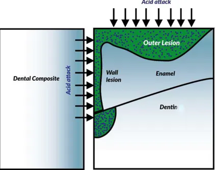
Secondary caries may develop rapidly around and below a broken restoration, or slower and more localized on the enamel along the cavosurface margin. (Figure 10) [76] A third type of secondary caries is called “cavity wall caries (Figure 10).
39 Figure 10. Histopathology of secondary caries lesions next to restoration. Secondary caries may present as an outer lesion, a cavity-wall lesion or as a lesion consisting of both an outer and cavity-wall lesion.
On the basis of the in vitro study of Hals and Nernaes [77] secondary caries lesions consist of two regions: the outer lesion and the cavity wall lesion. The outer lesion has a progressing front parallel to the outer surface of the tooth surface and is histologically similar to a primary lesion, a localized process of both demineralizations of enamel and dentin and enzymatic and bacterial degradation of dentin. [19] The wall lesion develops at the interface between restoration and tooth and progresses perpendicularly to the tooth-restoration interface.
Any site along the cavosurface margin will demineralize if the local conditions change to an acidic environment, that depends on the biomass of specific cariogenic bacteria on the more or less polished restoration surface, or rather to the intermediary salivary glycoproteins that first form a pellicle to this surface. [78] Therefore, a minimum critical amount of mature biofilm is required to create an acidic environment, and there are few alternative hypotheses regarding the origin of the acidic environment. Indeed, it has been discussed whether secondary caries would initiate on the external surface and progress to the gap between tooth structure and
40
restoration and/or would (also) be started within this interface by the diffusion of bacteria or their products. (Figure 11) [79] [80] [81] [5]
Figure 11. Schematic representation of injuries related to the secondary caries lesions and external wall. Image adapted from Kuper et al. [82]
According to the hydrodynamic flow theory, when the marginal areas of a restoration deteriorate with time, this may result in the existence of a gap or defect at the cavity wall. Subsequently, a biofilm can establish itself in this defect along the tooth-restoration interface and secondary caries can develop within the gap. This theory recognizes the not bonded tooth-restoration interface as a sensitive site, subjected to opening and closing forces that create a hydrodynamic flow. The dissolution products move with this flow allowing a new acid attack. [83] Consistent with this theory, it is clear that a wall lesion can develop in any gap, but the wider the gap, the higher the risk that it will occur.
Recently, several alternative theories have been proposed to explain the gaps/lesion wall relationship. [84] In particular, the "theory of micro-infiltration" indicates, as the cause of the demineralization and the consequent lesion wall, the passage of small amounts of fluid into the
41
gaps of the tooth/restoration interface. Bacteria carried with the fluid are able to reach the sensible site and cause pathogenicity. In addition, the "theory of macro-infiltration" suggests the necessity of a stable settlement of the biofilm in the gap to have considerable demineralization and lesion development. Gap’s dimensions are expected to be from 225 micrometers to 400 micrometers to allow bacteria settlement. [83] [85] [86]
In this contest, several microleakage studies attest to the existence of this “weak link” or “microspace” between the tooth and the restoration. [77] Nelsen et colleagues in 1952 reported that droplets with a diameter up to 44 µm developed along the restoration cavosurface margins in extracted teeth upon rapidly freezing and thawing. [87] The authors defined the phenomenon “marginal percolation” attributed to differences in thermal expansion of the tooth and the restorative material. However, only in 1961, it was reported the use of the term “marginal leakage”, [88] while the catchy term “microleakage”, still currently used, was published in 1966. [89] However, numerous variables influence the microleakage experiment outcomes such as the source and type of teeth or tooth specimens, the choice of storage substrate and time, the type of intra- or extra-coronal restoration, including the location of the cavosurface margin and angulation of cavity walls. [90] [91] [92] [93] The handling, placement technique and polishing of the restorative material also influence the extent of observable microleakage. [94] Moreover, the characteristics of the dye or tracer, such as its diameter, and the chemical properties of the solute and solvent as a function of concentrations and exposure time appear also to play a central role. [95] [96] Most tracers have a molecular radius of less than one nm, and it is not improbable that the microleakage in many cases is simply a manifestation of capillary action phenomena.
42
The minuscule molecular dimension of dyes allows penetration into inter- and intra-prismatic microporesin enamel considered to be 1–30 nm wide, as well as in the peri- or inter-tubular dentin having 0.8–2.5 µm wide dentinal tubules.
Therefore, as a consequence of a microleakage, there may be the seepage of saliva, which drains the bacterial cells into the treated tooth. These cracks also act as an ecological niche for the growth of anaerobes. [97] However, a general belief is that the cariogenic biofilm for primary and secondary caries are similar, and consist mainly of Streptococcus mutans,
Lactobacilli and Actinomyces naeslundii, [98] [99] [100] although a
contrary opinion based on observations made in an in situ experiment has been proposed. [101]
In addition, the individual’s risk for secondary caries is also modified by the saliva quantity and qualities, which in particular comprise the salivary buffering ability. Patients with xerostomia for whatever reason experience more secondary caries, as a reflection of a higher risk of all forms of caries. In this contest, the oral hygiene habits of the patient are one of the primary factors that determines if secondary caries develops.
However, despite efforts to reduce the effects of caries, population-based studies reveal that the prevalence of caries remains stubbornly high. An example of this is seen within the United Kingdom population, where 84% of dentate adults were found to have at least one restoration. Of these adults each had, on average, 7.2 filled teeth. Analysis of the survival of dental restorations from within a large database of dental treatments within UK dental practice reveals that further intervention is required: [102]
43
•within 20% of fillings after 3 years of placement •within 50% of fillings after 10 years of placement.
Reasons for this can include marginal defects, secondary caries, fracture of the restoration or adjacent tooth substance and, in the case of tooth-colored restorations, unacceptable appearance. [103] Although the recent innovation on dental restoration materials, the percentage of replacement restorations in adults still accounted for about 50% (with a range of 45 to 55 percent), remaining constant since to 2001. [104] [103] This percentage is higher for amalgam than for resin-based composite restorations. Moreover, the percentage of replaced restorations because of the diagnosis of recurrent caries is much higher in general dental practice than in controlled clinical trials, in which recurrent caries represents 2 to 3 percent of the failures. In particular, the ratio of restoration replacement to primary restorations in general dental practice has been reported to be as high as 80:20 for resin-based composite restorations and 70:30 for amalgam restorations. [105] More recent studies indicate that this ratio is about 50:50 for restorations in permanent teeth. [83] Many factors affect this ratio, including the age of the population studied and the position of the restoration. [106] In addition, all factors that enhance the accumulation of biofilm mass or impede biofilm removal may be considered as risk factors for secondary caries. It is probably the reason why the location of the restoration played a major role in the occurrence of secondary caries. [107] Cervical composite restorations (class V) were the least affected by secondary caries, which obviously may be related to the fact that many class V studies were set up to evaluate the clinical effectiveness of adhesives in non-carious cervical lesions in patients with low caries risk and good oral hygiene. The highest overall incidences were
44
found in the posterior region, although there are few studies that reported the vulnerability of anterior restorations (classes III and IV). [107] However, also the gingival margins of all types of Class II through Class V restorations are more prone to develop secondary caries, due to the possible contamination from gingival fluid and saliva leaking between the matrix and the cavosurface margin, especially if a rubber dam is not used. Moreover, corrosion and biodegradation products originating from the restorative material may influence the biofilm, [108] even if this correlation is complex and not yet fully understood. [109]
45 2.2 DIAGNOSIS AND MANAGEMENT OF SECONDARY CARIES
Accurate detection of secondary lesions is crucial for estimating the true burden of the disease and allocating appropriate treatments. Currently, there is no standard to be recommended for performing such detection, with dentists using a variety of methods, with the even greater heterogeneity of subsequent treatment decisions.
Several conventional and newer methods are available to detect secondary lesions. [110] Most of the techniques used for detecting primary caries have also been used to detect secondary caries and artificial caries-like lesions adjacent to restorations. Visual or visual-tactile examinations, often combined with bitewing radiography, are still the most common. [111] [112]
Traditionally, secondary caries lesions were assessed via tactile examination. This method seemed to be specific (specificity increased even further if only clearly detectable ditches were regarded as lesions) but insensitive. In clinical terms, only few secondary lesions would be detected, while the risk of false-positive detections was not drastically decreased compared with, for example, visual detection. For clearly cavitated secondary lesions, the tactile assessment might well be a useful method, as both sensitivity and specificity are presumably increased. However, the presence of marginal ditching, staining, discoloration of the dental tissues and gaps at the tooth restoration interface are unreliable predictors for secondary caries. [76] [113] [114] [115] Therefore, visual detection of secondary caries is a challenge for the dentist [116] and may be confused with microleakage, that can be visualized as a line of stain
46
around the restoration, or with residual (arrested) caries, which can show a grey discoloration involving the restoration.
Radiographic assessment is regularly performed to screen for proximal primary or secondary caries lesions. Secondary caries at proximal or gingival locations in restorations are diagnosed by X-rays radiography with a variable angle in relation to the lesion. [117] However, the radio-opacity of restorative materials are radiopaque may hide the lesion completely or partially. [118] The risk stemming from such non detection largely depends on the progression speed of such lesions, which is so far not fully understood. The visual and radiographic assessment might be complementary when nonproximal and proximal surfaces are checked, respectively. Laser fluorescence-based instruments have been developed as an adjunct to visual lesion detection, not causing any radiation and allowing easy reexamination and monitoring of lesions and their activity. Overall accuracy was similar to that of radiographic detection, which makes it a potential alternative, especially in children. Quantitative light-induced fluorescence (QLF) generates images of the analyzed areas, with presumable carious tissues being less fluorescent than sound areas or restoration materials. [119] However, the value of this method for detecting secondary lesions might be limited in clinical routine since QLF is currently available for visible (nonproximal) surfaces. Moreover, given the fact that even on these visually assessable surface, QLF led to false-positive detections in nearly 4 of 10 cases, there should be severe doubts toward the suitability of this method for the outlined purpose. Moreover, the burnout that frequently occurs at the cervical margin also makes the interpretation difficult. [120]
47
Marginal defects and staining around the restoration are not predictive for secondary caries, [114] [121] [122] and are likely the main factors that lead to misinterpretations and possible overtreatment. For example, black and brown marginal staining can be misinterpreted as initial lesions and are more often detected in tooth-colored resin restorations than in amalgam restorations. [76] [91] Therefore, the diagnosis of recurrent caries lacks consistency, and the diagnostic variations among clinicians are impressive. [123] These differences reflect the subjective disparities that characterize the subsequent treatment.
To date, the clinical diagnosis of secondary caries invariably has resulted in the replacement of the restoration affected, on the basis of the often proclaimed advice: “in doubt, take it out”. However, the new guidelines recommend to repair and refurbish any localized defects at restoration margins, including clinically diagnosed secondary caries, rather than performing a total replacement. In the era of minimally invasive operative dentistry, the replacement of restorations should be preferably the last alternative for patients with a defective restoration, based on the available evidence for monitoring, refurbishment and repair of restorations. [124] For secondary caries, diagnostic criteria should reflect the best options for management based on the presence of cavitation and lesion activity, ensuring the best health outcome for the patient. [125] In this contest, the treatment has to take into account the extent of the lesion, examining three characteristics of carious lesions and specifically softening of the tissues, discoloration and wetness of the lesions. Subsequently, a small part of the resin-based composite material adjacent to the stained margins are removed and, if the defects did not extend deep into the tooth-restoration interface, the cavities are considered
48
suitable for repair using a conventional restorative technique. [126] Dental teaching programs related to localized defects on restorations, including secondary caries, indicate that repair of the restoration is adopted frequently as an alternative to total replacement. [83] The majority of dental schools consider repair a definitive measure and reported that an acceptable life span of repaired restorations is four years. [83]
49 2.3 PREVENTION OF SECONDARY CARIES BY SILVER
The oral hygiene habits of the patient are the primary factor that determines if secondary caries develops, not whether the restoration along the cavosurface margin can be considered as ‘excellent’, ‘adequate’ or ‘deteriorated’. The prevention and preservation approach is significant as the caries development is a slow process. Hence, preventing early carious lesions by the removal of biofilm as well as the application of silver or placement of sealants is advised. [127] Nowadays, silver ions (Ag+) are one of the most effective methods to control bacterial growth in a variety of medical applications including the prevention of caries disease.
Since ancient times, the silver ion has been known to be effective against a broad range of microorganisms. [15] For instance, vessels made of Ag have been used for water disinfection and food preservation since the time of the Persian kings [128]. Later, the Phoenicians, Greeks, Romans, and Egyptians adopted this practice. In 1869, Raulin described, for the first time, the antimicrobial activity of silver against Aspergillus niger. [128] It is currently accepted that Ag+ is responsible for the antibacterial properties although they are relatively reactive. The binding of silver ions in the form of insoluble precipitates (AgCl), or during the interactions with proteins (e.g., albumin), causes a significant decrease of its antibacterial efficacy. The dynamic development of nanotechnology in recent years has provided new challenges for fabricating silver nanoparticles (AgNPs). In general, silver atoms (Ag0) on the surface of AgNPs, interacting with molecular oxygen or with other redox-active compounds, can be oxidized to silver oxide. [129] [130] [131] [132] [133] [134] The oxidation of silver oxide allows the release of silver ions in the environmental, and biological media
50
carrying out its antibacterial action. Therefore, AgNPs can also be used as a source of Ag+ through the release process.
Due to the oligodynamic effect, the antibacterial activity of Ag+ is directly proportional to its environmental concentration. Jung and colleagues compared the antibacterial activity of silver ions obtained in various ways and showed that silver ions produced in an electrolytic way are better antibacterial agents than those obtained by dissolving the silver compounds. [57]
Despite numerous studies conducted over the last decade, there are still considerable gaps in our knowledge about the specific mechanisms of antibacterial\antibiofilm action of silver ions. Several proposals have been developed to explain the inhibitory effects of Ag+ ions on bacteria. Furthermore, the precise basis of their antibacterial activity has yet to be defined. This is mainly due to the pleiotropic effects of nano-silver on bacterial cells, which suggests multiple mechanisms of action on several cellular targets (Figure 12).
The most noticeable are:
(i) interaction with the bacterial cell envelope (destabilization of the membrane—loss of K+ ions and a decrease of ATP level); (ii) interaction with molecules inside the cell (e.g., nucleic acids and
enzymes);
51 Figure 12. Schematic representation of the silver nanoparticle mechanism of action on the biofilm forming a microbial cell. Ag+ adhere to the microbial cell surface and results in membrane damage and disruption of electron transport chain; Ag+ penetrate inside the microbial cells and affect cellular machinery interacting with DNA bases and enzymes.
The positive charge of Ag+ interacts with the negative charge of the microbial cell wall leading to disruptions in the structural morphology of the microbial cell. [135] [136] [137] [138] Jung et al. proved that the accumulation of Ag+ in the bacterial cell envelope is followed by the separation of the cytoplasmic membrane from the cell wall in both Gram-positive and Gram-negative bacteria. [57] According to reference Jung et al., carboxyl groups (–COOH) in glutamic acid and phosphate groups in teichoic acid are mostly responsible for the binding of silver ions. However, the thickness and composition of the microbial wall is also one of the crucial factors in deciding the potency of the antimicrobial Ag. [139] The presence of a 30-nm-thick negatively charged peptidoglycan layer in gram-positive bacteria such as Staphylococcus aureus makes it less vulnerable to the silver compared to gram-negative bacteria such as E. coli
![Figure 1. The process of tooth remineralization. Adapted from Dodds et al. [20]](https://thumb-eu.123doks.com/thumbv2/123dokorg/2756990.940/12.892.174.744.453.1024/figure-process-tooth-remineralization-adapted-dodds-et-al.webp)


![Figure 8. Salivary glands and saliva function. Adapted from Dodds et al. [20]](https://thumb-eu.123doks.com/thumbv2/123dokorg/2756990.940/31.892.177.786.259.534/figure-salivary-glands-saliva-function-adapted-dodds-et.webp)

![Figure 14. Schematic representation of LDH structure (Reproduced from Tronto et al., 2013[258])](https://thumb-eu.123doks.com/thumbv2/123dokorg/2756990.940/78.892.196.769.305.716/figure-schematic-representation-ldh-structure-reproduced-tronto-et.webp)

