Transglutaminase 2 is involved in
autophagosome maturation
Manuela D’Eletto
Dottorato di Ricerca in
Biologia Cellulare e Molecolare
UNIVERSITÀ DEGLI STUDI DI
ROMA
"TOR VERGATA"
FACOLTA' DI SCIENZE MATEMATICHE FISICHE E
NATURALI
DOTTORATO DI RICERCA IN BIOLOGIA
CELLULARE E MOLECOLARE
XXII CICLO
Transglutaminase 2 is involved in autophagosome
maturation
Manuela D'Eletto
A.A. 2009/2010
Docente Guida/Tutor: Prof. Piacentini
Coordinatore: Prof. Cesareni
ABSTRACT...3
INTRODUCTION ...5
1. TRANSGLUTAMINASES ...5
2. TYPE 2 TRANSGLUTAMINASE ...7
2.1 TG2 FUNCTIONS IN THE CELLS ...10
2.2 EFFECT OF TG2 DELETION ...12
2.3 TG2 IN DISEASE STATES...13
3. AUTOPHAGY ...15
3.1 AUTOPHAGY AND NEURODEGENERATIVE DISEASE ...20
3.2 AUTOPHAGY AND CARDIAC DISEASE...20
3.2 AUTOPHAGY AND TG2 ...21
AIM OF THE THESIS ...23
MATERIALS AND METHODS ...25
1. MATERIALS...25
2. CELL CULTURE...25
3. WESTERN BLOT ANALYSIS ...26
4. FLUORESCENCE MICROSCOPY ...26 5. ELECTRON MICROSCOPY...27 6. MORPHOMETRIC ANALYSIS ...27 7. TG2 KNOCKOUT RECONSTITUTION ...27 8. ANALYSIS BY FACS ...28 9. DATA ANALYSIS ...28 10. ANIMALS ...28 RESULTS...29
1. EFFECT OF TG2 ON AUTOPHAGY INDUCTION ...29
2. THE LACK OF TG2 IS ASSOCIATED WITH AN IMPAIRMENT OF VESICLE ...35
3. EFFECT OF TG2 KNOCKOUT MEFS RECONSTITUTION AND CROSS-LINKING ACTIVITY INHIBITION ON THE AUTOPHAGOSOME MATURATION...37
4. THE ABSENCE OF TG2 INHIBITS THE FUSION OF AUTOPHAGOSOMES WITH LYSOSOMES ...39
DISCUSSION ...47
ABSTRACT
Autophagy is a highly conserved cellular process responsible for the degradation of long-lived proteins and organelles. Autophagy occurs at low levels under normal conditions, but it is enhanced in response to stress, e.g., nutrient deprivation, hypoxia, mitochondrial dysfunction and infection.
“Tissue” transglutaminase (TG2) accumulates, both in vivo and in vitro, to high levels in cells under stressful conditions. Therefore, in this study, we investigated whether TG2 could also play a role in the autophagic process.
To this end, we used TG2 knockout mice and cell lines in which the enzyme was either absent or overexpressed. The ablation of TG2 protein both in vivo and in vitro, resulted in an evident accumulation of microtubule-associated protein 1 light chain 3 cleaved isoform II (LC3 II) on pre-autophagic vesicles, suggesting a marked induction of autophagy.
By contrast, the formation of the acidic vesicular organelles in the same cells was very limited, indicating an impairment of the final maturation of autophagolysosomes. In fact, the treatment of TG2 proficient cells with NH4Cl, to inhibit lysosomal activity, led to a marked accumulation of LC3 II and damaged mitochondria similar to what we observed in TG2-deficient cells.
These data indicate a role for TG2-mediated post-translational modifications of proteins in the maturation of autophagosomes accompanied by the accumulation of many damaged mitochondria.
INTRODUCTION
1. TRANSGLUTAMINASES
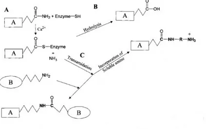
The human transglutaminases (TGase) are a widely distributed and peculiar group of enzymes that catalyse the post-translational modification of proteins by the formation of isopeptide bonds. This may occur either through protein cross-linking via ε-(γ-glutamyl) lysine bonds or through incorporation of many primary amines at the level peptide-bound glutamine residues (Folk and Finlayson, 1977) (Figure 1).
The cross-linked protein products are highly resistant to mechanical challenge and proteolytic degradation, and their accumulation is found in a number of tissues, including skin, hair, blood clotting and wound healing (Lorand and Conrad, 1984).
Figure 1. (A) TGases display a Ping Pong-based mechanism, whereby the active, Ca2+
stabilized conformation of the enzyme forms a covalent intermediate between the active-site thiol residue and a glutamine residue in the first protein substrate, with concomitant activation of the glutamine acyl moiety and release of ammonia. (B) This active thioester undergoes either hydrolysis (an unfavourable reaction), releasing glutamic acid in the substrate protein, or (C) an acyl transfer to a primary amine, which can be either a small molecule (like a polyamine) or protein-bound (the ε-amino group of a lysine residue).
In mammals, nine distinct TGase isoenzymes have been identified at the genomic level (Grenard et al., 2001); however, only six have so far been isolated and characterized at the protein level, after purification either from natural sources or as recombinant proteins. The fully characterized enzymes include (a) the circulating zymogen Factor XIII, which is converted, by a thrombin-dependent proteolysis, into the active TGase Factor XIIIA, (plasma TGase) involved in stabilization of fibrin clots and in wound healing; (b) the keratinocyte TGase (type 1 TGase) which exists in membrane-bound and soluble forms, is activated severalfold by proteolysis and is involved in the terminal differentiation of keratinocytes; (c) the ubiquitous tissue TGase (type 2 TGase); (d) the epidermal/hair follicle TGase (type 3 TGase), which also requires proteolysis to become active and, like type 1, is involved in the terminal differentiation of keratinocytes; (e) the prostatic secretory TGase (type 4 TGase) (Dubbink et al., 1998), essential for fertility in rodents; and (f) the characterized type 5 TGase, involved
in keratinocyte differentiation
(Candi et al., 2001). All mammalian forms have considerable structural homology, are products of different genes arising from duplication, rearrangement and chromosomal shifts, and are members of the papain-like superfamily of cysteine proteases (Makarov et al., 1999).In addition to these well-studied enzymatic actions, some members of the TG family participate in a plethora of other biological processes through actions unrelated to their transamidase catalytic activity (Lorand and Graham, 2003). For example, TG2 (Im and Graham, 1990), TG4 (Spina et al., 1999), and TG5 (Candi et al., 2004) bind and hydrolyze GTP: GTP binding causes a transition to the compact, inactive conformation and the inhibition of TG activity (Liu et al., 2002). To date, however, only TG2 has been shown to utilize this function in its role as a high-molecular-weight G protein (Gh) involved in signalling by certain G protein-coupled receptors
(Nakaoka et al., 1994). FXIIIA (Ueki et al., 1996) and TG2 (Lorand et al., 1988) also function as adaptor proteins to facilitate matrix interactions during cell adhesion. Finally, TG2 has also been reported to act as a serine/threonine kinase (Mishra and Murphy, 2004), a protein disulfide isomerase (Hasegawa et al., 2003; Mastroberardino et al., 2006), and an extracellular ligand for the orphan GPCR, GPR56, which is involved in tumor suppression (Xu et al., 2006), although involvement of these latter functions in physiology and/or disease remains unclear.
All members of this superfamily possess a catalytic triad of Cys-His-Asp or Cys-His-Asn. A few clusters are easily identifiable on the basis of
sequence homology, which include the non-enzymic erythrocyte band 4.2 proteins, TG4, Factor XIII, TG5, TG7, TG3 and finally, TG2. The tissue content of the different isoenzymes is tightly regulated at the transcriptional level (Polakowska et al., 1999; Lee et al., 1996).
Early structural studies on TGases, performed by high-resolution crystallography on the zymogenic A subunit of plasma factor XIII, which needs proteolytic processing by thrombin to generate the active dimeric enzyme, revealed that each Factor XIIIA subunit is composed of four domains (termed N-terminal β-sandwich, core domain, containing the catalytic and regulatory sites, and C-terminal β-barrels 1 and 2). This organization in four domains is highly conserved during evolution among the TGase isoforms.
Deregulation of enzyme activity, generally associated with major disruptions in cellular homoeostatic mechanism, has resulted in these enzymes contributing to a number of human diseases, including neurodegeneration, autoimmune diseases, infectious diseases, progressive tissue fibrosis and diseases related to the assembly of the stratum corneum of the epidermis of the skin (Kim et al., 2002).
2. TYPE 2 TRANSGLUTAMINASE
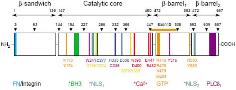
Tissue or type 2 transglutaminase (TG2) represents the most ubiquitous isoform belonging to TGases family. This protein is found throughout the body due to its constitutive expression in endothelial cells, smooth muscle cells, and fibroblasts as well as in a number of organ-specific cell types (Fesus and Arato, 1986; Thomazy and Fesus, 1989). The human TG2 gene has been localized to chromosome 20q12 by in situ fluorescence hybridization (Gentile et al., 1994). The gene of human TG2 has been reported to be 32.5 kb in size and contains 13 exons and 12 introns (Gentile et al., 1991; Fraij and Gonzales, 1997). Full length TG2 is 687 aminoacids in length and the predicted molecular mass is about 78 kDa.
The structure of TG2, crystallized in a dimer form in a complex with GDP has been reported (Liu et al., 2002); like to the other transglutaminases (as mentioned above), TG2 has four distinct domains (Figure 2): a N-terminal β-sandwich, with fibronectin and integrin binding site, a catalytic core, containing the catalytic triad for the acyl-transfer reaction, and two C-terminal β-barrel domains.
Figure 2. The four structural domains are indicated by arrows with amino acid positions (top). Exon boundaries of the gene encoding TG2 are indicated by arrowheads with numbers corresponding to the last amino acids of each exon-encoded region. Exon 10, which contains residues forming the GDP-binding site, is marked.
A unique guanidine nucleotide-binding site, which has not been found in any other protein, is located in a cleft between the catalytic core and the first b-barrel; this sequence is coded by exon 10 of the TG2 gene, which has very poor sequence homology with the same exons in other TGases.
The arrangement of the aminoacids of the catalytic centre (Cys277, His335
and Asp358) in a charge-relay catalytic triad, analogous to that of thiol
proteinases such as papain (Pedersen et al., 1994), confers high reactivity on Cys277 to form thioesters with peptidylglutamine moieties in the protein
substrate or to react with relatively mild chemicals such as acrylamide (Bergamini and Signorini 1988). The high reactivity of Cys277 has been
employed to develop a wide range of active-site-directed irreversible inhibitors of the enzyme. In the absence of Ca2+, the enzyme assumes the
basic latent conformation and the reactivity of Cys277 is decreased either by
hydrogen-bonding with the phenolic hydroxy group of Tyr516 or by
formation of a disulphide with a neighbouring cysteine residue, namely Cys336 (Noguchi et al., 2001).
TG2 is located in the extracellular matrix (ECM) or at the cell surface in association with the ECM (Gaudry et al., 1999) and also intracellularly, where it is mostly cytosolic, but is also found associated with the inner face of the plasma (Im and Graham, 1990) or nuclear membrane (Singh et al., 1995), or in the mitochondria (Krasnikov et al., 2005).
The transamidase activity of TG2 plays an extracellular role in matrix stabilization, which is important in wound healing, angiogenesis, and bone remodeling, and an intracellular role, predominantly in the cross-linking of proteins during apoptosis. TG2 has five reported functions in addition to its transamidase activity. These include GTPase activity and intracellular G protein signalling via the α1B/α1D adrenergic receptors (Nakaoka et al.,
1994), the TPα thromboxane A2 receptor (Vezza et al., 1999), and the oxytocin receptor (Park et al., 1998). An extracellular stabilizing function by being able to form tight ternary TG2-fibronectin-collagen complexes (Radek et al., 1993), and an adapter function to facilitate cell adhesion to fibronectin by interacting directly with β1/β3/β5 integrins (Akimov et al., 2000), with heparan sulfate chains of the heparan sulfate proteoglycan receptor, syndecan 4 (Telci et al., 2008), or with the orphan G protein-coupled cell-adhesion receptor GPR56 (Xu et al., 2006). TG2 has also been reported to possess intracellular serine/threonine kinase activity, with insulin-like growth factor binding protein 3 (IGFBP), p53 tumor suppressor protein, or histones (Mishra and Murphy, 2004) as substrates, as well as extracellular protein disulfide isomerase (PDI) activity (Hasegawa et al., 2003; Mastroberardino et al., 2006). The transglutaminase and G protein signaling activities of this protein are reciprocally regulated by the binding of Ca2+ and
GTP, respectively (Nakaoka et al., 1994): calcium-activated TG2 assumes an expanded ellipsoid structure, while GTP binding promotes transition to the compact, transamidase-inactive conformation (Begg, Carrington et al., 2006; Pinkas et al., 2007) (Figure 3).
Figure 3. Three-dimensional ribbon structure of the compact, inactive GDP-bound form and the expanded irreversible peptide inhibitor-bound form of TG2. A remarkably large conformational change, from the compact form to an extended ellipsoid structure that exposes the TG active site, accompanies activation of TG2.
2.1 TG2 FUNCTIONS IN THE CELLS
TG2 subcellular locations as well as its identified substrates clearly suggest multiple roles for this enzyme. In fact extensive analysis carried on in different cellular populations under physiological and pathological settings lacked to provide a unique paradigm: while some cell types (e.g. endothelial and smooth muscle cells) express constitutive high TG2 level (Thomazy and Fesus, 1989), in other cell types it is induced by distinct signalling pathways, targeting specific response elements in the regulatory region of the gene. Retinoic acid (RA), TGFβ, NF-κB and AP responsive sites and regions have been functionally identified and all of them are related to induction of cellular defence mechanisms and cellular maturation (Szegezdi et al., 2000).
Once expressed, the regulation of TG2 activities relies on multiple heterogeneous factors. As a G-coupled protein, the role of TG2 in transmitting signals from seven transmembrane-helix receptors to phospholipase C has been clearly described (Iismaa et al., 2000), being phospholipase C activated following TG2 binding of GTP (Murthy et al., 1999). High Ca2+ levels can induce the release of GTP/GDP molecules,
inhibiting signalling and promoting transglutaminase activity. Interaction of TG2 with specific molecules (e.g. with sphingosylphosphocholine) might reduce the Ca2+ requirement for the transglutaminase activity (Lai et al.,
1997).TG2 activity is also strongly influenced by nitric oxide: up to 15 of the 18 cysteine residues can be nitrosylated and denitrosylated in a Ca2+
-dependent manner, inhibiting and activating the enzyme, respectively (Lai et al., 2001).
After Ca2+ activation, TG2 interacts and modifies major components of
the cytoskeleton. In response to RA treatment, TG2-dependent transamidation of RhoA results in the increased binding of RhoA GTPase to ROCK-2 protein kinase, autophosphorylation of ROCK-2 and phosphorylation of vimentin which can lead to the formation of stress fibers and increased cell adhesion (Singh et al., 2001).These events are prevented by TG2 inhibition. TG2 can interact with β-tubulin and with microtubule-binding proteins (Piredda et al., 1999) including tau, which can be crosslinked by the enzyme (Murthy et al., 1998). As a matter of fact, recently it has been suggest a role for TG2-mediated cross-linking in stabilizing particulate material shed from the placenta and, more generally, in the organization and turnover of trophoblast plasma membrane and associated cytoskeleton (Robinson et al., 2007). An interesting aspect of TG2 function is its capability to translocate to the nucleus under certain conditions: in fact,
through
the two nuclear localization sequences (NLS) (Figure 2) and, presumably with the help of importin-a3, TG2 comes in the nucleus (Peng et al., 1999), where it can function either as a G protein or as a transamidase activated by nuclear Ca2+-signals to crosslink histones, retinoblastoma andSP1 proteins. This suggests that TG2 could have a direct role in chromatin modifications or gene expression regulation.
TG2 is induced in cells undergoing apoptosis in vivo (Fesus et al., 1987). Its overexpression primes cells for suicide and inhibition of its expression by antisense strategy results in decreased cell death (Oliverio et al., 1999). It has been reported that TG2 sensitizes cells for apoptosis by interacting with mitochondria (Piacentini et al., 2002), shifting them to a higher polarized state and altered redox status. This might provoke activation of transglutaminase crosslinking activity. During the late stage of apoptosis, the massive increase of cytosolic Ca2+ determines the switch of TG2 to its
crosslinking configuration in all subcellular compartments leading to extensive polymerization of intracellular proteins (including actin and Rb) and formation of detergent-insoluble structures (Oliverio et al., 1997; Fesus et al., 1989). These protein scaffolds stabilize the structure of the dying cell
before its clearance by phagocytosis, limiting the release of harmful intracellular components and consequently inflammatory or autoimmune responses (Piredda et al., 1997). Under pathological conditions the death of cells expressing high amounts of TG2 can occur as a result of a ‘mummification’ event caused by extensive crosslinking of cytosolic proteins without signs of either apoptosis or necrosis (Griffin and Verderio, 2000).
2.2 EFFECT OF TG2 DELETION
Considering the multifunctionality and unique cellular biochemistry of TG2, it has been unexpectedly found out that homozygous deletion of TG2 does not result in an embryonic lethal phenotype (De Laurenzi and Melino, 2001; Nanda et al., 2001). The homozygous null animals are viable, of normal size and weight, and born with mendelian frequency. The most probable explanation for the lack of severe phenotypes is that other transglutaminases in mammalian tissues can compensate for the loss of TG2. However, such compensation is necessarily partial, since the other mammalian transglutaminases do not bind GDP/GTP and, with the exception of FXIIIA, they have not been found on the cell surface.
Therefore, alterations are expected in TG2–/– mice, especially under
certain stresses and pathological conditions. In fact, decreased adherence of primary fibroblasts (Nanda et al., 2001) and impaired wound healing related to altered cytoskeletal dynamics of fibroblasts have been observed in these mice, consistent with the suggested extra- and intra-cellular functions of TG2. Moreover, when cell death is induced in TG2-/- mice, the clearance of
apoptotic cells by phagocytosis is defective in the thymus and the liver and inflammatory as well as autoimmune reactions develop (Szondy et al., 2003).
TG2-deficient mice also show glucose intolerance and hyperglycaemia because of reduced insulin secretion, a phenomenon similar to a subtype of diabetes called MODY (for maturity-onset diabetes of the young) (Bernassola et al., 2002). Further, the ablation of TG2 in mice causes an increased vulnerability of cardiomyocytes to ischemia/reperfusion injury (Szondy et al., 2006). This effect is associated with a decreased capacity of TG2-/- mice to synthesize ATP, due to a reduced activity of mitochondrial
2.3 TG2 IN DISEASE STATES
TG2 has been implicated in the pathogenesis of a number of diseases, such as celiac sprue (Molberg et al., 2000), neurodegenerative disorders (Hoffner and Djian, 2005), diabetes (Bernassola et al., 2002), liver cirrhosis and fibrosis (Mirza et al., 1997; Issa et al., 2004), renal scarring (Johnson et al., 2003), and certain types of cancer (Mangala and Mehta, 2005). Importantly, it is the enzymatic function of TG2 that is thought to contribute to the pathology or etiology of most of the aforementioned diseases. Coeliac disease is a malabsorbtion syndrome characterized by almost total atrophy of villi in the jejunum on exposure to dietary glutens.
TG2 is involved in generating T cell stimulatory gluten peptides through deamidation of specific glutamines (Willemijn et al., 2002). In HLA-DQ2 or HLA-DQ8 settings, the TG2-formed disease-triggering epitopes provoke a pathological immune response that destroys the jejunal epithelium. In parallel, a T-cell-mediated autoimmune response is initiated, producing IgA-type autoantibodies against TG2 (Dieterich et al., 1997), the detection of which has become a widely used diagnostic marker of coeliac disease.
Dysregulation of the suggested functions of TG2 in various pathological settings might significantly contribute to the development of fibrosis in susceptible organs such as the lung, liver and kidney (Skill et al., 2001).
Alzheimer's disease, the most common age-related neurodegenerative disorder, is associated with the selective damage of brain regions and neural circuits, including but not exclusively, neurons in the neocortex, hippocampus, and amygdala. Dysfunction and loss of neurons in these neural circuits result in impaired memory, thinking and behavior. Two major defining hallmarks of Alzheimer's disease pathology are the extracellular neuritic senile plaques and the intraneuronal neurofibrillary tangles. Senile plaques and neurofibrillary tangles are formed by abnormally polymerized proteins in the brain, and both of these lesions are extremely insoluble structures. Purification and analysis have demonstrated that senile plaques contain amyloid fibrils composed of the amyloid β-protein (Aβ), a 39-42 amino acid peptide which is proteolytically derived from a larger transmembrane glycoprotein, the amyloid precursor protein (APP) (Kang et al., 1987; Masters et al., 1985). Neurofibrillary tangles are composed primarily of paired helical filaments (PHFs) (Delacourte and Defossez, 1986; Kosik et al., 1986), and a major component of the PHFs is the hyperphosphorylated form of the microtubule associated protein tau (Goedert et al., 1992; Lee et al., 1991). It has been hypothesized that TG2
may be involved in the pathogenesis of Alzheimer's disease by facilitating the formation of one or both of these insoluble lesions. TG2 has been localized by biochemistry and immunocytochemistry in the human brain, particularly in neurons (Appelt et al., 1996; Miller and Anderton, 1986). Miller and Anderton (1986) also investigated the cross-linking of neurofilaments by TG2, and extended the previous findings by demonstrating that all three neurofilaments polypeptides are TG2 substrates and can be cross-linked into insoluble, but non-filamentous aggregates.
The major component of the neurofibrillary tangles, is an abnormally phosphorylated form of the microtubule-associated protein tau and not neurofilament; therefore, it was hypothesized that the pathological aggregation of tau into insoluble neurofibrillary tangles may be enzymatically facilitated by TG2, in fact, several studies have demonstrated that tau is readily cross-linked by TG2 (Appelt et al., 1996; Appelt and Balin, 1997).
Huntington disease (HD) is a dominantly inherited disorder that is characterized by a progressive impairment of coordination and/or motor neuron degeneration, and is associated with variable mental syndromes (Vonsattel and DiFiglia, 1998). This disorder is caused by the expansion of CAG trinucleotide repeats in the gene encoding huntingtin (htt) (Ross, 1995).The CAG trinucleotide repeats are found in the coding region, and are translated to a stretch of polyglutamine in the expressed protein. In the normal population the number of CAG repeats varies from 6 to 35, whereas lengths of 40 and over invariably cause HD (Gusella et al., 1993) and the length of CAG repeat significantly correlate with the severity of clinical symptoms in HD patients (Andrew et al., 1993).
Several studies have proposed that Huntington's disease could be caused in part by abnormal protein-protein interactions related to the elongated polyglutamine stretch in huntingtin. The neuronal intranuclear and cytosolic inclusions composed of mutant huntingtin are present in Huntington brain (Davies et al., 1997; DiFiglia et al., 1997), although it is not clear whether they are harmful or beneficial (Saudou et al., 1998). The cytoplasmic and intranuclear inclusions of mutant huntingtin that occur in Huntington's disease brain are likely to play a role in the process of the disease and there is evidence which indicates that TG2 may contribute to the formation, growth and/or stabilization of these aggregates (Gentile et al., 1998).
One of the proposed mechanisms of htt aggregation is based on the action of TG2 because expanded polyglutamine repeats are excellent glutaminyl-donor substrates for TG2-catalyzed cross-linking (Lesort et al., 2000).
By crossing HD R6/1 transgenic mice with TG2–/– mice, a reduction in
cell death was observed in R6/1/TG2–/– compared with HD R6/1 mice,
together with the potentiation of the formation of htt aggregates and significant improvement in both motor performance and survival (Mastroberardino et al., 2002) suggesting that the involvement of TG2 in the loss of neurons in HD is not related to the formation of htt aggregates.
Although TG2’s role in the aggregate formation is not yet well established, it is clear that TG2 could contribute to the pathogenesis in HD through mechanisms other than aggregate formation. The interplay between TG2 and mitochondrial function could be one of the mechanisms for the pathogenesis of HD, although supporting evidence for this hypothesis is still lacking. Impaired mitochondrial function, which has been one pathological mechanism for HD, resulted in a significant increase of TG activity in situ (Lesort et al., 2000). At the same time, studies showed that TG2 might act as a 'sensitizer' towards apoptotic stimuli by modulating mitochondrial function (Piacentini et al., 2002). Since HD is characterized by impaired mitochondrial function together with increased TG2, there is likely an important interplay between TG2 and mitochondrial function which could contribute to the pathogenesis of HD.
Recent studies have proved that autophagy has a protective role towards these deseases, being the foundamental mechanism to remove proteic aggregates (Winslow and Rubinsztein, 2008).
3. AUTOPHAGY
Autophagy is a degradative pathway mostly implicated in the recycling of portions of cytosol and in the removal of superfluous or damaged organelles. In addition to proteins, this transport route is uniquely able to catabolize other cellular constituents such as lipids, carbohydrates and nucleic acids.
This process occurs at a basal level in most tissues and contributes to the routine turnover of cytoplasmic components. However, it can also be massively induced by a change in the environmental conditions or by cytokines and other signaling molecules to adapt and/or cope with various physiological and pathological situations.
As a result, autophagy is important for cellular remodelling and development, and is involved in preventing ageing and controlling cell growth (Levine and Klionsky, 2004). Moreover, it plays a protective role in several human diseases such as cancer, neurodegeneration (Huntington’s,
Parkinson’s and Alzheimer’s diseases) and muscular disorders (Huang and Klionsky, 2007; Levine and Kroemer, 2008).
Autophagy also defends cells from invasion by certain pathogenic bacteria such as Mycobacterium tuberculosis, viruses such as the herpes simplex virus and the tobacco mosaic virus, and intracellular parasites like Toxoplasma gondii (Amano et al., 2006; Huangand Klionsky, 2007; Levine and Kroemer, 2008). Finally, autophagy may be the central player of type II programmed cell death and in some cases appears to be regulated in conjunction with apoptosis (Gorski et al., 2003).
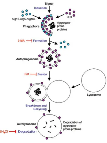
The autophagic pathway involves the delivery of cytoplasmic cargo sequestered inside double-membrane vesicles to the lysosome. Initial steps include the formation (vesicle nucleation) and expansion (vesicle elongation) of an isolation membrane, which is also called a phagophore. The edges of the phagophore then fuse (vesicle completion) to form the autophagosome, a double-membraned vesicle that sequesters the cytoplasmic material. This is followed by fusion of the autophagosome with a lysosome to form an autolysosome where the captured material, together with the inner membrane, is degraded (Figure 4).
Nutritional status, hormonal factors, and other cues like temperature, oxygen concentrations, and cell density are important in the control of autophagy. One of the key regulators of autophagy is the target of rapamycin, TOR kinase, which is the major inhibitory signal that shuts off autophagy in the presence of growth factors and abundant nutrients.
Figure 4. Schematic diagram of the steps of autophagy. Autophagy begins with the formation of the phagophore or isolation membrane (vesicle nucleation step). The concerted action of the autophagy core machinery proteins at the phagophore assembly site (PAS) is thought to lead to the expansion of the phagophore into an autophagosome (vesicle elongation). The autophagosome can engulf bulk cytoplasm non specifically, including entire organelles, or target cargos specifically. When the outer membrane of the autophagosome fuses with an endosome (forming an amphisome before fusing with the lysosome) or directly with a lysosome (docking and fusion steps), it forms an autophagolysosome. Finally, the sequestered material is degraded inside the autophagolyosome (vesicle breakdown and degradation) and recycled.
The class I PI3K/Akt signaling molecules link receptor tyrosine kinases to TOR activation and thereby repress autophagy in response to insulin-like and other growth factor signals (Lum et al., 2005). Some of the other regulatory molecules that control autophagy include 5′-AMP-activated protein kinase (AMPK), which responds to low energy; the eukaryotic initiation factor 2α (eIF2α), which responds to nutrient starvation, double-stranded RNA, and endoplasmic reticulum (ER) stress; BH3-only proteins that contain a Bcl-2 homology-3 (BH3) domain and disrupt Bcl-2/Bcl-XL
inhibition of the Beclin 1/class III PI3K complex; the tumor suppressor protein, p53; death-associated protein kinases (DAPk); the ER membrane-associated protein, Ire-1; the stress-activated kinase, c-Jun-N-terminal kinase; the inositol-trisphosphate (IP3) receptor (IP3R); GTPases; Erk1/2;
ceramide; and calcium (Meijer and Codogno, 2006; Rubinsztein et al., 2007).
Downstream of TOR kinase, there are more than 20 genes in yeast (known as the ATG genes) that encode proteins (many of which are
evolutionarily conserved) that are essential for the execution of autophagy (Mizushima and Klionsky, 2007). These include a protein serine/threonine kinase complex that responds to upstream signals such as TOR kinase (Atg1, Atg13, Atg17), a lipid kinase signaling complex that mediates vesicle nucleation (Atg6, Atg14, Vps34, and Vps15), two ubiquitin-like conjugation pathways that mediate vesicle expansion (the Atg8 and Atg12 systems), a recycling pathway that mediates the disassembly of Atg proteins from mature autophagosomes (Atg2, Atg9, Atg18), and vacuolar permeases that permit the efflux of amino acids from the degradative compartment (Atg22).
In mammals, proteins that act more generally in lysosomal function are required for proper fusion with autophagosomes,such as the lysosomal transmembrane proteins, LAMP-2 and CLN3, and for the degradation of autophagosomal contents, such as the lysosomal cysteine proteases, cathepsins B, D, and L.
The identification of signals that regulate autophagy and genes that execute autophagy has facilitated detection and manipulation of the autophagy pathway. Phosphatidylethanolamine (PE) conjugation of yeast Atg8 or mammalian LC3 during autophagy results in a nonsoluble form of Atg8 (Atg8-PE) or LC3 (LC3 II) that stably associates with the autophagosomal membrane (Figure 5).
Consequently, autophagy can be detected biochemically (by assessing the generation of Atg8-PE or LC3 II) or microscopically (by observing the localization pattern of fluorescently tagged Atg8 or LC3) (Mizushima and Klionsky, 2007). These approaches must be coupled with ancillary measures to discriminate between two physiologically distinct scenarios increased autophagic flux without impairment in autophagic turnover (i.e., an increased “on-rate”) versus impaired clearance of autophagosomes (i.e., a “decreased offrate”), which results in a functional defect in autophagic catabolism.
Autophagy can be pharmacologically induced by inhibiting negative regulators such as TOR with rapamycin (Rubinsztein et al., 2007), and pharmacologically inhibited by targeting the class III PI3K involved in autophagosome formation with 3-methyladenine, by targeting the fusion of autophagosomes with lysosomes, using inhibitors of the lysosomal proton pump such as bafilomycin A1, or by decreasing the lysosomal protease activities using NH4Cl that neutralize lysosomal pH (Rubinsztein et al.,
Figure 5. Aggregate-prone proteins like mutant huntingtin can also be sequestered in this way. The Atg12-Atg5-Atg16L complex and LC3 localize to the phagophore throughout its elongation process. Upon completion of autophagosome formation, the Atg12-Atg5-Atg16L complex dissociates from the membrane while LC3 II remains on it. Autophagy can be inhibited by drugs like 3-methyladenine (3-MA) at the formation of autophagic vacuole stage, by bafilomycin A1 (Baf) at the fusion stage between autophagic vacuole and lysosome and by NH4Cl at the degradation stage after the autolysosome formation.
3.1 AUTOPHAGY AND NEURODEGENERATIVE DISEASE
Early reports demonstrating that autophagosomes accumulate in the brains of patients with diverse neurodegenerative diseases, including Alzheimer’s disease, transmissible spongiform encephalopathies, Parkinson’s disease, and Huntington’s disease (Rubinsztein et al., 2007; Williams et al., 2006), led to the initial hypothesis that autophagy contributed to the pathogenesis of these disorders. In mice with cerebellar degeneration due to mutations in glutamate receptor, autophagy was also postulated to be a mechanism of nonapoptotic cell death (Yue et al., 2002). In contrast, more recent studies provide compelling evidence that at least in model organisms autophagy protects against diverse neurodegenerative diseases and that the accumulation of autophagosomes primarily represents the activation of autophagy as a beneficial physiological response or, in the case of Alzheimer’s disease, the consequence of a defect in autophagosomal maturation (Rubinsztein et al., 2007; Williams et al., 2006).
Beyond its role in the clearance of misfolded proteins spontaneously generated during routine protein turnover, autophagy likely plays an important role in the clearance of aggregate-prone mutant proteins associated with several different neurodegenerative diseases. These include proteins with polyglutamine (polyQ) expansion tracts such as those seen in Huntington’s disease, mutant α-synucleins that cause familial Parkinson’s disease, and different forms of tau including mutations causing frontotemporal dementia (Williams et al., 2006). Because substrates need to be unfolded to pass through the narrow pore of the proteasomal barrel, oligomeric and aggregated proteins are poor substrates for proteasomal degradation and better targets for autophagic degradation (Figure 5).
The mechanism by which these proteins exert their cellular toxicity is still controversial, but it is generally believed that they are particularly toxic in oligomeric complexes and that higher-order protein aggregates may be formed as a last attempt to prevent toxicity in the absence of a properly functioning quality-control system (Martinez-Vicente and Cuervo, 2007).
3.2 AUTOPHAGY AND CARDIAC DISEASE
Defective autophagy (due to impaired autophagosome-lysosome fusion) may play a role in relatively rare forms of inherited diseases of the heart
(e.g., Danon disease, Pompe disease). Of greater medical significance is the possibility that autophagy may constitute an important physiological or pathophysiological response to cardiac stresses such as ischemia or pressure overload, which are frequently encountered in patients with coronary artery disease, hypertension, aortic valvular disease, and congestive heart failure.
The accumulation of autophagosomes has been noted in cardiac biopsy tissues of patients with these disorders, rodent models of these cardiac diseases, and isolated stressed cardiomyocytes (Terman and Brunk, 2005).
Prior to genetic studies, it was largely assumed that autophagy invariably contributed to myocyte degeneration in such settings. However, more recent data challenge this view; the cytoprotective effects of autophagy (either via ATP production, protein and organelle quality control, or other mechanisms) may predominate in certain settings. The cardiomyocyte, similar to the neuron, is a postmitotic cell in which basal autophagy may be important in protein and organelle quality control.
The heart consumes more energy per gram than any other organ in the body, and common cardiac disorders (e.g., cardiac ischemia and heart failure) are characterized by a reduction in the availability of energy substrates, a factor that contributes to transient or sustained impairment of cardiac function. Furthermore, when cardiac stresses are sustained for long periods of time, myocytes remodel their cellular architecture (e.g., undergo elongation and hypertrophy) to adapt to stress. The needs of the stressed heart for more energy substrates and for cellular remodeling may be met in part through the autophagy pathway; cardiac-specific deficiency of atg5 early in cardiogenesis does not result in any phenotypic abnormalities under basal conditions but results in more severe cardiac dysfunction following treatment with pressure overload or β-adrenergic stress.
These data suggest that upregulation of autophagy in failing hearts is an adaptive response that protects against hemodynamic or neurohormonal stresses.
3.2 AUTOPHAGY AND TG2
A recent work has been shown that inhibition of PKC-delta by rottlerin or knockdown of TG2 protein by a TG2-specific siRNA, resulted in a marked increase in autophagy shown by presence of autophagic vacuoles in the cytoplasm, formation of the acidic vesicular organelles, membrane association of microtubule-associated protein 1 light chain 3 (LC3) with
autophagosomes, and a marked induction of LC3-II protein, important hallmarks of autophagy (Akar et al., 2007).
These results revealed that PKC-delta plays a critical role in the expression of TG2, which in turn may regulate autophagy, suggesting a novel mechanism of TG2-mediated autophagy. However, previous studies carried out in TG2-/-/ HD transgenic mice support a positive regulation of
autophagy by TG2 (Mastroberardino et al., 2002). These mice showed that the HD onset is associated by a large reduction in nonapoptotic cell death associated with an increased number of protein nuclear inclusions, clearly suggesting an impairment in their autophagic pathway.
AIM OF THE THESIS
Transglutaminase 2 is a multifunctional enzyme able to catalyse Ca2+
-dependent post-translational modification of proteins, it may also act as a G protein in transmembrane signaling, as a kinase, as a protein disulphide isomerase and as a cell surface adhesion mediator (Fesus and Piacentini, 2002). The vast array of biochemical functions catalysed by TG2 distinguishes it from the other members of the transglutaminase family.
Multiple lines of evidence suggest an involvement of the enzyme in neurodegenerative diseases, such as Huntington (HD) and Parkinson (PD), heart failure, infectious diseases and cancer (Mastroberardino et al., 2002; Fesus and Piacentini, 2002).
Interestingly, autophagy has been shown to play an essential role, whether the pathogenesis of all these diseases or in the response to them, thus suggesting a possible participation of TG2 in this “self-digestion” pathway (Rubinsztein et al., 2007).
In particular, recent reports have demonstrated that multiple forms of cardiovascular stress, including pressure overload, chronic ischemia and infarction-reperfusion injury, provoke an increase in autophagic activity in cardiomyocytes (Terman and Brunk, 2005). Autophagy, or cellular self-digestion, is a cellular pathway involved in protein and organelle degradation, including damaged mitochondria (Levine and Klionsky, 2004).
Autophagy occurs at low basal levels under normal conditions in the heart, but is rapidly upregulated in response to stress, e.g., nutrient deprivation, hypoxia and pressure overload. It is widely accepted that an impaired autophagy might represent a prominent feature of myocardial ischemia and reperfusion. In fact, in the heart, autophagy plays an essential role in maintaining cellular homeostasis under normal conditions (Terman and Brunk, 2005).
Upregulation of autophagy may be beneficial to the cell by recycling of proteins to generate free amino acids and fatty acids needed to maintain energy production, by removing damaged organelles, and by preventing accumulation of protein aggregates.
In fact, autophagy is considered the major clearance route for intracellular aggregate-prone proteins causing neurodegenerative diseases (Williams et al., 2006). Recently, it has been shown that inhibition of PKC-delta, by
rottlerin, or knockdown of TG2 protein, by siRNA in pancreatic cancer cells, resulted in a marked increase in autophagy (Akar et al., 2007).
These results revealed that PKC-delta plays a critical role in the expression of TG2, which in turn may regulate autophagy, suggesting a novel mechanism of TG2-mediated autophagy. However, previous studies carried out in TG2-/-/ HD transgenic mice support a positive regulation of
autophagy by TG2. In these mice the HD onset is associated by a large reduction in nonapoptotic cell death associated with an increased number of protein nuclear inclusions, clearly suggesting an impairment in their autophagic pathway (Mastroberardino et al., 2002).
Considering the important role of TG2 in neurodegenerative diseases and heart homeostasis, we decided to investigate the function of TG2 in the regulation of autophagy. This study shows that the enzyme plays an important role in the later stages of autophagy.
MATERIALS AND METHODS
1. MATERIALS
LysoTrackerRed was obtained from Molecular Probes. Anti-β-tubulin and anti-actin were from Sigma, anti-LC3 was from MBL, anti-LC3 was from Novus Biologicals, anti-TG2 was from NeoMarkers, anti-p62 was from BD Transduction Laboratories, ANT was from Calbiochem, anti-COXIII was from Molecular Proes and anti-caspase3 was from Cell Signaling. Secondary antibodies were from Jackson Immunoresearch Labs. Earle’s balanced salt solution (EBSS), rapamycin, 2-deoxy-D-glucose, bafilomycin A1, NH4Cl and acridine orange were obtained from Sigma.
DMEM and FCS were obtained from Gibco; ECL detection system (Chemi- Glow) was from Alpha Innotech.
2. CELL CULTURE
MEFs (mouse embryo fibroblasts) were isolated by trypsinization of littermate embryos from wild-type and knockout TG2 mouse at E14. The dissociated cells were plated and grown to near-confluence and were passed every 3 or 4 days until reaching “crisis” period (in which there is no increase in cell number of the cultures). The cultures spontaneously resume a rapid rate of proliferation (between 90–100 days) and these immortal cultures will continue to divide indefinitely.
All cells were cultured in DMEM supplemented with penicillin/streptomycin and 10% foetal calf serum. For amino acid starvation, cells were washed two times in Earle’s balanced salt solution (EBSS) and incubated in EBSS for the indicated periods. For autophagy induction cells were incubated in full medium in the presence of 1 μM rapamycin for 48 h or 10 mM 2-deoxy-D-glucose for 36 h. Bafilomycin A1 (100 nM) and NH4Cl (20 mM) treatments were administered in DMEM. For TG2 inhibitor treatment cells were incubated in full medium in the presence of 200 μM R283 for 18 h (Skill et al., 2004)
3. WESTERN BLOT ANALYSIS
Myocardium tissue fragments, from WT and TG2-/- mice fed ad libitum
or subjected to 24 or 48 hrs starvation, were removed immediately after killing and homogenized in 50 mM Tris-HCl pH 7.4, 50 mM NaCl, 1% Triton X-100, 10% glycerole 320 mM sucrose plus protease inhibitor. All cells cultured and subjected to different treatments as previously described, were lysed in 20 mM Tris-HCl pH 7.4, 150 mM NaCl, 1% Triton X-100 with protease inhibitor. Mitochondria were prepared by differential centrifugation: unbroken cells, nuclei and large membranes were removed through a centrifugation at 500g. The supernatant was further centrifugated at 9000g. The pellet, which constituted the mitochondrial enriched fraction, was resuspended in 50 mM Tris-HCl pH 7.4, 1 mM EDTA with protease inhibitor. Protein were quantified with standard Bradford staining. Aliquots of total protein extracts (30 μg) from cells after different treatments were separated on a 12% pre-cast SDS-polyacrylammide gel (Invitrogen) and transferred to nitrocellulose membrane. Membranes were probed with the above described antibodies. The signal was detected by enhanced ECL chemiluminescent detection system.
4. FLUORESCENCE MICROSCOPY
Sub-confluent cells (MEFs) grown on coverlips were transiently transfected with pLPCX-GFP-LC3 vector using lipofectamineTM 2000 (Invitrogen) according to the manufacturer’s protocols. Human LC3 cDNA was obtained by PCR amplification from a human cDNA library (Clontech) and inserted into EcoRI and SalI sites of pEGFP/C2 (Clontech). GFP-LC3 fusion cDNA was then excised from the pEGFP plasmid by cutting with NheI and SalI restriction enzymes and inserted into pLPCX digested with EcoRI and SalI, with NheI and EcoRI DNA ends blunted to allow the ligation. Acidic compartments were labelled by incubating the cells with 75 nM LysoTracker Red (Molecular Probes) in the culture medium at 37°C for 20 min. Nuclei were stained with 10 μg/ml Hoechst 33342 for 20 min. Cells were then fixed with 4% paraformaldehyde for 20 min at room temperature. The coverslips were mounted on microscope slides, sealed with an antifade solution and examined with a image workstation DeltaVision (AppliedPrecision) Olympus 1X70 microscope.
5. ELECTRON MICROSCOPY
Ultrastructural analysis was performed on heart samples and on cultured cells. Myocardium tissue fragments, from WT and TG2-/- mice fed ad libitum
or subjected to 48 h starvation, were fixed with 2.5% glutaraldehyde in 0.1 M cacodylate buffer for 1 h at 4°C. Post-fixation was performed with 1% OsO4, for 2 h at 4°C. Samples were then dehydrated in a graded ethanol series and embedded in Spurr resin. Mouse embryonic fibroblasts (MEFs), derived from WT and TG2-/- mice, cultured in different conditions to induce
autophagy, were fixed with 2.5% glutaraldehyde in 0.1 M cacodylate buffer for 1 h at 4°C, and postfixed in 1% osmium tetroxide in 0.1 M cacodylate buffer for 1 h. The cells were then dehydrated in graded ethanol and embedded in Spurr resin. Ultrathin sections were stained with 2% uranyl acetate and observed under a Zeiss EM900 transmission electron microscope. Images were captured digitally with a Mega View II digital camera (SIS).
6. MORPHOMETRIC ANALYSIS
The number of autophagic vacuoles per cell were counted under Zeiss EM900 transmission electron microscope at direct magnification of 12,000x (48 μm2) for the various treatment conditions. Electron micrographs (25–100
images per treatment condition) were examined, and values are expressed as AVs per field. Autophagic vacuoles were classified according to whether they were autophagosomes or autolysosomes based on the following criteria: Autophagosomes have double membranes and uncompacted cytoplasmic material including organelles such as mitochondria and ribosomes, whereas autolysosomes are single or double-membrane-limited vesicles with densely compacted amorphous or multilamellar contents. All numerical values are expressed as mean ± SEM.
7. TG2 KNOCKOUT RECONSTITUTION
Human TG2 cDNA was obtained by PCR amplification from a human cDNA library (Clontech) and cloned in XhoI and EcoRI sites of the pLPCX vector (Clontech). TG2-/-MEF were transiently transfected with pLPCX-TG2
using lipofectamineTM 2000 (Invitrogen) according to the manufacturer’s protocols.
8. ANALYSIS BY FACS
To evaluate the acidity of the lysosomal compartment, cells were incubated with 1 μg/ml Acridine Orange for 20 min at room temperature, with protection from light. Cells, treated as described above, were analyzed on a FACScan (Becton Dickinson) cytometer. CellQuest software was used for analysis.
9. DATA ANALYSIS
All the data reported were verified in at least three different experiments and reported as mean ± SEM. Only p-values of less than 0.01 were considered significant.
10. ANIMALS
All animals, C57B6 wild-type and knockout mice (De Laurenzi and Melino, 2001), were housed and farmed according to the guidelines proposed by the Italian National Research Council. Animals were sacrificed through cervical dislocation.
RESULTS
1. EFFECT OF TG2 ON AUTOPHAGY INDUCTION
Some works have suggested a possible role for TG2 in autophagy during neurodegenerative processes (Mastroberardino et al., 2002; Malorni et al., 2008). This hypothesis has been recently investigated in pancreatic cancer cells in which the enzyme plays an inhibitory role on the induction of autophagy mediated by PKC delta (Ozpolat et al., 2007).
Prompted by these findings, we decided to investigate the role of TG2 in autophagy in physiological settings utilizing WT and TG2 knock-out mice and embryonal fibroblasts (MEF) derived from both wild-type (WT MEF) and TG2 knockout (TG2-/- MEF) mice.
In a first set of experiments we analyzed the role of TG2 on autophagy in the cardiac muscle obtained from WT and TG2-/- mice by comparing the
LC3 II post-translational modification under resting conditions as well as after 24-48 hours of mice starvation (STV). It is well known that heart muscle shows major changes during starvation; in fact, the number and the size of autophagosomes increased drastically after 48 h of fasting (Mizushima et al., 2004).
In addition, it was previously reported that TG2 plays a protective role in cardiomyocytes; accordingly, the TG2-/- mice heart is more sensitive to
ischemia/reperfusion injury than is its wild-type counterpart (Szondy et al., 2006). This cardio-protective effect is paralleled by a defect in ATP synthesis since significantly lowered ATP levels have been found in TG2
-/-hearts, as result of impaired mitochondrial production (Szondy et al., 2006). During bioenergetic stress, autophagy provides nutrients to the cell through degradation and release of products from the lysosomes; these products then can be recycled and used to synthesize new proteins and produce energy (ATP) (Nishida et al., 2009).
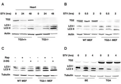
We monitored autophagy in vivo by analysing the lipid modification of LC3, leading to the membrane-associated isoform II of 15-16 kDa, which is a typical hallmark of autophagosome formation. Both LC3 I and LC3 II are rather unstable, the latter being rapidly degraded by lysosomal proteases once the autophagolysosome is formed. As reported in Figure 6A, after 24 hours of STV there is a slight reduction of TG2 in the heart of WT mice followed by the accumulation of the LC3 II after 48 hours of food deprivation. Interestingly, the hearts of TG2-/- mice show an earlier and more
pronounced accumulation of LC3 II when compared with the WT mice. In order to investigate the mechanism by which the absence of TG2 induces accumulation of LC3 II we studied the role of TG2 in autophagy using immortalized MEFs obtained both by WT and TG2-/- mice in which
autophagy was induced by different stimuli such as nutrient deprivation, treatment with rapamycin and 2-Deoxy-D-glucose (2-DG).
Figure 6. TG2 modulates the expression of autophagy-associated proteins LC3. (A) Myocardium tissue fragments were removed from WT and TG2-/- mice under resting
conditions as well as after 24-48 h of starvation (STV) and then homogenized. Proteins were separated by SDS-PAGE and analyzed by immunoblotting with anti LC3 and TG2 antibodies. The cardiac muscle of TG2-/- mice shows an earlier and more pronounced accumulation of
LC3 II during food deprivation, when compared with the WT mice. (B) Effect of nutrient depletion on autophagy in the WT and TG2-/- MEFs. Cells cultured in an amino acid-free and
serum-free medium (EBSS) were collected after 0, 5 or 2 h, and the proteins were subjected to immunoblotting for expression of LC3. The levels of the 18 kDa LC3 I rapidly decreased in WT compared with TG2-/- MEFs. Since LC3 II located in the inner autophagosomal
membrane is degraded upon fusion with lysosomes, the accumulation of LC3 II observed in the absence of TG2 might suggest a downregulation of the autophagic flux both in vivo and in isolated MEFs due to a block in late phases of autophagic process. (C) WT and TG2-/- MEFs
after induction of autophagy by using rapamycin or 2-DG. All autophagic stimuli used induce a greater level of LC3 II accumulation in TG2-/- versus WT MEFs. (D) Human neuroblastoma
cell lines expressing low (BE) or high (TGA) levels of TG2 protein under starvation conditions. The cells with a lower level of the enzyme (BE) show an evident accumulation of LC3 II, confirming an inverse correlation with TG2 expression. Cells cultured in an amino acid-free and serum-free medium (EBSS) were collected after 2 or 4 h, lysed and proteins were subjected to immunoblotting for expression of LC3. All blots were probed for tubulin or actin to verify protein loading.
First we analyzed the effect of nutrient depletion on autophagy in the WT and TG2-/- MEFs incubated in an amino acid-free and serum-free medium for
different time intervals; autophagy was monitored by detecting the LC3 I lipidation by western blot analysis.
As reported in Figure 6B, the levels of the 18 kDa LC3 I rapidly decreased in WT compared with TG2-/- MEFs, confirming the in vivo data
obtained in TG2-/- mice. It is interesting to note that the LC3 II levels was
already elevated in the TG2-/- control cells. This indicates that the absence of
TG2 affects the level of autophagy also under steady state conditions. It is important to note that LC3 II located in the inner autophagosomal membrane is degraded upon fusion with lysosomes (Kabeya et al., 2000). Thus the accumulation of LC3 II form, observed in the absence of TG2, might suggest that the autophagic flux is downregulated in the absence of the enzyme, both in vivo and in isolated MEFs.
In order to confirm the differential response of WT and TG2-/- MEFs to
autophagy induction we also compared their sensitivity to rapamycin or 2-Deoxy-D-glucose (2-DG). Rapamycin is a well-known lipophilic macrolide antibiotic which is widely used to induce autophagy by inactivating mTOR (Noda and Oshumi, 1998), while the 2-DG is a structural analogue of glucose differing at the second carbon atom by the substitution of hydrogen for a hydroxyl group (Aft et al., 2002). It has been shown to inhibit glycolysis and the growth of cultured human cells, depleting cell’s ATP content by 25–30% (Jain et al., 1985). Considering the role for TG2 in the mitochondrial homeostasis (Battaglia et al., 2007), and given the function of 2-DG in regulating cell’s ATP content, we decided to analyze 2-DG’s effect on autophagy in the absence of TG2.
We hypothesized that interfering with the glucose metabolism by a nonmetabolisable analogue would lead to increased autophagy, particularly in the absence of TG2. Results reported in Figure 6C confirmed this hypothesis and indicate that all used autophagic stimuli induce a greater level of LC3 II accumulation in TG2-/- versus WT MEFs.
To further investigate the role of TG2 in the regulation of autophagy we extended our study to human neuroblastoma SK-N-BE cell lines expressing low (BE) or high (TGA) TG2 levels. Figure 6D clearly indicates that upon induction of autophagy by STV the BE cell line, which expresses low levels of TG2, shows an evident accumulation of LC3 II; on the other hand, this isoform was detected in lower amounts in cells overexpressing TG2 (TGA), confirming the above described inverse correlation with TG2 expression. The cellular level of LC3 II has been regarded as a marker for the induction
of autophagy. However it is important to consider that LC3 II is rapidly degraded by the lysosomal proteases following the formation of autophagolysosomes (Kabeya et al., 2000). Therefore, an increase of LC3 II levels can be interpreted as representing both an induction and a block in autophagy.
In order to obtain further insight regarding the impact of TG2 on the autophagic process we employed electron microscopy to analyze the effects of the enzyme on autophagy. Observation of myocardium sections from WT mice subjected to starvation for 48 h, revealed the presence of autophagosomes in the cytoplasm of myocytes together with accumulation of residual bodies, higly electron-dense organelles containing indigestible products (Figure 7A and C). The indigestible products were also observed at the periphery of the cells before being released into the extracellular space (Figure 7E). All these features are indicative of an intense autophagocytotic degradation process.
By contrast, upon starvation the myocardium from TG2-/- mice shows
accumulation of autophagic vacuoles containing only partially degraded organelles and myeline figures (Figure 7B) associated with an extensive vacuolation (Figure 7D). Observation at higher magnifications revealed that these large vacuoles originated from degenerating mitochondria; in fact, mitochondria showed progressive loss of cristae, swelling and myeline figures (Figure 7F). Taken together, these observations seem to indicate that the accumulation of LC3 II observed upon stimulation of autophagy in the absence of TG2, is probably the result of an impairment of autophagolysosomes maturation and a block in their content degradation.
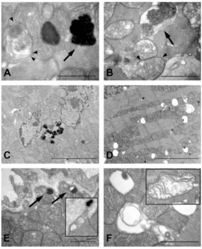
Figure 7. Transmission electron micrographs of hearts taken from WT (A, C and E) and TG2-/- (B, D and F) mice, housed under starvation conditions for 48 h. (A) Autophagic
vacuoles were detected in myocardium of WT mice; vacuoles contain morphologically intact cytoplasmic material wrapped by typical double membrane (arrowheads), together with late autophagic vacuoles, or residual bodies, were observed (arrow). (C) Intense autophagic activity resulted in accumulation of numerous lysosomes containing electron dense material in the cytoplasm of WT myocytes. (E) Vesicles containing the indigestible products (arrows) were found at the periphery of WT cardiomyocytes, before delivering their content into the extracellular space (insert). (B) TG2-/- cardiomyocytes displayed the presence of early
autophagic vacuoles, in which their content is still morphologically intact (arrowheads), and also of autophagosomes with partially degraded material (arrows). (D) Ultrastructural features of myocardium from TG2-/- mice consisted of extensive intracytoplasmic vacuolations. (F)
Large vacuoles appeared to originate from swollen mitochondria; degeneration of mitochondrial matrix and disruption of cristae is shown in the insert. Bars: (A, B, E and F) = 1 μm; (C and D) = 5 μm.
2. THE LACK OF TG2 IS ASSOCIATED WITH AN IMPAIRMENT OF VESICLE
To understand whether the observed accumulation of LC3 II and the defective autophagy is associated with an impairment in the lysosome acidification, WT and TG2-/- MEF cells were stained with acridine orange
(AO) to reveal massive accumulation of acidic vesicular organelles.
Acridine orange is a lysosomotropic agent, a weak base that moves freely across biological membranes when uncharged, while its protonated form accumulates in acidic compartments, where it forms aggregates that fluoresce in bright red. Interestingly, the quantification of acidic vesicular organelles by flow cytometry after starvation or 2-DG treatment, revealed a lower percentage of fluorescent-positive cells, in TG2-/- vs. WT MEFs
(Figure 8A).
These data indicate that, upon autophagy induction, the low LC3 II expression observed in WT MEFs is associated with increased acidification, while in the TG2-/- MEFs the high LC3 II levels were not followed by
acidification.
These data confirm that the LC3 II accumulation in TG2-/- MEFs is due to
an impairment in later phases of autophagic process, that can be caused by either a failed fusion between autophagosomes and lysosomes or by a lack of degradation of cytoplasmic material inside the autophagolysosome.
Figure 8. Quantitative detection of acidic vesicular organelles in WT and TG2-/- MEFs by
acridine orange staining using FACS analysis. The percentage of fluorescent-positive cells, after starvation or 2-DG treatment, was significantly lower in TG2-/- than in WT MEFs
(indicating that in the TG2-/- MEFs the high LC3 II levels were not followed by acidification).
Values are means ± SE M of 3–4 determinations; p < 0.05 (*) respect to TG2-/- MEF in the
To test this hypothesis we performed autophagy induction experiments in the presence of NH4Cl which inhibits the activation of the lysosomal
enzymes, hence blocking the degradation process. Western blot analysis (Figure 9B and C) showed that in the presence of NH4Cl the difference in
LC3 II accumulation observed in TG2-/- vs. WT MEFs was no longer
detected, irrespective of the agent used to induce autophagy. Taken together, our data show that TG2 plays a critical role in the later phases of the autophagic process. Therefore accumulation of autophagosomes in cells lacking TG2 is due to the inhibition of autophagic flux rather than to an induction of the process.
Figure 9. WT and TG2-/- MEFs under autophagic stimuli (starvation condition or D-dg
treatment) in the absence or the presence of NH4Cl, which inhibits the activation of the
lysosomal enzyme and blocks the degradation process. In the presence of NH4Cl the
difference in LC3 II accumulation observed in TG2-/- vs. WT MEFs was no longer detected.
Cells, cultured in EBSS for 2 or 18 h or treated with 10 mM 2-DG for 36 h, were lysed and the proteins subjected to immunoblotting for expression of LC3.
3. EFFECT OF TG2 KNOCKOUT MEFs RECONSTITUTION AND
CROSS-LINKING ACTIVITY INHIBITION ON THE
AUTOPHAGOSOME MATURATION
To confirm the TG2 involvement in the regulation of autophagy, we rescued TG2 into TG2-/- MEFs and inhibited TG2 cross-linking activity in
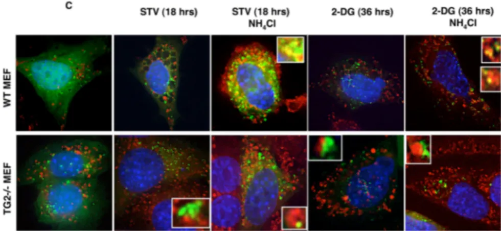
WT MEF cells. Interestingly, the transfection of WT human TG2 cDNA into TG2-/- MEFs (Figure 10A, insert) restored the capacity of these cells to
complete the autophagic process, resulting in the acidification of autophagosomes (Figure 10A).
A further demonstration of TG2’s role in the autophagic process was obtained by using a specific sitedirected TG inhibitor (R283), that resulted in a marked decrease in the number of acidic vesicular organelles detected by AO staining, as observed in the TG2-/- cells (Figure 10B). It is important to
note that R283 specifically inhibits the transglutaminases cross-linking activity (Skill et al., 2004).
Figure 10. (A) Transient reconstitution of TG2-/- MEF with TG2 (as shown in the insert)
restores the capacity of these cells to complete the autophagic process. TG2-/- MEF were
transiently transfected with pLPCX-TG2 using lipofectamine, cultured in EBSS for 0.5 and 2 h, and analyzed by acridine orange staining using FACS analysis. TG2 transfection restores the acidification of autophagolysosomes, resulting in the capacity to complete the autophagic process. (B) Effects of nutrient depletion on WT MEF treated with R283, a specific irreversible site-directed TG inhibitor. Analysis of acridine orange staining by FACS shows a reduced acidification, like that observed in TG2-/- cells. Cells were treated with 200 μM R283
for 18 h, cultured in EBSS for 0.5 or 2 h and incubated with Acridine Orange. Values are means ± SEM of at least three determinations; p < 0.01 (*) respect to cells untreated with TG2 inhibitor (R283).