PhD in Biochemistry
XXXII Cycle (2016/2019)
Insights into the mechanism(s) of action and
therapeutic applications of Esculentin-1a-derived
antimicrobial peptides
Tutor Coordinator Prof. Maria Luisa Mangoni Prof. Stefano Gianni
PhD student
Maria Rosa Loffredo
1
2
Index
• List of papers relevant for this thesis
………..6• List of other papers
………...7Abstract
……….8Abbreviations………...11
1. Introduction
………...121.1. THE ANTIMICROBIAL RESISTANCE………...12
1.1.1. Mechanisms, origins and evolution in bacteria……….... 13
1.2. PSEUDOMONAS AERUGINOSA PATHOGEN………....17
1.2.1. Pseudomonas aeruginosa biofilm development………...18
1.3. ANTIMICROBIAL PEPTIDES (AMPs)………....22
1.3.1. Structure-function properties………....24
1.3.2. Mode of action of cationic AMPs………...31
1.3.2.1. Membrane-permeabilizing AMPs………....33
1.3.2.2. Non-membrane-permeabilizing AMPs: Focus on AMPs-intracellular targets interactions………...37
1.3.3. AMPs as therapeutic agents………..40
1.4. FROG SKIN AMPs………...42
1.4.1. Esculentins and Esculentin-1a(1-21)NH2………...44
1.5. STRATEGIES TO IMPROVE THE THERAPEUTIC POTENTIAL OF ESCULENTIN-1a(1-21)NH2………...48
1.5.1. Modification of the primary structure: Focus on the diastereomer Esc(1-21)-1c……… 48
1.5.2. Functionalization of biomaterials with Esculentin-1a(1-21)NH2 and Esc(1-21)-1c [Esc peptides]………..52
3
2. AIMs of the work
………. 563. Results
………....573.1. MECHANISM(S) OF ACTION OF ESC PEPTIDES…………...57
3.1.1. Focus on the microbial membrane as target………57 3.1.1.1. Confocal microscopy analysis and killing activity
against the mutant P. aeruginosa PAO1 ΔpdvT strain…...57 3.1.1.2. Cytoplasmic membrane permeabilization…………....62 3.1.1.3. Activity on spheroplasts of P. aeruginosa…………....65 3.1.1.4. Leakage assay on large unilamellar vesicles (LUVs) of different composition……….. 67 3.1.1.5. Circular Dichroism (CD) spectroscopy………....70 3.1.2. Focus on non-membranous targets………...74 3.1.2.1. Identification of intracellular targets in the mechanism of antimicrobial activity of Esc peptides: Antibiofilm activity at dosages below the minimum inhibitory growth concentration (sub-MIC)………...74 3.1.2.2. Peptides’ effect on P. aeruginosa motility………75 3.1.2.3. Gene-expression analysis and role of ppGpp nucleotide in biofilm formation……….77
3.2. ENGINEERED NANOPARTICLES (NPs) FOR PROLONGED THERAPEUTIC EFFICACY OF ESC PEPTIDES………...81 3.2.1. Properties of Esc peptide-loaded NPs………...81 3.2.2. In vitro release kinetics……….83 3.2.3. Diffusion of NPs across artificial CF mucus and bacteria biofilm prototype………...84 3.2.4. In vitro antipseudomonal activity of Esc peptide-loaded NPs………...85
4
3.2.5. In vivo antimicrobial efficacy of Esc Peptide-loaded NPs……87
3.2.6. In vivo effect of Esc Peptide-loaded NPs on immune response and pulmonary toxicity……….... 88
3.3. IMMOBILIZATION OF ESC PEPTIDES ON HYDROGEL CONTACT LENSES (CLs) TO PREVENT AND/OR TREAT P. AERUGINOSA-ASSOCIATED OCULAR SURFACE INFECTIONS………....91
3.3.1. Covalent immobilization of Esc peptides on CLs and their amino acids quantification………..91
3.3.2. Killing activity and inhibition of bacterial adhesion to the surface of Esc peptide-coated CLs………...92
3.3.3. Effect of Esc peptide immobilization on CLs parameters…….94
3.3.4. In vitro toxicity of Esc peptide-coated CLs………...95
4. Discussion
………....975. Conclusion and Future Perspectives
………...1086. Material and Methods
……….1106.1. MATERIALS……….110
6.2. MICROORGANISMS………...111
6.3. MAMMALIAN CELLS………....111
6.4. METHODS……….112
6.4.1. Studies of mechanism of action………..112
6.4.1.1. Confocal microscopy analysis and killing activity against mutant P. aeruginosa PAO1 ΔpdvT strain……….….112
6.4.1.2. Cytoplasmic membrane permeabilization: Sytox Green assay………..112
5
6.4.1.3. Spheroplasts preparation for
3(4,5-Dimethylthiazol-2yl)2,5-diphenyltetrazolium bromide (MTT) assay……..113
6.4.1.4. Preparation of LUVs……….. 115
6.4.1.4.1. ANTS/DPX leakage assay………...116
6.4.1.4.2. CD spectroscopy……….... 117
6.4.1.5. Antibiofilm activity………....118
6.4.1.5.1. Motility………...118
6.4.1.5.2. Gene expression………... 119
6.4.2. PVA-engineered PLGA NPs………....119
6.4.2.1. Synthesis and characterization………... 119
6.4.2.2. In vitro release kinetics of Esc peptides………. 121
6.4.2.3. In vitro transport of NPs through mucus and biofilm models………... 121
6.4.2.4. In vitro activity of NPs against P. aeruginosa……... 123
6.4.2.5. In vivo effect in a murine lung infection model and lung toxicity/inflammation………....123
6.4.3. Covalent immobilization of Esc peptides to CLs and quantification………...125
6.4.3.1. Antimicrobial activity of Esc-immobilized CLs……125
6.4.3.2. Cytotoxicity………....126
6.5. STATISTICAL ANALYSIS………...127
7. References
………...1288. Acknowledgments
………1556
List of papers relevant for this thesis
• Loffredo MR, Ghosh A, Harmouche N, Casciaro B, Luca V, Bortolotti A, Cappiello F, Stella L, Bhunia A, Bechinger B, Mangoni ML. “Membrane perturbing activities and structural properties of the frog-skin derived peptide Esculentin-1a(1-21)NH2 and its Diastereomer Esc(1-21)-1c: Correlation with their antipseudomonal and cytotoxic activity.” Biochimica
et biophysica Acta 2017;1859(12):2327-2339.
• Casciaro B, Lin Q, Afonin S, Loffredo MR, de Turris V, Middel V, Ulrich AS, Di YP, Mangoni ML. “Inhibition of Pseudomonas aeruginosa biofilm formation and expression of virulence genes by selective epimerization in the peptide Esculentin-1a(1-21)NH2.” FEBS Journal 2019; 286(19):3874-3891. • Casciaro B, d'Angelo I, Zhang X, Loffredo MR, Conte G, Cappiello F,
Quaglia F, Di YP, Ungaro F, Mangoni ML. “Poly(lactide- co-glycolide)
nanoparticles for prolonged therapeutic efficacy of Esculentin-1a-derived antimicrobial peptides against Pseudomonas aeruginosa lung infection: in
vitro and in vivo studies.” Biomacromolecules 2019; 20(5):1876-1888. • Casciaro B, Dutta D, Loffredo MR, Marcheggiani S, McDermott AM,
Willcox MD, Mangoni ML. “Esculentin-1a derived peptides kill
Pseudomonas aeruginosa biofilm on soft contact lenses and retain
antibacterial activity upon immobilization to the lens surface.” Peptide
Science 2018; 110:e23074.
• Casciaro B, Loffredo MR, Luca V, Verrusio W, Cacciafesta M, Mangoni ML. “Esculentin-1a derived antipseudomonal peptides: Limited induction of
resistance and synergy with Aztreonam.” Protein and peptide letters 2018; 25(12):1155-1162.
• Casciaro B, Cappiello F, Loffredo MR, Ghirga F, Mangoni ML. “The potential of frog skin peptides for anti-infective therapies: The case of Esculentin-1a(1-21)NH2.” Current medicinal chemistry. 2019; In press.
7
List of other papers
• Casciaro B, Calcaterra A, Cappiello F, Mori M, Loffredo MR, Ghirga F, Mangoni ML, Botta B, Quaglio D. “Nigritanine as a new potential antimicrobial alkaloid for the treatment of Staphylococcus aureus-induced infections.” Toxins 2019; 11(9): E511
• Merlino F, Carotenuto A, Casciaro B, Martora F, Loffredo MR, Di Grazia A, Yousif AM, Brancaccio D, Palomba L, Novellino E, Galdiero M, Iovene MR, Mangoni ML, Grieco P. “Glycine-replaced derivatives of [Pro3, DLeu9]TL, a Temporin L analogue: Evaluation of antimicrobial, cytotoxic and hemolytic activities.” European journal medicinal chemistry. 2017 Oct
20; 139:750-761.
• Buommino E, Carotenuto A, Antignano I, Bellavita R, Casciaro B, Loffredo MR, Merlino F, Novellino E, Mangoni ML, Nocera FP, Brancaccio D, Punzi P, Roversi D, Ingenito R, Bianchi E, Grieco P. “The
outcomes of decorated prolines in the discovery of antimicrobial peptides from Temporin-L.” ChemMedChem. 2019 Jul 3; 14(13):1283-1290.
8
Abstract
Cationic α-helical antimicrobial peptides (AMPs) hold promise for treatment of the raising multi-drug resistant microbial infections, due to their broad spectrum of activity and membrane-perturbing mechanism of action. Compared to conventional antibiotics, these features make them newsworthy molecules that hardly induce microorganisms to acquire resistance to them.
Among these pathogens, Pseudomonas aeruginosa is the most clinically relevant Gram-negative bacterium known to cause serious human infections, e.g. pneumoniae, especially in immune-compromised patients, such as cystic fibrosis (CF) sufferers and keratitis, associated to contact lens (CL) wear. This is due to the unique ability of this pathogen to adhere to different types of inert materials or biological tissues, and to grow in a more resistant and dangerous sessile life form, called biofilm.
Recently, two Esculentin-1a-derived antimicrobial peptides i.e. Esc(1-21) and its D-amino acids containing Esc(1-21)-1c, [Esc peptides], have been fully characterized for their powerful antipseudomonal activity against both planktonic and biofilm forms.
The diastereomer showed a higher bactericidal activity than the all-L isomer against the more dangerous Pseudomonas biofilm phenotype; a lower cytotoxicity and higher biostability. However, when tested in vitro against the free-living form of this pathogen, it displayed a weaker bactericidal effect.
Here, to investigate the reason accounting for this discrepancy, mechanistic studies on intact bacterial cells were initially carried out. Then to further understand the effect of packing parameters, i.e. composition, charge, shape
9
and negative intrinsic curvature of membrane phospholipids in the membrane-permeabilizing activity of Esc peptides, leakage assays and circular dichroism spectroscopy analysis were carried out.
Our results have suggested that the weaker in vitro antibacterial activity of Esc(1-21)-1c on the planktonic phenotype of the Gram-negative bacterium P.
aeruginosa is mainly correlated to a slighter ability in permeabilizing both
outer and inner bacterial membranes.
Notably, experiments with lipid vesicles have suggested that if electrostatic interactions between negatively-charged membrane phospholipids and positively-charged peptide molecules do play a crucial role in the peptides’ membrane perturbing activity, this latter is hampered by the bilayer structure packing parameters including hydrogen bonding and intrinsic curvature, associated to phosphatidylserine (PE), especially for the diastereomer compared to all-L parent peptide.
In parallel, we explored the molecular mechanism underlying the biofilm inhibition activity of Esc peptides when used at dosages below the minimal growth inhibitory concentration (1/8 MIC), by studying the peptides’ effect on the expression of key genes involved in the bacterial virulence and motility, as well as the peptide’ interaction with the bacterial signaling nucleotide ppGpp. Our findings revealed that the two D-amino acids containing Esc(1-21)-1c, confer the peptide the ability to downregulate the expression of biofilm-associated genes, likely as a result of increased peptide stability and prolonged binding to ppGpp compared to the all-L peptide.
10
Furthermore, we reported two different applicative strategies to ameliorate the biological properties of these two AMPs: (i) encapsulation in poly(lactide-co-glycolide) (PLGA) nanoparticles; and (ii) covalent conjugation to soft CLs.
In the first case, to enhance the peptides’ bioavailability and to optimize their translocation to the target infectious site, Esc peptides were loaded into PLGA nanoparticles (NPs) engineered with polyvinyl alcohol (PVA). The peptides-loaded NPs were found to be more efficient in diffusing through artificial CF mucus and simulated bacterial extracellular matrix compared to the free peptides. Moreover, they were more efficient in inhibiting P. aeruginosa growth under both in vitro and in vivo conditions at long term.
In the second case, Esc peptides were covalently immobilized to hydrogel soft CLs and tested for their ability to reduce bacterial colonization. The antimicrobial CLs were able to cause more than four log reduction in the number of bacterial cells within 20 min and to reduce bacterial adhesion to their surface in 24 hours.
Finally, the ability of both peptides to limit the onset of microbial resistance was also evaluated by exposing Pseudomonas strains to multiple cycles of treatment at sub-MIC dosages. Interestingly, in contrast with conventional antibiotics, Esc peptides did not induce resistance in P. aeruginosa cells.
Overall, besides providing knowledges on the molecular mode(s) of action the two esculentin-derived AMPs, our data suggest that Esc peptides, particularly Esc(1-21)-1c, have great potential to be developed as novel drugs for treatment and prevention of P. aeruginosa pneumonia and keratitis.
11
Abbreviations
AMP, antimicrobial peptide; ANTS, 8-aminonaphthalene-1,3,6-trisulfonic acid, disodium salt; BAL, bronchoalveolar lavage; CD, circular dichroism; CF, cystic fibrosis; CFU, colony-forming units; CL, contact lens; CTRL control; CV, crystal violet; DH, hydrodynamic diameter; DIC, differential interference contrast; DLS, dynamic light scattering; DMEM, Dulbecco’s modified Eagle’s medium; DPC dodecylphosphocholine; DPX, p-xylene-bis-pyridinium bromide; DTPA, diethylenetriaminepentaacetic acid; EDC, 1-ethyl-3-(3-dimethylaminopropyl) carbodiimide hydrochloride; EDTA ethyl-enediaminetetraacetic acid; ELISA, enzyme-linked immunosorbent assay; ELS, electrophoretic light scattering; Esc peptides collectively Esc(1-21) and Esc(1-21)-1c; LB, Luria-Bertani broth; LPS, lipopolysaccharide; LTA, lipoteichoic acids; LUV large unilamellar vesicles; MAA, methacrylic acid; MTT, 3-(4,5-dimethylthiazol-2-yl)-2,5-diphenyltetrazolium bromide; NMR, nuclear magnetic resonance; NPs, PVA-engineered PLGA nanoparticles; OD, optical densit y; PBS, phosphate buffered saline; PDI, polydispersity index; PLGA, poly(lactide-co-glycolide); POPC, 1-palmitoyl-2-oleoyl-sn-glycero-3-phosphocholine; POPE, 1-palmitoyl-2-oleoyl-sn-glycero-3-phosphoethanolamine; POPG, 1-palmitoyl-2-oleoyl-sn-glycero-3-phosphoglycerol; ppGpp guanosine-30,50-bisdiphosphate; PVA, poly(vinyl alcohol); PVD, pyoverdine; QS, quorum sensing; RF, respirable fraction; Rho-Esc, rhodamine-B-labeled Esc peptides; Rho-Esc_NPs, rhodamine-B-labeled Esc(1-21) NPs; RP-HPLC, reverse phase high-performance liquid chromatography; SAB, sodium acetate buffer; SD standard deviation; SDS, sodium-dodecyl sulfate; SEM standard errors of the mean; SILF, simulated interstitial lung fluid; THR, trehalose; TOCL, tetra-oleoyl cardiolipin.
12
1. Introduction
1.1. THE ANTIMICROBIAL RESISTANCEIn the 1900’s the discovery of antibiotics as compounds to treat infections revolutionized the healthcare community. Indeed, antibiotics acquired relevant support for clinical approaches in the surgical procedures, organ transplantation and management of cancer patients [WHO 2014; Reygaert 2018].
Thanks to the antibiotic’s discovery and to the rapidity by which they were introduced to the pharmaceutical market, infectious diseases became a public health problem [Michael et al 2014].
Currently, antimicrobial resistance (AMR) has been recognized by The World Health Organization (WHO) as one of the three most important public health threats of the 21st century [WHO 2015]. In accordance with the Centre for Disease Control and Prevention (CDC), the amount of deceased people in USA owing to antibiotic-resistant infections is at least 23,000 per year. Future perspectives are certainly not more optimistic; indeed, new United Kingdom reports estimated that antibiotic resistant infections will cause around 10 million premature deaths per year by 2050, as well as significant economic damage of up to US$3.5 billion per year [OECD 2018].
Interestingly, a recent report outlined a plan of simple rules to reduce the resistance state: better hygiene, fewer and more careful antibiotic prescriptions, quick testing to discriminate bacterial from viral infections and educating the public by media campaigns.
13
1.1.1. Mechanisms, origins and evolution in bacteria
Antimicrobials are compounds endowed with specific activity against microbes, while the antibiotics are antibacterial compounds derived from microorganisms. AMR refers to the lack of susceptibility of microorganisms to a specific compound. It is generally evaluated on the basis of the minimum inhibitory concentration (MIC), i.e. the minimal concentration of compound that can inhibit the growth of that bacterium. The resistance to a specific compound can be due to the inability of this latter to hinder the bacterial growth of or to kill the bacterial species [Kidd et al 2018].
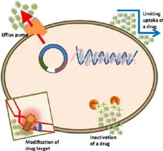
The genotypical resistance can be distinguished in intrinsic, acquired and adaptive. The first one includes mutations in gene(s) causing an altered outer membrane permeability, an over-expression of efflux pumps that extrude the antimicrobial molecules as well as the synthesis of antibiotic-inactivating enzymes [Figure 1] [Reygaert 2018].
14
Figure 1. A schematic representation of the intrinsic antibiotic resistance mechanisms in
bacteria [Reygaert 2018].
The acquired resistance can be achieved by two different pathways: vertical evolution or horizontal evolution, whereby de novo mutations, involved in the antibiotic sensitivity, are transmitted to the descendants [Figure 2a], or the external antibiotic resistance genes are acquired through horizontal gene transfer (HGT) from another bacterium (donor) [Breidenstein et al 2011] [Figure 2b].
Although the relative contribution of each of these evolutionary paths to the development of clinical bacterial resistance is still unknown, one of the most important factors is the HGT [Henrichfreise et al 2007;Pang et al 2019].
15
Figure 2. Mechanisms of acquisition of resistance genes in bacteria. a) Vertical evolution,
transmission of the de novo mutations in the bacterial genome to the daughter bacterial cells. The antibiotic-sensitive bacterial cells are depicted in blue, whereas the antibiotic-resistant cells in red icon. b) Horizontal acquisition, also known as horizontal gene transfer (HGT), can involve the phage transduction, conjugation or transformation. All these processes are based on the transmission carriers of resistance mutations and/or genes of mutations (indicated by red DNA routes) from the donor bacterial cell (in red) to the target bacteria (in blue) [Sommer et al 2017].
The third one is the adaptive resistance. Compared to the intrinsic and acquired resistance mechanisms, the adaptive resistance is dependent on transient alterations in genes and/or protein expression as a response to environmental stimuli or chemical and physical stresses [Sandoval-Motta and Aldana, 2016].
Among environmental stimuli associated with this type of resistance there are antibiotics, pH, heat shock, DNA stress, anaerobiosis, polyamines, cations and nutrient deficiency states and group behavioural adaptations such as biofilm formation and swarming motility [Fernández et al 2011].
16
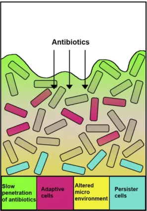
Biofilm-mediated resistance to antibiotics (simplified in Figure 3), is a topic that is currently attracting attention. The pathogens involved in the biofilm infections are Staphylococcus aureus, Staphylococcus epidermidis,
Pseudomonas aeruginosa, Burkholderia cepacia complex and Haemophilus influenzae, among others [Folkesson et al 2012; Fernández et al 2011;
Bjarnsholt et al 2009].
An important role in adaptive resistance is also given by “persister” cells (PC), subset of phenotypic variants in an isogenic bacterial population that, unlike drug-resistant bacteria, survive the antibiotic attacks by decreasing their metabolism and entering into a non-growing/quiescent state [Lewis 2010].
Generally, PC arise as adaptation to stress environmental factors, including oxidative stress, DNA damage, nutrients and oxygen deprivation, and antibiotics [Wang et al 2017]. Indeed, PC typically increase in stationary-phase cultures and biofilm lifestyle, wherein PCs represent as much as 1% of the total bacterial population [Grassi et al 2017] [Figure 3].
17
Figure 3. The biofilm-mediated antibiotic resistance. Limited penetration of antibiotics within
the biofilm (green); some biofilm cells exhibit an adaptive response as a result of antibiotic attack (pink); an altered microenvironment in biofilm (yellow) leads to lower growth of bacteria. This results in reduced antibiotic uptake and the emergence of multidrug-susceptibility “persister” cells (in blue). [Pang et al 2019].
1.2. PSEUDOMONAS AERUGINOSA PATHOGEN
One of the human pathogens that most easily acquire resistance to conventional antibiotics is the Gram-negative bacterium Pseudomonas aeruginosa
(P.aeruginosa). Pulmonary infections due to P.aeruginosa are the main cause
of lung decline and death in patients suffering from cystic fibrosis (CF) [Parker et al 2016; Blanc et al 1998; Sadikot et al 2005].
Cystic fibrosis (CF) is a genetic disorder that is caused by recessive mutations in the gene encoding the cystic fibrosis transmembrane conductance regulator
18
(CTFR), which regulates chloride transport across the airway epithelium and submucosal glands. The most common mutation is a deletion of phenylalanine at position 508, leading to hyperinflammation and formation of a thick and viscous mucus layer with impaired mucocillary clearance. Thus, these conditions predispose the lung of CF patients to a favourable environment for bacterial growth and colonization [Nixon et al 2001; Maurice et al 2018]. Despite aggressive antibiotic treatments, Pseudomonas often grows in the CF lung and leads to chronic and recalcitrant infections characterized with robust host inflammatory response [Rudkjøbing et al 2012; Bjarnsholt et al 2009].
P. aeruginosa is among the most virulent opportunistic pathogens capable of
surviving in nutrient-poor environments and colonizing biological or abiotic surfaces e.g. medical implants such as catheters, prostheses or contact lenses (CLs), making its eradication even more complex [Willcox 2011].
Among medical devices, CLs wear is one of the most important risk factors for the development of microbial keratitis. This is because Pseudomonas can easily colonize the hydrogel or silicone hydrogel lens surface, forming biofilms which can rapidly colonize the cornea tissue, once the CL is placed in the eye [Willcox 2011].
1.2.1. Pseudomonas aeruginosa biofilm development
Adaptive antibiotic resistance represents a huge obstacle in the treatment of
Pseudomonas biofilm infections as compared to the free-swimming,
planktonic counterpart of this microbial pathogen [Bhagirath et al 2016, Pletzer et al 2016].
Biofilms are complex sessile communities of microbes found either attached to an inert or living surface and integrated in a self-produced extracellular
19
matrix as three-dimensional aggregates [Costerton et al 1999; Roy et al 2018]. Exopolysaccharides, proteins, nucleic acids, and other cellular debris, collectively named extra polymeric substances (EPS), are the general constituents of this matrix.
It is known that biofilm formation in vitro starts with an irreversible adhesion of planktonic bacterial cells to surfaces favoured by pili and flagella. Type IV pili, for example, are filamentous protein complexes that are of vital importance for the initial attachment of cells as well as for the regular biofilm maturation [Figure 4].
Attachment of cells to a surface is then followed by microcolonies formation, secretion of EPS and biofilm maturation by quorum sensing intercellular communication.
It is notable that in the maturation step, the twitching motility, which is the bacterial movement resulting from extension and retraction of pili including the Type IV, plays a direct role in the cell-to-cell interactions [Skercer and Berg 2001; Giraud et al 2011].
Furthermore, besides swimming and twitching motility, P. aeruginosa can also swarm through viscous environments. The swarming is a complex type of motility, depending on flagella, type IV pili and bacterial surfactants, e.g. rhamnolipids which contribute to the early stage of biofilm formation and colonization of adjacent semi-solid surfaces [Overhage et al 2007].
20
Figure 4. Biofilm development process. (1) Initial attachment of bacterial cells to a surface.
Gene expression changes lead to down-regulation of polar flagella and up-regulation of Type IV pili. Bacteria start to adhere to the surface and to produce extracellular matrix, (2) Microcolonies formation through different cycles of bacterial cell division. The increased expression of Type IV pili and secretion of extracellular matrix components allow a higher attachment of cells to a surface as well as a strong association with other cells leading to protection from the external environment. (3) Development of mature biofilm structure with subpopulations of cells with different morphologies between the external and internal layers of biofilm microcolonies. (4) Dispersion of some cells, which revert to the motile phenotype and dissociate, from the outer surface of the biofilm by quorum-sensing pathway, external signals or physical breakdown [Taylor et al 2014].
Biofilm differentiation and maturation is a highly regulated program associated to expression of dozens of genes and regulatory circuits. A critical regulatory circuit is the stringent response triggered by the cellular and starvation stress (i.e. nutrients deficiency, carbon source, iron or lipid starvation, heat and oxidative stresses) and mediated by the synthesis of small signalling nucleotides, i.e. the guanosine diphosphate 3’-diphosphate (ppGpp) and 5’-triphosphate 3’-diphosphate (pppGpp), collectively denoted nucleotide alarmones tetra- and penta-phospate (p)ppGpp [Potrykus and Cashel 2008; Romling et al 2013].
In Gram-negative bacteria the synthesis of these molecules is regulated by global stress response regulator enzymes, RelA and SpoT; while a single bifunctional enzyme Rsh is associated to the Gram-positive bacteria. As
21
illustrated in Figure 5, in response to amino acid starvation, RelA binds to the ribosome, which is blocked by uncharged tRNA molecules, and triggers the production of cellular alarmones ppGpp. Contrariwise, under other type of stresses including iron starvation, SpoT starts ppGpp synthesis. SpoT is alternatively a bifunctional enzyme that can hydrolyze ppGpp. Subsequently, the rapid intracellular accumulation of the alarmones ppGpp and its following binding to the RNA polymerase, influences the transcription causing a passage from cell growth to survival. Anti-biofilm peptides are able to block the intracellular accumulation of ppGpp [De la Fuente-Nunez et al 2014; Pletzer et al 2016].
Figure 5. Stringent response in biofilm formation mediated by alarmones ppGpp. RelA is
expressed as a response to amino acid deprivation and binds to the ribosome triggering the synthesis of cellular alarmones ppGpp. Instead, SpoT causes the production of ppGpp as response to iron starvation. ppGpp signalling molecules bind to RNA polymerase, thus influencing the transcription. [Pletzer et al 2016].
22
Another global regulatory system involved in the biofilm development is the quorum sensing (QS) that involves cell-to-cell communication.
In P. aeruginosa, the principal QS systems are the las, rhl and pqs. They regulate the response of bacterial cells to the surrounding environment through the production of signalling molecules implicated in the expression of several virulence factors including toxins and proteases [De Kievit et al 2001].
Among these virulence factors, an example is the endogenous siderophore pyoverdine (PVD), which acts as both an iron carrier and a virulence-related signalling molecule. Indeed, it has been demonstrated that it is in turn implicated in biofilm control, cell-to-cell communication, and in the expression of other virulence factors, including exotoxin A, exoprotease PrpL, and PVD itself [Imperi et al 2009].
1.3. ANTIMICROBIAL PEPTIDES (AMPs)
Natural antimicrobial peptides (AMPs), also called host defence peptides, are a class of evolutionally conserved components of the innate immunity that are expressed as a first-line of defence against microbial pathogens in many multicellular organisms, ranging from prokaryotes to humans [Zanetti 2004; Powers and Hancock 2003; Zhang and Gallo 2016].
In 1922, Alexander Fleming identified the first human antimicrobial protein, called Lysozyme from nasal mucus, while in 1940s this finding was tarnished by the advent of the “Golden Age of Antibiotics” with the discovery of penicillin.
However, in the 1960s, with the increased emergence of antibiotic resistance, the development of novel therapeutic strategies directed against biofilm-related infections became urgently needed.
23
The AMPs’ discovery began in 1980 when Hans G Boman discovered how the injection of bacterial cells in Cecropia silk moth was able to induce the synthesis of an AMP, called cecropin. [Hultmark et al 1980]. Another relevant breakthrough occured in 1987 when Michale Zasloff and co-workers isolated magainins from the skin of the African frog Xenopus laevis [Zasloff 1987]. In 1994 the increased scientific and clinical interest towards AMPs led to the discovery of cathelicidins in the mammalian host defense by Bob Lehrer’s group [Gallo et al 1994].
Even if more than 5,000 AMPs have been discovered or synthesized up to date, these likely depict only a tiny part of all gene-encoded antibiotic peptides existing in nature.
Many AMPs are small cationic molecules which act mainly by killing pathogenic bacteria, fungi, viruses and protozoa via direct non-receptor mediated membrane damage.
In addition, or in support to their membrane-perturbing activity, AMPs can also have non-membranous targets, interfering with intracellular biochemical processes like the inhibition of nucleic acids, proteins and cell wall synthesis [Brodgen 2005].
Furthermore, besides their well-known antibacterial activity, several AMPs have been characterized as innate immunity modulators anti-diabetic agents and anti-tumor agents. Melittin and NK-2, a bee venom and porcin NK-lysin-derived peptides respectively, have been identified as antitumor molecules. Particularly, while melittin inhibits the tumour cell metastasis by decreasing cell motility and migration, NK-2 allows cancer cell death by interaction with
24
negatively charged phosphatidylserine (PS) on the tumour cell surface. [Pushpanathan et al 2013].
1.3.1. Structure-function properties
Generally, AMPs are relatively small molecules. They can vary from 6 to 60 amino acid residues with a cationic character at neutral pH.
The target cell selectivity of AMPs is the result of sensitive interplay between structural and functional features, as summarised below.
• Peptide’s charge: It is the sum of all charges of the ionizable groups. Most AMPs are positively charged with a net charge ranging between + 2 to + 9 due to arginine (Arg) and/ or lysine (Lys) residues [Hancock and Sahl 2006]. The positive charge is required for the initial interaction of AMPs with the components of the outer surface of microorganisms (e.g. lipopolysaccharides, LPS, in Gram-negative bacteria or lipoteichoic acids, LTA, in Gram-positive bacteria). Some anionic AMPs (AAMPs) (net charge -1 to -7), rich in aspartic or glutamic acids residues, have also shown an antimicrobial efficacy. They use metal ions (e.g. Zn2+) to establish salt bridges with the anionic components of microbial membranes [Harris et al 2011]. Their mechanism of action, even if poorly understood, mainly includes translocation across the membrane to specific intracellular targets (e.g. ovine and SAAPs) or pathogen’ membranes perturbation via pore formation (e.g. cyclotides) or in a carpet-like manner, as in the case of human-β-defensins prior to interact with intracellular targets. However, the mechanisms of their antimicrobial activity are unclear.
25
Peptide’s conformation and sequence: Generally, according to their
secondary structures in a membrane-mimicking environment, AMPs are classified in the following groups: (i) α-helical AMPs, such as the cathelicidin LL-37, melittin, magainin [Figure 6B]; (ii) β-stranded peptides connected by two or more disulfide bonds (e.g., defensins and protegrins) [Figure 6A]; (iii) β-hairpin or loop AMPs interconnected by a single disulphide bond and/or cyclization of the peptide backbone (e.g., gramicidin S, thanatin) [Figure 6D]; (iv) linear peptides rich in tryptophan (e.g., indolicidin), proline and arginine (e.g., PR-39), glycine and histidine (e.g., histatins) [Figure 6C]. Most of them are usually unstructured in aqueous solution and adopt an amphiphatic helical conformation in membrane mimetic environment. In the numerous AMPs’ sequences, the prevalent amino acid residues are the cationic Lys and Arg, which are fundamental for their electrostatic interaction with the negatively-charged lipid head-groups of microbial membranes at lipid/water interface, as well as the aromatic tryptophan (Trp) which favors AMPs’ anchorage into membranes [Shagaghi et al 2018]. Several α-helical AMPs contain glycine (Gly) as first amino acid because it is considered an excellent N-capping residue for α-helices, thus limiting proteolytic degradation by aminopeptidases [Tossi et al 2000]. On the other hand, AMPs rarely contain residues as aspartic acid (Asp) or glutamic acid (Glu).
26
Figure 6. Structural classes of antimicrobial peptides. (A) β-sheet, tachyplesin I, defensins,
protegrins; (B) α-helical structure of human cathelicidin LL-37 or magainin 2; (C) linear extended indolicidin, PR39, histatins; (D) β-hairpin or loop peptides, e.g. thanatin, gramicidin S. In yellow are represented the disulfide bonds [Powers and Hancock 2003].
• Peptide’s amphipathicity and hydrophobicity: The amphipathicity or
hydrophobicity refers to the distribution of hydrophobic and hydrophilic moieties within the peptide sequence. There is a strong correlation between the amphipathic structure of AMPs and their mechanism of action, particularly by van der Waals interactions: the hydrophilic domains of AMPs interact with the negatively-charged membrane components whereas the hydrophobic face interacts with the membrane phospholipids leading to peptides’ penetration into the membrane bilayer [Hancock and Chapple 1999; Dathe et al 1997]. Approximately 50% hydrophobic residues are found in many naturally occurring AMPs [Yeaman and Yount 2003; Tossi et al 2000] and assist their partition into the membrane. Importantly a direct correlation
27
between and mammalian cell toxicity has been reported [Strandberg et al 2015].
• Target cell membrane composition: an attractive feature of many
AMPs is their cell selectivity i.e., the peptide ability to kill bacterial cells at concentrations significantly lower than those causing damage to cells of the host organism [Hancock and Sahl 2006].
Since cellular membranes are the primary target for the mechanism of action of AMPs, it is worth mentioning that selectivity results from a different composition between the membrane of eukaryotic cells and bacterial cells [Wang 2017].
In nature, bacteria can be divided into two groups, Gram-positive and Gram-negative. Gram-positive bacteria are characterized by a thick peptidoglycan and LTA layer (40-80 nm) surrounding the cytoplasmic membrane. Gram-negative bacteria, on the contrary, have a thinner peptidoglycan layer (8 nm thick) adjacent an outer membrane with an asymmetric composition: phospholipids are the main components of the inner leaflet, while the outer layer is mostly made of LPS [Wada et al 2012].
By contrast, eukaryotic cells only have the plasma membrane, with asymmetric lipid composition in the two leaflets of the bilayer.
The major components of bacterial membranes include negatively-charged phospholipids. Specifically, in Gram-negatives, both membranes contain phosphatidylglycerol (PG, ~20%) and cardiolipin (CL, ~5%); the content of anionic lipids in Gram-positive bacteria is much higher and PG and CL are again the most important components [Malanovic and Lohner 2016].
28
Furthermore, the positively charged L-lysyl phosphatidylglycerol, (l-lysyl-PG or LPG) can also be present in the membranes of Gram-positive.
In both bacteria types, the zwitterionic phospholipid is phosphatidylethanolamine (PE), and no sterols are present [Epand et al 2016] [Figure 7]. Obviously, bacterial membrane composition is influenced by specific strain and growth conditions [Bobone and Stella 2019].
Human cells contain cholesterol and have no anionic phospholipids in the outer leaflet of their cell membrane. In comparison with eukaryotic cells, bacterial membranes have more anionic lipids in the outer surface of their bilayers and their transmembrane potential is more negative than that of mammalian cells [Yeaman and Yount 2003].
Figure 7. Schematic representation of the cell wall of Gram-positive and Gram-negative
bacteria [Epand et al 2016].
For these reasons, electrostatic interactions between positively-charged peptide molecules and negatively-charged membrane components of bacterial cells are prevalent.
29
Greater amounts of “non-bilayer lipids” are contained in the bacterial membranes and are characterized by negative (for PE, CL and phosphatidic acid, PA) or positive (LPG) values for the “intrinsic curvature”[Malanovic and Lohner 2016].
This feature is related to the relative sizes of the phospholipid head-groups and acyl chains: lipids with a cross-sectional area composed by similar head-groups and tails (e.g., phosphatidylcholine PC, PG or PS) have a cylindrical shape and pack well in flat bilayer structures (zero intrinsic curvature). On the contrary, lipids with head-group shorter than the tails, as for PE and PA, favour concave shapes of the monolayer (negative curvature). Lipids with larger polar heads, such as LPG or phosphatidylinositol PI, have positive curvature [Koller and Lohner 2014] [Figure 8].
By investigating interactions between AMPs and different types of model membranes mimicking the composition of natural bilayers is helpful in order to understand the AMPs’ selectivity [Bocchinfuso et al 2011; Savini et al 2018].
As demonstrated by Matsuzaki association of cationic peptides to phospholipid bilayers and their perturbation is enhanced by the presence of anionic lipids [Matsuzaki et al 1989; Russell et al 2010; Golbek et al 2017]; while, the positively charged lipid LPG, contained into Gram positive bacteria, hinders AMP activity [Andra et al 2011]. Although the knowledge regarding the effect of the presence of negative curvature lipids in the bacterial membranes, such as PE, is not yet well-defined, different groups, including Dr. Matsuzaki and Allende showed that PE inhibits pore formation by magainin, melittin,
30
alamethicin, PMAP23 and mastoparan X [Matsuzaki et al 1998; Allende et al 2005; Bobone et al 2012].
By contrast, other works showed that the presence of PE favours the activity of some AMPs [Schröder-Borm et al 2003; Epand et al 2006; Leite et al 2015].
The inhibition of AMPs’ activity by PE can be understood considering that AMPs act by introducing into the head-groups of the membrane phospholipids, triggering a positive curvature strain and, as consequence, a pore-like mechanism of membrane perturbation. In comparison, lipids with a negative intrinsic curvature would hinder this mechanism [Matsuzaki et al 1998; Lee et al 2005].
However, it is difficult to definite an exact role of PE in AMPs’ selectivity, and future studies regarding the role of negative curvature will be needed to understand this effect.
Figure 8. Molecular shape and curvature strain of primary lipids of bacterial membranes
31
1.3.2. Mode of action of cationic AMPs
Most of the cationic AMPs exert their antimicrobial action by inducing an irreversible alteration of the cellular structure and/or function through different and complex mechanisms [Powers and Hancock 2003; Brogden 2005; Jenssen et al 2006; Teixeira et al 2012].
All mechanisms involve key steps such as attraction, attachment and insertion of AMPs into the target cell’s membrane, thus causing a lytic or a non-lytic effect [Nijnik and Hancock 2009; Shagaghi et al 2018].
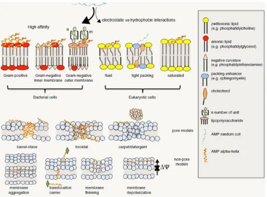
Unlike conventional antibiotics, the initial binding is mediated by electrostatic interactions between the cationic residues of AMPs and the negatively-charged outer envelope of Gram-negative bacteria (i.e. anionic phospholipids and LPS) or the LTA on the Gram-positive bacterial surface [Malanovic and Lohner 2016] [Figure 9, 10].
Once AMPs interact with LPS, divalent cations such as Mg2+ and Ca2+ bound to LPS are dislocated, thus leading to a local disorder of the outer membrane and promotion of peptide translocation into the cytoplasmic membrane, according to the self-promoted uptake hypothesis [Bechinger and Gorr 2017; Anunthawan et al 2015].
32
Figure 9. Molecular basis and general mechanisms of cell selectivity of AMPs [Lee et al
2019].
In addition to this primary mechanism of action, many AMPs exert immunomodulatory functions. Indeed, AMPs can protect the host by enhancing recruitment or activation of leukocytes and monocytes at the site of infection or by modulating the host-cell responsiveness to Toll-like receptor (TLR) ligands [Figure 10].
33
Figure 10. Biological functions of AMPs. AMPs can bind to bacterial membranes through
electrostatic interactions either to permeabilize the membrane or to penetrate the bacterium causing inhibition of intracellular functions. Furthermore, AMPs can also have immunoregulatory functions by recruiting/activating immunocytes or by affecting of Toll-like receptor (TLR) neutralization of microbial products and nucleic acids release upon tissue damage. DC, dendritic cell; LPS, lipopolysaccharide; LTA, lipoteichoic acid; MAVS, mitochondrial antiviral signaling protein. [Zhang and Gallo 2016].
1.3.2.1. Membrane-permeabilizing AMPs
After AMPs target and bind to the outer membrane of pathogen, the peptides’ ability to traverse the lipid bilayer can be influenced by the threshold concentration of the peptide itself, which is usually investigated in experimental protocols as peptide-to-lipid ratio (P/L). This depends on many aspects including, peptide concentration, peptide tendency to self-assemble or to oligomerize, phospholipid membrane composition, fluidity, head group type, as well as pH, temperature and ionic strength [Teixeira et al 2012; Huang 2000].
Generally, at low peptide/lipid ratios AMPs lie parallel to the lipid bilayer, while once high peptide/lipid concentrations are reached, they begin to
34
penetrate and to dispose themselves perpendicularly to membranes thus causing the formation of transmembrane channels, pores and/or extensive membrane lesions [Brogden 2005; Ciumac et al 2019].
The general models proposed to explain the membranolytic mode of action of AMPs are reported below:
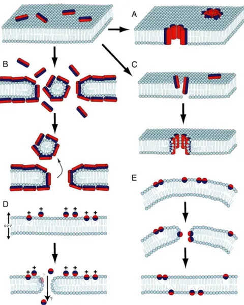
• “Barrel-stave”: Peptide helices shape a bundle with a central lumen into the bilayer, like a barrel wherein the helical peptides are the staves. The hydrophobic peptide surfaces interact with the fatty acid chains of the membrane phospholipids, while the hydrophilic regions point inward forming the interior region of the channel. The leakage of intracellular components through these pores subsequently causes cell death. It has been noted that a transmembrane pore can be made of a minimum of three molecules. In this model, the membrane does not show significant curvature and the hydration of the membrane remains unchanged [Li et al 2017]. The mechanism of action of alamethicin is defined according to this model [Figure 11 A] [Shagaghi et al 2018; Leitgeb et al 2007; Yang et al 2001].
• “Toroidal-pore (worm hole)”: AMPs helices insert into the bilayer inducing a positive curvature strain in the lipid monolayers (membrane thinning) to bend through the pore wherein the water core is lined by peptide molecules and lipid head groups. Similarly, to the previous model, in toroidal-pore structure the polar faces of the peptides are tied with the polar head groups of the lipids. This type of model has been proposed for AMPs which target intracellular components; pore disintegration would then lead to AMPs translocation into the
35
cytoplasmic space (i.e. magainins, melittin and protegrins) [Figure 11 C] [Hallock et al 2003; Matsuzaki et al 1996; Yang et al 2001]
• “Disordered toroidal pore model”: Pore formation is more stochastic and involves fewer peptides [Sengupta et al 2008].
• “Carpet mechanism”: Peptide are electrostatically attracted to the anionic phospholipid head groups covering the membrane surface like a carpet. Once critical threshold concentrations of AMPs have been reached, AMPs form toroidal transient holes in the membrane. This is then followed by membrane micellization in a detergent like manner with collapse of the membrane packing into fragments and cell lysis. Cecropins and ovisprin employ this model [Figure 11 B] [Teixeira et al 2010; Shai 1999; Ciumac et al 2019].
• “Membrane thinning/thickening affection model”: When the thickness of the bilayer is affected by the presence of the peptides, or the membrane itself is remodelled to form anionic rich domains surrounding the peptide.
On the other hand, some AMPs can permeabilize the membrane by a non-lytic mechanism accompanied by transient pore formation. This includes: (i) aggregate-channels, wherein the peptides form disordered aggregates associated with water molecules, allowing ion diffusion (e.g. cyclic peptide bactenecin) [Hancock and Chapple 1999]; (ii) molecular electroporation, wherein AMPs form an electric field across the membrane thus resulting in pore formation (e.g. oligo-arginine cell-penetrating peptides, CPP) [Figure 11 D] [Cahill 2010]; (iii) sinking-rafts, wherein the amphipathic AMPs bind to particular lipid sites and sink into the bilayer causing transient pores with
36
peptide translocation to the cytoplasmic membrane (e.g. polyphemusin) [Figure 11 E][Haney et al 2010].
Figure 11. Mechanisms of action of AMPs. A) barrel-stave, B) carpet, C) toroidal pore, D)
molecular electroporation, E) sinking raft. Hydrophilic and hydrophobic surface of peptides are indicated in red and in blue, respectively [Shagaghi et al 2018].
37
1.3.2.2. Non-membrane-permeabilizing AMPs: Focus on AMPs-intracellular targets interactions
In the last years the number of reports explaining non-lytic killing mechanisms of AMPs has been quickly augmented [Yeung et al 2011; Mojsoska and Jenssen 2015].
Different evidences seem to indicate that AMPs have single or multiple intracellular targets, including inhibition of protein synthesis, alteration of cytoplasmic membrane septum formation, DNA binding affecting transcription and/or replication, cell wall biosynthesis inhibition and inactivation of enzyme activities [Scocchi 2016] [Figure 12].
Numerous studies with the insect and mammalian proline-rich AMPs (PR-AMPs) have shown that this group inhibits the protein synthesis. Initially, Boman discovered that PR-39 hinders the translation and synthesis of DNA [Boman et al 1993]; later, the PR-AMPs Bac7, was found to interfere with the stress genes expression involved in protein synthesis when used at sub-lethal concentrations [Tomasinsig et al 2004]. Recently, other studies have indicated that binding to ribosome subunits and inhibition of protein synthesis represent additional intracellular targets of PR-AMPs family.
A correlation between DNA binding capability and antimicrobial activity was initially observed with Buforin II. This is a well-known example of AMP that binds DNA after its translocation inside E. coli without membrane damage [Park et al 2000].
Furthermore, different AMPs have been found to target components of the cell wall peptidoglycan, e.g. the lantibiotics, a class of post-translationally modified bacteriocins derived from Gram-positive bacteria [Hechard and Sahl 2002]. Despite being membrane-active compounds, many bacteriocins have
38
the peptidoglycan precursor lipid II as one of the major targets. Since peptidoglycan is absent in eukaryotic cells these peptides have attracted interest for therapeutic field [Willey and Van der Donk 2007].
In recent studies, other membrane-independent mechanisms of microbicidal activity have been reported for AMPs including: (i) the release of autolytic enzymes from lipoteichoic acids by θ-defensins group leading to cell wall breaking [Wilmes, M., 2014]; and (ii) the phosphatidylserine externalization to outer membrane surface with DNA fragmentation and chromatin condensation leading to apoptosis-like death by magainin-2 peptide [Lee and Lee 2014].
Since many studies regarding non-lytic mechanisms of action have not investigated the state of membrane integrity, it is not yet clear whether the killing effect is related only partially or totally to the intracellular action or whether it is triggered by membrane permeabilization.
The activity of AMPs and their related mechanism of action depends on various factors including concentrations of AMPs, bacterial growth phase and bacterial membrane composition. Several AMPs can have two or more inhibitory functions and a peptide that exerts a bactericidal effect via membrane perturbation may also kill microbes by a non-membranolytic manner or viceversa [Hale and Hancock 2007].
For example, Bac7, when used at low concentration it can inhibit bacterial growth by binding to intracellular targets; while if used at several high MIC concentrations it can also kill bacteria through a secondary direct membranolytic mechanism [Podda et al 2006].
39
Furthermore, several AMPs when used at sub-MIC can act as inhibitors of biofilm formation by opportunistic bacterial pathogens (e.g. P. aeruginosa). This could be the result of inhibition of genes expression by specific intracellular signal molecules, i.e. (p)ppGpp which is involved in the control of bacterial motility as well as in the stress-induced biofilm development and maintenance [de la Fuente-Nunez et al 2012; Lin et al 2018]
Figure 12. Illustration of possible targets of membrane-active and non-membrane
40
1.3.3. AMPs as therapeutic agents
Against the background of rapidly increasing resistance development to conventional antibiotics all over the world, efforts to bring AMPs into clinical use are accelerating.
Interestingly, several AMPs, due to their broad antimicrobial spectrum and high bactericidal activity, are currently evaluated in clinical trials as novel anti-infectives, but also as new pharmacological agents to modulate the immune response, to promote wound healing, and to prevent post-surgical infections [Mahlapuu et al 2016; Lei et al 2019].
To date, only a few AMPs have been approved by the Food and Drug Administration (FDA).
Polymyxins, introduced already in the 1950s, are the most characterized. They are used for intravenous treatment of drug-resistant infections caused by Gram-negative pathogens but are also applied as topicals formulations in the prevention and treatment of local infections.
Furthermore, daptomycin, was approved and marketed in 2003 as an anionic antibacterial peptide for the treatment of skin infections caused by Gram-positive bacteria. This peptide has also shown growth inhibitory effects on typhoid bacillus and Staphylococcus aureus with high drug resistance.
To date, as indicated in the Table 1, there are several AMPs under clinical development in different therapeutic areas. They hold promise to confirm the therapeutic advantage of these novel candidates for a market authorization of new AMP-based drugs [Mwangi et al 2019;Mahlapuu et al 2016].
41
Table 1. Examples of AMPs in clinical phase of development.
AMP Description Phase Indication
Pexigan Analog of magainin (skin
of African clawed frog) Phase III Infected diabetic foot ulcers
Omiganan Derived from indolicidin
(bovine) Phase II/III
Catheter infections and rosacea
Lytixar Synthetic antimicrobial
peptidomimetic Phase I/II Gran-positive skin infections
Novoexatin Derived from defensins
(humans) Phase II
Onychomycosis (fungal nail infection)
LL-37 LL-37 (humans) Phase I/II Hard-to-heal venous leg ulcers
Iseganan (IB-367)
Derived from protegrin 1
(porcine leukocytes) Phase III
Oral mucositis in patients receiving radiotherapy for head and neck malignancy
42
1.4. FROG SKIN AMPs
Among the sources of natural AMPs, amphibian skin is considered one of richest [Conlon 2011].
They are mainly located at high concentrations in dermal serous glands of skin dorsal region of the animal acting as protector agents against invading microorganisms. As reaction to stress or tissue damage, adrenergic stimulation of myocytes, surrounding dermal glands, causes AMPs release of their content onto the skin surface, by a holocrine mechanism [Mangoni 2006; Konig et al 2015; Simmaco et al 1998; Conlon et al 2004].
Since the discovery of magainins from the skin of Xenopus laevis [Zasloff 1987] a large number of AMPs has been isolated and characterized from the skin secretion of various Anuran species [Coccia et al 2011; Conlon 2011a]. From various Rana genera, many AMPs have been classified into several families, e.g. magainins, temporins, brevinines -1 and -2, ranalexins, ranacyclins, bombinins and esculentins-1 and -2 [Conlon et al 2009; Pantic et al 2017; Mangoni et al 2003; Morikawa et al 1992] on the basis of their structural similarities [Ladram and Nicolas 2016] (Table 2).
43
Table 2. Primary structure of some frog-skin AMPs.
Peptide Sequence Genus
Magainin-1 GIGKF LHSAG KFGKA FVGEI MKS Xenopus
Magainin-2 GIGKF LHSAK KFGKA FVGEI MNS Xenopus
Bombinin GIGAL LSAAK VGLKG LAKGL AEHFA N-NH2* Bombina
Bombinin H1 IIGPV LGMVG SALGG LLKKI-NH2* Bombina
Temporin A FLPLI GRVLS GIL-NH2* Rana
Brevinin-1 FLPVL AGIAA KVVPA LFCKI TKKC Rana
Brevinin-2 GLLDS LKGFA ATAGK CVLQS LISTA SCKLA KTC Rana
Ranalexin FLCCL IKIVP AMICA VTKKC Rana
Ranaciclin-T GALRG CWTKS YPPKP CK-NH2* Rana
Esculentin-1 GIFSK LGRKK IKNLL ISGLK NVGKE VGMDV
VRTGI DIAGC KIKGE C Rana
Esculentin-2a GILSL VKGVA KLAGK GLAKE GGKFG LELIA
CKIAK QC Rana
44
1.4.1. Esculentins and Esculentin-1a(1-21)NH2
Esculentin-1 family is a set of biologically active compounds previously isolated by Pelophylax lessonae/ridibundus (previously known as Rana
esculenta) [Conlon 2008] [Figure 13] and afterwards detected also in other
different frog species such as the related North American species Lithobates
palustris/areolatu [Basir et al 2000], or obtained by screening skin-derived
cDNA libraries [Wang et al 2012; Li et al 2007; Iwakoshi-Ukena et al 2007; Conlon et al 2014].
Figure 13. A specimen of Pelophylax lessonae/ridibundus
All members of this family have 46 amino acids with a C-terminal loop stabilized by a disulphide bridge forming an hepta peptide ring [Simmaco et al 1994]. They have a net charge of +5 at neutral pH, an identical 16-46 region and an amphipathic α-helical structure in membrane mimetic environments [Mangoni et al 2015; Wang et al 2016].
The primary structures of some members of esculentis-1 family and their derivatives are showed in Table 3.
45
Moreover, they are endowed with a broad spectrum of activity against Gram-positive and Gram-negative bacteria, including P. aeruginosa and fungal species, such as Candida albicans (lethal concentration, LC, ranging from 0.1 to 1.5 µM) with a low toxicity towards mammalian cells [Simmaco et al 1993; Simmaco et al 1994].
After discovering that the fragment isolated from skin secretions of P.
lessonae/ridibundus and corresponding to the 19-46 amino acids of
esculentin-1 peptides, was devoid of antimicrobial activity (probably due to its low net positive charge at neutral pH) [Wang et al 2016] the antimicrobial activity of 1-18 portion of the parent full-length peptide was tested.
This synthetic peptide, named esculentin-1b(1-18)NH2, i.e. Esc(1-18) (Table 3), was amidated at the carboxyl-end (C-terminal) in order to preserve a net positive charge of +5 at neutral pH and to prevent its proteolytic degradation. It showed a lower hemolytic activity and a comparable antimicrobial activity to that of the full-length parent peptide Esculentin-1b [Mangoni et al 2003]. It was also found that Esc(1-18) adopted an α-helical conformation in anionic lipid vesicles. Since the minimum length for a peptide in α-helical structure to span a phospholipid bilayer (~ 30 Å thick) is 20 amino acids [Gamberi et al 2007], a slightly longer analog of esculentin-1a, named escuentin-1a(1-21)NH2, i.e. Esc(1-21) (Table 3) was subsequently synthesized and characterized for its biological activity.
Esc(1-21) shares the first 20 amino acids with the natural esculentin-1a peptide, followed by a glycinamide at its C-terminus [Islas-Rodriguez et al 2009]. Differently from Esc(1-18) it carries the substitution Leu-11-Ile and three additional C-terminal residues (Leu-Lys-Gly), giving it higher net positive charge (+6) (Table 3).
46
Esc(1-21) has a strong antimicrobial activity, mainly against Gram-negative bacteria, e.g. P. aeruginosa including both free-living and sessile forms of this microbial pathogen. Specifically, it has a similar efficacy against both reference and clinical isolates of P. aeruginosa with a MIC of 4 µM and a rapid bactericidal activity (15 min) causing 99.9% bacterial killing at a concentration ranging from 0.5 µM to 1 µM [Luca et al 2013].
Differently, compared to Esc(1-18), a weaker activity against Gram-positive bacterial strains with MIC values ranging from 1 µM to 64 µM [Kolar et al 2015] and a lower toxicity against human erythrocytes [Isla-Rodriguez et al 2009] have been detected.
Recently, further in-depth in vivo studies against mouse models of acute
Pseudomonas-induced pneumonia were also carried out to investigate its
antipseudomonal efficacy. An intratracheal administration of Esc(1-21) significantly prolonged survival of mice (25%) compared to vehicle-treated animals [Luca et al 2013].
The in vivo activity of this peptide was also examined in mouse models of
Pseudomonas-induced sepsis, wherein intravenous administration of
Esc(1-21) resulted in 40% prolonged survival mice with respect to controls [Luca et al 2013]. Similarly, when the peptide was locally applied to the ocular surface of mouse models of Pseudomonas-induced keratitis, it decreased the eye infection degree, thus protecting the cornea from perforation compared to non-treated infected animals [Kolar et al 2015].
Besides displaying antimicrobial properties, Esc(1-21) was found to possess the ability (i) to hamper the secretion of the pro-inflammatory cytokine TNF-α from P. aeruginosa LPS-stimulated macrophages and [Di Grazia et al 2015] (ii) to promote migration of lung epithelial cells causing ~ 100% coverage of
47
the pseudo-“wound” field produced in a monolayer of bronchial epithelial cells within 20 h at a concentration of 10 µM [Cappiello et al 2016]. This is expected to recover the injured bronchial epithelium.
Table 3. Primary structure of Esculentin-1 analogs and derivatives.
Peptide Sequencea Net charge at
neutral pH
Esculentin-1a GIFSK LAGKK IKNLL ISGLK NVGKE
VGMDV VRTGI DIAGC KIKGE C +5
Esculentin-1b GIFSK LAGKK LKNLL ISGLK NVGKE
VGMDV VRTGI DIAGC KIKGE C +5
Esculentin-1a(1-21)NH2 GIFSK LAGKK IKNLL ISGLK G-NH₂ +6
Esculentin-1a(19-46) LKNVG KEVGM DVVRT GIDIA GCKIK
GEC +1
Esculentin-1b(1-18)NH₂ GIFSK LAGKK LKNLL ISG-NH₂ +5
aBasic and acidic amino acids are indicated by red and blue letters, respectively.
48
1.5. STRATEGIES TO IMPROVE THE THERAPEUTIC POTENTIAL OF ESCULENTIN-1a(1-21)NH2
Despite the encouraging hallmarks that make AMPs a valid group of compounds for the new generations of antibiotics, Esc(1-21) is not devoid of the limitations common to all them: (i) the susceptibility to proteolytic degradation; (ii) the cytotoxicity at high concentrations and (iii) the limited ability to overcome biological barriers before reaching the site of infection at effective concentrations [Casciaro et al 2017a; Kang et al 2014; Haney et al 2017; Gordon et al 2005; Barreto-Santamaria et al 2019].
Recent research on AMPs has focused on different rational biochemical and computational approaches based on amino acids replacement [Fjell et al 2011; Porto et al 2017; Biswas et al 2019].
Below is a list of some of these approaches:
1.5.1. Modification of the primary structure: Focus on the diastereomer Esc(1-21)-1c
✓ Replacement with natural amino acids: Some amino acids have unique properties, such as proline, due to its low propensity to form α-helical structures. A change in the proline content may cause alteration of the peptide conformation with reduction in cytotoxicity [Zhang et al 1999].
✓ Acetylation and/or Amidation of N- and C-terminal ends: One common method to enhance the peptide’s stability against proteases is the N-acetylation [Nguyen et al 2005; Papo and Shai 2004]. The C-amidation is frequently used to improve peptide efficacy and to decrease its hemolytic activity by
49
increasing the overall net positive charge of the primary structure of the peptide and by stabilizing its amphipathic helix formation [Kim et al 2011; Nguyen et al 2010].
✓ Cyclization by linking the N- and C-terminus: A well-known method to improve both microbicidal activity and serum stability, in comparison to the linear peptide form [Giuliani et al 2007; Oyston et al 2009].
✓ Incorporation of D-amino acids and non-natural amino acids: Since host proteases can recognize and hydrolyze natural L-amino acids, incorporation of D-L-amino acids into the peptide sequence represents a valuable alternative to protect the peptide from proteolytic degradation without affecting the peptide’s functionality [Giuliani and Rinaldi 2011]. Moreover, different types of non-natural amino acid are used to increase the α-helix stability as well as the spectrum of activity of AMPs, including β-didehydrophenylalanine [Gupta and Chauhan 2011] and α-aminoisobutyric acid (Aib) [De Zotti et al 2012].
Considering the existence of a direct correlation between the α-helical content of a peptide and its antimicrobial/cytotoxic activity [Zang et al 2016; Huang et al 2014] an analog of Esc(1-21), containing three α-aminoisobutyric acid (Aib) and named [Aib1,10,18]-Esc(1-21) was initially synthesized (Table 4). The Aib-Esc(1-21) revealed a higher α-helical content; a higher activity against Gram-positive bacteria but stronger cytotoxicity compared to the parent peptide [Biondi et al 2017].
Subsequently, with the aim to protect Esc(1-21) from proteolytic degradation and to reduce its cytotoxicity, another analog carrying two D-amino acids,