Postoperative respiratory failure in liver
transplantation: Risk factors and effect on
prognosis
Alfonso Wolfango AvolioID1,2☯*, Rita Gaspari2,3☯, Luciana TeofiliID2,4,
Giuseppe BiancoID1, Giorgia Spinazzola3, Paolo Maurizio Soave3, Gianfranco Paiano3, Alessandra Gioia Francesconi3, Andrea Arcangeli2,3, Nicola Nicolotti5,
Massimo Antonelli2,3
1 Fondazione Policlinico Universitario A. Gemelli IRCCS, Department of Surgery -Transplantation Service,
Rome, Italy, 2 UniversitàCattolica del Sacro Cuore, Rome, Italy, 3 Fondazione Policlinico Universitario A. Gemelli IRCCS, Department of Anaesthesiology and Intensive Care Medicine, Rome, Italy, 4 Fondazione Policlinico Universitario A. Gemelli IRCCS, Institute of Hematology, Rome, Italy, 5 Fondazione Policlinico Universitario A. Gemelli IRCCS, Institute of Hygiene and Epidemiology, Rome, Italy
☯These authors contributed equally to this work.
Abstract
Background
Postoperative respiratory failure (PRF, namely mechanical ventilation>48 hours) signifi-cantly affects morbidity and mortality in liver transplantation (LTx). Previous studies ana-lyzed only one or two categories of PRF risk factors (preoperative, intraoperative or postoperative ones).
The aims of this study were to identify PRF predictors, to assess the length of stay (LoS) in ICU and the 90-day survival according to the PRF in LTx patients.
Methods
Two classification approaches were used: systematic classification (recipient-related preop-erative factors; intraoppreop-erative factors; logistic factors; donor factors; postoppreop-erative ICU fac-tors; postoperative surgical factors) and patient/organ classification (patient-related general factors; native-liver factors; new-liver factors; kidney factors; heart factors; brain factors; lung factors). Two hundred adult non-acute patients were included. Missing analysis was performed. The competitive role of each factor was assessed.
Results
PRF occurred in 36.0% of cases. Among 28 significant PRF predictors at univariate analy-sis, 6 were excluded because of collinearity, 22 were investigated by ROC curves and by logistic regression analysis. Recipient age (OR = 1.05; p = 0.010), female sex (OR = 2.75; p = 0.018), Model for End-Stage Liver Disease (MELD, OR = 1.09; p<0.001), restrictive lung pattern (OR = 2.49; p = 0.027), intraoperative veno-venous bypass (VVBP, OR = 3.03; p = 0.008), pre-extubation PaCO2(OR = 1.11; p = 0.003) and Model for Early Allograft Function
a1111111111 a1111111111 a1111111111 a1111111111 a1111111111 OPEN ACCESS
Citation: Avolio AW, Gaspari R, Teofili L, Bianco G,
Spinazzola G, Soave PM, et al. (2019) Postoperative respiratory failure in liver transplantation: Risk factors and effect on prognosis. PLoS ONE 14(2): e0211678.https://doi. org/10.1371/journal.pone.0211678
Editor: Jose Ignacio Herrero, Universidad de
Navarra, SPAIN
Received: June 26, 2018 Accepted: January 20, 2019 Published: February 11, 2019
Copyright:© 2019 Avolio et al. This is an open access article distributed under the terms of the
Creative Commons Attribution License, which permits unrestricted use, distribution, and reproduction in any medium, provided the original author and source are credited.
Data Availability Statement: All relevant data are
within the paper and its Supporting Information files.
Funding: The authors received no specific funding
for this work.
Competing interests: The authors have declared
that no competing interests exist.
Abbreviations: BMI, body mass index; CI,
confidence intervals; CIT, Cold Ischemia Time; D-MELD, Donor Model for End-stage Liver Disease;
(MEAF, OR = 1.37; p<0.001) resulted independent PRF risk factors. As compared to patients without PRF, the PRF-group had longer LoS (10 days IQR 7–18 versus 5 days IQR 4–7, respectively; p<0.001) and lower day-90 survival (86.0% versus 97.6% respectively, p<0.001).
Conclusion
In conclusion, MELD, restrictive lung pattern, surgical complexity as captured by VVBP, pre-extubation PaCO2and MEAF are the main predictors of PRF in non-acute LTx patients.
Introduction
Postoperative pulmonary complications occur in 5 to 10% of surgical patients, and in 9 to 40% abdominal surgical patients [1–3]. Infectious and non-infectious pulmonary complications are the main cause of early postoperative morbidity, early mortality, and increased hospital stay [1–3]. Postoperative respiratory failure (PRF), defined as the need for mechanical ventilation for more than 48 hours after surgery is among the most serious postoperative pulmonary com-plications [3–4].
Candidates to liver transplantation (LTx) exhibit several concomitant morbidities and have to face the effects of an extremely invasive surgery, sometimes complicated by massive bleed-ing. Moreover, the transplant procedure itself implies bilateral transection of the abdominal muscles, insult on chest wall related to retractor, and diaphragmatic impairment [5–9]. Finally, in patients with primary non-function of the graft and related dysfunction of other organs (severe encephalopathy, hemodynamic instability, renal failure), the need for an urgent re-transplant contraindicates the liberation from mechanical ventilation.
Several factors including age, female sex, degree of liver decompensation, previous lung abnormalities, renal impairment, diabetes, and preoperative donor data have been variably associated with pulmonary complications or specifically with PRF [6,10–18]. In general, com-parison among studies is impaired by the different definitions and timings adopted for end-point evaluation. In addition, no study has defined the relevance of potential risk factors for PRF by excluding collinearity and Odd Ratios (OR) are available only in few studies [12–16]. Overall, intraoperative surgical complexity has been investigated only in terms of transfusion needs [11,13–16], while the relationship between PRF and postoperative graft recovery has been scarcely explored [11,13,19].
The aim of this study is to investigate risk factors for PRF in LTx patients, as well as the PRF impact on the prognosis.
Materials and methods
Study design
This retrospective study was designed to identify potential risk factors for PRF (primary end-point) and to estimate the length of stay (LoS) in ICU and day 90-survival according to PRF (secondary end-points) in LTx patients. Adult patients admitted to the postoperative Intensive Care Unit (ICU) of Fondazione Policlinico Universitario A.Gemelli IRCCS of Rome between January 2010 and August 2017 were included. LoS was calculated as the difference in days from the day of discharge (ICU or hospital) and the day of transplant. Patients who died in ICU or hospital were excluded from LoS calculation. The study was approved by the
EF, Extubation Failure; FiO2, Fraction of Inspired
Oxygen; HCC, Hepatocellular Carcinoma; ICU, Intensive Care Unit; IQR, Interquartile Range; LoS, Length of Stay; LTx, Liver Transplantation; MEAF, Model for Early Allograft Function; MELD, Model for End-Stage Liver Disease; MELDNa, Model for End-Stage Liver Disease Sodium; OR, Odd Ratio; PaCO2, partial pressure of arterial CO2; PaO2,
partial pressure of arterial oxygen; pod, postoperative day; PRF, Postoperative Respiratory Failure; RIFLE, Risk Injury Failure Loss End-stage of kidney disease; ROC, Receiver Operator Characteristic; SAPS, Simplified Acute Physiology Score; SD, Standard Deviation; VVBP, veno-venous bypass; WF, Weaning Failure.
Institutional Review Board of “Istituto di Anestesiologia e Rianimazione” of Fondazione Poli-clinico Universitario A.Gemelli IRCCS, Rome, Italy.
Data
We used a routinely-collected anonymous data set on liver transplants, prospectively gathered at our institution. We counted the number of missing data for each variable and we included only variables with a percentage of missing less than or equal to 8%. Variables relevant to the study purpose are shown inS1 Table. Multivariable analysis was performed on the same num-ber of observations.
Echocardiography, pulmonary function tests, and arterial blood gas analyses were obtained as a part of the routine preoperative evaluation. The pulmonary defect pattern was defined as obstructive in presence of a forced expiratory flow in 1 second / forced vital capacity ratio �70% of the predicted value, or as restrictive if the total lung capacity was <80% of the pre-dicted value [20]. Restrictive pattern was defined according to the percentage of predicted total lung capacity: mild (�70%), moderate (60–69%) and severe (<60%) [20]. Pleural effusion was defined as moderate-severe when the estimation by ultrasonography suggested a volume greater than 500 mL [21]. Ascites was defined as mild (<5 L) or moderate-severe (�5 L) according to the intraoperative aspiration. Hepatic encephalopathy grade was defined accord-ing to West Haven criteria [22]. Intraoperative fluid balance was monitored through Swan-Ganz catheter and esophageal Doppler. Crystalloids were administered (3–5 mL/Kg/h) to maintain a central venous pressure target of 5 mmHg and avoid bleeding from liver bare areas; each major hemodynamic change was counteracted by crystalloids and/or vasoactive drugs administration. Postoperatively, a slightly negative fluid balance was maintained. The value of 9 g/dL of hemoglobin was adopted as target for transfusion.
Patients were considered ready for weaning from mechanical ventilation according to fol-lowing criteria: hemodynamic stability, body temperature <38˚ C, pressure support �8 cm H2O with positive end-expiratory pressure �5 cm H2O, SaO2>90% on FiO2�0.4, respiratory
rate �35 breaths/min, maximal inspiratory pressure �20 cm H2O, tidal volume >5 ml/Kg,
rapid shallow breathing index <105 breaths/min/L, no respiratory acidosis and adequate level of consciousness [23]. PRF was defined as the need for mechanical ventilation for more than 48 hours after transplantation or the reinstitution of mechanical ventilation (invasive or non-invasive) at any time during the ICU stay after liver transplantation [16]. PRF patients were further grouped as weaning failure (WF) patients if not fulfilled weaning criteria at 48 h after transplant or as extubation failure (EF) patients if they were extubated within 48 hours but required the reinstitution of mechanical ventilation, either through reintubation or non-inva-sive ventilation, according to the tolerability of non-invanon-inva-sive ventilation interface and gas exchange efficacy [24]. Tracheotomy was performed in patients needing ventilation for more than 10 days. Infectious postoperative pulmonary complications were defined according to the American Society of Infectious Disease guidelines criteria [25].
Scores
Eleven scores were included in the analyses. Seven scores refer to organ functions and include Model for End-stage Liver Disease (MELD) [26] and MELDNa at listing [27], MELD and MELDNa at transplant, MELD at day 3 post operation (3-pod) [28], Model for Early Allograft Function (MEAF) [29], and Risk Injury Failure Loss End-stage of kidney disease (RIFLE) [30]. Two additional scores, donor age x MELD (D-MELD) [31], and BAlance of Risk [32] regard the match between quality of the donor and disease severity of the patient. One score, the Sim-plified Acute Physiology Score (SAPS II), refers to a multisystem evaluation of critical patients
[33]. The Dindo-Clavien score obtained at hospital discharge [34] was used only to provide an efficacious stratification among groups and subgroups in terms of complication prevalence.
Allocation, surgery, graft perfusion and immunosuppressive therapy. Cirrhotic
patients were prioritized for transplantation according to the disease severity and ranked by MELD score [26]. Patients with hepatocellular carcinoma (HCC) were equalized to cirrhotic ones according to the Italian allocation system, deserving attention to donor-recipient match [31,35–38]. The operation was performed in order to minimize the ischemia time. The hepa-tectomy started when the donor team was on the way back. Cases with delay in the graft avail-ability were managed at the end of hepatectomy by VVBP or termino-lateral temporal porto-caval anastomosis. Split livers (n = 2) were performed in situ. The grafts were perfused with University of Wisconsin or Histidine-Tryptophan-Ketoglutarate solution, as previously reported [39]. All patients received immunosuppressive therapy consisting of the combination of calcineurin inhibitors (cyclosporine or tacrolimus), mofetyl-micophenolate and low-dose steroids starting on postoperative day 0. In cases with renal impairment, calcineurin inhibitors’ administration was postponed and introduced at adjusted doses according with renal function recovery. Biopsy proven rejection episodes were treated with steroid boluses and, if resistant, with anti-thymocyte polyclonal globulins.
Risk assessment
The overall risk was assessed combining risk factors according to a systematic classification and to a patient- and organ- specific classification (Table 1).
The competitive contribution of different factors to PRF was investigated. Non-collinear variables resulting more robustly associated with PFR were then combined in logistic analyses to quantify the impact of strongest factors in each category.
Statistical analysis
Data were expressed as continuous or dichotomous variables. Continuous variables were reported as mean± SD or as median and IQR. Dichotomous variables were reported as abso-lute (number) and relative (percentage) frequency. According to guidelines on statistical stud-ies in organ transplantation [42], missing data relative to study covariates involved always less than 8.0% of cases. The incidence of PRF was compared between different patient groups using Chi squared test. Continuous variables were compared between patients with and with-out PRF using Student’s t-test. Multivariate logistic regression analysis was performed to iden-tify factors associated with PRF.
The relationship between factors and PRF was reported as Odd Ratio (OR) and 95% confi-dence intervals (CI). Due to the large number of potential factors, only those with p�0.1 at univariate analysis were considered. According to the backward stepwise selection approach, variables with p>0.1 were eliminated. The goodness of fit of the final model was assessed using the Hosmer-Lemeshow test [43].
In order to avoid multicollinearity among similar parameters (for example, MELD vs MELDNa at the transplant) the potentially most performing parameters were identified using variance inflation factor statistics and ROC curve methodology [44]. Survival was expressed as patient survival and assessed by Kaplan Meier method and log-rank test. All analyses were per-formed with SPSS version 25.0 (Chicago, IL).
Results
Overall, 212 consecutive transplants performed in 210 adult patients were identified. Twelve transplants were excluded (8 acute liver failure, 1 death at ICU arrival, 1 tracheotomy before
LTx, and 2 early re-transplants due to primary non-function of the graft). In total, 200 trans-plants in 200 patients were studied. (S1 Fig). Among 200 patients, 7 had been previously admitted to the ICU (2 pneumonia, 2 variceal bleeding and 3 sepsis) and discharged before LTx. In contrast, 3 patients requiring hemofiltration were transplanted while staying in ICU. All patients arrived to ICU intubated and mechanically ventilated as per our protocol. Patients’ characteristics of the study population are summarized inTable 2.
Predictors of PRF at univariate analysis
Clinical characteristics of patients with and without PRF are summarized inTable 2andS1 Table. PRF was observed in 72 (36.0%) out of 200 transplants. PRF and no-PRF cases had simi-lar age, BMI and prevalence of diabetes and HCC. A higher proportion of cases in the PRF group were female, required pre-transplant or post-transplant hemofiltration, presented grade Table 1. Dual-perspective approach to potential risk factors.
A. Systematic approach Potential risk factors
A1. Preoperative factors related to the recipient
Age at transplant; Sex; BMI; Indication; Diabetes; MELD at transplant; MELDNa at transplant; Hemofiltration; Left Ventricular Ejection Fraction percentage; Systolic pulmonary artery pressure; Diastolic dysfunction; pH; PaO2and PaCO2at listing; Restrictive or Obstructive pattern at Pulmonary
Function Tests; Encephalopathy grade; Hepatopulmonary syndrome; Portopulmonary syndrome
A2. Intraoperative factors VVBP; Porto-caval anastomosis; transfusion requirements (Packet red blood cells, Fresh frozen plasma, Platelets); Operation time
A3. Logistic factors D-MELD; BAR; CIT
A4. Donor factors Donor age; Standard donor/non-standard donor [40]; Extended criteria donor/non-extented criteria donor [41]
A5. Postoperative ICU factors Hemofiltration or Hemodialysis; SAPS II; Mechanical Ventilation; PaO2;
PaCO2;PaO2/FiO2ratio; Post-operative Pulmonary Complications A6. Postoperative surgical factors MEAF; MELD at the 3rdpod; RIFLE at the 3rdpod (2–3 versus 0–1);
creatinine at the 3rdpod B. Patient or organ based approach
B1. General factors (patient) Age at transplant; Sex; BMI; Diabetes; SAPS II
B2. Native-liver factors Indication; MELD at listing; MELD at transplant; MELDNa at listing; MELDNa at transplant; Hepatopulmonary syndrome; Portopulmonary syndrome; VVBP; Transfusion requirements (Packet red blood cells, Fresh frozen plasma, Platelets)
B3. New-liver factors Donor Age; Standard donor/non-standard donor [40]; Extended criteria donor/non-extented criteria donor [41]; D-MELD; BAR; MEAF; MELD at the 3rdpod; CIT; Operation time
B4. Kidney factors Hemofiltration; RIFLE at the 3rdpod; creatinine at the 3rdpod
B5. Heart factors Left Ventricular Ejection Fraction percentage; Systolic Pulmonary Artery Pressure; Diastolic dysfunction
B6. Brain factors Encephalopathy grade
B7. Lung factors Mechanical Ventilation; pH, PaO2,and PaCO2at listing; pre-extubation
PaO2, PaCO2and PaO2/FiO2ratio; post-extubation PaO2,PaCO2and PaO2/
FiO2ratio; Restrictive or Obstructive pattern at Pulmonary Function Tests;
Post-operative Pulmonary Complications
BMI: body mass index, MELD: Model for End-stage Liver Disease, PaO2: partial pressure of arterial oxygen, PaCO2:
partial pressure of arterial CO2, VVBP: Veno-Venous Bypass, D-MELD: Donor Model for End-stage Liver Disease,
BAR: BAlance of Risk score, CIT: Cold Ischemia Time, ICU: Intensive Care Unit, SAPS: Simplified Acute Physiology Score, FiO2: Fraction of Inspired Oxygen, MEAF: Model for Early Allograft Function, RIFLE: Risk Injury Failure
Loss End-stage of kidney disease
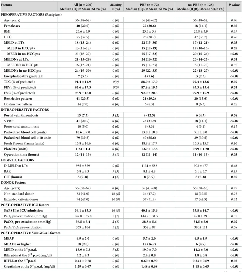
Table 2. Characteristics of the study population and comparison between PRF and no-PRF cases (univariate analysis).
Factors All (n = 200)
Median (IQR/ Mean±SD/n (%)
Missing n (%)
PRF (n = 72) Median (IQR)/ Mean±SD/n (%)
no-PRF (n = 128) Median (IQR)/ Mean±SD/n (%)
P value
PREOPERATIVE FACTORS (Recipient)
Age (years) 56 (48–62) 0 (0) 56 (48–62) 56 (48–62) 0.90 Female sex 40 (20.0) 0 (0) 22 (30.6) 18 (14.1) 0.05 BMI 25.6± 3.9 0 (0) 25.3± 3.9 25.8± 3.9 0.37 HCC 75 (37.5) 0 (0) 28 (38.9) 47 (36.7) 0.76 MELD at LTx 18 (13–24) 0 (0) 22 (15–30) 17 (12–21) 0.05 MELD in HCC pts 13 (11–18) 0 (0) 15 (12–19) 12 (10–15) 0.02 MELD in no-HCC pts 21 (16–27) 0 (0) 25 (17–32) 20 (15–24) <0.01 MELDNa at LTx 21 (15–28) 0 (0) 24 (16–32) 20 (14–25) 0.01 MELDNa in HCC pts 16 (12–21) 0 (0) 19 (14–23) 15 (11–20) 0.07 MELDNa in no-HCC pts 24 (19–30) 0 (0) 29 (22–33) 22 (18–27) <0.01 Encephalopathy grade �2 7 (3.5) 0 (0) 4 (5.6) 3 (2.3) <0.01 TLC (% of predicted) 91.4± 14.9 0(0) 88.0± 17.0 92.4± 13.6 0.02 FEV1(% of predicted) 92.6± 17.3 0(0) 87.8± 19.5 95.3± 15.4 0.01 FVC (% of predicted) 96.9± 18.0 0 (0) 92.0± 20.5 99.9± 15.9 <0.01 Restrictive pattern 41 (20.5) 0 (0) 21 (29.2) 20 (15.6) <0.01 Obstructive pattern 14 (7.0) 0 (0) 6 (8.3) 8 (6.3) 0.82 INTRAOPERATIVE FACTORS
Portal vein thrombosis 15 (7.5) 5 (2) 9 (12.5) 6 (4.7) 0.04
VVBP 41 (20.5) 0 (0) 23 (31.9) 18 (14.1) <0.01
Porto-caval anastomosis 10 (5.0) 0 (0) 6 (8.3) 4 (3.1) 0.11
Packed red blood cell (units) 10.6± 9.0 0 (0) 13.0± 10.0 9.1± 8.0 <0.01
Packed red blood cell >10 units 79 (39.5) 0 (0) 40 (55.6) 39 (30.5) <0.01
Fresh Frozen Plasma (units) 16.8± 16.6 0 (0) 18.8± 17.7 15.5± 15.7 0.16
Platelets (units) 1.24± 1.4 0 (0) 1.69± 1.58 0.99± 1.20 <0.01
Operation time (hours) 12 (11–13) 3 (1) 12 (11–14) 11 (10–13) 0.03
LOGISTIC FACTORS D-MELD at LTx 985± 529 0 (0) 1131± 586 903± 477 0.46 BAR 6.8± 4.3 7 (3) 8.1± 4.8 6.1± 3.7 0.13 CIT (hours) 8 (7–8) 4 (2) 8 (7–9) 8 (7–8) 0.05 DONOR Factors Age (years) 55 (38–67) 0 (0) 56 (43–68) 55 (38–66) 0.95 Non-standard donor 82 (41.0) 16 (8) 34 (47.2) 48 (37.5) 0.21
Extended criteria donor 94 (47.0) 16 (8) 37 (51.4) 57 (44.5) 0.31
POST-OPERATIVE ICU factors
SAPS II at ICU admission 36.1± 15.3 16 (8) 40.1± 15.6 33.8± 14.7 <0.01
PaO2pre-extubation (mmHg) 147.8± 35.8 5 (2) 144.2± 31.5 149.0± 39.0 0.37
PaCO2pre-extubation (mmHg) 36.5± 5.4 2 (1) 38.8± 5.6 34.5± 5.0 0.02
PaO2/FiO2pre-extubation 369± 104 5 (2) 352± 87 380± 111 0.08
POST-OPERATIVE SURGICAL factors
MEAF 4.9± 2.0 0 (0) 5.7± 2.0 4.5± 1.9 <0.01
MEAF 8 or higher 18 (9.0) 0 (0) 12 (16.7) 6 (4.7) <0.01
MELD at the 3rdp.o.d. 15.9
± 7.3 7 (3) 19.0± 7.0 14.2± 7.0 <0.01
Bilirubin at the 3rdp.o.d(mg/dl) 5.2± 4.3 0 (0) 2.4± 0.8 1.8± 0.8 <0.01
RIFLE at the 3rdp.o.d. 0.43± 0.78 0 (0) 0.60± 0.90 0.33± 0.69 0.03
Creatinine at the 3rdp.o.d. (mg/dl) 1.29
± 0.67 0 (0) 1.48± 0.68 1.18± 0.65 <0.01
�2 encephalopathy or hepatopulmonary syndrome. Pulmonary function tests were altered in 55 out of 200 patients (restrictive pattern in 41 cases, 20.5% and obstructive pattern in 14 cases, 7.0%). Overall, the presence of restrictive pattern was significantly associated with PRF (Table 2). Among PRF patients with restrictive pattern, moderate-severe pleural effusion was observed in 35 cases (85.3%) and moderate-severe ascites was present in 12 cases (53.6%). On the whole, PRF cases exhibited higher values of MELD and MELDNa at transplant. In particu-lar, MELD was higher in PRF patients independently from the HCC status, whereas, MELD in PRF patients with HCC was lower than in PRF without HCC (Fig 1).
PRF cases showed a higher incidence of portal thrombosis, received a higher number of packed red blood cell and platelet units, and more frequently required veno-venous bypass (VVBP). Furthermore, PRF patients displayed higher D-MELD, whilst Cold Ischemia Time (CIT) was moderately increased. The operation time in the PRF group was longer than in no-PRF group. Similarly, no-PRF cases had higher SAPS II scores at ICU admission. After surgery, no-PRF patients exhibited higher pre-extubation PaCO2levels. Overall, 22 patients (11.0%) developed
pneumonia, mostly in the PRF group (20, 27.8%). MEAF (evaluated as continuous variable and as percentage of patients with MEAF score >8), MELD at the 3rdpod, bilirubin at the 3rdpod, RIFLE and creatinine at the 3rdpod were higher in PRF cases. Accordingly, PRF cases suffered a higher incidence of severe complications as assessed by the Clavien-Dindo stratification.
Predictors of PRF at ROC and multivariate analysis
Among the 28 PRF predictors at univariate analysis, hemofiltration and pre-LTx mechanical ventilation were not included for the exiguous number of positive cases. Six parameters were Table 2. (Continued)
Factors All (n = 200)
Median (IQR/ Mean±SD/n (%)
Missing n (%)
PRF (n = 72) Median (IQR)/ Mean±SD/n (%)
no-PRF (n = 128) Median (IQR)/ Mean±SD/n (%)
P value OTHER DATA (available after 48 hours)
PaO2post-extubation (mmHg) 115.8± 34.8 15 (8) 103.1± 33.9 123.4± 32.3 <0.01
PaCO2post-extubation (mmHg) 37.5± 6.0 15 (8) 39.2± 6.8 36.4± 5.1 <0.01
PaO2/FiO2post-extubation 283± 93 15 (8) 255± 100 300± 85 <0.01
Mechanical Ventilation (hours) 22 (17–44) 0 (0) 61 (35–91) 20 (16–25) <0.01
Non-infectious lung involvement 113 (56.5) 0 (0) 53 (73.6) 60 (46.9) <0.01
Pneumonia 22 (11.0) 0 (0) 20 (27.8) 2 (1.6) <0.01 Clavien-Dindo stratification Grade 0 60 (30.0) 0 (0) 7 (9.7) 53 (41.4) <0.01 Grade 1 49 (24.5) 0 (0) 15 (20.8) 34 (26.6) 0.36 Grade 2 37 (18.5) 0 (0) 15 (20.8) 22 (17.2) 0.52 Grade 3A 13 (6.5) 0 (0) 7 (9.7) 6 (4.7) 0.17 Grade 3B 12 (6.0) 0 (0) 7 (9.7) 5 (3.9) 0.05 Grade 4 15 (7.5) 0 (0) 9 (12.5) 6 (4.7) 0.04 Grade 5 14 (7.0) 0 (0) 12 (16.7) 2 (1.6) <0.01
Grade 3B and higher 41 (20.5) 0 (0) 28 (38.9) 13 (10.2) <0.01
PRF: Postoperative Respiratory Failure, IQR: interquatile range, BMI: body mass index, LTx: Liver Transplantation, HCC: Hepatocellular carcinoma, MELD: Model for End-stage Liver Disease, LVEF%: Left Ventricular Ejection Fraction percentage, SPAP: Systolic Pulmonary Arterial Pressure, PPS: porto-pulmonary syndrome, PaO2:
partial pressure of arterial oxygen, PaCO2: partial pressure of arterial CO2, TLC: Total Lung Capacity, FEV1: Forced Expiratory Flow in 1 second, FVC: Forced Vital
Capacity, VVBP: Veno-Venous bypass, D-MELD: Donor Model for End-stage Liver Disease, BAR: BAlance of Risk score, CIT: Cold Ischemia Time, ICU: Intensive Care Unit, SAPS: Simplified Acute Physiology Score, FiO2: Fraction of Inspired Oxygen, MEAF: Model for Early Allograft Function, RIFLE: Risk Injury Failure Loss
End-stage of kidney disease
excluded because of collinearity. The remaining 22 parameters were investigated by ROC curve (Fig 2,S2 Table) and logistic regression analyses.
In addition, recipient age was included for epidemiologic reasons. Logistic regression analy-sis showed the following independent risk factors: recipient age (OR = 1.05; 95%CI 1.01–1.09; Fig 1. Frequencies of MELD in no-HCC and HCC patients. (A) Histograms of MELD according to the outcome (PRF vs no-PRF) are reported. For each subset
mean± SD and median (IQR) are reported. (B) Frequencies and (percentages) are reported in PRF and no-PRF patients according to MELD �22 and MELD <22.
Fig 2. ROC curve analysis. The Areas Under the Curve and Standard Errors are reported under each subset.
p = 0.010), female sex (OR = 2.79; 95%CI 1.19–6.52; p = 0.018), MELD at transplant
(OR = 1.09; 95%CI 1.04–1.14; p<0.001), lung restrictive pattern (OR = 2.49; 95%CI 1.11–5.61; p = 0.027), VVBP (OR = 3.03; 95%CI 1.33–6.90; p = 0.008), pre-extubation PaCO2(OR = 1.11;
95%CI 1.03–1.18; p = 0.003) and MEAF (OR = 1.37; 95%CI 1.15–1.63; p<0.001). Overall, 198 out of 200 observations with a full set of confounders were included in the multivariate analy-sis, The analysis displayed an excellent Hosmer-Lemeshow test value indicative of goodness of fit (p = 0.88). Detailed data are reported inS3 Table.
Univariate analysis according to the type of PRF
Among PRF cases, 44 (61.1%) were attributable to WF and 28 (38.9%) to EF. Main causes of WF were hemodynamic instability, alveolar-interstitial pulmonary edema (frequently transfu-sion-associated circulatory overload in patients with massive bleeding), neurological
impairment, and pending surgical issues (abdominal packing). In contrast, in EF cases, reinsti-tution of mechanical ventilation was mainly due to copious tracheal secretions with ineffective cough and atelectasis and concomitant respiratory muscle fatigue.
Demographic and clinical characteristics of EF and WF patients are shown inTable 3and inS4 Table.
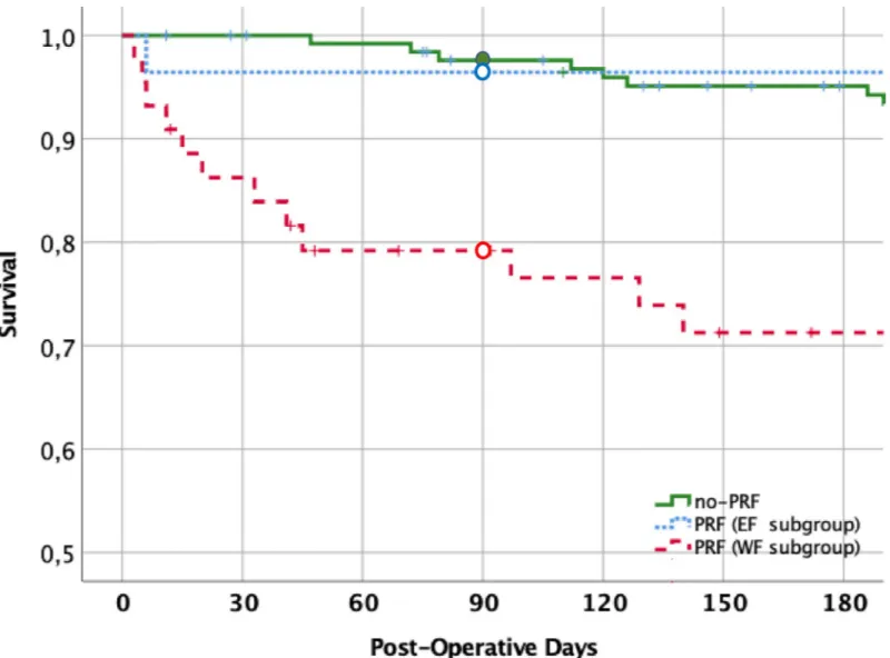
On the whole, WF cases showed significantly higher values of MELDNa than EF patients, while MELD values were only slightly increased. Regarding intraoperative variables, WF cases showed a higher prevalence of VVBP (p = 0.04). Among additional postoperative data, EF cases displayed lower post-extubation PaO2/FiO2 ratio, lower rate of severe surgical complica-tions, and lower ICU mortality rate (Fig 3).
Length of stay, discharge and survival
Overall, LoS in ICU was 6 days (IQR 5–10 day). No-PRF cases had a median LoS of 5 days (IQR 4–7 day) and were all discharged from ICU. In contrast, PRF ones had a significantly higher LoS (10 days, IQR 7–18 day, p<0.001) in comparison with no-PRF patients. The LoS was similar in EF and WF groups (Table 3). The day-90 survival was 97.6%±1.4 in the no-PRF group (one patient died at the 80thpostoperative day for intractable ascites and sepsis) and 86.0%±4.1 in the entire PRF group (p<0.001). Among PRF patients, 90-day survival was 96.4%±3.5% and 79.2%±6.2% in the EF and WF subgroups, respectively (p = 0.047) (Fig 3).
Discussion
The reported incidence of PRF in liver transplantation ranges between 11% and 42% due to the different thresholds used to define “prolonged” mechanical ventilation (from 24 hours to 7.5 days), and to the different inclusion criteria [11–18]. Overall, the incidence of PRF in our study population is 36.0%. We found that PRF is affected by seven independent variables, including MELD at transplant, restrictive lung pattern, use of VVBP, MEAF, pre-extubation PaCO2, patient age and sex. Remarkably, many variables considered in our analysis, such as
intraoperative surgical factors (portal thrombosis and porto-caval anastomosis) or logistic risk factors (D-MELD, BAlance of Risk, CIT) have been never investigated, neither collinearity was ruled out in previous studies [6,11–18]. Furthermore, this is the first study including the MEAF in a multivariable prediction model for PRF.
Among the identified PRF risk factors, MELD at transplant, restrictive lung pattern and use of VVBP resulted the most relevant. The role of MELD [13,14,17] and restrictive lung pattern [16] has been previously evidenced. Notably, our study population includes two well-defined groups: high-MELD no-HCC patients and low-MELD HCC patients, and both groups have similar PRF prevalence. Regarding the restrictive lung pattern, it mainly resulted from pleural
Table 3. Characteristics of PRF patients according to extubation and weaning failure (univariate analysis).
Factors Extubation failure (n = 28)
Median (IQR) / Mean± SD/n (%) Weaning failure (n = 44) Median (IQR) / Mean±SD /n (%) P value
PREOPERATIVE FACTORS (recipient)
Age (years) 58 (50–62) 54 (47–62) 0.90 Female sex 8 (28.6) 14 (31.8) 0.77 BMI >30 2 (7.1) 9 (20.5) 0.13 HCC 11 (39.3) 17 (38.6) 0.96 MELD at LTx 20 (14–26) 23 (15–31) 0.06 MELDNa at LTx 23 (16–30) 27 (15–33) 0.01 Encephalopathy grade �2 2 (7.1) 2 (4.5) 0.22 Restrictive pattern 7 (25.0) 14 (31.8) 0.56 Obstructive pattern 3 (10.7) 3 (6.8) 0.62 INTRAOPERATIVE factors
Portal Vein thrombosis 2 (7.1) 7 (15.9) 0.26
VVBP 5 (17.9) 18 (40.9) 0.04
Packed red blood cell (units) 11.3± 9.9 14.1± 11.3 0.24
Packed red blood cell >10 units 12 (42.9) 28 (63.6) 0.08
Fresh Frozen Plasma (units) 14.5± 14.8 22.0± 19.1 0.07
Platelets (units) 1.60± 1.80 1.75± 1.43 0.73
Operation time (hours) 12 (12–14) 13 (12–14) 0.58
LOGISTIC FACTORS
D-MELD at LTx 1067± 556 1172± 606 0.56
BAR 7.1± 4.0 8.8± 5.3 0.12
CIT (hours) 8 (7–8) 8 (7–9) 0.23
POST-OPERATIVE ICU FACTORS
SAPS II at the ICU admission 37.8± 13.3 41.6± 16.9 0.33
PaCO2pre-extubation (mmHg) 37.4± 5.6 38.0± 6.1 0.68
PaO2/FiO2pre-extubation 372± 88 337± 85 0.13
POST-OPERATIVE SURGICAL FACTORS
MEAF 5.3± 2.0 5.9± 1.9 0.23
MEAF 8 & over 3 (10.7) 9 (20.5) 0.28
MELD on 3rdp.o.d. (mg/dl) 17.6± 6.2 18.9± 8.4 0.17
Bilirubin on 3rdp.o.d. (mg/dl) 5.7
± 4.3 8.0± 5.6 0.06
Creatinine on 3rdp.o.d. (mg/dl) 1.32
± 0.57 1.58± 0.73 0.10
OTHER DATA (available after 48 hours)
PaO2post-extubation (mmHg) 89.0± 32.0 113.9± 31.6 <0.01
PaCO2post-extubation (mmHg) 39.2± 7.8 39.3± 6.1 0.96
PaO2/FiO2post-extubation 209± 93 290± 92 <0.01
Mechanical Ventilation (hours) 21 (14–39) 73 (66–119) <0.01
Non-infectious lung involvement 17 (60.7) 36 (81.8) 0.22
Pneumonia 7 (25.0) 13 (29.5) 0.68
Clavien-Dindo Grade 3B and higher 5 (17.9) 23 (52.3) <0.01
LoS in ICU post LTx (days) 9 (7–15) 10 (7–18) 0.81
Death in ICU 1 (3.6) 10 (22.7) 0.03
PRF: Postoperative Respiratory Failure, BMI: body mass index, HCC: Hepatocellular carcinoma, MELD: Model for End-stage Liver Disease, LTx: Liver Transplantation, VVBP: Veno-Venous bypass, D-MELD: Donor Model for End-stage Liver Disease, BAR: BAlance of Risk score, CIT: Cold Ischemia Time, ICU: Intensive Care Unit, SAPS: Simplified Acute Physiology Score, PaO2: partial pressure of arterial oxygen, PaCO2: partial pressure of arterial CO2, FiO2: Fraction of Inspired Oxygen, MEAF:
Model for Early Allograft Function, RIFLE: Risk Injury Failure Loss End-stage of kidney disease, LoS: Length of stay.
effusion (with or without ascites) causing basal atelectasis. Actually, while ascites is absent or drained in the early postoperative days, pleural effusion may even worsen due to the lung dys-function and/or diaphragm trauma.
Regarding VVPB, in our experience it is used in case of high surgical complexity due to var-ious conditions such as grade-III/IV portal thrombosis, Budd Chiari syndrome, huge liver in polycystic liver disease, late re-transplant. In addition, we occasionally used VVBP as a rescue procedure for massive bleeding, when the graft is not yet ready to be implanted (delay in the organ transport, complex reconstruction on the back table). The proportion of patients receiv-ing VVBP in our study is comparable to that previously reported [16]. Moreover, in our series of patients, it can be considered as a surrogate marker of portal hypertension/thrombosis, it is not collinear with MELD, neither it was routinely used in our high-MELD cases. VVBP pro-longsper se the operation time, activates fibrinolysis and platelets’ consumption [45]. Although the operation time showed the highest area under the curve at ROC analysis, VVBP was more Fig 3. Survival analysis according with the PRF status. Patient survival at 90 days was 97.6%±1.4% in the no-PRF group (continuous line), 96.4%±3.5% in the EF subgroup (dash-interrupted line), and 79.2%±6.2% in the WF subgroup (dot-interrupted line). Survival was significantly different between PRF and no-PRF groups (p<0.001) and, within PRF patients, between EF and WF- subgroups (p = 0.047). WF, but not EF patient’ survival, differed from that of no-PRF patients.
predictive in all decile categories at the Hosmer-Lemeshow test. We are aware that in centers not using VVPB, other indicators might be predictive for surgical complexity.
In our population, PRF risk markedly increased along the MEAF score. The association between graft recovery and PRF has been scarcely investigated. High transaminases have been associated with PRF [11,18] or PaO2/FiO2�300 mmHg [19]. This is the first study
investigat-ing the PRF impact of the MEAF score, obtained by bilirubin, transaminase and INR, and ranging continuously from 1 to 10 [29]. More recently, a novel score predicting graft recovery has been developed, based on bilirubin, transaminase, INR and platelet count [46]. Although more performant than MEAF, it includes parameters gathered until pod 10, and it is not suit-able to evaluate PRF at 48 hours.
The mean value of pre-extubation PaCO2,even though still normal, was higher in PRF
patients. This finding has been previously reported [6] and it is conceivable that it results from a problematic weaning: notwithstanding the exact underlying mechanisms is presently unknown, the contribution of graft dysfunction/non-function or phrenic and/or diaphrag-matic surgical trauma cannot be excluded.
As previously reported, we observed a negative prognostic effect for recipient age and female sex at multivariate analysis [17,47]. It has been suggested that females are less likely to receive intraoperative low-tidal protective ventilation during surgery, and this may yield a higher rate of postoperative respiratory complications [47]. Moreover, as compared to male patients, MELD score in females underestimates liver-kidney dysfunction up to 3 points, prob-ably for the lower dietary intake and the lower tubular secretion of creatinine [48].
Of note, we failed to demonstrate any impact on the PRF occurrence of diabetes, BMI and, for the exiguous number of cases, of re-transplant [11–18,49,50].
Noteworthy, we observed that PRF, as usually defined [3,16], includes two populations of patients with different 90-day survival. Notably, the EF patients exhibited similar overall sur-vival as no-PRF patients, whereas they did not differ from WF patients regarding main base-line clinical characteristics and PRF prognostic factors. It could be argued that these patients may have been extubated a quite bit early, as suggested by their lower post-extubation PaO2/
FiO2. The assessment for readiness for extubation is based on several static measures, even
though the dynamic nature of the weaning makes difficult to definitely predict its success [23]. Indeed, the EF occurs more frequently in patients unable to manage copious tracheal secretion due to ineffective cough, or respiratory fatigue secondary to poor patient cooperation or mal-nutrition. However, all these issues cannot be anticipated before extubation. Accordingly, EF patients compared with WF patients for LoS in ICU, whilst they had a significantly lower 90-day survival.
Although these observations have been gathered in a small subset of patients, we can hypothesize that EF group represents a low-risk subset of LTx patients, with lower prevalence of severe complications (Clavien-Dindo grade 3B and over), resulting in better survival. Over-all, these findings show that differentiating among PRF patients those with EF from those with WF, can have a significant prognostic relevance.
Our study suffers from some limitations. It is based on a single-center experience, and has a retrospective design, whereas, to the best of our knowledge, PRF has not been investigated in multi-center prospective cohorts. According to previous studies, the role of PRF was investi-gated at 48 hours categorizing PRF as a dichotomic variable.[3–4] Nevertheless, our findings pave the way to future studies exploring the prognostic role of PRF as a time-dependent s vari-able. In our series of patients, functional capacity, frailty and sarcopenia were not prospectively recorded and were not included in the analyses, whereas BMI, a surrogate method to evaluate sarcopenia, was not predictive. The Dindo-Clavien grading of complications was used instead of the new Comprehensive Complication Index [51]. Furthermore, we did not test the Donor
Risk Index [10], which is not applicable in Italy for the shorter distance between donor and recipient hospitals and higher donor age [52]. Finally, the present study does not include pedi-atric patients, adult patients transplanted for acute liver failure and living-donor transplants.
The multidisciplinary approach to predict PRF in the complex context of LTx deserves con-sideration. The methodology adopted may provide hepatologists and surgeons an adequate tool to estimate the ICU prognosis of listed patients. Patients with high MELD, restrictive lung pattern and/or high surgical complexity should be informed on their high PRF risk, eventually increased by early allograft dysfunction. Likewise, our study may help surgeons and intensi-vists to identify risk-mitigation strategies in specific patients, such as a wider use of non-inva-sive ventilation soon after the extubation. Accordingly, the frequent use of non-invanon-inva-sive ventilation, avoiding endotracheal reintubation, can explain the lower pulmonary infection rate of our population in comparison with other studies [6,16]. In fact, non-invasive ventila-tion prevents basal atelectasis due to abdominal distension, alteraventila-tion of diaphragmatic func-tion and contractility, promotes lung recruitment and decreases work of breathing [24].
Conclusions
On the whole, our analysis carried out according to the organ-based perspective, lung restric-tive pattern, narestric-tive liver (MELD), surgical complexity, as captured by VVBP, and new liver function (MEAF) are the main determinants for PRF in non-acute LTx patients. Remarkably, donor variables, as evaluated before the transplant, do not always reflect the postoperative donor-related risk, which can be actually established only in the early postoperative days.
Supporting information
S1 Fig. Consort diagram. Eligibility, exclusion and study population
(TIF)
S1 Table. Characteristics of the study population and comparison between PRF and no-PRF cases at univariate analysis. no-PRF: Postoperative Respiratory Failure, IQR: interquatile
range, BMI: body mass index, LTx: Liver Transplantation, HCC: Hepatocellular carcinoma, MELD: Model for End-stage Liver Disease, LVEF%: Left Ventricular Ejection Fraction per-centage, SPAP: Systolic Pulmonary Arterial Pressure, PPS: porto-pulmonary syndrome, PaO2:
partial pressure of arterial oxygen, PaCO2: partial pressure of arterial CO2, TLC: Total Lung
Capacity, FEV1: Forced Expiratory Flow in 1 second, FVC: Forced Vital Capacity, VVBP:
Veno-Venous bypass, D-MELD: Donor Model for End-stage Liver Disease, BAR: BAlance of Risk score, CIT: Cold Ischemia Time, ICU: Intensive Care Unit, SAPS: Simplified Acute Physi-ology Score, FiO2: Fraction of Inspired Oxygen, MEAF: Model for Early Allograft Function,
RIFLE: Risk Injury Failure Loss End-stage of kidney disease. (DOCX)
S2 Table. List of variables investigated by ROC curve analysis.
(DOCX)
S3 Table. Details of the multivariate analysis (logistic regression). The number of events
(70) allowed the identification of 7 predictive factors. Hosmer-Lemeshow test: Chi2= 3.78, p = 0.88). Age, MELD at transplant and pre-extubation PaCO2showed a broad IQR
(Inter-quartile Range) excursion, explaining how the low OR (Odd Ratio) is indicative of a strong sta-tistical effect. MELD: Model for End-stage Liver Disease, VVBP: Veno-Venous bypass, PaCO2:
partial pressure of arterial CO2, MEAF: Model for Early Allograft Function.
S4 Table. Characteristics of PRF patients according to extubation and weaning failure (univariate analysis). PRF: Postoperative Respiratory Failure, BMI: body mass index, HCC:
Hepatocellular carcinoma, MELD: Model for End-stage Liver Disease, LTx: Liver Transplanta-tion, VVBP: Veno-Venous bypass, D-MELD: Donor Model for End-stage Liver Disease, BAR: BAlance of Risk score, CIT: Cold Ischemia Time, ICU: Intensive Care Unit, SAPS: Simplified Acute Physiology Score, PaO2: partial pressure of arterial oxygen, PaCO2: partial pressure of
arterial CO2, FiO2: Fraction of Inspired Oxygen, MEAF: Model for Early Allograft Function,
RIFLE: Risk Injury Failure Loss End-stage of kidney disease, LoS: Length of stay. (DOCX)
Acknowledgments
The Authors thank Prof. Salvatore Agnes for performing the largest part of surgeries, Dr. Ric-cardo Inchingolo for the helpful interpretation of pulmonary function tests, Dr. Giuseppe Marrone for the useful discussion on clinical implications of portal hypertension and to Dr. Alessandro Vitale for the statistical supervision.
Author Contributions
Conceptualization: Alfonso Wolfango Avolio, Rita Gaspari, Luciana Teofili, Massimo
Antonelli.
Data curation: Alfonso Wolfango Avolio, Rita Gaspari, Luciana Teofili, Giuseppe Bianco,
Giorgia Spinazzola, Paolo Maurizio Soave, Gianfranco Paiano, Alessandra Gioia Frances-coni, Nicola Nicolotti.
Formal analysis: Alfonso Wolfango Avolio, Rita Gaspari, Giuseppe Bianco, Nicola Nicolotti. Investigation: Alfonso Wolfango Avolio, Rita Gaspari.
Methodology: Alfonso Wolfango Avolio, Rita Gaspari, Massimo Antonelli. Supervision: Alfonso Wolfango Avolio.
Validation: Alfonso Wolfango Avolio, Rita Gaspari. Visualization: Alfonso Wolfango Avolio.
Writing – original draft: Alfonso Wolfango Avolio, Rita Gaspari, Luciana Teofili. Writing – review & editing: Alfonso Wolfango Avolio, Rita Gaspari, Luciana Teofili,
Giu-seppe Bianco, Giorgia Spinazzola, Paolo Maurizio Soave, Gianfranco Paiano, Alessandra Gioia Francesconi, Andrea Arcangeli, Nicola Nicolotti, Massimo Antonelli.
References
1. Smetana GW, Lawrence VA, Cornell JE. Preoperative pulmonary risk stratification for non-cardiotho-racic surgery: systematic review for the American College of Physicians. Ann Intern Med 2006; 144:581–595. PMID:16618956
2. Canet J, Gallart L, Gomar C, Paluzie G, Vallès J, Castillo J, et al. Prediction of postoperative pulmonary complications in a population-based surgical cohort. Anesthesiology 2010; 113:1338–1350.https://doi. org/10.1097/ALN.0b013e3181fc6e0aPMID:21045639
3. Arozullah AM, Daley J, Henderson WG, Khuri SF, Daley J. Multifactorial risk index for predicting postop-erative respiratory failure in men after major noncardiac surgery. Ann Surg 2000; 232:242–253. PMID:
10903604
4. Svensson LG, Hess KR, Coselli JS, Safi HJ, Crawford ES. A prospective study of respiratory failure after high-risk surgery on the thoraco abdominal aorta. J Vasc Surg 1991; 14:271–282. PMID:1880835
5. Sinclair M, Gow PJ, Grossmann M, Angus PW. Review article: sarcopenia in cirrhosis-aetiology, impli-cations and potential therapeutic interventions. Aliment Pharmacol Ther 2016; 43:765–777.https://doi. org/10.1111/apt.13549PMID:26847265
6. Bozbas SS, Eyuboglu FO, Ozturk Ergur F, Gullu Arslan N, Sevmis S, Karakayali H, et al. Pulmonary complications and mortality after liver transplant. Exp Clin Transplant 2008; 6:264–270. PMID:
19338487
7. Zimmerman JE, Wagner DP, Seneff MG, Becker RB, Sun X, Knaus WA. Intensive care unit admissions with cirrhosis: risk-stratifying patient groups and predicting individual survival. Hepatology 1996; 23:1393–1401.https://doi.org/10.1002/hep.510230615PMID:8675156
8. Rodrı´guez-Roisin R, Krowka MJ, Herve´ P, Fallon MB. Pulmonary-Hepatic vascular Disorders (PHD). Eur Respir J 2004; 24:861–880. PMID:15516683
9. Mc Alister VC, Grant DR, Roy A, Brown WF, Hutton LC, Leasa DJ, et al. Right phrenic nerve injury in orthotopic liver transplantation. Transplantation 1993; 55:826–830. PMID:8475559
10. Feng S, Goodrich NP, Bragg-Gresham JL, Dykstra DM, Punch JD, DebRoy MA, et al. Characteristics associated with liver graft failure: the concept of a donor risk index. Am J Transplant 2006; 6:783–790.
https://doi.org/10.1111/j.1600-6143.2006.01242.xPMID:16539636
11. Glanemann M, Langrehr J, Kaisers U, Schenk R, Mu¨ller A, Stange B, et al. Postoperative tracheal extu-bation after orthotopic liver transplantation. Acta Anaesthesiol Scand 2001; 45:333–339. PMID:
11207470
12. Huang CT, Lin HC, Chang SC, Lee WC. Pre-operative risk factors predict post-operative respiratory fail-ure after liver transplantation. PLoS One 2011; 6:e22689.https://doi.org/10.1371/journal.pone.0022689
PMID:21829646
13. Kleine M, Vondran FW, Johanning K, Timrott K, Bektas H, Lehner F, et al. Respiratory risk score for the prediction of 3-month mortality and prolonged ventilation after liver transplantation. Liver Transpl 2013: 19:862–871.https://doi.org/10.1002/lt.23673PMID:23696476
14. Pedersen MR, Choi M, Brink JA, Seetharam AB. Pretransplant Factors and Associations with Postoper-ative Respiratory Failure, ICU Length of Stay, and Short-Term Survival after Liver Transplantation in a High MELD Population. J Transplant 2016; 2016:6787854.https://doi.org/10.1155/2016/6787854
PMID:27980860
15. Yuan H, Tuttle-Newhall JE, Chawa V, Schnitzler MA, Xiao H, Axelrod D, et al. Prognostic impact of mechanical ventilation after liver transplantation: a national data base study. Am J Surg 2014; 208:582– 590.https://doi.org/10.1016/j.amjsurg.2014.06.004PMID:25151187
16. Levesque E, Hoti E, Azoulay D, Honore I, Guignard B, Vibert E, et al. Pulmonary complications after elective liver transplantation-incidence, risk factors, and outcome. Transplantation 2012; 94:532–538.
https://doi.org/10.1097/TP.0b013e31825c1d41PMID:22885879
17. Wang A, An X, Xia VW. Female gender of the Recipient Is Independently Associated With prolonged Ventilation Time and Hospital Stay After Liver Transplantation. Transplant Proc 2016; 48:120–122.
https://doi.org/10.1016/j.transproceed.2016.01.004PMID:26915855
18. Garutti I, Sanz J, Olmedilla L, Tranche I, Vilchez A, Fernandez-Quero L, et al. Extravascular Lung Water and Pulmonary Vascular Permeability Index Measured at the End of Surgery Are Independent Predictors of Prolonged Mechanical Ventilation in Patients Undergoing Liver Transplantation. Anesth Analg 2015; 121:736–745.https://doi.org/10.1213/ANE.0000000000000875PMID:26218864 19. Faenza S, Ravaglia MS, Cimatti M, Dante A, Spedicato S, Labate AM. Analysis of the causal factors of
prolonged mechanical ventilation after orthotopic liver transplant. Transplant Proc 200; 38:1131–1134.
20. Lung function testing: selection of reference values and interpretative strategies. Am Thoracic Society. Am Rev Respir Dis 1991; 144:1202–1218.
21. Roch A, Bojan M, Michelet P, Romain F, Bregeon F, Papazian L, et al. Usefulness of ultrasonography in predicting pleural effusions>500 mL in patients receiving mechanical ventilation. Chest 2005; 127:224– 232.https://doi.org/10.1378/chest.127.1.224PMID:15653988
22. Ferenci P, Lockwood A, Mullen K, Tarter R, Weissenborn K, Blei AT. Hepatic encephalopathy-defini-tion, nomenclature, diagnosis, and quantification: final report of the working party at the 11th World Con-gresses of Gastroenterology, Vienna, 1998. Hepatology 2002; 35:716–721.https://doi.org/10.1053/ jhep.2002.31250PMID:11870389
23. Boles JM, Bion J, Connors A, Herridge M, Marsh B, Melot C, et al. Weaning from mechanical ventilation. Eur Respir J 2007; 29:1033–1056.https://doi.org/10.1183/09031936.00010206PMID:17470624 24. Antonelli M, Conti G, Bufi M, Costa MG, Lappa A, Rocco M, et al. Non invasive ventilation for treatment
of acute respiratory failure in patients undergoing solid organ transplantation: a randomized trial. JAMA 2000; 283:235–241. PMID:10634340
25. Mandell LA, Wunderink RG, Anzueto A, Bartlett JG, Campbell GD, Dean NC, et al. Infectious Diseases Society of America/American Thoracic Society consensus guidelines on the management of commu-nity-acquired pneumonia in adults. Clin Infect Dis 2007; 44Suppl 2:27–72.
26. Wiesner R, Edwards E, Freeman R, Harper A, Kim R, Kamath P, et al. Model for end-stage liver disease (MELD) and allocation of donor livers. Gastroenterology 2003; 124:91–96.https://doi.org/10.1053/gast. 2003.50016PMID:12512033
27. Yun BC, Kim WR, Benson JT, Biggins SW, Therneau TM, Kremers WK, et al. Impact of pretransplant hyponatremia on outcome following liver transplantation. Hepatology 2009; 49:1610–1615.https://doi. org/10.1002/hep.22846PMID:19402063
28. Cucchetti A, Ercolani G, Cescon M, Ravaioli M, Zanello M, Del Gaudio M, et al. Recovery from liver fail-ure after hepatectomy for hepatocellular carcinoma in cirrhosis: meaning of the model for end-stage liver disease. J Am Coll Surg 2006; 203:670–676.https://doi.org/10.1016/j.jamcollsurg.2006.06.018
PMID:17084328
29. Pareja E, Cortes M, Herva´ s D, Mir J, Valdivieso A, Castell JV, et al. A score model for the continuous grading of early allograft dysfunction severity. Liver Transpl 2015; 21:38–46.https://doi.org/10.1002/lt. 23990PMID:25204890
30. Park MH, Shim HS, Kim WH, Kim HJ, Kim DJ, Lee SH, et al. Risk Scoring Models for Prediction of Acute Kidney Injury after Living Donor Liver Transplantation: A Retrospective Observational Study. PLoS One 2015; 10:e0136230.https://doi.org/10.1371/journal.pone.0136230PMID:26302370 31. Avolio AW, Cillo U, Salizzoni M, De Carlis L, Colledan M, Gerunda GE, et al. Balancing donor and
recip-ient risk factors in liver transplantation: the value of D-MELD with particular reference to HCV reciprecip-ients. Am J Transplant 2011; 11:2724–2736.https://doi.org/10.1111/j.1600-6143.2011.03732.xPMID:
21920017
32. Dutkowski P, Oberkofler CE, Slankamenac K, Puhan MA, Schadde E, Mu¨llhaupt B, et al. Are there bet-ter guidelines for allocation in liver transplantation? A novel score targeting justice and utility in the model for end-stage liver disease era. Ann Surg 2011; 254:745–753.https://doi.org/10.1097/SLA. 0b013e3182365081PMID:22042468
33. Le Gall JR, Loirat P, Alperovitch A, Glaser P, Granthil C, Mathieu D, et al. A simplified acute physiology score for ICU patients. Crit Care Med 1984; 12:975–977. PMID:6499483
34. Clavien PA, Barkun J, de Oliveira ML, Vauthey JN, Dindo D, Schulick RD, et al. The Clavien-Dindo clas-sification of surgical complications: five years experience. Ann Surg 2009; 250:187–196.https://doi.org/ 10.1097/SLA.0b013e3181b13ca2PMID:19638912
35. Cillo U, Burra P, Mazzaferro V, Belli L, Pinna AD, Spada M, et al. A Multistep, Consensus-Based Approach to Organ Allocation in Liver Transplantation: Toward a "Blended Principle Model". Am J Transplant 2015; 15:2552–2561.https://doi.org/10.1111/ajt.13408PMID:26274338
36. Avolio AW, Agnes S, Cillo U, Lirosi MC, Romagnoli R, Baccarani U, et al.http://www.D-MELD.com, the Italian survival calculator to optimize donor to recipient matching and to identify the unsustainable matches in liver transplantation. Transpl Int 2012; 25:294–301.https://doi.org/10.1111/j.1432-2277. 2011.01423.xPMID:22268763
37. Avolio AW, Halldorson JB, Burra P, Dutkowski P, Agnes S, Clavien PA. Balancing utility and need by means of donor-to-recipient matching: a challenging problem. Am J Transplant 2013; 13:522–523.
https://doi.org/10.1111/ajt.12031PMID:23282243
38. Avolio AW, Agnes S, Chirico AS, Cillo U, Frongillo F, Castagneto M. Successful transplantation of an injured liver. Transplant Proc 2000; 32:131–133. PMID:10700996
39. Avolio AW, Agnes S, Nure E, Maria G, Barbarino R, Pepe G, et al. Comparative evaluation of two perfu-sion solutions for liver preservation and transplantation. Transplant Proc 2006: 38:1066–1067.https:// doi.org/10.1016/j.transproceed.2006.03.009PMID:16757265
40. Avolio AW, Agnes S, Gasbarrini A, Nure E, Siciliano M, Castagneto M. Prognostic value of MELD score and donor quality in liver transplantation: implications for the donor recipient match. Transplant Proc. 2006; 38(4):1059–62.https://doi.org/10.1016/j.transproceed.2006.03.008PMID:16757263
41. Cameron AM, Ghobrial RM, Yersiz H, Farmer DG, Lipshutz GS, Gordon SA, et al Optimal utilization of donor grafts with extended criteria: a single-center experience in over 1000 liver transplants. Ann Surg. 2006: 243(6):748–55.https://doi.org/10.1097/01.sla.0000219669.84192.b3PMID:16772778 42. Jacob M, Lewsey JD, Sharpin C, Gimson A, Rela M, van der Meulen JH. Systematic review and
valida-tion of prognostic models in liver transplantavalida-tion. Liver Transpl 2005; 11:814–825.https://doi.org/10. 1002/lt.20456PMID:15973726
43. Hosmer DW, Lemeshow S, Rodney XS: Applied Logistic Regression. 3rd ed. New York, John Wiley and Son; 2013.
44. Allison PD. Logistic Regression Using the SAS System: Theory and Application. Cary, NC: Wiley-SAS; 2001
45. Cleland S, Corredor C, Ye JJ, Srinivas C, McCluskey SA. Massive haemorrhage in liver transplantation: Consequences, prediction and management. Word J Transplant 2016; 6:291–305.
46. Agopian VG, Harlander-Locke MP, Markovic D, Dumronggittigule W, Xia V, Kaldas FM, et al. Evalua-tion of Early Allograft FuncEvalua-tion Using the Liver Graft Assessment Following TransplantaEvalua-tion Risk Score Model. JAMA Surg 2018; 153(5):436–444.https://doi.org/10.1001/jamasurg.2017.5040PMID:
29261831
47. Gajic O, Dara SI, Mendez JL, Adesanya AO, Festic E, Caples SM, et al. Ventilator-associated lung injury in patients without acute lung injury at the onset of mechanical ventilation. Crit Care Med 2004; 32:1817–1824. PMID:15343007
48. Cholongitas E, Marelli L, Kerry A, Goodier DW, Nair D, Thomas M, Patch D, Burroughs AK. Female liver transplant recipients with the same GFR as male recipients have lower MELD scores—a system-atic bias. Am J Transplant 2007; 7:685–692.https://doi.org/10.1111/j.1600-6143.2007.01666.xPMID:
17217437
49. Avolio AW, Agnes S, Chirico AS, Castagneto M. Primary dysfunction after liver transplantation: donor or recipient fault? Transplant Proc 1999; 31:434–436. PMID:10083176
50. Barone M, Viggiani MT, Avolio AW, Iannone A, Rendina M, Di Leo A. Obesity as predictor of postopera-tive outcomes in liver transplant candidates: Review of the literature and future perspecpostopera-tives. Dig Liver Dis 2017; 49:957–966.https://doi.org/10.1016/j.dld.2017.07.004PMID:28801180
51. Slankamenac K, Nederlof N, Pessaux P, de Jonge J, Wijnhoven BP, Breitenstein S, et al. The compre-hensive complication index: a novel and more sensitive endpoint for assessing outcome and reducing sample size in randomized controlled trials. Ann Surg 2014; 260:757–762.https://doi.org/10.1097/SLA. 0000000000000948PMID:25379846
52. Avolio AW, Gruttadauria S, Grieco A, De Feo TM. Comment to "Liver Match: a prospective observa-tional cohort study on liver transplantation in Italy". Dig Liver Dis 2011; 43:921.https://doi.org/10.1016/j. dld.2011.05.016PMID:21752740