Super
vi
sor
:
Pr
of
.
Mar
i
a
Beat
r
i
ce
Bi
t
ont
i
Pr
of
.
Mi
Co-
eke
super
Van
vi
Li
sor
j
sebet
:
t
ens
Coor
di
nat
or
:
Pr
of
.
Anna
Mar
i
a
I
nnocent
i
Uni
ver
si
t
y
of
Cal
abr
i
a,
Depar
t
ment
of
Ecol
ogy
Alla mia Famiglia
per avermi concesso un altro punto di vista sulla Vita
e
a tutti coloro che mi sono fedeli
nell’Amicizia e Amore…
Al Governo Berlusconi per aver tagliato i fondi per la Ricerca
e a quello Prodi per averci provato…
“L’Italia è una Repubblica fondata sul lavoro”
A tutti coloro che lavorano per passione e necessità o sono in cerca di proposte
e stabilità.
Grazie.
Viviamo ancora in tempi interessanti?
:-)
AIM OF THE WORK ... 2
INTRODUCTION ... 3
ARABIDOPSIS THALIANA AS A MODEL PLANT... 3
The history of Arabidopsis thaliana... 3
Arabidopsis nomenclature, characteristics and ecotypes... 6
Timetable for growth-stage based analysis... 7
LEAF DEVELOPMENT... 8
The maintenance of the SAM’s identity and the initiation of leaf primordium ... 8
Leaf morphogenesis in flowering plants... 9
Primary morphogenesis: acquisition of the dorsiventral asymmetry ... 10
Secondary morphogenesis: acquisition of the proximal-distal and lateral polarity... 11
Hormones, light and leaf development ... 12
Heteroblasty in Arabidopsis ... 12
THE TRANSCRIPTION MACHINERY OF EUKARYOTES... 13
Gene expression in eukaryotes... 13
The transcription cycle and the formation of a transcript ... 13
General transcription factors and the eukaryotic promoters ... 14
Formation of the PIC (Pre-Inititation Complex) assembly ... 15
RNAPII initiation regulation ... 17
RNAPIIa/o, Mediator, Elongator and the transcript elongation... 17
The Elongator complex ... 18
Plant Elongator ... 18
Transcript termination ... 19
SUCROSE IN PLANTS... 20
General characteristics... 20
Sucrose transport... 20
Sucrose carriers in Arabidopsis ... 21
Sucrose synthesis and cleavage: sucrose synthases and invertases... 21
Sucrose metabolism... 22
Sugar-sensing mechanisms and general gene regulation... 23
RESULTS ... 24
CYTOLOGICAL INVESTIGATIONS OF THE ARABIDOPSIS THALIANA ELO1 MUTANT GIVE NEW INSIGHTS INTO LEAF LATERAL GROWTH AND ELONGATOR FUNCTION... 24
CONCLUSION ... 34
REFERENCES ... 36
ANNEX: VIB’S WORK ... 42
RESULTS... 42
1. AtELP genes and T-DNA insertion lines ... 42
2. Morphological comparison between first and third leaves of the DRL1 overexpression line 10B5 (in Ler background) and the wild-type Ler. PCN analysis on RON1 first and third leaves ... 54
3. Map Based Cloning (MBC): the TRN1 experience... 61
4. Bioinformatics: AtELP1p homologue of Saccharomyces cerevisiae ELP1/TOT1/IKI3p... 64
MATERIALS AND METHODS... 65
REFERENCES... 70
Aim of the work
My PhD work has been focused on the cytophysiological characterization of the
Elongator complex in Arabidopsis thaliana. The holo-Elongator complex is made of two
subcomplexes: ELP1, ELP2 and ELP3 that compose the core-Elongator, and ELP4, ELP5
and ELP6 that constitute the accessory subcomplex. The predicted function of the complex
is suggested by ELP3, which contains a HAT domain (histone acetyltransferase activity)
that causes cromatin relaxation through histone acetylation and promotes transcription
elongation of several genes.
Several experimental approaches have led to a model of Elongator's function in
transcription elongation and overall energetic metabolism regulation, in a stage-specific
and sucrose-mediated way.
Initially, in my first year work, performed in Ghent (Belgium) under the supervision
of Prof. Mieke Van Lijsebettens, several genes presumably involved in the Elongator
complex were analyzed for T-DNA insertions and heterozygosity at the locus, to find new
alleles for the Arabidopsis thaliana homologues of the Yeast Elongator complex.
Phenotype scoring and germination tests were done to further ascertain the mutant
genotypes and phenotypes.
In addition, in order to investigate leaf lateral growth and mechanisms such as cell
division and expansion, the DRL1ox10B5 (an overexpression line of DEFORMED ROOTS
AND LEAVES1) and RON1 (ROTUNDA1) leaf mutants were investigated through
morphometric analysis. Finally, under the supervision of Dr. Cnops Gerda, I participated at
the final step of the map-based cloning of the TRN1 (TORNADO1), amplifying and
sequencing a small genomic region in order to find, through sequence alignment, the
putative TRN1 locus.
Next, at the University of Calabria, I investigated the relationships between leaf
development, growth conditions and Elongator’s function by studying growth responses,
under different nutrient and environmental conditions, in both wild type and elo1 ‘narrow
leaves’ mutants.
These mutants belong to the elongata class and have a mutation in one of the
components of the histone acetyl transferase Elongator complex. Through germination
assays, morphometric analysis and electron microscopy I show that light response, stress
tolerance and sucrose metabolism are affected in the Elongator mutant.
Performing an analysis of the genes differentially expressed in the elo mutants (data
not shown for VIB’s patenting reasons), which have been identified by Fleury Delphine
(pers. comm.) I propose a functional model for the Elongator complex that is
sucrose-dependant and stage-specific.
My PhD work has been achieved through a collaboration with Prof. Mieke Van Lijsebettens, who works at the VIB (Flemish Interuniversity Institute for Biotechnology, Department of Plant Systems Biology, Technologiepark 927, 9052 Ghent, Belgium) and thanks to the supervision of Prof. Maria Beatrice Bitonti of the Università della Calabria (Dipartimento di Ecologia, Via ponte P. Bucci, Cubo 6B, 87036, Arcavacata di Rende, CS, Italia).
This project was funded by the European Union in the frame of the PREDEC project (HPMT-CT-2000 00088) and the MIUR (ex 60% grant).
Introduction
Arabidopsis thaliana as a model plant
The history of Arabidopsis thaliana
The flowering dicotyledonous plant named Arabidopsis thaliana (mouse-ear cress or wall cress) and ‘Arabetta comune’ in Italian is now widely used as a model system in molecular and developmental biology, as well as in physiology and cell biology. Arabidopsis provides an ideal model system for studying genetic and cellular interactions during development, but also genetic basis of species introductions, range expansion and adaptation to broad geographies, all important aspects of modern plant ecology and evolutionary biology.
Indeed, Arabidopsis is to plant biologists what the white mouse is to medical researchers. The organism is highly amenable to conventional and molecular genetic approaches [1], its architecture and development are well characterized and the sequence of the entire genome is accessible since year 2000. Since it is currently considered such an important weed for plant research and it is used as a favorite experimental organism for many aspects of plant biology, its history becomes fascinating and necessary to be told [2, 3].
Arabidopsis was first discovered in the 16th century by Johannes Thal (1542-1583) in the Harz
Mountains in Germany and was initially named Pilosella siliquosa.
In 1753, thanks to the Swedish botanist Carolus Linneaeus (1707-1778) its name became Arabis
thaliana, in honor of Johannes Thal.
It was only in 1841 that taxonomist Gustav Heynhold changed definitively Arabis thaliana in
Arabidopsis thaliana (L.) Heynh.
In 1873, Alexander Braun (1809-1877) described the first Arabidopsis mutant, found in a field near Berlin [4]. Its phenotypical description (lack of pistils and stamens, transformation of stamens to petals and rise of a new flower in place of the gynoecium) is considered nowadays as the result of a mutation in the AGAMOUS gene, cloned in 1990.
The end of the 19th century became very important for plant biology: in 1894, Eduard Strasburger (1844-1912), who is currently considered the founder of modern cytology, published ‘Lehrbuch der Botanik für Hochschulen’, (‘Textbook of Botany for Universities’, 35th edition still in publication), while other scientists in 1900 rediscovered Mendel’s work on heredity. Cytology and Genetics raised new scientific challenges and for a few decades new suitable animal and plant models were studied to facilitate scientific investigations.
On this basis, Friedrich Laibach (1885–1967), which was a Strasburger's graduate student in Bonn, Germany, accurately observed that Arabidopsis had only 5 chromosomes (2n=10), the lowest odd number known up at that time for a plant. In 1907 he published his PhD thesis ‘The question of individuality of chromosomes in plant kingdom’ and gave an account of the chromosome number of several plants [5]. Laibach, upon graduation, continued to work intermittently with Arabidopsis in the next 30 years, but only in 1943 he described for the first time the potential of this plant as a model organism for genetic studies [6].
Actually, he was in complete disagreement with the Russian genetist N.N. Titova, who went in 1935 on an expedition to find a model plant that could be used in genetics and cytogenetics and finally discarded
Arabidopsis because of its small chromosome number (incorrectly stated by Titova, who thought that the
chromosome haploid number was three, instead of 5), the small size of the plant (which was considered a drawback), and the inability to distinguish different chromosome pairs. It does not appear that Arabidopsis was ever used in the laboratory by Titova and her colleagues.
In 1937 F. Laibach, who was particularly interested in natural variation and the effects of light quality and quantity on flowering time and seed dormancy, started to collect Arabidopsis ecotypes, being very attracted from the large variation in physiological traits among accessions.
In 1947 Laibach's graduate student E. Reinholz published a detailed study on X-ray mutagenesis, which led to the first collection of Arabidopsis mutants. Nonetheless, he also discovered that a late flowering plant could be induced in an early flowering type by those mysterious rays.
Since these first results on mutant plants a large set of new mutant collections was created in the ‘50s and ‘60s. In those years, researchers such as Langridge, Napp-Zinn's, Hussein, Cetl, Rédei, Van der Veen, Veleminsky, Röbbelen, Lawrence and Müller demonstrated the utility of Arabidopsis for laboratory studies.
Soon after, it became evident that the Arabidopsis research community had to organize a common scientific platform and a conference to exchange information on Arabidopsis: in 1964 a newsletter called AIS,
Arabidopsis Information Service was founded by F. Laibach, A. Müller, G. Rédei and J. Veleminsky
(constituting the first advisory board, while G. Röbbelen served as first editor) and in 1965, the First International Arabidopsis Conference was held in Göttingen, Germany. In the same year, F. Laibach decided to retire and Röbbelen went for the curator role for Laibach's Arabidopsis ecotype collection.
In 1976, Bennet and Smith demonstrated that Arabidopsis had the smallest nuclear DNA content of all the Angiosperms analyzed.
The widespread adoption of Arabidopsis as a model plant, followed by the current revolution in plant genetics, physiology, and molecular genetics, occurred in the 1980s.
The idea that plant biologists should concentrate on a model organism was then under intense discussion and a number of proposals were made such as using petunia because of its ease of transformation and the availability of haploid lines, or using tomato because of the availability of mutants.
One of the most influential Arabidopsis papers from the early 1980s was a report from Meyerowitz’s lab that Arabidopsis had only ~70 Mb of nuclear DNA. Many people were excited by the low DNA content of
Arabidopsis because of the technical difficulties of doing Southern blots on plants and of cloning genes from
organisms with large genomes.
What swung the balance in favor of Arabidopsis is not certain, though several contributions can be pointed out. One was the demonstration that mutational analysis could be done to saturation in laboratory conditions, and therefore that informative mutations in any gene could be obtained in screens of a practicable size. Another was the demonstration that Arabidopsis has a very small genome and is therefore convenient for gene cloning, which at that time was difficult for large-genome organisms; yet another was the demonstration that Arabidopsis could be transformed with exogenous DNA.
These discoveries followed the publication of the first complete linkage map of Arabidopsis (a map showing distances between mutated genes in terms of recombination frequency), published by Koorneef and co-workers in 1983. This map had 76 morphological markers. Such maps allowed researchers to see the approximate positions of heritable factors (genes and regulatory elements) on chromosomes. In addition, it was clear from even earlier work that embryo lethals could be produced and studied in detail, and that
Arabidopsis could be used as a model system for genetic analysis of plant embryo development.
Reasons to adopt Arabidopsis as a model system for plant development, physiology, and molecular genetics were published very closely from 1985 to 1987, giving strength and conviction to a fast growing plant researcher community.
Production of tagged mutant collections began and methods were developed making it possible to genetically engineer Arabidopsis using Agrobacterium tumefaciens.
The first gene sequences were published in 1986 and T-DNA-mediated transformation of Arabidopsis was also first established in 1986. This was followed by the first restriction fragment-length polymorphism (RFLP) map in 1988, T-DNA insertional cloning, map-based cloning and the extremely efficient vacuum infiltration method of transformation. Indeed, the production of physical maps based on restriction fragment length polymorphisms (RFLPs) began during this time, allowing genes to be located and characterized even when their identity was unknown.
In 1990, scientists outlined a long-range plan for the Multinational Coordinated Arabidopsis thaliana Genome Research Project. The widespread adoption of Arabidopsis as a laboratory model system in plant biology has led to additional meetings.
In the fall of 1996, the Arabidopsis Genome Initiative began a collaborative effort to determine the complete sequence of the Arabidopsis thaliana genome, which finally was completely sequenced in year 2000. The sequencing of the genome has the potential to lead to the understanding of the function of the proteins coded by those genes.
Scientists from the National Science Foundation have collectively set a goal to understand the functions of all 25,000 Arabidopsis genes by the year 2010. The publication of this goal has only resulted in the number of labs and institutions growing even more.
Currently, over 11,000 researchers and over 4000 organizations around the world are studying
Arabidopsis.
The study of this plant has been made so effective partly through the Arabidopsis Information Resource (TAIR) [7], an online website where scientists share much experimental information on
Arabidopsis.
The goal of this website is to facilitate interaction within a research community that aims to understand
Arabidopsis better.
Arabidopsis nomenclature, characteristics and ecotypes
Nomenclature: Eukaryota; Viridiplantae; Streptophyta; Streptophytina; Embryophyta; Tracheophyta; Euphyllophyta; Spermatophyta; Magnoliophyta; Eudicotyledons; Core Eudicotyledons; Rosids; Eurosids II; Brassicales; Brassicaceae; Arabidopsis.
Fig.2. Morphological characteristics of the flowering plant A. thaliana (a, b) and its seed production in siliques (c). Arabidopsis thaliana (Angiosperm, Dycot) is a small (15-30 cm) annual herb, member of the mustard
family (Brassicaceae, previously named Cruciferae, which includes cabbage, radish, brussel sprouts) and is self-fertilizing with a short life cycle, from 6 weeks to three months (Fig. 2a).
It has bisexual flowers and is typified by a cross-shaped corolla, tetradynamous stamen (4 long and 2 short ones) (Fig. 2b) and capsular fruits named siliques (Fig. 2c). A single plant can produce 5,000-10,000 seeds in 6-8 weeks. It is a facultative long-day plant (flowering is accelerated when days are long) and even if it has no agronomic significance, it offers important advantages for basic research in genetics and molecular biology.
Indeed, Arabidopsis is exceptionally suited to genetic analysis, since its unique characteristics allow for the rapid growth and analysis of a large number of individuals in a minimum of space (100 plants per 0.5 m²) and subsequent amplification of useful genotypes for further study.
Its genome has been entirely sequenced [8], with the exception of some regions around telomeres, centromeres and the ribosomal RNA gene repeat region (115.4 Mbp out of 125 Mbp sequenced).
The plant has five chromosomes, which contain 125 Mbp of DNA and 25,498 identified proteins grouped in 11,000 families.
It has extensive genetic and physical maps of all five chromosomes (the size of the chromosomes vary from 17.5 Mbp to 29.1 Mbp). The genome consists for 80% of single and low-copy DNA. Gene density is one gene every 4.5 Kb, while the average gene length is 2 kb.
Ribosomal DNA accounts for 6% of the genome and is located around the top of chromosomes 2 and 4, while about 60% of plant’s genes have a homologous counterpart elsewhere within the genome. 14% of the genome is made up of transposable elements and GC content it up to 35%. Methylation occurs in 6% of the cytosine basis. Plastid and mitochondria genomes are small, and encode a further 79 and 58 protein genes, respectively. Duplications occurred in all five chromosomes.
Most importantly, Arabidopsis can be transformed easily and efficiently by simply spraying flowers with bacteria (Agrobacterium tumefaciens) that contain a gene of interest in a plasmid. In particular, this has allowed the creation of large collections of insertion mutants based upon a random integration of T-DNA inserts into the plant nuclear genome.
Arabidopsis originated from Eurasia and North Africa; nowadays it is collected from a wide range of
habitats distributed primarily over most of the northern hemisphere. It has 750 natural accessions collected from around the world, quite variable in terms of form, development and physiology (e.g. disease resistance, flowering time, etc).
A DNA sequence polymorphism of up to 1.4% in low copy DNA is present between ecotypes, which are nowadays all available from the two major seed stock centers, ABRC (Arabidopsis Biological Resource Centre, http://www.biosci.ohio-state.edu/~plantbio/Facilities/abrc/abrchome.htm) and NASC (The European -Nottingham- Arabidopsis Stock Centre, http://Arabidopsis.info).
Researchers around the world are using these differences in natural accessions to uncover the complex genetic interactions such as those underlying plant responses to environment and evolution of morphological traits. While many collections of natural accessions may not meet a strict definition of an ecotype, they are commonly referred to as ecotypes in the scientific literature. Most common accessions are Col-0
(Columbia-0), used as standard for the genome sequence, the laboratory strain Ler (Landsberg erecta) and Ws
Timetable for growth-stage based analysis
The analysis of Arabidopsis growth and development provides a framework methodology for identifying and interpreting phenotypic differences in plants resulting from genetic variation and/or environmental stress. Thirty growth stages cover the development of the plant from seed imbibition through the completion of flowering and seed maturation (Fig.3). These growth stages span the entire life cycle of the plant, thereby maximizing the ability to detect subtle changes that affect only a limited aspect of development. This data set is a robust representation of wild-type development with which all mutants and environmentally stressed plants may be compared [9].
Fig.3. The following growth stages [9] are for Columbia plants grown in soil in 16 hour days/ 8 hour nights with
temperatures of 22C during the day and 20°C at night. Lighting was provided with fluorescent bulbs giving an average light intensity of 175 micromoles/meter²*sec. Seeds were cold treated (stratification) for 3 days at 4°C after imbibition to synchronize germination. Days until each stage are approximate and will vary according to growth conditions and genetic background. Approximate dates given include the three day stratification.
Leaf development
The body of Angiosperms is generally constituted of two distinct classes of organs having either an overall radial symmetry (roots and stems) or a distinct asymmetrical development (floral organs and leaves) [10-14]. The leaf is a synchronized medley of developmental domains and is the key unit of the shoot system. Its morphology (Fig. 4) is achieved through an harmonization of processes, cell divisions/enlargements and differentiations [15-17].
Fig.4. Terminology used for the description of leaf morphology. Left, gross morphology of the fifth rosette leaf of Arabidopsis. Upper right, magnified views of the leaf surface. Lower right, magnified view of cross section of the leaf
blade. Figure taken from [18].
The capacity of the shoot apical meristem (SAM) to incessantly produce new organs depends on the activity of its stem cell populations, which are located at the meristem tip. In the SAM, leaves arise in succession from organ-founder cells recruited on the flanks of the shoot, a lateral region of relatively high mitotic index called the meristem “peripheral zone” (PZ) [19].
In Arabidopsis, leaf development can generally be divided into two main genetically and environmentally regulated processes: initiation of the leaf primordium, starting from the SAM, [15, 20, 21] and leaf morphogenesis, which consists in the acquirement of suborgan identities through tissue differentiation through a dorsal-ventral, proximal-distal and lateral leaf polarity [11, 22] and, finally, the development of a marginal meristem [23-33].
Arabidopsis leaves are very suitable material for studies of leaf morphogenesis because of their simple
and stable form and the ease with which genetic analysis can be performed.
The maintenance of the SAM’s identity and the initiation of leaf primordium
Primordium initiation in the flowering plant Arabidopsis starts soon after embryogenesis from the peripheral zone of the shoot apical meristem. The SAM is characterized by a population of about 1000 cells and is divided into cytologically defined zones (Fig. 5): the central zone (CZ) is at the very apex, the peripheral zone (PZ) where new leaf and flower primordia originate is on the sides, while the rib zone (RZ) is in the central parts of the meristem [34-37].
Fig.5. Arabidopsis SAM. Cytological domains of the shoot apical meristem. L1 and L2 form the tunica while L3 forms the
The SAM maintains a relatively high rate of cell proliferation [38] and preserves a balance between cell proliferation and commitment to make leaf primordia, between self-renewal and organ initiation. Indeed, the balance of cell production in the SAM and the rate of cell loss through integration of cells into leaves and stem dictate the size of the SAM.
In other words, under the input of key genes, such as the three CLAVATA (CLV) genes [39-55], WUSCHEL (WUS) [56-71], SHOOTMERISTEMLESS (STM) [62, 72-75] or the KNOTTED-like homeobox of Arabidopsis thaliana1 (KNAT1) and ASYMMETRIC LEAVES (AS1) genes [24, 27, 30, 75-95], founder cells of SAM’s tunica (L1 and L2) [96, 97] and corpus (L3) layers decide whether to start or not periclinal or anticlinal cell division and differentiate into epidermis, mesophyll (palisade and spongy layer) and vascular tissues.
Basically, the maintenance of the SAM is controlled by the opposing functions of two pathways: a WUSCHEL-based and a CLAVATA-based pathway. The positively acting pathway based on the WUS gene promotes meristem growth, acting on the central stem-cell population in the SAM, while the negatively acting pathway based on the CLV gene products suppresses meristem growth. WUS expression downregulates the espression of CLV genes expression and vice versa. Thus, any tendency for increased SAM growth via increased WUS activity leads automatically to suppression of WUS activity via the CLV loop. Mutations at CLV1 and CLV3 lead to enlarged meristems and accumulation of excess undifferentiated cells in the meristems (20, 21).
CLV and WUS genes are necessary for regulating meristem size, but class-1 KNOTTED-like homeobox (KNOX) genes, which are expressed in overlapping domains within the SAM and turned off in developing leaves, are required for the maintenance of meristem identity and when downregulated control leaf initiation [24, 75, 92, 98-102]. Loss- and gain-of-function mutations indicate that KNOX (Knotted-like homeobox) genes such as the STM gene [79] are important regulators of the function of the SAM. Indeed, KNOX1 genes, which constitute a gene family in plants, are excluded from the presumptive leaf primordium (P0). The regulation of KNOX gene expression seems to be complex and likely occurs at the level of transcription, translation, alternate splicing, and extracellular trafficking. Analyses of molecular phylogeny, based on the sequences and patterns of expression of transcripts revealed that KNOX genes can be divided into two families in plants: class I KNOX genes, only expressed in the SAM, and class II KNOX genes, which have more diverse patterns of expression and a yet not determined function.
Six KNAT genes have been identified in Arabidopsis [27-33, 103, 104].
Leaf morphogenesis in flowering plants
In Arabidopsis, leaves initiate post-embryonically in the PZ of the SAM according to a radial pattern. The first two rosette leaves are in opposite positions; the third leaf is perpendicular to the axis between the first two leaves; and the fourth and subsequent leaves are at angles of 137° in a so-called spiral phyllotaxis.
Leaf formation consists in the attainment of suborgan identities through tissue differentiation and the establishment of three leaf axis (Fig. 6): the proximal-distal axis from petiole’s base (which is the attached end) to blade’s tip (the free end), the dorsal-ventral (or adaxial-abaxial/upper-bottom) axis, with one side forming close to (ad) and the other side away from (ab) the SAM and, finally, the lateral or left–right axis, typical of an expanded lamina around a central midvein.
These asymmetries are important for the function of the leaf, in particular the ad/abaxial axis concerning the two leaf surfaces, since the adaxial surface is optimized for light capture and photosynthesis, while the abaxial surface is fit to exchange gasses and transpire water for plant cooling and circulation.
Primary morphogenesis: acquisition of the dorsiventral asymmetry
Leaves tend to be flattened perpendicular to the stem axis and may show an asymmetric distribution of cell types, with tissues specialised for light harvesting on the ‘adaxial’ side and those specialised for gas exchange on the ‘abaxial’ side.
The establishment of dorsiventrality starts in the incipient leaf primordium once class-I KNOX genes, such as KNAT1 or KNAT2 in Arabidopsis or KN1 in Zea, are downregulated in SAM’s peripheral zone.
Four tissues are specified along the dorsiventral axis: the upper epidermis and palisade parenchyma, with dorsal identity; the spongy parenchyma and the lower epidermis, with ventral identity.
In addition, in the mesophyll the vascular bundle has its own asymmetry consisting in an upper xylem and a lower floem.
Nowadays, it is believed that the initial establishment of ad/abaxial polarity may result from a signal deriving from the SAM, which induces or maintains adaxial identity: in the absence of this signal, abaxial identity may be the default.
Waites and Hudson [24] proposed that after primordium initiation, lamina outgrowth requires the juxtaposition of a gradient that generates adaxial and abaxial domains such that leaves lacking one of these domains will necessarily be radial.
Essentially, the future adaxial tissue becomes defined by a set of peculiar transcription factor activities, as does the abaxial tissue, and it is the juxtaposition of these two different tissue identities that triggers lateral growth of the organ to engender the classical flattened and expanded leaf lamina.
Different classes of putative transcription factors and gene families have been shown to be involved in this process: the ARP (AS1, RS2 and PHAN) genes to start leaf differentiation, the PHABULOSA (PHB) [12, 105-115], PHAVOLUTA (PHV) [12, 111, 112, 114, 116, 117] and REVOLUTA (REV) [12, 106, 107, 111] genes of the III HD-ZIP family, to acquire adaxial identity and genes of the KANADI [12-14, 111, 113, 118-122] and YABBY [21, 87, 123-127] families to specify abaxial identity.
These transcripts seem to be uniformly expressed throughout the leaf primordium, but soon after they become limited either to the adaxial domain of the leaf (PHB/PHV/REV) or to the abaxial domain (KANADI, YABBY).
In general, the expression of the adaxial identity genes seems to preclude that of the two abaxial identity family genes and, conversely, ectopic expression of abaxial identity genes leads to suppression of adaxial identity.
The first plant affected in dorsiventral patterning was the cold-sensitive phantastica (phan) mutant of
Antirrhinum [24, 25]. The mutations was found in the PHANTASTICA locus, whose product (a MYB
transcription factor) is nowadays part of the ARP (ASYMMETRIC LEAVES1/ROUGH SHEATH2/ PHANTASTICA) protein class [128], only present in lateral organ primordia. This mutation leads to loss of dorsiventrality in leaves, which were radial and completely abaxialized.
It was observed that the regulation of the knox gene family occurred through the ARP proteins: in
Arabidopsis, through the AS1 gene [83, 129-131] the ortholog of RS2 [128, 130, 132, 133] and PHAN
[134-139], the expression of 2 KNOX genes, KNAT1 and KNAT2 [27, 30, 77], were downregulated in leaf primordia.
Expression of the AS1 gene is, moreover, negatively regulated by the SHOOT MERISTEMLESS gene in the SAM [72, 131].
Soon after new interesting gain-of-function (PHB, PHV) and loss-of-function mutants affected in the establishment of dorsiventrality were studied. The first resulted in adaxialized radial leaves (with similar additional axillary meristems around the base), while the seconds resulted in abaxialized radial cotyledons [114].
McConnell [114, 115] proposed a model wherein the phb-1d mutation promotes the development of both the SAM and the adaxial region of a leaf primordium in parallel.
In addition, other gain-of-function alleles (KANADI) resulted also in radial abaxialized leaf primordia [113, 122]
The KAN gene whose transcript encodes a GARP domain, which binds to a specific region of DNA, was originally identified as an enhancer of the abnormal dorsiventrality of carpels associated with the crabs claw (crc) mutation [14], [122]. Members of the KANADI gene family in Arabidopsis thaliana regulate abaxial identity and laminar growth of lateral organs [118]. Ectopic expression of either KANADI1 (KAN1)
or KAN2 throughout leaf primordia resulted in the transformation of adaxial cell types into abaxial ones and the abolishment of lateral expansion [113, 126].
The partitioning of organ primordia into discrete abaxial and adaxial domains involves gradual separation of the KAN expressing domain from the PHB-like–expressing domain.
The pinhead/zwille mutant exhibits defects in leaf dorsiventrality similar to those of the phb-1d mutant, but with more and severe defects ranging from abnormal floral organs, embryos and embryogenesis [140]. PINHEAD (PNH) is expressed in the central domain of the developing plant: the provascular tissue, the shoot apical meristem, and the adaxial (upper) sides of lateral organ primordia. Ectopic expression of PNH on the abaxial (lower) sides of lateral organs results in upward curling of leaf blades. This phenotype correlates with a loss of cell number coordination between the two surfaces of the blade, indicating that ectopic PNH can cause changes in cell division rates.
More strikingly, moving PNH expression from the central to the peripheral domain of the embryo causes transformation of the determinate cotyledon axis to an indeterminate state [141].
Another gene family involved in dorsiventral development and in the abaxialization of lateral organs comprises the YABBY genes [142].
YABBY genes encode a zinc finger and a helix-loop-helix motif, that resembles the first two helices of the high mobility group proteins and are predicted to be transcriptional regulators.
Mutations in members of this family cause abaxial patterning defects that are relatively mild and frequently limited to specific organs (often because of their redundancy).
A schematic representation of several important genes involved in the development of leaf primordium in
Arabidopsis is presented in Fig. 7.
Fig.7. Genes involved in the development of leaf primordium in Arabidopsis. Schematic representation of patterns of
expression of genes required for early steps in leaf development. Cross sections of a shoot apex with two young leaf primordia and one predicted area of a leaf primordium are shown. Regions in which the indicated genes are expressed are shaded. Taken and modified from [18].
Secondary morphogenesis: acquisition of the proximal-distal and lateral polarity
The formation of an expanded lamina along the proximal-distal and lateral axis are controlled by a few genes which seem to specify the leaf lamina cell size and number in a polarity-dependent manner [9, 16, 143, 144].
These genes are the AN (ANGUSTIFOLIA) gene [22, 145-151], the ROT3 (ROTUDIFOLIA3) gene [22, 147, 152-156], which encodes a cytochrome P450 and the ELP (Elongator) genes of Arabidopsis.
The AN gene is thought to be a key gene in regulation of the polar elongation of leaf cells in the leaf-width direction specifically.
The phenotype, specific to leaves and floral organs (modified leaves), is caused not by a reduction in cell number, but by a specific defect in cell elongation in the transverse (leaf-width) direction of the leaf [22].
The ROT3 gene appears to be the key gene that regulates the elongation of leaf cells (and floral organs cells) in the leaf-length direction, along the proximal-distal axis, without any change in the normal number of cells.
Another class of mutants involved in the expansion of the leaf lamina are the elo mutants belonging to the elongata class [157, 158]. These mutants show a “narrow leaves” phenotype, not depending on cell expansion polarity but on cell number, which is reduced.
The products of these genes belong to a 6 subunit complex involved in the process of transcription elongation, namely the Elongator complex [159, 160].
The description and function of the Elongator complex will be discussed further on in another paragraph.
Hormones, light and leaf development
Hormones are closely implicated in leaf development and adaptation to changes in environmental factors.
Cytokinins, i.e. are able to organize the development of SAM (through the expression of a subset of KNOX genes [29]), stimulate phyllotaxy and leaf expansion, mainly through action on cell division [161-163].
Leaf initiation involves an increased rate of cell division on the flank of the meristem and the signalling by cytokinins and auxin appears important in this process.
High localized auxin concentrations promote organ initiation, determine the size and position of the future primordium and regulate, in a reiterative process, phyllotaxy. A possible role for an auxin gradient in organ asymmetry patterning is suggested by the dynamic distribution of auxin transporters in the developing organ [164].
Brassinosteroids are important hormones for cell elongation and leaf expansion [165-167]: mutants with an affected brassinosteroid biosynthesis develop smaller leaves than the wild type.
GA dwarf mutants have an overall size reduction, which is evident also for leaves [168]. Abscisic acid (ABA) is recognized as the hormone that controls stomatal closure [169-172].
The PHYTOCHROME (PHY) gene is a light-dependent gene that controls the expansion of leaf blades in Arabidopsis [173-175].
Heteroblasty in Arabidopsis
Arabidopsis is a heteroblastic or heterophilic plant: it has different leaf morphologies between
juvenile, early adult and late adult phase [22, 176].
The Col ecotype has 2 small cotyledons, with no trichomes, 11 foliage, rosette leaves of different size, with trichomes and a complex vascular system and, finally, 3 cauline leaves, with no petiole [22, 155], produced later than rosette leaves (Fig. 8).
The transcription machinery of eukaryotes
Gene expression in eukaryotes
The expression of a gene (Fig. 9) starts with the formation of a copy of a coding DNA sequence into an RNA transcript (Transcription), which itself is afterwards used as the template for the synthesis of a protein (Translation) [178].
Transcription is the first step and a key control point in gene expression, since transcriptional regulation underlies all aspects of cellular metabolism.
Fig.9. Transcription and translation in eukaryotes. Figure taken from the web.
The transcription machinery of eukaryotes is much more complex than that of prokaryotes or archaea (which have only one polymerase, while eukaryotes use three nuclear enzymes, RNA polymerase I, II, and III to synthesize different classes of RNA), but the general mechanisms of transcription and its regulation are conserved [178].
The transcription of protein-coding genes in eukaryotes is performed by RNA polymerase II (RNAPII).
Transcription is strongly regulated at all levels, including reorganization and modification of the chromatin template. One of these modifications, i.e. histone acetylation, has long been correlated with transcriptional activity, and most known histone acetyltransferases (HATs) are believed to act prior to the initiation of transcription [179, 180].
The transcription cycle and the formation of a transcript
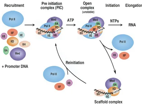
The primary phases of transcript generation constitute a three-step transcription cycle (Fig. 10) including the formation of the Pre-Initiation Complex (PIC) assembly at the promoter, the initiation of transcription, through promoter clearance and, eventually, the reinitiation of a new transcript.
After initiation is achieved, the transcript is elongated in a process named elongation, thanks to the Transcript Elongation Complex (TEC).
Finally, the transcript is terminated in a process named termination.
Current models suggest that during initiation, RNA polymerase II assembles at promoters together with coactivator complexes and general transcription factors.
During elongation, other accessory factors are thought to function as part of the active transcriptional machinery.
Fig.10. The eukaryotes transcription cycle. Formation of the PIC, initiation, elongation and reinitiation of transcription.
Figure taken from the web.
General transcription factors and the eukaryotic promoters
For protein coding genes, transcription by RNAPII requires the function of various accessory factors that associate with the enzyme during initiation and elongation.
Unlike prokaryote polymerase, eukaryote DNA-dependent RNA polymerases are by themselves unable to initiate and complete the transcription of a gene: they’re all dependent on the presence of accessory factors and General Transcription Factors (GTFs), required by all promoters (the minimal DNA sequence needed to specify non-regulated or basal transcription) used by RNAPII [181-183].
Eukaryotic RNA polymerases lack the sigma factor found in the prokaryotic enzyme. Therefore, it's not too surprising that promoters in the two groups are different.
Instead of the TATAAT sequence at the -10 location, which is found in prokaryotes, a TATA box, located at approximately -25, is found in eukaryotes. Additional sequences are located upstream of this site, with many promoters having a CAAT box and some containing a GC box, both at around -40 to -110 bases upstream. The location of these elements can vary, and they can be present on either strand.
The nucleation of transcription factors at the core promoter requires a complex network of protein-protein and protein-protein-DNA interactions. Formation of this promoter-bound complex is sufficient for a basal level of transcription. However, for activator-dependent (or regulated) transcription, general cofactors are often required to transmit regulatory signals between gene-specific activators and the general transcription machinery.
The GTFs may activate or repress gene expression and transcription through the modulation of the chromatin structure and binding to a core promoter (Fig. 11).
Enhancer or silencers sequences (cis-acting regulatory sequences, located several thousand bases upstream, downstream, or in the middle of the transcribed region) bind proteins (activators or repressors) which stimulate or repress transcription. These are often tissue- and species-specific, explaining the regulation of genes in some tissues. Activators may bind to coactivators, which directly interact with basal transcription factors (Fig. 11).
GTFs are needed to form the PIC (Pre-Inititation Complex, analogous to the bacterial Closed Complex), which is constituted of almost 60 polypeptides, but also to start the transcript elongation and to terminate and eventually restart the transcription.
Fig.11. Regulation of transcription at the core promoter through enhancers and silencers, cis-acting regulatory sequences,
and formation of the PIC assembly, made by coactivators, basal transcription factors and RNAPII. Figure taken from web.
Formation of the PIC (Pre-Inititation Complex) assembly
The model of PIC assembly starts with promoter binding of TFIID, followed by TFIIA, TFIIB, TFIIE, TFIIH and the remaining GTFs, including RNAPII, plus several additional cofactors (Fig. 12) [184-187].
TFIID is composed of TBP and about 14 TAFs (TBP Associated Factors), nearly all of which have been conserved through evolution, which function in the specific recognition of promoters and activator stimulated transcription [188, 189].
TFIIA and TFIIB are the two general factors that interact specifically and independently with TBP. TFIIA stabilizes TBP-DNA binding and strongly promotes binding of TFIID to DNA through an anti repression mechanism by competing with the TAF1 N-terminal domain (TAND) that occludes the DNA binding surface of TBP when TFIID is not bound to DNA [186, 188]. TFIIB plays a central role in PIC formation, interacting with TBP, RNAPII, TFIIF, and DNA on either side of the TATA box [186, 190-193]. The sequence TATA is located -30’ nucleotides upstream of the Transcription Start Point (TSP). In addition, there are also some weakly conserved features including the B-Recognition Element (BRE), approximately five nucleotides upstream of the TATA box. Occasionally there is no TATA box at the promoter. In this case a TAF will bind sequence specifically, and force the TBP to bind non sequence specifically. TAFs are highly variable, and add a level of control to the initiation.
In this state, RNAPII and the general factors are all bound to the promoter but are not in an active conformation to begin transcription. These factors can act indirectly on the transcription machinery by recruiting factors that modify chromatin structure, or directly by interacting with components of the transcription machinery. Not surprisingly, the promoters of class II eukaryotic genes contain elements to direct the binding of GTFs [192].
The transcription start site usually contains a pyrimidine-rich initiator element (INR), with the consensus sequence (Py)2A+1A/T(Py)2, that binds TFIID.
Fig.12. Transcription initiation.
As before mentioned, approximately 25-30 bp upstream from INR lays the TATA box (Consensus sequence TATA A/T A A/T), an element recognised and bound by the TBP (TATA-binding protein) subunit of TFIID. Binding of TFIID to the TATA box causes an 80° kink in the promoter DNA thereby enabling the binding of TFIIB to the adjacent BRE element (Consensus: G/C G/C G/A CGCC), which lies 35 bp upstream from INR. Binding of TFIIB stabilises the binding of TFIID to DNA, forming the interface for recruitment of the RNAPII/TFIIF complex [194].
Upon addition of ATP, the PIC undergoes a large conformational change resulting in separation of the DNA strands surrounding the transcription start site and the insertion of the template DNA strand into the active centre of RNAPII to form the unstable Open Complex [186, 193]. In this step, the general transcription factors TFIIH, TFIIE and TFIIF are thought to play major roles by inducing torsional strain in the DNA, promoting DNA strand separation, and positioning of the DNA strands within the Open Complex [195, 196] Indeed, formation of an Open Complex between RNAP II and the DNA template is a prerequisite for transcription initiation.
Melting of the double-stranded DNA into a single-stranded bubble is an ATP-dependent process and requires the action of two GTFs, TFIIE and TFIIH [197-199].
TFIIE binds selectively to the nonphosphorylated form of RNAPII (IIa) and this interaction is mediated by the 56-kD subunit of TFIIE. TFIIE also interacts with both subunits of TFIIF and with TFIIH, a multisubunit basal factor reported to catalyze RNA polymerase II CTD phosphorylation. Protein affinity assays demonstrate that TFIIE binds directly to ERCC-3, a DNA repair protein associated with TFIIH [199].
Initiation ends once the open complex is established and the first phosphodiester bonds of the RNA transcript is formed [200].
RNA synthesis begins with DNA-NTP base pairing, phosphodiester bond formation, and movement of the template DNA RNA hybrid within the active centre of RNAPII. Very early transcription complexes are unstable until the nascent transcript is elongated to nine nucleotides, about the length of the RNA–DNA hybrid in the transcription bubble. In many systems, multiple short abortive RNA products of 3–10 bases are synthesized before RNAPII starts transcribing full length RNAs.
RNAPII elongation complexes enter a second period of instability when the nascent transcript is 20–50 nucleotides in length. After synthesis of about 30 bases of RNA, RNAPII is thought to release its contacts with the core promoter and the rest of the transcription machinery and enter the stage of transcription elongation. Elongation complexes lack basal factors, with the exception of TFIIF, which in addition to being required for initiation [201, 202] is also an elongation factor and acts during the early stages of elongation to decrease the frequency of abortive transcription. It has been proposed that TFIIF functions to stabilise the tight interaction of the PIC with promoter DNA and facilitates its unwinding during the formation of the open complex [203, 204].
Before RNAPII becomes engaged into productive transcript elongation, it must pass through a stage known as promoter clearance, which marks the transition between transcription initiation and elongation and phosphorylation of the C-terminal domain (CTD) of Rpb1, the largest subunit of RNAPII [199, 205]. Only non-phosphorylated RNAPII (RNAPIIa) can form initiation complexes. During this stage, the PIC is partially disassembled: a subset of GTFs remains at the promoter in the Scaffold Complex, enabling the fast recruitment of the remaining general factors to speed up a new transcription reinitiation.
RNAPII initiation regulation
Initiation is regulated by many mechanisms. These can be separated into two main categories: protein interference and chromatin structure inhibition.
Protein interference is the process where some signalling protein interacts, either with the promoter or some stage of the partially constructed complex, to prevent further construction of the polymerase complex, so preventing initiation. This is generally a very rapid response and is used for fine level, individual gene control and for 'cascade' processes for a group of genes useful under specific conditions (i.e. DNA repair genes or heat shock genes).
These methods of control can be combined in a modular method, allowing very high specificity in transcription initiation control.
RNAPIIa/o, Mediator, Elongator and the transcript elongation
RNAPII is a 550 Kda complex of 12 subunits. Despite its obvious structural complexity, this multisubunit enzyme requires two groups of auxiliary proteins to solve two critical biochemical problems: the recognition of target promoters and the modulation of the RNA transcripts production of individual genes in response to developmental and environmental signals.
The largest subunit of RNAPII (Rpb1) has a domain at its C-terminus that is called the CTD (carboxy-terminal domain). The CTD can be considered as a platform for transcription factors and is the action site of CTD kinases (two cyclin dependent kinases, namely CDK7 and CDK8 are components of the PIC assembly) and phosphatases, thus allowing attraction and rejection of factors that have a function in the transcription process (elongation, mRNA maturation, surveillance, and export [184]).
The CTD is composed of a tandemly repeated amino acid motif, a heptapeptide consisting of the following consensus sequence: Y1S2P3T4S5P6S7, (Tyr-Ser-Pro-Thr-Ser-Pro-Ser). The amount of the tandem repeats varies between species, ranging from 15 in microsporidia to 26 in yeast, 32 in Caenorhabditis elegans, 45 in Drosophila and 52 in Mammals [206].
During the transcription cycle, Ser2 and Ser5 undergo waves of phosphorylation and dephosphorylation. Indeed, many different combinations of phosphorylations are possible along the repeats. Only the phosphorylated (IIo) form of RNAPII can participate in transcriptional elongation through the RNAPII transcript elongation complex (TEC), which plays important roles in mRNA maturation, pre-mRNA capping, splicing, 3’-end processing and export, while the non-phosphorylated CTD (RNAPIIa) is essential for the formation of a pre-initiation complex (PIC).
The RNAPIIo form marks the transition from initiation to elongation: phosphorylation destabilizes the PIC assembly, leads to the formation of the Scaffold complex and promotes the exchange of factors associated with the polymerase[207].
CTD phosphorylation at Ser5 has been correlated with transcription initiation and early elongation (promoter clearance), whereas Ser2 phosphorylation is associated with RNAPII already elongating the transcript.
Elongation is stimulated by a large variety of factors (TFIIS, Spt16/Pob3, elongin, ELLs, TFIIF and the Elongator complex)[208-215], of which some prevent pausing or stalling of the RNAPII complex and others model the chromatin for transcription. The degree of chromatin condensation is modulated by histone acetyltransferases and deacetylases [216, 217]. The transition to elongation also involves the replacement of Srb/Mediator, which is tightly associated with RNAIIa with an elongation complex termed Elongator, associated with the hyperphosphorylated RNAPIIo form [208, 217].
The Mediator complex of about 24 proteins, originally isolated from yeast, but present in all eukaryotes examined, is a coactivator complex limited to initiation complexes [218], essential for both basal and activated transcription [219].
Its subunits are generally divided in 3 domains or modules (head, middle, tail): the products of the five SRB genes, characterized as interacting with the RPB1 CTD; the products of four genes identified as global suppressors, and six members of a new protein family, termed Med, implicated in transcriptional activation.
Reconstitution experiments in vitro have shown mediator to stimulate the phosphorylation of CTD by the TFIIH kinase 30- to 50-fold. Since Mediator binds only hypophosphorylated CTD, this function may serve to release itself from the initiation complex once the signal for elongation is received. This is supported by evidence for the recycling of Mediator to initiation complexes and the absence of Mediator from elongating polymerases [220, 221].
The Elongator complex
The elongating RNAPII holoenzyme is copurified with a multisubunit complex, Elongator, whose stable interaction depends on the hyperphosphorylated state of the RNAPII C-terminal domain [208].
Purified holo-Elongator was initially identified in yeast and was found made of 6 proteins organized into 2 subcomplexes [222, 223]. The first one consists of ELP1 [208], ELP2 (a WD40 repeat protein) [222], and ELP3 (a histone acetyltransferase activity directed against histones H3 and H4, highly conserved between yeast and man, that together with other factors facilitates movement of RNA polymerase II through nucleosomes) [217]. The other consists of ELP4 [224], ELP5 [225], and ELP6 [223] constituting the HAP complex, made of HAT Accessory Proteins.
It was suggested that the HAP complex interacts preferentially with the core complex rather than with RNAPII and was therefore proposed to have a regulatory function. The largest subunit, Elp1, was previously isolated from a genetic screen for resistance to Kluyveromyces lactis toxin and was initially named TOT1 (Toxin-Target1). Other TOT genes were soon discovered.
Deletion of TOT/ELP genes in yeast cells results in a variety of phenotypes consistent with a role for the factor in transcription elongation, which include slow growth adaptation, delayed gene activation, and temperature sensitivity.
In addition, the elp mutants also are delayed in the G1 phase of the cell cycle and are hypersensitive to calcofluor white, 6-azauracil, and caffeine [208, 222-224].
Deletion of ELP3 is synthetically lethal with deletion of the tail of histone H4, or of the Rpb9 subunit of RNA polymerase II and confers a severe growth defect with the gene encoding the Gcn5 subunit of the SAGA complex [213, 226].
It is to be noted however, that despite the growing evidence for Elongator function, recent work in yeast has been unable to detect Elongator associated with RNAPII under any conditions, and revealed that the majority of Elongator is cytoplasmic [220].
Plant Elongator
The Elongator complex has been identified in different species (Saccharomycers cerevisiae, Mus
musculus, Rattus norvegicus, Homo sapiens, etc) and recently also in Plants [159, 160].
Namely, in Arabidopsis, through an initial large-scale screen of EMS (ethane methylsulfonate) leaf mutants of the elongata class (elo1, elo2, elo3, and elo4) with abnormally shaped leaves were investigated.
Mutations were finally mapped and found to be located in genes constituting the Elongator complex in Plants and involved in its regulation (DRL1) (Table1).
Table1. AtElongator genes. The A. th. homologues of the Yeast Elongator genes are presented together with DRL1
(Deformed Root and Leaves1) in the table with their accession number and their function. AtELP3 has recently been colocalized with the elo3 mutant [160]. AtELP1, AtELP4 and DRL1 have been colocalized respectively as elo2, elo1 and
elo4.
The elo1, elo2, and elo3 mutations colocalized with the genes encoding the ELP4, ELP1, and ELP3 homologues, respectively. Each of the elo mutants carried a point mutation in one of these Elongator genes. The sequencing of the ELO2 gene in elo2 plants revealed a single nucleotide change (G to A) that caused a premature stop codon (TGG to TAG) toward the 3’ end of the third exon.
In the elo3 mutant, a single nucleotide change (G to A) in the fifth exon of the ELO3 gene changed an aspartic acid residue, which is conserved in all ELO3 homologues, into an asparagine (D to N). A single base pair change (G to A) at the acceptor splice site of the third intron of the ELO1 gene caused missplicing in the
elo1 mutant. Three different mRNAs of ELO1 were present in the elo1 plants, resulting from three different
splicing events: exon4 was spliced out, intron3 was spliced out incorrectly using the first AG in exon 4, or intron 3 was not spliced out.
The three splicing events had a similar effect on the formation of the putative protein: at the N terminus, 142 amino acids were identical to those of wild type, followed by divergent amino acids and a stop codon.
The elo4 mutation colocalized with the gene encoding the DRL1 (Deformed Roots and Leaves1) protein in Arabidopsis thaliana, which is the homologue of the Yeast TOT4/KTI12 protein associated with the Elongator complex.
The recessive mutation at the DRL1 locus causes growth defects and general organ disorganization: at the anatomic and cyto-histological level shoot, root, inflorescence and flower meristems were affected.
The leaf phenotype of the drl1-2 mutant, named “narrow leaves“, has been dissected at different morphological levels: compared to the wild-type Ler leaves the drl1-2 leaf had reduced lamina width and area and an increased lamina/petiole ratio or even an unclear transition between petiole and lamina. The “narrow leaves phenotype” is present in all elo mutants (Fig. 13).
Fig.13. 21 DAV wild-type Ler (left) and mutant elo1 (right) plantlets grown on GM+V+1% sucrose. Elo1 mutants,
belonging to the “elongata” class display a “narrow leaf” phenotype, with a slim lamina.
At a histological level the reduced lateral growth of the lamina has been correlated with a reduced (50%) Palisade Cell Number (PCN). DIC (Differential Interference Contrast) optics analysis showed a reduction in upper epidermis cell-size, but a significant increased palisade cell-size.
In serial transversal sections the palisade cells were larger and more irregularly shaped and intercellular spaces were present all over the mesophyll; the lamina was thicker and the midvein less pronounced, which possibly indicates leaf ventralization, even if marker gene analysis showed that the drl1-2 leaves had clear dorsal and ventral domains.
Transcript termination
After initiation and elongation, the last step in the transcription cycle is transcript termination. At this stage, the mRNA is cleaved, polyadenylated and transported to the cytoplasm, where it will be translated [227, 228].
Little regulation occurs at termination, although it has been proposed that newly transcribed RNAs are kept in place if proper termination is inhibited, allowing very fast expression of genes given a stimulus, but this has not been demonstrated in eukaryotes yet.
Sucrose in plants
All plants use sucrose, the universal carbohydrate of systemic transport, not only as nutrient but also as general regulator (signal molecule) of growth and development, since it is involved in gene expression through the so-called sugar-sensing pathway [229-234].
General characteristics
Sucrose (common name: table sugar, cane sugar, beet sugar, or maple sugar, depending upon its natural source; also called saccharose; saccarosio in Italian) is a non-reducing L-fructoside disaccharide which is the result of an acetal oxygen bridge in the alpha orientation between an α-D-glucose and a ß-D-fructose (Fig.14). The osmotic effect of a substance is tied to the number of particles in solution, so an mL of sucrose solution with the same osmolarity as glucose will have twice the number of carbon atoms and therefore about twice the energy. Thus, for the same osmolarity, twice the energy can be transported per mL. As a non-reducing sugar, sucrose is less reactive and more likely to survive its transport in the phloem. Sucrose has the same molecular formula, C12H22O11, as lactose and maltose, but differs from both in structure. Its systematic name is α-D-glucopyranosyl-(1→2)-β-D-fructofuranose and it melts at 186°C.
Fig.14. Sucrose molecule: alpha form of D-glucose + beta form of D-fructose
Sucrose transport
Sucrose is an important energy vector for plant sink organs or for fast developing sink tissues unable to perform photosynthesis, like i.e. roots and fruits, where it is stored as sucrose or starch or metabolized. It results either from a surplus of carbohydrates after photosynthesis in mature leaves, which are the predominant sites of photosynthesis in higher plants or from root uptake from soil (but eventually also from a lab germination medium).
Carbohydrate exporting tissues are often referred to as ‘source tissues’ while importing tissues as ‘sink tissues. Specific plants may also translocate sugar alcohols (polyols like mannitol or sorbitol) or sucrose derivatives, i.e., raffinose, verbascose and stachyose, but these plants simultaneously translocate at least an equal amount of sucrose [235, 236].
Sucrose is transported in solution via a specific part of the vasculature, the phloem, which represents a long distance distribution network for assimilates in order to supply nonphotosynthetic organs with energy and carbon skeletons. It is actively transported into the phloem by the companion cells.
Beginning with the synthesis of sucrose, the first event is the transport of sucrose in the vacuole, which determines the pool of sucrose available for export (sucrose is temporarily stored into the vacuole). Then, sucrose has to exit the mesophyll cell and, from the apoplasm, enter the phloem cells. When sucrose is unloaded into the apoplasmic space, it can then be taken up as sucrose into the sink cells or cleaved by an invertase to hexoses that are transported by specific carriers.
Sucrose diffuses into the neighboring sieve tube cells, through a high number of plasmodesmata [237-239]. Sieve elements lose their nucleus and many organelles during differentiation, but stay connected to companion cells, which have a high metabolic activity. Sieve elements are connected to form sieve tubes that oppose very little resistance to the flow of sap. In most species, at least crop species, the sieve element/ companion cell complex (SE-CCC) is symplasmically isolated from the surrounding cells. Water molecules in the xylem diffuse into the sieve tube cells increasing the pressure in these cells. It is this pressure that forces the sugar and water through the phloem.
In addition to diffusion, there are two principal pathways for the delivery of sucrose into the minor vein SE-CCC: symplastic and apoplastic loading, in which sucrose can move from phloem into the cytoplasm of sink cells with or without crossing the plasmamembrane or the cell. In the first way sucrose passes the entire route from the leaf mesophyll cells to the sieve element–companion cell complex in the so-called ‘symplast’, moving from cell to cell via plasmodesmata while in the second sucrose is released from the
mesophyll cells and then actively taken up by sucrose transporters located in the sieve element–companion cell complex.
An interesting experiment showed that sucrose is also present in plastids [240].
Sucrose carriers in Arabidopsis
Three types of sucrose carriers (H+/sucrose symporters and antiporters) have been identified in plants: 1) plasma membrane influx transporters, responsible for sucrose entry into cells;
2) tonoplast (vacuole) transporters which are sucrose/H+ antiporters, since the vacuole is acidic compared to the cytoplasm;
3) plasma membrane effux transporters, antiporters responsible for the unloading of sucrose in sink organs or for sucrose exit from the mesophyll cells, in close vicinity to the phloem.
Nine sucrose transporter genes AtSUC1-9 have been identified in Arabidopsis thaliana (Fig. 15) [238, 241-243], seven of which encode functional proteins, whereas two genes, AtSUC6 and AtSUC7, are pseudogenes encoding aberrant proteins [241]. Most of these sucrose transporters are localized in the phloem (AtSUC3 and AtSUC5, with AtSUC2 and AtSUC4, predominantly in minor veins), other in cells close to phloem (AtSUC3), but others in flower tissues (AtSUC1, AtSUC8 and atSUC9).
Fig.15. A. thaliana’s sucrose carrier’s phylogenetic tree.
Sucrose synthesis and cleavage: sucrose synthases and invertases
Free sugars in plants are released from polysaccharides, beginning with disaccharides [244]. In higher plant cells, sucrose synthesis and cleavage are essential reactions for the provision and allocation of carbon resources or initiation of hexose-based sugar signals in importing structures. The breakdown is performed through the regulated activity of three important key enzymes, plus their isoenzymes: acid and alkaline invertases [245-255], also named sucrases, which catalize the following reaction: sucrose + H2O = glucose + fructose and sucrose synthases [230, 256], catalizing the following reaction: sucrose + UDP = UDPglucose + fructose. Sucrose synthases are known to catalyze sucrose synthesis as well as sucrose cleavage.
Plants contain two unrelated families of invertases: acid forms that derive from periplasmic invertases of eubacteria and are found in cell wall and vacuole, and neutral/alkaline forms evolved from the cytosolic invertases of cyanobacteria [257]. Invertases are able to alter sugar signals by producing glucose rather than UDPglucose, thus compared to sucrose synthases two-fold more hexoses. Vacuolar sites of cleavage could allow temporal control via compartmentalization. Together with transcriptional control, the action of invertases may also be regulated at the enzyme level by inhibitor proteins [247, 258].
At the enzyme level, sucrose synthases can be regulated by rapid changes in sub-cellular localization, phosphorylation, and carefully modulated protein turnover [245].
In general, hexoses favor cell division and expansion, whereas sucrose favors differentiation and maturation [259], thus invertases mediate the initiation and expansion of many new sink structures, often with vacuolar activity preceding that in cell walls.
Sucrose metabolism
During the day, plants produce sucrose and starch in their leaves as an energy source for the coming night. Photosynthesis is the main process in leaves, but in higher plants not all cells are photosynthetically active. Indeed, root cells or cells of reproductive structure, developing organs and storage tissue rely entirely on the import of carbohydrates synthesized in leaves. Primary carbon metabolism is divided between the chloroplast and the cytosol. Photosynthesis is regulated as a two-way process by the rate of utilization of photosynthate in the rest of the plant [260-262]. Light regulates the expression of genes for photosynthesis and the activity of the gene products (feedforward control). Other environmental variables such as temperature and nutrition determine the rate at which end-products from the TCA cycle are used (feedback control) [263].
In particular, photosynthesis is inhibited when the production and accumulation of carbohydrates in source leaves exceeds the rate of utilization of these photosynthates by sink organs [264].
Schematically, sucrose metabolism can be summarized as follows: the fixation of CO2 in the chloroplast yields triose-phosphates, which are needed in the TCA cycle for the regeneration of the ribulose-1,5-bisphosphate (CO2 acceptor) and for starch synthesis.
All triose-phosphates in excess are used essentially for sucrose formation in the cytosol (through gluconeogenesis to generate fructose, glucose and, finally, through sucrose synthasess, sucrose, which is exported to sink tissues where it is stored in the vacuole) and starch synthesis in the chloroplast. Starch and sucrose synthesis must be adjusted to the photosynthetic rate to ensure continuous phosphate recycling. When exported into the cytoplasm from chloroplast. Triose-phosphates can go through glycolysis and respiration, to produce ATP. The fructose-2,6-bisphosphate (Fig.16) is considered a key enzyme in maintaining a balance between glycolysis and gluconeogenesis [265].
Sugar-sensing mechanisms and general gene regulation
Though lots of sugars and their intermediates have been discovered in plants, only sucrose and hexoses have been found to act as signal molecules. Sugar-induced signals will interact with other sensing and signaling pathways [229, 231].
Three different sugar-sensing systems have been identified in plants:
(a) an hexokinase-sensing system, similar to the one described for yeast and animals; (b) a hexose transport-associated sensor, again similar to the situation in yeast; (c) a sucrose-specific pathway, which may involve a signaling sucrose transporter.
These three mechanisms represent the initial step of signal transduction. It is believed that phosphatases and/or kinase may regulate downstream other proteins that directly bind to promoters or coactivators/corepressors of specific set of genes, such as photosynthetic genes. A sucrose cycling through acid invertase and hexokinases may be a fine-tuned mechanism for gene expression regulation [261].
In plants, elevated levels of cellular sugar upregulate genes involved in the synthesis of polysaccharides, storage proteins, pigments, as well as genes associated with defense responses and respiration. Genes that are upregulated when nutrient availability is high are named ‘feast’ gens, while gens upregulated in nutrient depletion are named ‘famine’ genes.
Sugar depletion enhances the expression of ‘famine’ genes involved in photosynthesis and resource remobilization, such as starch, lipid and protein catabolism [256, 266, 267].
It has also been proposed that reduced photosynthesis is the result of increased hexose production and cytosolic phosphate (Pi) depletion or the feedback inhibition of sucrose phosphate synthase that results in the accumulation of phosphorylation intermediates, the depletion of stromal Pi, and the decrease of ATP synthesis [262, 268].
Sugar abundance enhances the expression of ‘feast’ genes, which in turm enhance gluconeogenesis, fatty acid synthesis, but in general all genes involved in anabolism and storage processes.