Università Degli Studi di Siena
Dipartimento di Biotecnologie Mediche Dottorato di Ricerca in
Biotecnologie Mediche
Ciclo XXIII
Coordinatore Prof. Lorenzo Leoncini
Maximizing disinfection procedures in
endodontics
Settore Scientifico-Disciplinare: Endodonzia
Dottorando: Dott. Raffaele Paragliola
Tutor: Prof. Simone Grandini
Anno Accademico 2020/2021
Contents
Introduction
Chapter 1. The role of instrumentation
1.1 Root canal infection 1.2 Root canal instrumentation 1.2.1 The crown down approach 1.2.2 Working length determination 1.2.3 Size of apical preparation 1.2.4 Preparation techniques 1.2.5 Hand file instrumentation 1.2.6 Patency filing 1.2.7 The era of nickel titanium 1.2.8 Rotary file systems 1.2.9 Reciprocating systems 1.2.10 Which system is best?Chapter 2. The role of irrigation
2.1 Type of irrigant 2.2 Factor influencing endodontic irrigants 2.3 Mode of delivery 2.4 Activation of irrigantChapter 3. The role of Smear layer
3.1 The significance of the smear layer 3.2 Should the smear layer be removed? 3.3 Methods to remove the smear layerChapter 4. The role of Enterococcus Faecalis
4.1 E. faecalis Characteristics and Strains 4.2 Prevalence in Secondary Root Canal Infections 4.3 Survival and Virulence Factors 4.4 Methods of Eradication 4.5 Conclusion
EXPERIMENTAL PART
Chapter 5. Final Rinse Optimization: Influence of Different Agitation
Protocols
5.1 Introduction 5.2 Materials and Methods 5.3 Results 5.4 Discussion
Chapter 6. Comparison of smear layer removal using four final-rinse
protocols
6.1 Introduction 6.2 Materials and Methods 6.3 Results 6.4 Discussion
Chapter 7. Influence of surfactant and PUI on the effectiveness of NaOCl for
final rinse optimization
7.1 Introduction 7.2 Materials and Methods 7.3 Results 7.4 Discussion
Chapter 8. The role of PUI on Enterococcus Faecalis
8.1 Introduction 8.2 Materials and methods 8.3 Results 8.4 Discussion
Chapter 9. Conclusions and future directions
Introduction
We are living in the age of evidence-based medicine. Any new concept and technique to be used on patients should ideally be assessed in randomized controlled clinical trials against their respective gold standards. This, however, poses a major problem particularly in endodontic research. A favourable outcome of root canal treatment is defined as the reduction of a radiographic lesion and the absence of clinical symptoms of the affected tooth after a minimal observation period of 1 yr. (1). Alternatively, the so called surrogate outcome (dependent) variables yielding quicker results, such as the microbial load remaining in the root canal system after different treatment protocols, can be defined. However, these do not necessarily correlate with the “true” treatment outcome (2). Endodontic success is dependent on multiple factors (3), and a faulty treatment step can thus be compensated. For instance if cultivable microbiota remain after improper canal disinfection, they can theoretically be entombed in the canal system by a perfect root canal filling (4), and clinical success may still be achieved (5). On the other hand, in a methodologically sound clinical trial, single treatment steps have to be randomized and related to outcome. Otherwise, the results do not allow any conclusions and no causative relationships may be revealed (6).
The above issues may be viewed as the reason (or as an excuse) for the fact that no randomized controlled clinical trials exist on the effect of irrigating solutions on treatment outcome in the endodontic literature. As of yet, we largely depend on data from in vitro studies and clinical trials with microbial recovery after treatment as the surrogate outcome. Clinical recommendations based on such findings are merely deductive and need to be interpreted with care.
Chapter 1
1.1 Root Canal Infection
A traditional concept that explains infectious processes occurring in humans suggests that diseases are produced as the result of the aggressive invasion of harmful microorganisms, which battle with the human host’s defences, triggering mechanisms that release antibodies and immune cells. The impact of such an approach generates a predisposition to search for those “most dangerous” microorganisms that can cause/ trigger the most severe damage to the host. In line with this view, infectious processes of the oral cavity were proposed to be caused by a relatively small number of organisms from the diverse collection of species found in the human mouth (7). In caries, for example, the frequent isolation of Streptococcus mutans from carious lesions (8–12) generated a considerable number of studies to explore the ex vivo features of this bacterium. Research findings showing the significant acid-tolerant capabilities of S. mutans defined this organism as “the” agent responsible for initial enamel and dentine demineralization. Similarly, in periodontal disease, the frequent recovery of proteolytic microorganisms from deep periodontal pockets, such as Porphyromonas gingivalis, increased the attention of periodontists to these bacteria because they were considered key etiological agents of the disease (11,12). The main disadvantage with this traditional view of the infectious process, especially in oral infections, is that the determination of true cause-and-effect relationships is not always possible. Consequently, the predominance of certain microorganisms at a given site may be the result of the disease itself rather than that of the initiating agent (13). Recently, the “ecological plaque hypothesis” (14–19) has improved on these classic infectious concepts to explain the aetiology of caries and periodontal disease. This hypothesis suggests that the organisms associated with the disease may also be present at sound sites, but at levels too low to represent a clinical threat. In other words, disease is produced as the result of changes in the local environmental conditions that will shift the balance of the resident flora.
Root canal infections have a different nature than that of caries or periodontitis because they become established in originally sterile compartments of the oral cavity. In many cases, this led to the concept that the aetiology of root canal infections involves only a single pathogen. For example, the predominance of certain proteolytic black-pigmented anaerobic organisms in cultures from infected root canals associated with acute symptoms suggested that these organisms are foremost etiological agents in such cases (20,21). Recently, the frequent recovery of Enterococcus faecalis in root canals associated with persistent infections brought
about an intense research interest in this bacterium. E. faecalis has become the ideal organism to test different irrigants, medicaments, and antiseptic solutions used in endodontics ex vivo, with findings that revealed its innate resistance capacity (22–24). This extensive interest in E. faecalis, perhaps driven by its ability to grow under almost any laboratory condition (25), resulted in the concept that the organism is the sole etiological agent for chronic endodontic infections. Consequently, the focus on E. faecalis resulted in much less information on the existence of other organisms in such infections that may possess similar tolerating characteristics to E. faecalis and that would shed light on the existence of a polymicrobial persisting community. Thus, it is not surprising that ecological parameters in root canal infections are not often discussed.
From an ecological perspective, the root canal can be considered a highly controlled environment with a limited number of niches. Although niches are composed by a variety of environmental factors that limit the growth of one species relative to others (26), the main limiting factors in root canal niches that influence bacterial colonization are, for instance, oxygen and nutrient availability (27). After root canal treatment, other limiting factors become involved, such as pH and the short/long-term effects of the antibacterial medicaments applied. Bacterial survival in such controlled environments, especially after root canal treatment, is based on the capacity of organisms to adapt to the existing conditions. Although traditional views suggest that the organisms surviving root canal treatment are a selected group of the “most robust” organisms, the application of ecological parameters indicates that bacterial survival after root canal treatment will depend not on the robustness of the organisms, but on how good an adaptor the organism is to the new limiting factors in their corresponding niches. Furthermore, as in every natural microenvironment, the adaptive capabilities of individual organisms are exponentially augmented when growing in biofilm communities. The foundation for this ecological approach to endodontic infections suggests that the most dangerous “pathogen” is not an individual species, but a polymicrobial entity that undergoes physiological and genetic changes triggered by changes in the root canal environment.
Currently, there is no substantial evidence indicating that certain microorganisms of the microbial flora in root canal infections are more virulent than others. With this in mind, Sundqvist and Figdor (28) stated that a proper definition for endodontic pathogens should include every organism capable of inducing the tissue destruction in apical periodontitis. In reality, however, the majority of endodontic-microbiology studies refer to the endodontic
pathogen as the bacterium isolated from a symptom-associated root canal that grows in the laboratory in a specific media. By this approach, the most frequently recovered species will assume the role of major endodontic pathogen. In persistent root canal infections, for example, the frequent occurrence of monocultures of E. faecalis has raised suspicion that this bacterium may be the sole organism persisting in the root canals. Considering that mono-infections rarely if ever occur in nature, it is possible that the apparent pure cultures of E. faecalis could be the result of sampling and culturing techniques that favour it over other organisms at the site that were either in low numbers or were physiologically inactive or dormant. For instance, in a commonly cited study (29), from the total 100 root- filled teeth with apical periodontitis sampled E. faecalis was reported as the most frequently recovered organism (32%), although in 32% of the cases with persistent lesion no microbe could be isolated. In yet nine root-filled teeth without periapical lesion that showed bacterial growth, the organism was found in one case. In a similar study, 25 root-filled teeth requiring retreatment were sampled and E. faecalis was found in 14 of those 20 teeth with bacterial growth (30). However, it would seem that this study was focused primarily in proving the occurrence of E. faecalis in root-filled teeth rather than in exploring the microbial flora in persisting infections. Similarly, in a recent study using a sophisticated nested PCR technique, the target bacterium E. faecalis was found in 41 of 50 (82%) untreated root canals and in 38 of 50 (76%) treatment failure associated root canals (31). As in other related works (32– 35), PCR methodology seems to be exclusively directed to find only E. faecalis, ignoring the rest of the flora present that may be as important as E. faecalis in provoking the treatment failures. On the other hand, recent investigations have confirmed the polymicrobial nature of root canal infections (36, 57). In a study with monkeys (36), different combinations of bacteria were experimentally inoculated in root canals and periapical lesions were induced. The teeth were treated endodontically and followed-up radiographically and histologically for 2 to 2.5 years. In the root canals with bacteria present when the root filling was removed, 30 of the 31 canals had persisting periapical lesions. Importantly, more of these non-healed lesions were associated with various combinations of bacterial strains, that is, mixed infections, than single strains. Previously, the same research group (38) also found that when an “eight-strain collection” of species, derived from one infected root canal, was re-inoculated in equal proportions into other monkey teeth, species such as Bacteroides oralis (now Prevotella oralis) dominated in mixed infections and showed a more potent capacity for tissue destruction. Furthermore, B. oralis could not be reisolated from inoculated root canals after the experimental period when inoculated as a pure culture. In another study using the tissue
cage model implanted subcutaneously in the backs of rabbits, the same col- lection of eight bacterial strains from monkey root canals were inoculated in different combinations and individual species. The combination of B. oralis, Fusobacterium necrophorum, Peptostreptococcus anaerobius, and Streptococcus milleri was the most predominant and induced higher titers of circulating antibodies than that obtained with individual inoculations, such as E. faecalis (39).
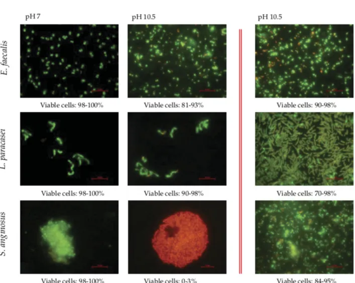
Even if we accept the polymicrobial nature of root canal infections, one of the major problems in understanding endodontic infections is that we still extrapolate between individual organisms growing in liquid (planktonic) cultures and the in vivo situation. A significant literature now exists demonstrating that the physiology of a bacterium in planktonic culture is profoundly different from that of the same organism growing on a surface in a biofilm [see review by Costerton et al. (40)]. For instance, planktonic bacteria are more sensitive to antimicrobial agents because of their ease of diffusion within the bulk fluid, whereas biofilm bacteria are notably resistant to these agents (41–45). In this context, the study of biofilms in root canal infections has included biofilms formed by mixed cultures of anaerobic bacteria in extracted teeth (46, 47) or by pure cultures of E. faecalis (48, 49). Biofilms of five root canal isolates have also been used to test the antimicrobial efficacy of endodontic irrigants, such as sodium hypochlorite (NaOCl) (2.25%), 0.2% chlorhexidine, 10% povidone iodine, and 5 ppm colloidal silver, with NaOCl shown to be the most effective agent of this group (50). In addition, Chavez et al. tested the alkaline tolerance of species isolated from chronically infected root canals and found that E. faecalis and other Gram-positive organisms, such as Lactobacillus paracasei, Olsenella uli, or Streptococcus gordonii, shared similarly high alkaline-tolerant capabilities when growing in planktonic conditions. S. anginosus, S. oralis, and F. nucleatum, on the other hand, were greatly affected by the alkaline stress (see Fig. 1) (51). Of importance, however, was the observation that this difference in alkaline tolerance was not apparent when the strains were tested in biofilms because all seven strains showed a similar high tolerance to alkaline pH (Fig. 1). These findings not only show the capacity of root canal bacteria other than E. faecalis to adapt to alkaline stress, but also provide further evidence that bacteria in surface-adhered biofilm consortia are more resistant to environmental stress than when grown in liquid culture.
Figure 1. Fluorescence micrographs using Live/Dead fluorescence staining for bacterial viability. Cells stained fluorescent green represent viable cells, whereas cells stained fluorescent red are nonviable or damaged. In the first column, images show planktonic cells of three root canal strains at neutral media (pH 7). The middle column shows planktonic cells after exposure to pH 10.5 for 4 hours, and the right column shows biofilm cells exposed to alkaline challenge (pH 10.5) for 4 hours. Bars, 2 m. Images are published with permission of Blackwell Publishing. International Endodontic Journal, Chávez de Paz et al. (65) As the host defense loses its access to the necrotic pulp space, opportunistic microorganisms selected by harsh ecological conditions and the low-oxygen environment aggregate in the root canal system (52). These microbial communities may survive on organic pulp tissue remnants and exudate from the periodontium (53, 54). Consequently, clusters of microorganisms in
necrotic teeth and teeth with failed root canal treatments are typically found in the apical root canal area, where they have access to tissue fluid (52). In long-standing infections, root canal bacteria can invade the adjacent dentin via open dentinal tubules (55, 56).
Primary root canal infections are polymicrobial, typically domi- nated by obligately anaerobic bacteria (53). The most frequently isolated microorganisms before root canal treatment include Gram-negative anaerobic rods, Gram-positive anaerobic cocci, Gram-positive anaerobic and facultative rods, Lactobacillus species and Gram-positive facultative Streptococcus species (53). The obligate anaerobes are rather easily eradicated during root canal treatment. On the other hand, facultative bacteria such as nonmutans Streptococci, Enterococci, and Lactobacilli, once established, are more likely to survive chemomechanical instrumentation and root canal medication (57). In particular Enterococcus faecalis has gained attention in the endodontic literature, as it can frequently be isolated from root canals in cases of failed root canal treatments (58, 59). In addition, yeasts may also be found in root canals associated with therapy-resistant apical periodontitis (60).
It is likely that all of the microorganisms able to colonize the necrotic root canal system cause periapical inflammatory lesions. Enterococci can survive in monoculture, but cause only minor lesions (38). Certain Gram-negative taxa appear to be more virulent (53). The outer membrane of Gram-negative bacteria contains endotoxin, which is present in all necrotic teeth with periapical lesions (61), and is able to trigger an inflammatory response even in the absence of viable bacteria (62). Furthermore, the levels of endotoxin in necrotic root canals are positively correlated to clinical symptoms such as spontaneous pain and tenderness to percussion (63). Virulent Gram-negative anaerobic rods depend on the presence of other bacteria in their environment to survive and establish their full pathogenic potential (38). Such aggregations of microorganisms in an extracellular polysaccharide matrix associated with a surface (in our case the inner root canal wall) are called biofilms (64). There is convincing evidence that microorganisms organized in this manner are far less susceptible to antimicrobial agents than their planktonic counterparts, which have traditionally been used to test the antimicrobial efficacy of substances in vitro (65, 66). If a bacterially inoculated broth is confronted with an antimicrobial fluid, the efficacy of that agent can appear to be very convincing, similar as with agar-diffusion tests. However, in the root canal system biofilms and infected dentinal tubules make disinfection much more difficult and thus study models such as standardized infected bovine dentin blocks (67) or in vivo models appear to be more valid than the above mentioned study designs. Furthermore, it has been shown that organic
and inorganic dentin components, which are suspended in the irrigant during chemomechanical instrumentation, inhibit most antimicrobial agents (68, 69).
In conclusion, the biofilm concept and the specific conditions in the pulpless root canal microniche cannot be overestimated when considering the actions of different irrigating solutions.
References
1. Ørstavik D. Time-course and risk analyses of the development and healing of chronic apical periodontitis in man. Int Endod J 1996;29:150 –5.
2. Peters LB, van Winkelhoff AJ, Buijs JF, Wesselink PR. Effects of instrumentation, irrigation and dressing with calcium hydroxide on infection in pulpless teeth with periapical bone lesions. Int Endod J 2002;35:13–21.
3. Ørstavik D, Qvist V, Stoltze K. A multivariate analysis of the outcome of endodontic treatment. Eur J Oral Sci 2004;112:224 –30.
4. Saleh IM, Ruyter IE, Haapasalo M, Ørstavik D. Survival of Enterococcus faecalis in infected dentinal tubules after root canal filling with different root canal sealers in vitro. Int Endod J 2004;37:193– 8.
5. Peters LB ,Wesselink PR. Periapical healing of endodontically treated teeth in one and two visits obturated in the presence or absence of detectable microorganisms. Int Endod J 2002;35:660 –7. 6. Alderson P, Green S, Higgins J. Cochrane Reviewer’s Handbook. The Cochrane Library, Chichester: John Wiley & Sons, Ltd., 2004. 7. Loesche WJ. Chemotherapy of dental plaque infections. Oral Sci Rev 1976; 9:65–107 8. Bowden GH. Microbiology of root surface caries in humans. J Dent Res 1990;69:1205– 10.
9. Loesche WJ. Role of Streptococcus mutans in human dental decay. Microbiol Rev 1986;50:353– 80.
10. Marsh PD. Microbial community aspects of dental plaque. In:Newman HN, Wilson M, eds. Dental plaque revisited. Cardiff, UK: BioLine; 1999:237–53.
11. Moore WE, Moore LV. The bacteria of periodontal diseases. Periodontology 1994;5:66 –77.
12. Socransky SS, Haffajee AD, Cugini MA, Smith C, Kent RL Jr. Microbial complexes in subgingival plaque. J Clin Periodontol 1998;25:134 – 44.
13. Marsh PD. Are dental diseases examples of ecological catastrophes ? Microbiology 2003;149:279 –94.
14. Bowden GH, Hardie JM, McKee AS, Marsh PD, Fillery ED, Slack GL. The microflora associated with developing carious lesions of the distal surfaces of the upper first premolars in 13–14 year old children. In: Stiles HM, Loesche WJ, O’Brien TC, eds. Microbial Aspects of Dental Caries. Washington, DC: Information Retrieval; 1976:233–
41.
15. Marsh PD. Sugar, fluoride, pH and microbial homeostasis in dental plaque. Proc Finn Dent Soc 1991;87:515–25.
16. Marsh PD. Microbial ecology of dental plaque and its significance in health and disease. Adv Dent Res 1994;8:263–71.
17. Marsh PD,Bradshaw DJ. Physiological approaches to the control of oral biofilms. Adv Dent Res 1997;11:176 – 85.
18. Marsh PD, Featherstone A, McKee AS, et al. A microbiological study of early caries of approximal surfaces in schoolchildren. J Dent Res 1989;68:1151– 4.
19. Newman HN. Plaque and chronic inflammatory periodontal disease. A question of ecology. J Clin Periodontol 1990;17:533– 41.
20. Chávez de Paz Villanueva LE. Fusobacterium nucleatum in endodontic flare-ups. Oral Surg Oral Med Oral Pathol Oral Radiol Endod 2002;93:179 – 83.
21. Haapasalo M, Ranta H, Ranta K, Shah H. Black-pigmented Bacteroides spp. in human apical periodontitis. Infect Immun 1986;53:149 –53.
22. Dahlén G, Samuelsson W, Molander A, Reit C. Identification and antimicrobial susceptibility of enterococci isolated from the root canal. Oral Microbiol Immunol 2000;15:309 –12.
23. Eddy RS, Joyce AP, Roberts S, Buxton TB, Liewehr F. An in vitro evaluation of the antibacterial efficacy of chlorine dioxide on E. faecalis in bovine incisors. J Endod 2005;31:672–5.
24. Portenier I, Waltimo T, Ørstavik D, Haapasalo M. The susceptibility of starved, stationary phase, and growing cells of Enterococcus faecalis to endodontic medi- caments. J Endod 2005;31:380 – 6.
25. Siren EK, Haapasalo MP, Waltimo TM, Orstavik D. In vitro antibacterial effect of calcium hydroxide combined with chlorhexidine or iodine potassium iodide on
Enterococcus faecalis. Eur J Oral Sci 2004;112:326 –31.
26. Kassen R, Rainey PB. The ecology and genetics of microbial diversity. Annu Rev Microbiol 2004;58:207–31.
27. SundqvistG.Ecologyoftherootcanalflora.JEndod1992;18:427–30.
28. Sundqvist G, Figdor D. Life as an endodontic pathogen. Ecological differences between the untreated and root-filled root canals. Endodontic Topics 2003;6:3–28.
apical periodontitis. Int Endod J 1998;31:1–7.
30. Peciuliene V, Balciuniene I, Eriksen HM, Haapasalo M. Isolation of Enterococcus faecalis in previously root-filled canals in a Lithuanian population. J Endod 2000;26:593–5.
31. Gomes BP, Pinheiro ET, Sousa EL ,et al. Enterococcus faecalis in dental root canals detected by culture and by polymerase chain reaction analysis. Oral Surg Oral Med Oral Pathol Oral Radiol Endod 2006;102:247–53.
32. Kaufman B, Spångberg L, Barry J, Fouad AF. Enterococcus spp. in endodontically treated teeth with and without periradicular lesions. J Endod 2005;31:851– 6.
33. Sedgley C, Nagel A, Dahlén G, Reit C, Molander A. Realtime quantitative polymerase chain reaction and culture analyses of Enterococcus faecalis in root canals. J Endod 2006;32:173–7.
34. Williams JM, Trope M, Caplan DJ, Shugars DC. Detection and quantitation of E. faecalis by real-time PCR (qPCR), reverse transcription-PCR (RT-PCR), and culti- vation during endodontic treatment. J Endod 2006;32:715–21.
35. Zoletti GO, Siqueira JF Jr, Santos KR. Identification of Enterococcus faecalis in root-filled teeth with or without periradicular lesions by culture-dependent and independent approaches. J Endod 2006;32:722– 6.
36. Fabricius L, Dahlén G, Sundqvist G, Happonen RP, Möller ÅJ. Influenceofresidual bacteria on periapical tissue healing after chemomechanical treatment and root filling of experimentally infected monkey teeth. Eur J Oral Sci 2006;114:278 – 85.
37. Rôças IN, Siqueira JF Jr, Aboim MC, Rosado AS. Denaturing gradient gel electrophoresis analysis of bacterial communities associated with failed endodontic treatment. Oral Surg Oral Med Oral Pathol Oral Radiol Endod 2004;98:741–9.
38. Fabricius L, Dahlén G, Hölm SE, Möller ÅJ. Influence of combinations of oral bacteria on periapical tissues of monkeys. Scand J Dent Res 1982;90:200 – 6.
39. Dahlén G, Fabricius L ,Hölm SE ,Möller ÅJ. Interactions with inacollection of eight bacterial strains isolated from a monkey dental root canal. Oral Microbiol Immunol 1987;2:164 –70.
40. Costerton JW, Lewandowski Z, DeBeer D, Caldwell D, Korber D, James G. Biofilms, the customized microniche. J Bacteriol 1994;176:2137– 42.
41. Costerton JW, Cheng KJ, Geesey GG, et al. Bacterial biofilms in nature and disease. Annu Rev Microbiol 1987;41:435– 64.
42. Gilbert P, Das J, Foley I. Biofilm susceptibility to antimicrobials. Adv Dent Res 1997;11:160 –7.
43. Johnson SA, Goddard PA, Iliffe C, et al. Comparative susceptibility of resident and transient hand bacteria to para-chloro-meta-xylenol and triclosan. J Appl Microbiol 2002;93:336 – 44.
44. Larsen T. Susceptibility of Porphyromonas gingivalis in biofilms to amoxicillin, doxycycline and metronidazole. Oral Microbiol Immunol 2002;17:267–71.
45. Shani S, Friedman M, Steinberg D. The anticariogenic effect of amine fluorides on
Streptococcus sobrinus and glucosyltransferase in biofilms. Caries Res 2000; 34:260 –
7.
46. Barrieshi KM, Walton RE, Johnson WT, Drake DR. Coronal leakage of mixed an- aerobic bacteria after obturation and post space preparation. Oral Surg Oral Med Oral Pathol Oral Radiol Endod 1997;84:310 – 4.
47. Clegg MS, Vertucci FJ, Walker C, Belanger M, Britto LR. The effect of exposure to irrigant solutions on apical dentin biofilms in vitro. J Endod 2006;32:434 –7.
48. Dunavant TR, Regan JD, Glickman GN, Solomon ES, Honeyman AL. Comparative evaluation of endodontic irrigants against Enterococcus faecalis biofilms. J Endod 2006;32:527–31.
49. George S, Kishen A, Song KP. The role of environmental changes on monospecies biofilm formation on root canal wall by Enterococcus faecalis. J Endod2005;31:867–72. 50. Spratt DA, Pratten J, Wilson M, Gulabivala K. An in vitro evaluation of the antimicrobial
efficacy of irrigants on biofilms of root canal isolates. Int Endod J 2001;34:300 –7. 51. Chávez de Paz LE, Bergenholtz G, Dahlén G, Svensäter G. Response to alkaline stress by
52. Nair PN. Pathogenesis of apical periodontitis and the causes of endodontic failures. Crit Rev Oral Biol Med 2004;15:348 – 81.
53. Sundqvist G. Taxonomy, ecology, and pathogenicity of the root canal flora. Oral Surg Oral Med Oral Pathol 1994;78:522–30.
54. Love RM. Enterococcus faecalis a mechanism for its role in endodontic failure. Int Endod J 2001;34:399 – 405. 55. Shovelton DS. The presence and distribution of microorganisms with nonvital teeth. Br Dent J 1964;117:101–7. 56. Armitage GC, Ryder MI ,Wilcox SE. Cemental changes in teeth with heavily infected root canals. J Endod 1983;9:127–30. 57. Chavez DePaz LE, Dahlén G, Molander A, Möller A, Bergenholtz G. Bacteria recovered from teeth with apical periodontitis after antimicrobial endodontic treatment. Int Endod J 2003;36:500 – 8.
58. Engström B. The significance of Enterococci in root canal treatment. Odontol Revy 1964;15:87–106.
59. Haapasalo M, Ranta K, Ranta H.Facultatice Gram-negative entericrods in persistent periapical infections. Acta Odontol Scand 1983;91:458 – 63.
60. Waltimo TM, Sirén EK, Torkko HL, Olsen I, Haapasalo MP. Fungi in therapy resistant apical periodontitis. Int Endod J 1997;30:96 –101.
61. DahlénG,BergenholtzG.Endotoxicactivityinteethwithnecroticpulps.JDentRes 1980;59:1033– 40.
62. Dwyer TG, Torabinejad M. Radiographic and histologic evaluation of the effect of endotoxin on the periapical tissues of the cat. J Endod 1980;7:31–5.
63. Jacinto RC, Gomes BP, Shah HN, Ferraz CC, Zaia AA, Souza Filho FJ.Quantification of endotoxins in necrotic root canals from symptomatic and asymptomatic teeth. J Med Microbiol 2005;54:777– 83.
64. Costerton JW, Lewandowski Z, DeBeer D, Caldwell D, Korber D, James G. Biofilms, the customized microniche. J Bacteriol 1994;176:2137– 42.
65. Nickel JC, Ruseska I, Wright JB, Costerton JW. Tobramycin resistance of Pseudo- monas aeruginosa cells growing as a biofilm on urinary catheter material. Antimi- crob Agents Chemother 1985;27:619 –24.
66. Wilson M. Susceptibility of oral bacterial biofilms to antimicrobial agents. J Med Microbiol 1996;44:79 – 87.
67. Haapasalo M, Ørstavik D. In vitro infection and disinfection of dentinal tubules. J Dent Res 1987;66:1375–9.
68. Portenier I, Haapasalo H, Rye A, Waltimo T, Ørstavik D, Haapasalo M. Inactivation of root canal medicaments by dentine, hydroxylapatite and bovine serum albumin. Int Endod J 2001;34:184 – 8.
69. Portenier I, Haapasalo H, Ørstavik D, Yamauchi M, Haapasalo M. Inactivation of the antibacterial activity of iodine potassium iodide and chlorhexidine digluconate against Enterococcus faecalis by dentin, dentin matrix, type-I collagen, and heat- killed microbial whole cells. J Endod 2002;28:634 –7.
1.2 ROOT CANAL INSTRUMENTATION
The objectives of mechanical preparation are two-fold:
1. To facilitate irrigation Conventional radiography does not enlighten the clinician about the true complexity of the root canal system. Lateral canals, fins, anastomoses and ramifications are invariably present, with some canals being joined by narrow isthmi. The main canal is rarely round, but often oval, ribbon- like or even ‘C’-shaped, depending upon the tooth. One seminal study has demonstrated up to 53% of the canal will remain unreached by instrumentation following preparation (1). Therefore, mechanical preparation facilitates penetration of irrigants into these complex anatomical spaces. Although some dentine-containing micro-organisms will be removed during mechanical preparation, research suggests that a considerable amount of the canal will not be contacted by a file, therefore irrigants play a crucial role in destroying micro-organisms, neutralizing endotoxin and removing organic tissue components (2).
2. To facilitate obturation as cleaning and shaping does not remove all micro-organisms from the canal, obturation aims to entomb any residual pathogens and limit recolonization by preventing the passage of nutrients from both coronal and apical aspects. Mechanical preparation facilitates obturation. Schilder’s principles of canal preparation still hold true today (3). The idea of creating a continuously tapering preparation, free from mechanical errors, allows the best chance of a well-condensed obturation, with the absence of voids.
1.2.1 The crown down approach
The majority of micro-organisms are in the coronal portion of the canal and pulp chamber (4). Thus, whatever instruments are used, a crown down approach and only initial scouting of the canal prior to working length determination is sensible. This technique involves shaping the canal from the coronal aspect first and progressively working more apically with smaller diameter instruments (5) (Figure 2).
Figure 2. The crown down approach: the coronal third of the canal system is enlarged using GG or orifice-shaping files. The enlargement is directed away from the furcation and has the simultaneous benefit of removing dentine overhanging the orifices to allow optimal straight-line access. Such an approach: • Minimizes the transportation of pathogens further into the canal system; • Allows a greater amount of irrigant to be held in the canal, facilitating debris removal and disinfection; • Removes coronal curvatures and facilitates straight-line access;
• Improves accuracy of working length determination as reduction of curvature after working length determination may alter the working length and result in a tendency to transport the canal and over-enlarge the apical foramen;
• Reduces file binding in the coronal portion of the canal, facilitating working length assessment and further reducing the risk of instrument separation through torsional failure. Traditionally, Gates Glidden (GG) instruments would be used for the crown down procedure but many rotary filling systems now have orifice shapers to begin the preparation. If clinicians elect to use GGs it is wise to remember a Size 6 GG has an apical diameter of 1.5 mm (ISO 150), with sizes stepping down in 0.2 mm increments to a Size 1 GG at 0.50 mm (ISO 50). As such, even the smallest of GGs can be very destructive if used carelessly. Avoid using sizes above GG 3 (0.90 mm: ISO 90). Whatever instruments are used, caution must be taken with regard to the furcation region, the instruments being used away from the furcation (anti-curvature
filing) (6). Despite the aforementioned advantages, it is easier to create blockages and ledges with an aggressive or careless crown down approach, thus highlighting the importance of recapitulation.
1.2.2 Working length determination
The apical extent of preparation should be kept within the canal system: over extension can reduce success up to 62% and, for every mm short of the apex, underextension reduces success by 12% (7). Methods used to estimate the maximum working length for instrumentation include apical gauging by tactile sensation, instrumentation without local anaesthetic, using pre-operative radiographs alone, the paper point technique, working length radiographs (WLRs) with files in situ and, most recently, the use of electronic apex locators (EALs). Historically, the most widely accepted method is by placing a file to the estimated length, then taking a confirmatory radiograph, but the radiographic apex rarely corresponds with the anatomical apex (8) It follows that WLRs can only give an estimation of the correct termination of preparation.
Modern impedance-based multifrequency EALs are reliable and accurate >90% of the time (9) These devices are only accurate at a ZERO reading. Any reading given other than ZERO should not be used as a marker of apical extent. The ZERO reading is reached when the file contacts the periodontal ligament. Thus, by definition this is over extended and, to calculate the working length, one must subtract 0.5 mm from the ZERO reading length (10). For more information readers are referred to other papers on the subject of EALs (11,12).
The 2013 Faculty of General Dental Practitioners Selection Criteria for Dental Radiography states ‘Unless there is confidence about working length(s) derived from an electronic apex
locator, at least one good-quality radiograph is necessary to confirm working length(s)’ (13).
From this one could extrapolate that WLRs are no longer always necessary. We recommend that a combination of techniques is used.
1.2.3 Size of apical preparation
There is equivocal evidence regarding the effect of the size of apical preparation on the success of endodontic treatment (14,15) Smaller apical preparation has the advantage of minimizing the risk of transportation and extrusion of debris and irrigant. Conversely, a more aggressive apical preparation will remove more infected dentine and allow greater access to irrigants but may increase the risk of perforation and extrusion of debris and irrigants. Traditional teaching advocated using a master apical file which was three sizes larger than the first file to bind (16) Subsequent work has shown this method to be inaccurate (17). In addition, most apical foramina are not round but ovoid in shape and it is questionable whether infected dentine needs to be removed as appropriate irrigation penetrates dentine and kills micro-organisms (18). A modern approach to apical enlargement focuses on irrigation. Irrigant must reach the apical 1 mm of the canal (19). Evidence suggests that irrigants do not flow greater than 1−2 mm past the syringe tip. Ideally, the irrigating syringe tip must be placed within 1−2 mm of the apex (20). A conventional 30 gauge needle corresponds to the tip of an ISO 30 file, therefore an apical preparation smaller than this may result in the inability to place the needle tip within the apical 2 mm and thus there may be inadequate irrigation in this area. We suggest that an apical preparation of 0.25−0.30 mm (ISO 25−30) should be considered a good target. In addition, it has been demonstrated that larger taper preparations enhance cleaning and irrigation and subsequently reduce bacterial load (21). One study has shown only modest increases in irrigation with taper increases beyond 0.04 (22). The clinician must therefore be aware that increasing taper carelessly may also increase the risk of excessive tooth structure removal and perforation without added benefit. If canals are sclerosed or very curved such large preparation may not be possible.
1.2.4 Preparation techniques
New endodontic instrumentation systems are being continually introduced on to the market, allowing clinicians to complete endodontic treatment with simpler protocols, faster. Accordingly, there has been a paradigm shift towards nickel titanium rotary file systems. Nonetheless the clinician must understand the importance of hand filing: the clinician that cannot hand file is handicapped in the ‘art of endodontics’.
1.2.5 Hand file instrumentation
Hand files afford the clinician greater tactile feedback than rotary instruments and are often invaluable in determining the direction and magnitude of curvatures and canal configurations. There are two main types of files: Hedstrom and K files. The former are machined stainless steel cylinders that cut aggressively. The latter are twisted stainless steel that are more flexible and less aggressive. The cross-section varies depending on the type of file. All have 16 mm fluted portions and follow ISO dimensions. New instruments are available in nickel titanium. These instruments are flexible and potentially safer but cannot be pre-curved and negate some of the benefit of hand filing in the early stages, especially in curved canals.
Shaping the canal with hand files can be undertaken in numerous ways, depending upon the canal anatomy. Techniques for total canal preparation with hand files includes ‘step-back’, ‘crown-down’, ‘double flare’ and ‘anticurvature filing’ (5,6,23,24). Techniques for manipulation of the files during preparation include circumferential filing, ‘balanced force’ (25), watchwinding and push-pull. Thus the former describe the strategy and the latter describes the method of achieving that. ‘Step-back’ and ‘double flare’ techniques both involve determining the working length and choosing a master apical file size, then using progressively larger files at shorter lengths in order to create a continuous taper. Stainless steel hand files are all standard 2% ISO taper. The operator can choose the degree of taper created by adjusting the lengths to which progressively larger files are inserted. Traditional step back, using increments of 1 mm creates a canal with a 5% taper. If the clinician wishes to develop a larger taper, then reducing the increments to 0.5 mm will result in a canal with a 10% taper. One common pitfall with both these techniques is under preparation of the middle third of the canal. This poses problems when obturating using cold lateral compaction techniques, as accessory points cannot penetrate past the coronal third, resulting in an obturation which resembles an ‘inverted wine bottle’.
The ‘balanced force’ technique involves turning the file clockwise up to 90° followed by an anti-clockwise movement of 180° or more whilst maintaining apical pressure (25). The first movement engages the dentine, whilst the second movement releases and cuts the canal wall. This permits predictable, centred dentine removal. Though ‘balanced force’ may be used in all canals, it is an especially effective and safe technique for hand filing curved canals. Circumferential and push-pull filing techniques are more suitable for straight, wide canals, C-shaped or ovoid canals: the walls of the canal are reamed with an oscillating apico-coronal
movement. As a rule, the use of stainless steel endodontic instruments should be avoided in rotary hand-pieces as they can be aggressive and are prone to breakage.
Stainless steel files may be pre-curved to the estimated shape of the canal, preferably with a designated instrument to avoid contamination. It is useful to indicate the direction of the curve by marking it with the pointer on the rubber stop. After using each successive file, always irrigate and recapitulate with a fine file, such as #10, to disrupt and to agitate the plug of ‘dentine mud’ which builds up apically which can result in loss of working length. 1.2.6 Patency filing Patency filing is the process of placing an ISO 10 file (or smaller) 0.5 mm passively beyond the apex (26). It is imperative that the file is not excessively rotated, as this can enlarge the apical foramen. This removes dentine plugs that can be compacted in the apical region. These can harbour bacteria and may result in deviation of the instrument tip if not cleared. Ensuring patency of canals improves the success of RCT7. 1.2.7 The era of nickel titanium The most notable development in endodontics in the last 25 years is the introduction of nickel titanium (NiTi) instruments (27) This alloy, composed of 55% nickel and 45% titanium has several properties which are desirable for endodontics; most notably, NiTi has super elasticity and shape memory. This helps to keep the file centred in the canal and reduces the risk of procedural errors. Although NiTi instruments are commonly associated with rotary techniques, many manufacturers also produce hand file versions of their rotary systems, which are designed to be used in the same sequence. The super elasticity of nickel titanium does, however, prevent these files being pre-curved. Recent advances in material technology now afford greater flexibility and cyclic fatigue resistance (28) These include M-wire (Dentsply, Tulsa) and HyFlex CM or Controlled Memory (Coltene/Whaledent, Germany).
M-wire is now used in the production of single file systems (see below). HyFlex CM instruments
can also be pre-bent, reducing the risk of ledging, transportation or perforation. This may potentially revolutionize nickel- titanium technology.
1.2.8 Rotary file systems
Since the introduction of nickel titanium it has been possible to prepare root canals using a motor safely and predictably. Rotary instrumentation increases cutting efficiency. Although speed reducing motor hand-pieces can be coupled to existing units, the use of dedicated electric endodontic motors is recommended. The torque and speed can be adjusted to match the instrument manufacturers’ specifications precisely and many have auto reverse to prevent files binding in the canal and exceeding the torque limit. Rotary files usually create preparations of greater taper than the conventional ISO 2%, with some systems exhibiting variable taper throughout the length of the file.
Although most practitioners will be familiar with the manufacturers’ protocol for such instruments, Table 1 offers a list of guidelines relevant to all using rotary instrumentation (29). Table 1. Tips for using rotary NiTi file systems modified from the AAEs Guidelines. Most manufacturers would recommend the use of a ‘glide path’ to ensure safe and efficient passage of the instruments to full working length. By taking an ISO 20 hand file to the length to which a NiTi instrument is to go will significantly reduce the risk of instrument fracture, as covered below. There are ranges of NiTi instruments that are advocated for developing a glide path (eg Pathfile (Dentsply, Tulsa, USA). The manufacturers indicate these for use in sclerosed or difficult to negotiate canals. These should be used at slow speeds and with caution. It remains sensible to create a glide path with hand instruments first. The finer details of file design and shape will not be covered in this paper but the clinician should be aware that
many of the properties of an instrument are not simply governed by the material but the shape of the instrument. It is important to know the cutting efficiency, the taper size, and the instrument diameters at the tip.
• Although rotary NiTi file systems can be advantageous for preserving the original canal anatomy, they have limitations: When straight files are placed into curved roots the instrument can straighten the canal, resulting in a ‘zip’ apically where the apex is expanded. This is virtually impossible to fill. Rotary instruments should not be left rotating for more than 3−4 pecks of the apex to prevent such zipping and the ensuing difficulties this presents for obturation.
• Rotary preparations are circular, thus they are less useful in ribbon and ‘C’-shaped canals, which are better prepared with hand files using circumferential techniques. • Rotary files have a propensity to separate by two mechanisms (30) First, torsional
failure can occur by the file continuing to rotate whilst one part of it is bound against the canal. Secondly, continuous rotation of the file in a curved canal can result in cyclical failure. The move to single use instruments reduces the risk of instrument separation but this will never mitigate the risks of poor technique. Always inspect the tips of instruments during use: if the threads are unwinding there is a risk of separation, so discard them. Nonetheless, NiTi rotary instrumentation is safe and effective if care is taken and manufacturer’s instructions are followed (31).
1.2.9 Reciprocating systems
Reciprocation involves the file rotating in both anti-clockwise and clockwise directions: essentially a form of mechanized ‘balanced force’. The anti-clockwise movement engages dentine following which the clockwise turn releases the file from the canal before re-engaging the canal wall, shearing dentine and creating the preparation. The reciprocating motion and single file system has several important benefits:
• Decreased risk of cyclical failure as the files are rotating at a lower RPM;
• Decreased risk of torsional failure as the filing motion repeatedly disengages the dentine, thus preventing binding and instrument fracture;
• More cost-effective endodontic treatment as the current reciprocating systems are ‘single file’.
canals.
Currently, there are two systems on the market, Wave One (Dentsply- Maillefer, Ballaigues, Switzerland) and Reciproc (VDW, Munich, Germany). Wave One utilizes an 170°:50° anti- clockwise: clockwise movement and Reciproc 150°:30°. This means that it will take three reciprocating movements for both file systems to rotate 360°. Although marketed as a single file system, the ecommended protocol for Wave One still involves the initial use of hand files (32). The manufacturers of Reciproc advocate that production of a glide path with hand files is not required in most cases (33). It remains good practice to establish a glide path with 0.20 ISO files before any NiTi instrument is used to working length. These instruments surpass conventional rotary instruments in resisting cyclical and torsion fatigue and, although similar in concept, Wave One has greater resistance to torsional fatigue than Reciproc and Reciproc has greater resistance to cyclical fatigue than Wave One (34). This means that Reciproc is more suited to curved canals and Wave One to narrow or sclerosed canals.
1.2.10 Which system is best?
The method of instrumentation used (hand or rotary) does not appear to influence success rates (7), although one study found better success rates with rotary instruments amongst general practitioners (39). Although manufacturers are becoming more aware of the importance of robust supporting evidence, clinicians must not be duped by the marketing and should research the systems independently, if possible. We recommend practitioners remain open-minded about using differing systems using extracted teeth to trial new filing systems. Finally, always remember the mantra ‘files shape and irrigants clean’: no system of instrumentation renders the canal bacteria free (40,41) Irrigation is the key to success in endodontics and will be discussed in the next chapter.
References
1. Peters OA, Laib A, Göhring TN et al. Changes in root canal geometry after preparation assessed by high-resolution computed tomography. J Endod 2001; 27(1): 1−6.
2. Hübscher W, Barbakow F, Peters O. Root canal preparation with FlexMaster: canal shapes analysed by micro-computed tomography. Int Endod J 2003; 36(11): 740−747.
3. Schilder H. Filling root canals in three dimensions. Dent Clin North Am 1967; 11(7): 723−744.
4. Shovelton D. The presence and distribution of microorganisms within non-vital teeth. Br Dent J 1964; 117(3): 101−107.
5. Marshall F, Pappin J. A crown-down pressureless preparation root canal enlargement technique. In: Technique Manual. Oregon Health & Sciences University, Portland, Oregon, 1980. 6. Abou-Rass M, Frank A, Glick D. The anticurvature filing method to prepare the curved root canal. J Am Dent Assoc 1980; 101(5): 792−794. 7. Ng Y, Gulabivala K, Mann V. A prospective study of the factors affecting outcomes of non-surgical root canal treatment: part 1 perapical health. Int Endod J 2011; 44: 583−609. 8. Kuttler Y. Microscopic investigation of root apexes. J Am Dent Assoc 1939; 50(5): 544−552. 9. Gordon M, Chandler N. Electronic apex locators. Int Endod J 2004; 37(7): 425−437. 10. Nekoofar M, Ghandi M, Hayes S et al. The fundamental operating principles of electronic root canal length measurement devices. Int Endod J 2006; 39(8): 595−609. 11. Gordon MPJ, Chandler NP. Electronic apex locators. Int Endod J 2004; 37(7): 425−437. 12. Ali R, Okechukwu N, Brunton P et al. An overview of electronic apex locators: part 2. Br Dent J 2013; 214(5): 227−231. 13. FGDP. Selection Criteria for Dental Radiography 3rd edn. London: FGDP, 2013. 14. Yared GM, Bou Dagher FE. Influence of apical enlargement on bacterial infection during treatment of apical periodontitis. J Endod 1994; 20(11): 535−537.
15. Ørstavik D, Kerekes K, Molven O. Effects of extensive apical reaming and calcium hydroxide dressing on bacterial infection during treatment of apical periodontitis: a pilot study. Int Endod J 1991; 24(1): 1−7.
16. Walton R, Torabinajad M. Principles 822 DentalUpdate and Practice of Endodontics 2nd edn. Philadelphia: WB Saunders Company, 1996.
17. Wu MK, Barkis D, Roris A et al. Does the first file to bind correspond to the diameter of the canal in the apical region? Int Endod J 2002; 35: 264−267.
18. Wu MK, R’oris A, Barkis D et al. Prevalence and extent of long oval canals in the apical third. Oral Surg Oral Med Oral Pathol Oral Radiol Endod 2000; 89(6): 739−743. 19. Chow T. Mechanical effectiveness of root canal irrigation. J Endod 1983; 9(11): 475−479. 20. Gulabivala K, Ng Y, Gilbertson M et al. The fluid mechanics of root canal irrigation. Physiol Meas 2010; 31(12): R49. 21. Falk KW, Sedgley CM. The influence of preparation size on the mechanical efficacy of root canal irrigation in vitro. J Endod 2005; 31(10): 742−745. 22. Brunson M, Heilborn C, Johnson DJ et al. Effect of apical preparation size and preparation taper on irrigant volume delivered by using negative pressure irrigation system. J Endod 2010; 36(4): 721−724.
23. Mullaney T. Instrumentation of finely curved canals. Dent Clin North Am 1979; 23(4): 575−592.
24. Fava LRG. The double-flared technique: an alternative for biomechanical preparation. J Endod 1983; 9(2): 76−80.
25. Roane J, Sabala C, Duncanson M. The “balanced force” concept for instrumentation of curved canals. J Endod 1985; 11: 203−211.
26. Buchanan L. Working length and apical patency: the control factors. Endod Rep 1987; Fall-Winter: 16−20.
27. Walia H, Brantley W, Gertein H. An initial investigation of the bending and torsional properties of nitinol root canal files. J Endod 1988; 14: 346−351.
28. Alapati SB, Brantley WA, Iijima M et al. Metallurgical characterization of a new nickel-titanium wire for rotary endodontic instruments. J Endod 2009; 35(11): 1589−1593.
29. AAE. Rotary Instrumentation: an endodontic perspective. J Endod 2008; 11: 203−211. 30. Sattapan B, Nervo GJ, Palamara JE et al. Defects in rotary nickel-titanium files after clinical use. J Endod 2000; 26(3):161−165.
31. Parashos P, Messer HH. Rotary NiTi instrument fracture and its consequences. J Endod 2006; 32(11): 1031−1043.
32. Dentsply. WaveOne. 2013 [13 Jan 2015]. Available from: https://www.dentsply.co.uk/Products/Endodontics/ Endodontic-Files/Reciprocating-Files/ WaveOne.aspx#
33. VDW-Dental. Reciproc one file endo. 2013 [13 Jan 2015]. Available from: http:// www.vdw-dental.com/en/products/ reciprocating-preparation/reciproc. html
34. Kim HC, Kwak SW, Cheung GS et al. Cyclic fatigue and torsional resistance of two new nickel-titanium instruments used in reciprocation motion: Reciproc versus WaveOne. J Endod 2012; 38(4): 541−544.
35. Akçay I, Yiğit-Özer S, Adigüzel Ö et al. Deformation of the self-adjusting file on simulated curved root canals: a time-dependent study. Oral Surg Oral Med Oral Pathol Oral Radiol Endod 2011; 112(5): e12−e17.
36. ReDentNova. SAF system: Clinical guidelines. 2013 [13 Jan 2015]. Available from: http://www.redent.co.il/ Guidelines
37. De-Deus G, Souza EM, Barino B etal. The self-adjusting file optimizes debridement quality in oval-shaped root canals. J Endod 2011; 37(5): 701−705.
38. Siqueira Jr JF, Rôças IN, Favieri A et al. Chemomechanical reduction of the bacterial population in the root canal after instrumentation and irrigation with 1%, 2.5%, and 5.25% sodium hypochlorite. J Endod 2000; 26(6): 331−334.
39. Molander A, Caplan D, Bergenholtz G et al. Improved quality of root fillings provided by general dental practitioners educated in nickel–titanium rotary instrumentation. Int Endod J
2007; 40(4): 254−260.
40. Dalton BC, Ørstavik D, Phillips C et al. Bacterial reduction with nickel-titanium rotary instrumentation. J Endod 1998; 24(11): 763−767.
41. Siqueira Jr JF, Lima KC, Magalhães FA et al. Mechanical reduction of the bacterial population in the root canal by three instrumentation techniques. J Endod 1999; 25(5): 332−335.
Chapter 2
IRRIGATION
During endodontic treatment mechanical debridement alone will not rid the root canals of bacteria (1) regardless of whether this is done by hand files or rotary instruments (2). First, instruments do not access the complex shape of the root canal system (3-6). Secondly, within these inaccessible regions complex biofilms can develop that are not easily disrupted. Thirdly, instrumentation creates a smear layer that further prevents decontamination of the canal surface dentine and prevents a good adaptation of the obturation material to the canal wall. A sound irrigation regimen can help to deliver antimicrobials to these inaccessible areas of the root canal system, penetrate and remove biofilm and smear layer and even penetrate the dentine.
2.1 Type of irrigant
A recent Cochrane Systematic Review showed no difference between different endodontic irrigants (7). However, these results should be interpretedwith caution. A ‘no difference’ result is a reflection of the paucity of well-conducted clinical studies rather than taking as fact that no difference exists. The irrigant has several primary goals: dissolution of organic tissue and pulpal remnants, be they vital or necrotic, dissolution of select inorganic components, killing of micro-organisms and neutralization of endotoxin. Many different irrigants and combinations of irrigants have been used in RCT to achieve these goals. These include: • Sodium hypochlorite; • Chlorhexidine; • Sterilox; • EDTA; • Iodine potassium iodide; • Hydrogen peroxide; • Local anaesthetic, saline and/or water; • Mixtures of irrigants (QMIX®).
See Table 1 for a summary of their differing properties (8). When used alone, very few irrigants offer a complete spectrum of ideal properties.
Whenever dentine is cut using hand or rotary instruments, the mineralized tissues are not shredded or cleaved but shattered to produce considerable quantities of debris. Much of this, made up of very small particles of mineralized collagen matrix, is spread over the surface to form what is called the smear layer. Identification of the smear layer was made possible using the electron microprobe with scanning electron microscope (SEM) attachment, and first reported by Eick et al. (1970). These workers showed that the smear layer was made of particles ranging in size from less than 0.5–15 lm. Scanning electron microscope studies of cavity preparations by Bra ̈nnstrÖm & Johnson (1974) demonstrated a thin layer of grinding debris. They estimated it to be 2–5 lm thick, extending a few micrometres into the dentinal tubules.
The smear layer in a cavity and in the root canal may not be directly comparable. Not only are the tools for dentine preparation different in coronal cavities, but in the root canal the dentinal tubule numbers show greater variation and there are likely to be more soft tissue remnants present. The first researchers to describe the smear layer on the surface of instrumented root canals were McComb & Smith (1975). They suggested that the smear layer consisted not only of dentine as in the coronal smear layer, but also the remnants of odontoblastic processes, pulp tissue and bacteria. Lester & Boyde (1977) described the smear layer as ‘organic matter trapped within translocated inorganic dentine’. As it was not removed by sodium hypochlorite irrigation, they concluded that it was primarily composed of inorganic dentine. Goldman et al. (1981) estimated the smear thickness at 1 lm and agreed with previous investigators that it was largely inorganic in composition. They noted its
presence along instrumented canal surfaces. Mader et al. (1984) reported that the smear layer thickness was generally 1–2 lm. Cameron (1983) and Mader et al. (1984) discussed the smear material in two parts: first, superficial smear layer and second, the material packed into the dentinal tubules. Packing of smear debris was present in the tubules to a depth of 40 lm. Bra ̈nnstro ̈m & Johnson (1974) and Mader et al. (1984) concluded that the tubular packing phenomenon was due to the action of burs and instruments. Components of the smear layer can be forced into the dentinal tubules to varying distances (Moodnik et al. 1976, Bra ̈ nnstro ̈ m et al. 1980, Cengiz et al. 1990) to form smear plugs (Fig. 2). However, Cengiz et al. (1990) proposed that the penetration of smear material into dentinal tubules could also be caused by capillary action as a result of adhesive forces between the dentinal tubules and the material. This hypothesis of capillary action may explain the packing phenomenon observed by Aktener et al. (1989), who showed that the penetration could increase up to 110 lm when using surface-active reagents in the canal during endodontic instrumentation. The thickness may also depend on the type and sharpness of the cutting instruments and whether the dentine is dry or wet when cut (Barnes 1974, Gilboe et al. 1980, Cameron 1988). In the early stages of instrumentation, the smear layer on the walls of canals can have a relatively high organic content because of necrotic and/or viable pulp tissue in the root canal (Cameron 1988). Increased centrifugal forces resulting from the movement and the proximity of the instrument to the dentine wall formed a thicker layer which was more resistant to removal with chelating agents (Jodaikin & Austin 1981). The amount produced during motorized preparation, as with Gates- Glidden or post drills, has been reported as greater in volume than that produced by hand filing (Czonstkow- sky et al. 1990). However, McComb & Smith (1975) observed under SEM that instrumentation with K-reamers, K-files and Giromatic reciprocating files created similar surfaces. Additional work has shown that the smear layer contains organic and inorganic substances that include fragments of odontoblastic processes, microorganisms and necrotic materials (Pashley 1992). The generation of a smear layer is almost inevitable during root canal instrumentation. Whilst a noninstrumentation technique has been described for canal preparation without smear formation, efforts rather focus on methods for its removal, such as chemical means and methods such as ultrasound and hydrodynamic disinfection for its disruption. Root canal preparation without the creation of a smear layer may be possible. A noninstrumental hydrodynamic technique may have future potential (Lussi et al. 1993), and sonically driven polymer instruments with tips of variable diameter are reported to disrupt the smear layer in a technique called hydrodynamic disinfection (Ruddle 2007).
When viewed under the SEM, the smear layer often has an amorphous irregular and granular appearance (Bra ̈ nnstro ̈ m et al. 1980, Yamada et al. 1983, Pashley et al. 1988) (Fig. 3). The appearance is thought to be formed by translocating and burnishing the superficial components of the dentine walls during treatment (Baumgartner & Mader 1987). 3.1 The significance of the smear layer Root canal treatment usually involves the chemomechanical removal of bacteria and infected dentine from within the root canals. The process is often followed by an intracanal dressing and a root filling. Amongst important factors affecting the prognosis of root canal treatment is the seal created by the filling against the walls of the canal. Considerable effort has been made to understand the effect of the smear layer on the apical and coronal seal (Madison & Krell 1984, Goldberg et al. 1985, 1995, Evans & Simon 1986, Kennedy et al. 1986, Cergneux et al. 1987, Saunders & Saunders 1992, 1994, Gencog ̆lu et al. 1993a, Karago ̈z-Ku ̈c ̧u ̈kay & Bayirli 1994, Tidswell et al. 1994, Lloyd et al. 1995, Behrend et al. 1996, Chailertvanitkul et al. 1996, Vassiliadis et al. 1996, Taylor et al. 1997, Timpawat & Sripanaratanakul 1998, Economides et al. 1999, 2004, von Fraunhofer et al. 2000, Froe ́s et al. 2000, Goya et al. 2000, Timpawat et al. 2001, Clark-Holke et al. 2003, Cobankara et al. 2004, Park et al. 2004). Workers have reached different conclusions, with current knowledge of interactions between the smear layer and factors such as filling technique and sealer type being limited. In addition, the methodology of studies, the type and site of leakage tests, and the sample size should be taken into account and consideration given to these variables before conclusions are reached (Shahravan et al. 2007). Some authors suggest that maintaining the smear layer may block the dentinal tubules and limit bacterial or toxin penetration by altering dentinal permeability (Michelich et al. 1980, Pashley et al. 1981, Safavi et al. 1990). Others believe that the smear layer, being a loosely adherent structure, should be completely removed from the surface of the root canal wall because it can harbour bacteria and provide an avenue for leakage (Mader et al. 1984, Cameron 1987a, Meryon & Brook 1990). It may also limit the effective disinfection of dentinal tubules by preventing sodium hypochlorite, calcium hydroxide and other intracanal medicaments from penetrating the dentinal tubules.