Table of contents
TRAIL AND CELL DEATH 6
1.1 AN OVERVIEW ON CELLULAR APOPTOSIS ... 6
1.2 TRAIL BIOLOGY ... 8
1.3 TNF-‐FAMILY MEMBERS TARGETING THE EXTRINSIC APOPTOTIC PATHWAY ... 9
1.4 PRECLINICAL AND CLINICAL DATA ON THE EFFECTS OF TRAIL AND TRAIL AGONISTS ... 12
1.5 RH-‐TRAIL-‐BASED COMBINATIONAL THERAPY ... 14
POTENTIAL ROLE OF TRAIL IN THE MANAGEMENT OF AUTOIMMUNE DIABETES 15 2.1 TYPE 1 DIABETES MELLITUS ... 15
2.2 AUTOIMMUNITY IN T1DM ... 15
2.3 IMMUNOSUPPRESSIVE STRATEGIES IN T1DM OR FROM CAUSE TO CURE ... 17
2.4 TRAIL, AUTOIMMUNE DISEASES, AND T1DM ... 18
2.5 POSSIBLE MECHANISMS WHEREBY TRAIL PROTECTS AGAINST T1DM ... 19
2.6 TRAIL AND DIABETIC COMPLICATIONS ... 21
HIGH-‐FAT DIET AND TYPE 2 DIABETES MELLITUS 22 3.1 OBESITY AND TYPE 2 DIABETES MELLITUS ... 22
3.2 LIPOTOXICITY AND T2DM ... 22
3.3 ANIMAL MODELS OF T2DM ... 22
3.4 HIGH-‐FAT FEEDING AS A MODEL FOR THE STUDY OF T2DM ... 23
3.4 HIGH-‐FAT FEEDING AS A MODEL FOR THE STUDY OF T2DM ... 25
INTRODUCTION AND AIM OF THE STUDY 29 MATERIALS AND METHODS 31 4.1 ANIMALS AND EXPERIMENTAL PROTOCOL ... 31
4.2 RECOMBINANT HUMAN TRAIL PRODUCTION ... 31
4.3 DETERMINATION OF BODY COMPOSITION ... 32
4.4 FOOD INTAKE AND ENERGY INTAKE ... 32
4.5 GLUCOSE, INSULIN, LIPIDS, TRAIL AND PRO-‐INFLAMMATORY CYTOKINES MEASUREMENTS ... 32
4.6 INDIRECT CALORIMETRY STUDIES ... 33
4.7 PALMITATE OXIDATION RATE ... 33
4.8 GENE EXPRESSION QUANTIFICATION BY REAL-‐TIME PCR ... 34
4.9 IN VIVO APOPTOSIS ... 34
4.10 STATISTICAL ANALYSIS ... 34
RESULTS 36 5.1. TRAIL SIGNIFICANTLY REDUCES THE INCREASED ADIPOSITY ASSOCIATED WITH A HIGH FAT DIET IN SPITE OF PROMOTING A HIGHER ENERGY INTAKE ... 36
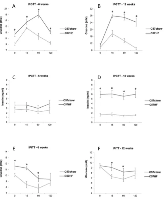
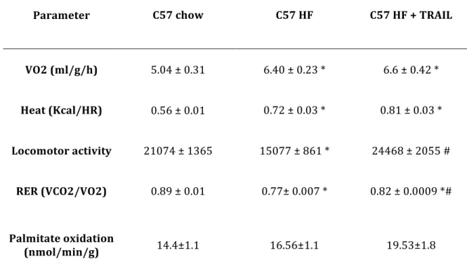
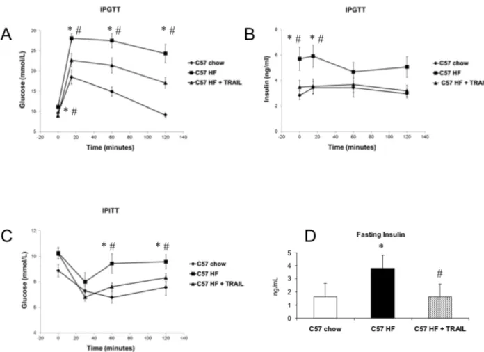
5.2. TRAIL SIGNIFICANTLY REDUCES HYPERGLYCAEMIA AND HYPERISULINEMIA DURING AN IPGTT AS WELL AS HYPERINSULINEMIA AT FASTING AND IMPROVES THE PERIPHERAL RESPONSE TO INSULIN IN HIGH FAT FED MICE 36 5.3. TRAIL TREATMENT SIGNIFICANTLY REVERSES THE CHANGES IN SUBSTRATE UTILIZATION INDUCED BY HFD AND AMELIORATES EX VIVO PALMITATE OXIDATION ... 37
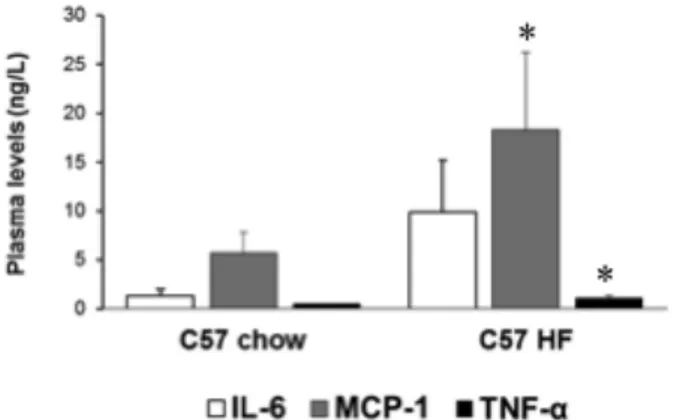
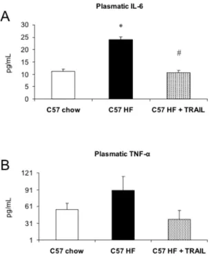
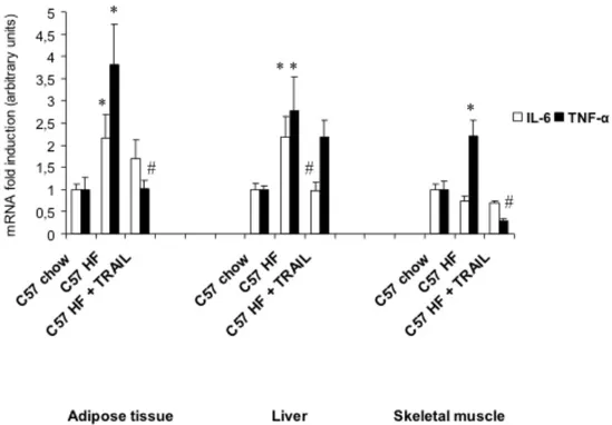
5.4. TRAIL SIGNIFICANTLY LOWERS CIRCULATING LEVELS AND GENE EXPRESSION OF PRO-‐INFLAMMATORY CYTOKINES ... 38
5.5. TRAIL TREATMENT MODULATES ADIPOSE TISSUE GENE EXPRESSION AND PROMOTES ADIPOSE TISSUE ... 39
DISCUSSION 44
LIST OF PUBLICATIONS (2010-‐2013) 48
REFERENCES 49
BACKGROUND
TRAIL and cell death
1.1 An overview on cellular apoptosis
For a single-‐cell organism, life with mutations is apparently better than no life at all, but in multicellular organisms the health of the organism takes precedence over the life of an individual cell and thus, when DNA damage occurs, the cells do not attempt to continue division but instead “commit suicide” by undergoing apoptosis. Apoptosis is a process leading to cell death, whereby unnecessary or damaged cells can be eliminated. According to the source of the pro-‐apoptotic signal, which can be either intracellular or extracellular, two separate ways of signalling take place, both converging to the activation of effector caspases and ultimately leading to cell death. These two ways of signalling have been divided into the intrinsic pathway, triggered by an intracellular signal, such as DNA damage, and the extrinsic pathway, triggered by an extracellular signal, which usually derives from cytotoxic cells of the immune system, as in (Figure 1)[Bernardi, 2012a].
Briefly, the activation of the intrinsic pathway depends on the balance between intracellular pro-‐apoptotic and anti-‐apoptotic proteins that control the release of mitochondrial cytochrome C, leading to the formation of the apoptosome, caspases activation, and then cell death (Figure 1). The proteins regulating the intrinsic pathway belong to the Bcl-‐ 2 family and they can be further divided into different subfamilies based on their function and structure. Some of them (e.g. Bax, Bak, and Bok) stimulate the release of cytochrome C, whereas other members (e.g. Bcl-‐2, Bcl-‐xL, and MCL-‐1) inhibit this process. A subfamily of these proteins is the BH3-‐only group, whose members share only the BH3 domain with the other proteins [Adams, 2007]. Bid is an important member of the BH3-‐only group since it connects the apoptotic pathways by transmitting apoptotic signals from the extrinsic to the intrinsic pathway. When pro-‐apoptotic molecules cause the permeabilization of the outer mitochondrial membrane, a protein called Smac/DIABLO is also released into the cytosol, which indirectly promotes apoptosis. Chai and colleagues have in fact demonstrated that the mitochondrial protein Smac/DIABLO interferes with the inhibitors of apoptosis proteins (IAP), thereby promoting the proteolytic activation of procaspase-‐3 [Chai, 2000; Wu, 2000].
On the other hand, the activation of the extrinsic pathway depends on the binding of specific pro-‐apoptotic ligands, such as FasL/CD95L, tumor necrosis factor-‐α (TNF-‐α) and TNF-‐ related apoptosis-‐inducing ligand (TRAIL) to their transmembrane receptors (Figure 1). This stimulates the trimerization of the transmembrane receptors and the recruitment of Fas associated death domain (FADD). Subsequently, FADD recruits both caspase-‐8 and caspase-‐10, which undergo auto-‐activation by proteolytic cleavage and in turn activate caspase-‐3, caspase-‐ 6, and caspase-‐7, which execute the apoptotic program. In some cells, called type 1, the extrinsic pathway generates a signal strong enough to initiate apoptosis by itself. However, in the majority of cells, called type 2, this signal needs to be amplified to induce apoptosis [Ozoren, 2002]. This amplification can be generally achieved by a cross-‐talk between the extrinsic and the intrinsic pathway.
Figure 1. Key components of the intrinsic and extrinsic apoptotic pathways
Intrinsic pathway: DNA damage activates p53, which stimulates the intrinsic pathway by upregulating Puma and Noxa, which inhibit the anti-‐apoptotic Bcl-‐2, Bcl-‐xL, and MCL-‐1 and thus activate the pro-‐apoptotic Bax and Bak. Bax and Bak then stimulate the release of cythocrome C, which binds to the adaptor Apaf-‐1 to recruit the initiator procaspase-‐9 into a signalling complex termed apoptosome, which subsequently undergoes activation. Caspase-‐9 then generates caspase-‐3, caspase-‐6, and caspase-‐7. In addition to the release of cytochrome C, the permeabilisation of the mithocondrial membrane causes the release of the protein Smac/DIABLO, which promotes apoptosis by blocking the inhibitor of apoptosis proteins (IAP), which normally inhibits the activity of several caspases. Extrinsic pathway: specific extracellular homotrimers interact selectively with their specific transmembrane death receptors. Ligand binding induces receptor clustering and recruitment of Fas-‐associated death domain (FADD), procaspase-‐8, and procaspase-‐10, which then undergo activation and form altogether the death-‐inducing signalling complex (DISC). This activates caspase-‐3, caspase-‐6, and caspase-‐7, which then trigger apoptosis. In addition, caspase-‐8 cleaves Bid and truncated Bid subsequently stimulates Bax and Bak to activate the intrinsic pathway. Of note, DISC formation may be blocked by several mechanisms such as the presence of c-‐FLIP. The cross-‐talk between the extrinsic and the intrinsic pathways relies on Smac/DIABLO and truncated Bid. (Adapted from Bernardi, 2012a)
1.2 TRAIL biology
TRAIL is one of the proteins binding to the pro-‐apoptotic transmembrane receptors, also called death receptors (DRs), which engage the extrinsic apoptotic pathway. TRAIL belongs to the TNF family of death-‐inducing ligands and was cloned on the basis of its sequence homology to TNF-‐α and FasL/CD95L [Wiley, 1995]. The percentage of identity with FasL/CD95L, TNF-‐α, lymphotoxin (Lt)-‐α, and Lt-‐β, which are the most related members of the TNF family, is 28%, 23%, 23%, and 22% respectively. The gene encoding for TRAIL has been mapped to the 3q26 chromosomal region. It spans approximately 20 kb, and has five exons and four introns. In humans, TRAIL is expressed as a type II transmembrane protein of 281 amino acids, whose extracellular C-‐terminal domain undergoes enzymatic cleavage. Subsequently, TRAIL is released as a soluble molecule with biological activity. The cysteine residue at position 230 (Cys230) allows TRAIL to interact and assemble with other two molecules of TRAIL forming a trimeric ligand. TRAIL homotrimers are one of the pro-‐apoptotic receptor agonists that bind to DRs on the surface of target cells and induce apoptosis (Figure 1)
Five human receptors for TRAIL have been identified so far, which can be divided into death receptors (DRs) and decoy receptors (DcRs). DRs are TRAIL-‐R1 (DR4/TNFRSF10A) [Pan, 1997a] and TRAIL-‐R2 (DR5/TNFRSF10B) [Pan, 1997b; Sheridan, 1997; Wu, 1997; Milani, 2003], which are both type I transmembrane proteins containing an intracellular death domain that classically stimulates apoptosis upon TRAIL binding. A major contribution of our group to the biology of the TRAIL/TRAIL-‐R system was the discovery that when TRAIL-‐R1/DR4 and TRAIL-‐R2/DR5 are activated they not only stimulate the extrinsic apoptotic pathway, but they may also activate survival/proliferation pathways, such as nuclear factor kB (NF-‐kB), ERK1/ERK2, and Akt [Harper, 2001; Secchiero, 2003; Zauli, 2005]. The second class of receptors includes the decoy receptors TRAIL-‐R3 (TRID/DcR1/TNFRSF10C) [Degli-‐Esposti, 1997a], TRAIL-‐R4 (DcR2/TNFRSF10D) [Degli-‐Esposti, 1997b; Marsters, 1997], and osteoprotegerin (OPG) [Zauli, 2009].
TRAIL is expressed on the surface of activated immune cells, such as natural killer (NK) cells, T cells, macrophages and dendritic cells, where it apparently functions as an immune effector molecule and it mediates anti-‐tumor cytotoxicity and immune surveillance [Di Pietro, 2004; Secchiero, 2008]. Studies on TRAIL -‐/-‐ mice have demonstrated that mice without TRAIL are viable and fertile but more susceptible to tumor metastases, indicating role of TRAIL
in immune surveillance and host defence against tumor initiation and progression [Cretney, 2002; Sedger, 2002]. In particular, TRAIL seems to mediate the ability of NK cells to block tumor growth and metastases development as well as some abilities of memory T cells [Janssen, 2005].
One of the unique aspects of TRAIL is that it has the ability to induce apoptosis preferentially in tumor cells, while it spares most normal cells [Ashkenazi, 2008a]. The balance between TRAIL death and decoy receptors was initially considered a possible mechanism whereby cells could negatively regulate TRAIL-‐induced cytotoxicity, therefore explaining TRAIL selectivity. Nevertheless, a correlation between this ratio and either protection or susceptibility to apoptosis has never been clearly demonstrated [Lincz, 2001; Wuchter, 2001; Mirandola, 2004; Kim, 2000] and it is still under debate.
1.3 TNF-‐family members targeting the extrinsic apoptotic pathway
Agents that promote or restore apoptosis through the extrinsic pathway have emerged as a promising anti-‐cancer therapeutic strategy. The vast majority of mechanisms leading to apoptosis resistance and non responsiveness to anti-‐cancer agents involve the intrinsic apoptotic pathway, possibly because it has greater involvement in the initial elimination of oncogene-‐transformed cells. These mechanisms include mutations related to p53, mutations modulating the ratio between pro-‐apototic and anti-‐apoptotic proteins, and/or overexpression of proteins belonging to the IAP family. So, agents that promote or restore apoptosis through the extrinsic pathway, such as pro-‐apoptotic receptor agonists, should be able to circumvent some of the mutations leading to apoptosis resistance and therefore to overcome the non-‐ responsiveness of tumor cells to anti-‐cancer therapies. For example, the activation of the extrinsic pathway induces apoptosis in tumors that are not responsive to chemotherapy because they have developed mutations of p53 [Ravi, 2004]. An additional reason that makes pro-‐apoptotic receptor agonists such an attractive therapeutic tool in the fight against cancer is that, given the interplay between intrinsic and extrinsic pathway, activating the extrinsic pathway may not only facilitate the outcome of conventional chemotherapies but it may also have a synergistic effect when combined to them (Figure 1), as many studies have demonstrated [Nagata, 1997].
Experimental evidence, however, has shown that not all the extracellular pro-‐apoptotic ligands could be used as anti-‐cancer agents, given that some of them display a low selectivity for tumors compared with normal cells [Nagata, 1997; Tartaglia, 1992]. For example, ever since the systemic delivery of Fas/Apo1/CD95 resulted in acute, lethal hepatotoxicity in mice this agent has been dismissed for cancer therapy [Verbrugge, 2009; Shiraishi, 2004]. Likewise, TNF-‐α systemic delivery caused pulmonary failure and coagulopathies as well as other symptomatic toxicities such as chills, fever, malaise, headache, myalgia, and nausea or vomiting in 13% of the patients studied. Therefore, these severe toxic effects that are elicited by FasL and TNF-‐α have precluded both ligands from use as systemic anti-‐cancer therapies [Nagata, 1997; Tartaglia, 1992].
In contrast to FasL and TNF-‐α, the pro-‐apoptotic ligand TRAIL, also known as Apo2L, seems to preferentially induce apoptosis in tumor cells rather than in normal cells in vitro [Ashkenazi, 1999]. Consistent with this, preclinical safety trials have demonstrated that this ligand does not cause toxic effects even at high doses. In particular, in their seminal study, Ashkenazi and colleagues demonstrated that the exposure of cynomolgus monkeys to recombinant human (rh)-‐TRAIL at 0.1-‐10 mg/Kg/day over 7 days did not induce detectable toxicity, whereas for comparison, TNF-‐α induced severe toxicity at 0.003 mg/Kg/day [Marsters, 1997]. On the basis of these properties, various agents/strategies targeting TRAIL-‐ R1/DR4 and TRAIL-‐R2/DR5 have been developed. Most of these have been so far extremely well tolerated in clinical safety trial. These include: recombinant Apo2L/TRAIL, TRAIL-‐R1/DR4 and TRAIL-‐R2/DR5 monoclonal antibodies, collectively called pro-‐apoptotic receptor agonists (PARAs), as well as TRAIL gene-‐delivering strategies and small molecules binding to death receptors.
Chronologically, the first of these molecules that had been developed were recombinant versions of the soluble Apo2L/TRAIL, including a non tagged protein and versions tagged with exogenous polypeptides to improve the stabilization of the homotrimeric structure, such as oligomeric TRAIL polypeptides joined through leucine zippers called LZ-‐TRAIL [Wiley, 2001]. Ashkenazi and colleagues developed an optimized, clinical-‐grade version of rh-‐TRAIL, consisting of amino acids 114-‐281 of the endogenous molecule, without any added tag [Ashkenazi, 2000] and with only a zinc molecule located at position 230 of each subunit helping to stabilize the trimeric protein structure. Subsequently, several TRAIL-‐R1/DR4 and TRAIL-‐R2/DR5 monoclonal antibodies have been developed [Chuntarapai, 2001; Ichikawa,
2001; Georgakis, 2005; Ashkenazi, 2001; Salcedo, 2003; Camellia, 2006]. Among these, LBY-‐ 135 is the only chimeric, rather than fully human, monoclonal antibody against TRAIL-‐R2/DR5 [Li, 2008] (Table 1). Although PARAs share the same mechanism of action, the pharmacological characteristics that they display are different and this may influence their therapeutic potential. For example, the serum half-‐life of rh-‐TRAIL is 30 to 60 minutes in humans, whereas the serum half-‐life of the antibodies ranges from 6 to 21 days. In addition, rh-‐ TRAIL binds not only to TRAIL-‐R1/DR4 and TRAIL-‐R2/DR5, like the monoclonal antibodies, but also to OPG, TRAIL-‐R3/DcR1 and TRAIL-‐R4/DcR2 [Wiezorek, 2010].
Table 1. Summary of the pro-‐apoptotic receptor agonists
Molecule Developed by Mechanism/Pharmacology
Ad5-‐TRAIL ___ Recombinant adenovirus encoding
human Apo2L/TRAIL AMG655 Conatumumab Amgen Human mAb targeting DR5
Apomab Genentech Human mAb targeting DR5
CS-‐1008/TRA8 Tigatuzumab Daiichi Sankyo Humanized version (CS-‐1008) of murine DR5-‐targeting antibody (TRA-‐8)
HGS-‐ETR1 Mapatumumab HGS GSK Human mAb targeting DR4 HGS-‐ETR2 Lexatumumab HGS Human mAb targeting DR5
LBY135 Novartis Chimeric mAb targeting DR5
PRO1762/AMG951 Dulanermin Genentech/Amgen rhApo2L/TRAIL targeting DR4 and DR5
PRO95780 Genentech Human mAb targeting DR5
DRAs ___ Small molecules activating DRs
Abbreviations: TRAIL, TNF-‐related apoptosis-‐inducing ligand; DRAs, death receptors agonists/activators; HGS, Human Genome Science; GSK, Glaxo Smith Kline; mAb, monoclonal antibody; DR4, death receptor 4; DR5, death receptor 5. (Adapted from Bernardi, 2012a).
1.4 Preclinical and clinical data on the effects of TRAIL and TRAIL agonists
The first preclinical data for the soluble, non tagged rh-‐TRAIL were published in 1999 by Ashkenazi and colleagues [Ashkenazi, 1999], demonstrating that this molecule induced apoptosis in cell lines derived from human colon, lung, breast, central nervous system, kidney and skin cancer, while it spared normal cells. Following studies showed that rh-‐TRAIL was able to induce apoptosis in other malignant cell lines derived from prostate cancer [Yu, 2000], thyroid cancer [Mitsiades, 2000], leukemia [Yao, 2007], multiple myeloma [Mitsiades, 2001] and non-‐Hodgkin’s lymphoma (NHL) [Daniel, 2007]. Consistent with these data obtained in vitro, TRAIL displayed a marked anti-‐tumor activity in vivo, in models of mouse xenografts of human colon or lung cancer, multiple myeloma, NHL, and glioma. Subsequently, the preclinical studies on the efficacy of TRAIL-‐R1/DR4 and TRAIL-‐R2/DR5 demonstrated that also these monoclonal antibodies were able to induce apoptosis in a wide variety of tumor cell lines and primary tumor explants [Ichikawa, 2001; Ashkenazi, 2008b].
Based on these promising preclinical results, the safety and the efficacy of pro-‐apoptotic receptor agonists have been evaluated in phase I and II clinical trials, as summarized in Table 2 [Herbst, 2010a; Soria, 2010; Tolcher, 2007; Hotte, 2008; Leong, 2009; Mom, 2009; Younes, 2010; Trarbach, 2010; Greco, 2008; Herbst, 2010b; Doi, 2011; Wakelee, 2010; Plummer, 2007; Camidge, 2010; Forero-‐Torres, 2010]. Overall, these studies have demonstrated that the pro-‐apoptotic receptor agonists are safe and well tolerated. These works have given ground for additional trials, some still ongoing [Ashkenazi, 2008c]. Focusing on rh-‐TRAIL, two main trials have been conducted so far. In the first one, which was a phase I, open-‐label, and dose-‐ escalation study, patients with advanced cancer were treated with rh-‐TRAIL at a dose ranging from 0.5 to 30mg/kg/day. Doses were given daily for 5 days, every 3 weeks. A total of 71 patients were treated, among whom only 2 drug-‐related dose-‐limiting adverse events were observed and a maximum tolerated dose was not reached during the dose escalation phase. In this study, rh-‐TRAIL demonstrated linear pharmacokinetic, with an estimated effective half-‐life of half an hour. Notably, one patient with chondrosarcoma exhibited a partial response with >80% tumor regression [Herbst, 2010a]. The second one was a phase Ib study in relapsed low-‐ grade NHL and non-‐small-‐cell lung carcinoma (NSCLC) [Soria, 2010]. In NSCLC, the combination of rh-‐TRAIL with carboplatin, paclitaxel and bevacizumab showed a 70% overall tumor response rate with 1 complete and 13 partial responses in the 18 patients treated.
Table 2. Development status of some of the pro-‐apoptotic receptor agonists
Phase/Treatment Patients Results
Conatumumab:
Phase Ia-‐
Monotherapy Advanced solid tumors (n=37) No DTL; no MTD up to 20 mg/kg Q2W; 1 PR in NSCLC Phase Ia-‐
Monotherapy Advanced solid tumors (n=18) No DTL; no MTD up to 20 mg/kg Q2W
Dulanermin:
Phase Ia-‐
Monotherapy Advanced solid tumors or NHL (n=71) 2 DLTs; no MTD up to 15mg/kg/d for 5d Q3W; 1 PR in chondrosarcoma
Phase Ib-‐+PAC,
CAR and BEV Stage IIIb/IV or recurrent NSCLC (n=24) No DLT; no MTD up to 20 mg/kg/d for 2d Q3W; 1 CR; 13 PRs
Lexatumumab:
Phase Ia-‐
Monotherapy Advanced solid tumors (n=37) 3 DTLs; MTD at 10mg/kg Q3W; no objective responses Phase Ia-‐
Monotherapy Advanced solid tumors (n=31) 1 DTLs; no MTD at 10mg/kg Q2W; no objectives responses
Mapatumumab:
Phase Ia-‐
Monotherapy Advanced solid tumors (n=49) 3 DTLs; no MTD up to 10 mg/kg Q2W; no objective responses Phase Ia-‐
Monotherapy Advanced solid tumors (n=41) No DTL; no MTD up to 20mg/kg Q4W; no objective responses Phase Ib-‐+PAC and
CAR
Advanced solid tumors (n=27) 2 DTLs; no MTD up to 20mg/kg Q3W; 5 PRs
Phase Ib-‐+GEM and CIS
Advanced solid tumors (n=49) 5 DTLs; no MTD up to 30mg/kg Q3W; 12 PRs
Phase II-‐
Monotherary Relapsed/refractory NHL (n=40) No DTL; no MTD up to 10mg/kg Q3W; 2CRs 1 PR Phase II-‐
Monotherapy
Relapsed/refrectory CRC (n=38) No DTLs; no MTD up to 20mg/kg Q2W; no objective responses Phase II-‐
Monotherapy Relapsed/refractory NSCLC (n=32) No DTLs; no MTD up to 10mg/kg Q3W; no objective responses
PRO95780:
Phase Ia-‐
Monotherapy Advanced solid tumors or NHL (n=50) 4 DTLs; no MTD up to 20mg/kg Q2W; no objective responses
Tigatuzumab:
Phase Ia-‐
Monotherapy Relapsed/refractory solid tumors/NHL (n=17) No DTL; no MTD up to 8mg/kg QW; no objectives responses Abbreviations: PAC, paclitaxel; CAR, carboplatin; BEV, bevacizumab; GEM, gemcitabine; CIS, cisplatin; NHL, non-‐Hodgkin lymphoma; NSCLC, non-‐small cell lung cancer; CRC, colorectal cancer; DLT, dose-‐limiting toxicity; MTD, maximum tolerated dose; QW, once per week; Q2W, every 2 weeks; Q3W, every 3 weeks; Q4W, every 4 weeks; PR, partial response; CR, complete response. (Adapted from Bernardi, 2012a)
1.5 rh-‐TRAIL-‐based combinational therapy
A large number of studies have revealed that combining TRAIL with either traditional or novel anti-‐cancer agents may reverse the resistance to monotherapy. Interestingly, it seems that low concentrations of chemotherapeutics can enhance the sensitivity to TRAIL or the TRAIL receptor antibody HGS-‐ETR1 by accumulating the TRAIL-‐R1/DR4 at the cell surface [Jin, 2007]. On the other hand, pre-‐treatment with chemotherapeutic agents of Bax-‐/-‐ human colon
cancer cells that are resistant to TRAIL has been shown to restore TRAIL sensitivity. For TRAIL-‐ based combinational therapy the agents/strategies that have been tested are listed on (Table
3) [Ravi, 2004; Frese, 2002; Jin, 2004; Hylander, 2005; Vignati, 2002; Cuello, 2001; Mizutani, 1999; Brooks, 2005; Johnson, 2003; Zhu, 2005; Ricci, 2007; Kim, 2008; Meng, 2007; Rosato, 2007; Zangemeister-‐Wittke, 2003; Ray, 2005; Li, 2004a; Arnt, 2002; Adams, 2007; Chai, 2000; Fulda, 2002; Li, 2004b; Guo, 2004; Maier, 2011; Croce, 2008; Garofalo, 2009]
Table 3. Summary of anti-‐cancer agents studied in combination with rh-‐TRAIL
Mechanism of
action Agent Experimental model
Cytotoxicity by traditional chemothrapies
Camptothecin, 5-‐fluorouracil, irinotecan, cisplatin, paclitaxel, doxorubicin, carboplatin, gemcitabine, adriamicin
In vitro
Paclitaxel, carboplatin, bevacizumab, cisplatin gemcitabine
In vivo Proteasome
inhibition Bortezomib In vitro
Kinase inhibition Sorafenib In vitro
Bcl-‐2 inhibition Genasense In vitro
BH3I-‐2’ In vitro
IAP inhibition Smac peptides In vitro and in vivo HDACs inhibition TSA, SAHA, LAQ824 In vitro
miRNAs
modulation Strategies increasing miR-‐212 and strategies decreasing miR-‐221 and miR-‐222
In vitro
Abbreviations: TRAIL, TNF-‐releated apoptosis-‐inducing ligand; BH3I, BH3 inhibitor; IAP, inhibitor of apoptosis proteins; HDACs, histone deacetylases; miRNA and miR, microRNA. (Adapted from Bernardi, 2012a)
Potential role of TRAIL in the management of autoimmune diabetes
2.1 Type 1 diabetes mellitus
Type 1 diabetes mellitus (T1DM) is an autoimmune disease whose incidence has been steadily increasing worldwide since the middle of the 20th Century. Recently, the increase in
T1DM incidence has been even faster than before and it is expected that the number of new cases diagnosed at or before 14 years of age will double in the next 15 years and the age of onset will be younger (0-‐4 years) [Harjutsalo, 2008]. It has been shown that T1DM results from an autoimmune β-‐cell destruction and when 70-‐80% of the β-‐cell mass has been destroyed the residual functional β-‐cells are insufficient in number to maintain glucose tolerance [Van Belle, 2011]. At this stage, the lack of insulin leads to hyperglycemia and T1DM becomes clinically apparent [Van Belle, 2011].
T1DM therapy is based on the use of insulin. Despite the improvements in insulin replacement regimens and devices, insulin remains a treatment that heavily affects patients’ quality of life and retains some important side effects, such as hypoglycemic events, seizures and coma. Moreover, T1DM is still associated with substantial morbidity and mortality. Consequently, during the past 20 years, a lot of interest has been focused on developing new strategies that aim to prevent, delay, and treat T1DM. These strategies are based on targeting the immune system [Bernardi, 2012b]. In contrast to the broadly immunosuppressive agents that were used initially, failed to produce lasting remission, and were associated with multiple long-‐term side effects, new rationally designed agents have shown to possess the characteristics to successfully prevent or delay T1DM development [Van Belle, 2011; Feutren, 1986; Dupré, 1988; Raz, 2001; Herold, 2005; Lugdvisson, 2012; Hu, 2007]. TRAIL is one of these agents, given that by regulating the homeostasis of the immune system it seems to delay T1DM natural history without causing adverse effects [Di Pietro, 2004; Secchiero, 2008].
2.2 Autoimmunity in T1DM
T1DM is an autoimmune disease in which CD4+ and CD8+ T lymphocytes infiltrate the
Islets of Langerhans resulting in a cell-‐mediated islets destruction, insulin deficiency and hyperglycemia. The concept that T1DM pathogenesis is immune-‐mediated dates back to the
sixties, when Burnet and Mackay included T1DM among several autoimmune diseases. Two years later, Gepts and colleagues found that the key pancreatic morphological feature in recent-‐ onset T1DM was an insulitis, suggesting that it is the immune system that is involved in the pathogenesis of the disease [Gepts, 1965]. Then, in 1974 the group of Nerup demonstrated that T1DM is a cell-‐mediated autoimmune disease by incubating porcine islets with peripheral blood mononuclear cells of diabetic patients and reporting cell migration inhibition in diabetics as compared to normal controls [Nerup, 1974].
The current view on the human immunology of T1DM could be summarized as follows: T1DM is a T cell-‐mediated disease. T cells are present in the islets, peri-‐pancreatic lymphnodes and systemic circulation of T1DM patients. These cells are mostly CD8+ lymphocytes, with only few macrophages and no T regulatory (Treg) cells, as to say that the distribution of T cell subsets is also abnormal [Buschard, 1980] and that circulating Treg cells display defective responsiveness [Roncarolo, 2007]. The importance of Treg cells in the maintenance of self-‐ tolerance and immune homeostasis has been clearly shown by the development of T1DM and other polyendocrinopathies in patients with immune-‐dysregulation polyendocrinopathy enteropathy X-‐linked syndrome, caused by impairment of Treg cell-‐mediated peripheral self-‐ tolerance [Sakaguchi, 2012]. On the other hand, the report that a patient with X-‐linked agammaglobulinemia developed T1DM diabetes shows that neither autoantibodies nor B cell function seem to be critically involved in the pathogenesis of this disease [Martin, 2001]. Although the delivery of rituximab, which is an anti-‐B cell antibody, protected NOD mice against T1DM development during the early stages of the disease [Hu, 2007], the same treatment did not change the disease course in early onset T1DM patients [Pescovitz, 2009], suggesting once again that cell-‐mediated autoimmunity in T1DM corresponds to a T cell-‐ mediated response.
As for the natural history of the disease, islet inflammation or insulitis generally precedes the onset of T1DM [Roep, 1986] and autoantibodies are usually detectable at this stage [Eisenbarth, 1986]. These autoantibodies are directed against several molecules of β-‐ cells, such as insulin, insulinoma-‐associated antigen (IA)-‐2, 65-‐kD isoform of glutamic acid decarboxylase (GAD-‐65), β-‐cell-‐specific zinc transporter (Znt8), and islet cell autoantibodies (ICA)-‐512. The role and/or significance of these autoantibodies is not entirely clear, given that fetal exposure to GAD-‐65 and/or IA-‐2 autoantibodies may protect against diabetes [Bottazzo, 1978]. Proinsulin, GAD-‐65, IA-‐2 and Znt8 are not only the major autoantibody targets but also
the main epitopes activating autoreactive T cells [Koezwara, 2004]. Once T cells have been activated by the recognition of islet autoantigens via MHC class I [Di Lorenzo, 2007] and II, β-‐ cells are destroyed either by CD8+ lymphocytes through the release of cytolitic granules containing perforin and granzyme, or by both CD8+ and CD4+ lymphocytes through Fas and Fas-‐ligand-‐dependent interactions or the release of IL-‐1β, TNF-‐α, γ-‐interferon (IFN-‐γ) [Hamilton-‐Williams, 2003].
2.3 Immunosuppressive strategies in T1DM or from cause to cure
Given that autoimmunity appears to be the main effector mechanism in T1DM, several immunosuppressive agents have been studied as potential therapies for patients with new-‐ onset T1DM. The first agents studied were broadly immunosuppressive drugs including cyclosporine, nicotinamide, anti-‐oxidant agents, heat shock proteins and anti-‐CD3 [Feutren, 1986; Dupré, 1988; Raz, 2001; Herold, 2005]. Unfortunately, most attempts either achieved minimal benefits or the adverse effects outweighed their benefits. One of the main side effects of cyclosporine was, for example, the induction of nephropathy [Feutren, 1986; Dupré, 1988]. So, the scientific interest moved to antigen-‐specific interventions, which are more targeted forms of immunosuppression, aiming at inducing selective immunological tolerance by the systemic delivery of the autoantigens involved in T1DM. The delivery of these autoantigens, such as GAD-‐65 and insulin, should in fact induce Treg cell responses (active tolerance) and anergize autoreactive T cells (passive tolerance). Consistent with this rationale, in one of these studies GAD-‐65 treatment displayed a short protective effect on C-‐peptide secretion in T1DM patients, who had been treated less than 6 months after diagnosis [Ludvigsson, 2008]. Nevertheless, the results coming from a more recent trial did not confirm the efficacy of such a treatment in the long-‐term, as GAD-‐65 delivery failed to ameliorate T1DM natural history over 15 months [Ludvigsson, 2012].
Beside these immunosuppressive strategies, another therapeutic strategy against T1DM, which aims at curing this disease rather than just treating it, is islet transplantation. Islet transplantation has been shown to successfully restore endogenous insulin production and glycemic stability in subjects with T1DM and instable control [Ricordi, 2003]. In particular, the islet transplantation performed according to the Edmonton protocol has achieved reproducible insulin independence, with more than 80% of recipients still insulin-‐free at 1 year after the
procedure [Shapiro, 2006]. Currently available data suggest that medium-‐ to long-‐term results are still not achievable mainly because of the high frequency of non-‐functioning grafts and secondary graft failure. Unfortunately, the methods that have been devised so far to protect β-‐ cells from immune destruction have only been able to delay, without preventing, the failure of the transplants and the consequent need of resuming the administration of insulin. Moreover all the adverse effects of long-‐term general immunosuppression and late graft loss overall represent an obstacle for any successful transplantation [Battaglia, 2006].
2.4 TRAIL, autoimmune diseases, and T1DM
A large amount of data has pointed out the ability of TRAIL to act as an immune system modulator [Di Pietro, 2004; Secchiero, 2008]. TRAIL best-‐known function is the induction of apoptosis, which occurs through the activation of the extrinsic apoptotic pathway, triggered by the binding of TRAIL to DRs, as discussed before. Beside its pro-‐apoptotic effect, TRAIL is also able to activate pathways involved in cell survival. It has in fact been demonstrated that TRAIL is able to stimulate the survival and proliferation of both neoplastic and non-‐neoplastic cells [Lancaster, 2003; Ehrhardt, 2003; Secchiero, 2004; Morel, 2005]. The TRAIL-‐mediated pro-‐ survival effect seems to be due to the recruitment of adaptor molecules that interact directly with DRs death domains. This would block the progression of the pro-‐apoptotic signal. In the meantime, TRAIL would be able to engage other intracellular pathways and mediators, including NF-‐κB, JNK, MAPK/ERK and Akt, leading to subsequent transcription of survival genes [Morel, 2005; Secchiero, 2004]. TRAIL ability of activating diametrically opposed pathways may actually be ascribed to cell responsiveness, rather than to the ligand properties. Cell responsiveness to TRAIL could result from the balance between death and decoy receptors expressed on the cell surface [Lincz, 2001; Wutcher, 2001; Mirandola, 2004; Kim, 2000], which in turn could come down to the cellular incorporation of specific intracellular proteins into “lipid rafts” [Hunter, 2006; Song, 2007]. Lipid rafts are platforms consisting of a dynamic pool of cholesterol and sphingolipids, which recruit signalling molecules including cell surface receptors. Independent studies have documented a redistribution of TRAIL receptors and DISC components from “non-‐rafts” into lipid rafts suggesting that this event could switch apoptosis instead of survival [Song, 2007]. However, a correlation between TRAIL receptors balance (i.e., death/decoy receptor ratio) and protection versus susceptibility to apoptosis has never been clearly demonstrated and it is still under debate.
Going back to T1DM, TRAIL is expressed on the surface of activated immune cells, such as NK cells, T cells, macrophages and dendritic cells, where it apparently mediates immune surveillance, particularly against hematological malignancies and autoimmune diseases [Secchiero, 2008]. Studies on TRAIL -‐/-‐ mice demonstrate that mice without TRAIL are viable and fertile, but more susceptible to autoimmune arthritis, experimental autoimmune encephalomyelitis and autoimmune diabetes, indicating a clear role for TRAIL in immune regulation. In addition, TRAIL involvement in immune surveillance has also been proved by the acceleration of experimental autoimmune encephalomyelitis (EAE) and autoimmune arthritis that followed TRAIL blockade [Cretney, 2002; Sedger, 2002]. Interestingly, in the models of EAE TRAIL-‐deficient mice display significantly lower Treg cells than wild-‐type mice, as to say that TRAIL not only inhibits autoreactive T cells but also promotes Treg cells [Ikeda, 2010].
Lamhamedi-‐Cherradi and colleagues are the first who have demonstrated that either blockade or TRAIL deficiency exacerbates T1DM [Lamhamedi-‐Cherradi, 2003]. The first animal model consisted of NOD mice injected with a soluble TRAIL receptor (sDR5/TRAIL-‐R2) in order to block TRAIL functions. In the mice injected with sDR5/TRAIL-‐R2, T1DM incidence increased significantly and its onset was also significantly accelerated with respect to the control group. In addition, the blockade of TRAIL by sDR5/TRAIL-‐R2 led to greater pancreatic inflammation as well as to enhanced cellular and humoral immune responses, with an increase in T cells proliferation and pro-‐inflammatory response as well as higher levels of anti-‐GAD-‐65 [Lamhamedi-‐Cherradi, 2003]. The second animal model consisted of TRAIL -‐/-‐ mice injected with streptozotocin, which is a drug that destroys β-‐cells and therefore cause diabetes. The findings observed in the diabetic TRAIL -‐/-‐ mice recapitulated those observed in the sDR5 treated NOD mice, since the incidence of T1DM increased significantly and its onset was significantly accelerated. Consistent with these observations, the administration of recombinant soluble TRAIL in streptozotocin-‐injected mice preserved pancreatic islets and significantly ameliorated the severity of T1DM, by lowering glucose levels [Zauli, 2010].
2.5 Possible mechanisms whereby TRAIL protects against T1DM
Having said that, the molecular mechanisms whereby TRAIL blockade exacerbates T1DM and, on the contrary, its delivery ameliorates the natural history of this disease, have
only partly been clarified. One of the studies addressing this issue showed that TRAIL suppressed the proliferation of autoreactive T cells isolated from diabetic NOD mice [Mi, 2003]. This effect was explained by the finding that TRAIL up-‐regulated p27 expression and simultaneously inhibited IL-‐2 production by autoreactive T cells. With respect to this issue, it has indeed been shown that the up-‐regulation of cdk inhibitors, such as p27, which sequester cyclin D2-‐cdk4 and cyclin E–cdk2 complexes, stops the progression of the cell cycle, preventing the progression of T cells through the G1 restriction point of the cell cycle, and therefore anergizing autorective T cells [Mi, 2003]. On the other hand, it has been shown that the cell exposure to IL-‐2 promotes the degradation of p27 and the entry into S phase [Appelman, 2001]; therefore IL-‐2 inhibition by TRAIL could be an additional mechanism whereby TRAIL anergizes diabetogenic T cells.
Recent works suggest that beside T cells anergy TRAIL could also promote β cell survival. In vitro experiments performed on rat insulinoma cells (INS-‐1), which are used as a model for the study of pancreatic β-‐cells, have shown that exposure to soluble TRAIL does not affect β-‐cell viability, but, by promoting the activation of NF-‐kB, it upregulates the expression of its decoy receptor TRAIL-‐R3 (DcR1), which in turns should prevent apoptosis [Kang, 2010a]. In this context, the selective increase of TRAIL and DcR1 expression in the pancreatic islets in NOD mice, which occurs in streptozotocin-‐induced T1DM, has been proposed as part of a defensive strategy of the β-‐cell against infiltrating leukocytes [Dirice, 2011].
Kang and colleagues have further investigated TRAIL protective effects in T1DM animal models, demonstrating that they could be partly attributed to the elevation of tissue inhibitor of metalloproteinase-‐1 (TIMP-‐1) levels that follows TRAIL delivery [Kang, 2010b]. In vivo experiments have in fact demonstrated that TRAIL-‐induced TIMP-‐1 elevation markedly reduced MMP-‐9 pancreatic activity, thus protecting against T1DM development/progression [Kang, 2010b], given that circulating MMP-‐9 is higher in diabetic patients and that MMP-‐9 cleaves insulin [Maxwell, 2001; Descamps, 2004; Xue, 2005]. The in vivo data are further supported by in vitro data showing that the addition of TIMP-‐1 significantly reduces INS-‐1 cells death [Kang, 2010b], consistent with a previous report showing that TIMP-‐1 prevents cytokine-‐mediated dysfunction and cytotoxicity in pancreatic islets and β-‐cell [Han, 2001].
2.6 TRAIL and diabetic complications
It is well known that T1DM morbidity and mortality are primarily due to diabetic complications. Diabetic complications are divided into macrovascular and microvascular and they include acute myocardial infarction, stroke, peripheral artery disease, diabetic nephropathy, retinopathy and neuropathy [Calcutt, 2009]. Experimental works suggest that TRAIL may protect against diabetic complications. With respect to cardiovascular diseases, our group has demonstrated that TRAIL delivery significantly reduced the development of atherosclerotic lesions and contributed to plaque stabilization by decreasing the number of infiltrating macrophages and increasing the number of vascular smooth muscle cells in an animal model of T1DM [Secchiero, 2006]. The concept that TRAIL might protect against cardiovascular diseases, is supported by the finding that circulating TRAIL is significantly reduced in different cohorts of patients with acute coronary syndrome and heart failure with respect to patients with stable angina or healthy coronary arteries [Michowitz, 2005; Schoppet, 2006; Secchiero, 2009]. In addition to the anti-‐atherosclerotic properties of TRAIL we have also demonstrated that TRAIL delivery protects against myocardial fibrosis and cell apoptosis in an animal model of diabetic cardiomyopathy [Toffoli, 2012], which corresponds to the stage in which ventricular dysfunction develops in diabetic patients in the absence of coronary atherosclerosis and hypertension.
In contrast to what has been found in experimental models of cardiovascular diseases, TRAIL unfortunately does not seem to protect againt diabetic microvascular complications. In an animal model of diabetic nephropathy Lorz and colleagues [Lorz, 2008] found that TRAIL gene and protein expression was significantly increased in both glomeruli and tubuli of diabetic kidneys. In following in vitro experiments it was found that TRAIL actually induced tubular cell apoptosis, which was enhanced by the concomitant presence of pro-‐inflammatory cytokines and high glucose levels. This would suggest that TRAIL could play an important role in the progression of diabetic nephropathy. The concept that TRAIL may play a role in the natural history of diabetic complications is further supported by the observation that patients with proliferative diabetic retinopathy exhibited lower levels of TRAIL in the conjunctival sac fluid with respect to patients with non-‐proliferative diabetic retinopathy or without diabetic retinopathy [Secchiero, 2011].
High-‐fat diet and type 2 diabetes mellitus
3.1 Obesity and type 2 diabetes mellitus
Type 2 diabetes mellitus (T2DM) represents a heterogeneous group of disorders characterized by insulin resistance and impaired insulin secretionand defined by a raised fasting or post-‐challenge blood glucose. T2DM incidence is increasing to the point that it is reaching epidemic proportions. Recent studies show that T2DM prevalence is strongly associated with overall obesity and central obesity, mainly due to unhealthy changes in lifestyle [Yang, 2010]. It has in fact been shown that obesity is an independent predictor of insulin resistance and T2DM [Collins, 2011] and that weight loss prevents it [Tuomilheto, 2011]. Among the multiple mechanisms linking obesity to diabetes, inflammation is a common feature that has been implicated in the pathophysiology of both diseases.
3.2 Lipotoxicity and T2DM
It has been argued that another mechanism explaining insulin resistance and compromised β-‐cell function in T2DM is a breakdown in lipids dynamics. This is often reflected by elevated levels of circulating free fatty acids (FFA) and tryglicerides, together with excessive deposition of fat in various extra-‐adipose tissues including the muscle bed and the β-‐cells. Increased tissue levels of FFA have been shown to cause the β-‐cell abnormalities of non-‐ diabetic obesity and ultimately result in obesity-‐dependent diabetes [Unger, 2011]. A western diet that is high in fat will therefore result in loss of β-‐cells, by pancreatic FFA deposition that in turn promotes lipoapoptosis as well as peripheral glucose intolerance due to lipid excess in skeletal muscle.
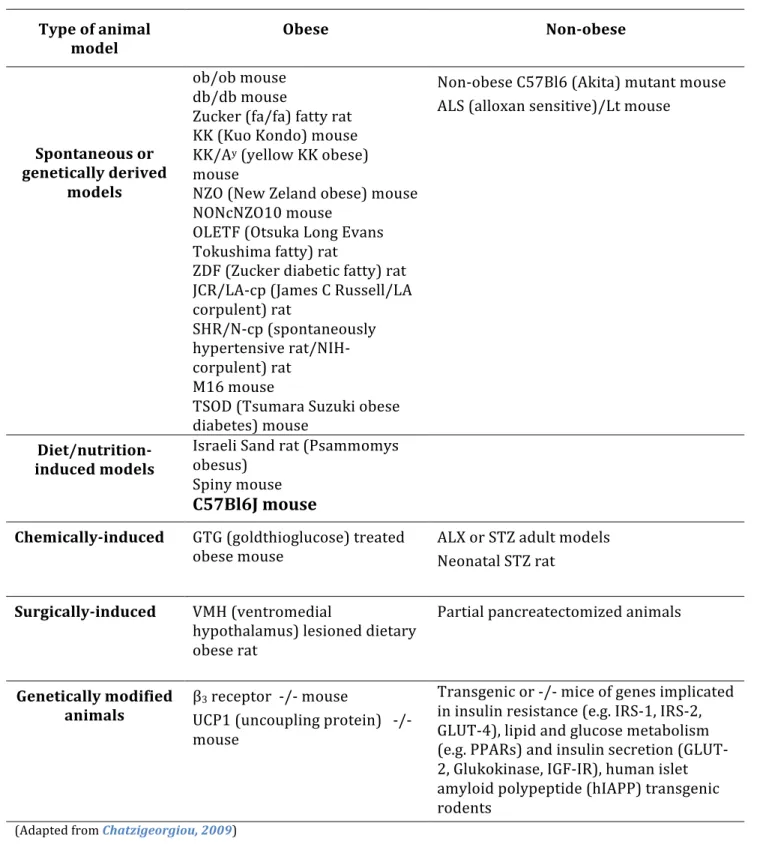
3.3 Animal models of T2DM
T2DM animal models are likely to be as complex and heterogeneous as the human condition. [Chatzigeorgiou, 2009] However, since obesity is the major environmental factor predisposing to T2DM (although 2/3 of obese subjects do not become diabetics), the ability of an animal model to develop firstly obesity and then T2DM is one of the most important criteria for selecting a model. T2DM murine models are summarized on Table 4.