L a p a to lo g ia v a sc o la re e la m e d ic in a ri a b ilit a ti va
PATOLOGIA VASCOLARE NELL’ESERCIZIO FISICO RIPETUTO
STRESS DELL’APPARATO MUSCOLO SCHELETRICO NELL’ESERCIZIO FISICO RIPETUTO
Cosimo Tudisco, M.D.; Salvatore Bisicchia, M.D. Cattedra e Scuola di Specializzazione in Ortopedia, Università degli Studi di Roma “Tor Vergata”
Introduction
Lower limb pain is very common in athletes, in articular leg pain accounts about 13% of all cases. It is very common in runners, but also in long distance walkers, jumpers and sports involving cutting movements [1].
Lower limb pain in athletes can be related with many different pathologies that must be differentiated.
Exertional compartment syndrome (CECS)
Chronic exertional compartment syndrome (CECS) is commonly overlooked as a cause of muscle pain. Studies of the etiology of chronic anterior leg pain indicate that CECS is the causative factor in 27% of cases [2,3]. The pathophysiology of CECS is related to a marked increase in tissue pressure within the confinement of a closed fascial space during exercise. Muscle volume can increase up to 20% of its resting size during exercise. Increased muscle volume causes an increase in the internal pressure within the fascial compartment [4,5]. The prevailing theory is that during progressive muscle activity rising intracompartmental pressures cause impaired muscle tissue perfusion [6,7]. Deoxygenation of muscle results in increased cell permeability, causing a shift of fluid into the interstitial space. Compromise of the microcirculation leads to ischemia and ultimately results in pain. There have been several theories refuting the ischemia theory [3,7] leading to theorization that pain is not from a lack of perfusion in the affected compartments, but rather from a disproportionate oxygen supply versus demand [5,8,9]. The actual incidence of the disease is not clear due to a mean 22 months delay in diagnosis, but is has the same frequency in
L a p a to lo g ia v a sc o la re e l a m e d ic in a ri a b ili ta ti
va males and females. The mean age at diagnosis is about 20. In 95% of the cases the anterior and lateral compartments of the leg are involved (Fig. 1), with few cases presenting in the other compartments of the leg, the tight and the forearm. Anabolic steroids, creatine use, eccentric exercises, poor running biomechanincs, overpronation of the foot can increase the risk of CECS [10]. Obtaining the history of compartment syndrome is very important because the physical exam is often unrevealing. A classic history is development of pain in a compartment of the leg at the same time, distance, or intensity of exercise. The pain increases in intensity as the patient continues exercising and resolves after a period of rest. Pain is bilateral in 70-80% of the cases. Other symptoms include numbness and tingling in the dermatomal distribution of the nerve running through the compartment or weakness of the affected muscle [10]:
1. Weakness of dorsiflexion–anterior compartment 2. Weakness of eversion–lateral compartment
3. Weakness on plantar flexion–posterior compartment
A definitive diagnosis of CECS can be done only measuring pressure in the affected compartments. A patient should perform the offending exercise until severe symptoms occur. They are then rested for 5 min and compartment pressures are taken. A resting pressure ≥15 mmHg and 5 min post-exercise pressures greater than 20 mmHg are diagnostic of compartment syndrome. Joint position of the ankle and knee should be standardized during the test because it can affect intracompartment pressures [4,5,8,10].
Conservative treatments include rest, discontinuing the activity that elicits symptoms or decreasing the intensity of training, icing, oral NSAIDs, arch support orthotics, stretching, deep tissue massage, ultrasound. Optimizing shoewear, running surfaces and running mechanics are other important factors [10]. The only definitive treatment of CECS is fasciotomy of the involved compartments [11]. Fasciotomy can be performed open, mini-open or endoscopic according to surgeon’s and patient’s features (Fig. 2). The reported success is 80% in the anterior and lateral compartments, 50% in the deep posterior compartment. Release of the superficial peroneal nerve should be done only if needed. After surgery a soft bandage is applied for 2-3 days and patients are encouraged to start range of motion immediately to avoid excessive scarring. The athlete can return to full activity in 6–8 weeks if they are symptom free and have recovered full strength and flexibility [10].
L a p a to lo g ia v a sc o la re e la m e d ic in a ri a b ilit a ti va
Medial tibial stress syndrome
This condition is characterized by bilateral leg pain that increases when the patient exercises. Tenderness on palpation of the medial border of the tibia is present even after a long period of resting (i.e. 30 days). It is thought to be an overuse syndrome due to repetitive traction of the flexor muscles on the medial border of the tibia. Diagnosis is mainly clinical, imaging studies show specific features of this disease with an hyperintensity on MRI sequences or definite uptake on late sequences on bone scan [12].
Conservative treatment includes resting, icing, NSAIDs, low impact sports (such as biking or running in water) and inserts in the shoes [13]. Surgical treatment consists of the release of the flexor muscles from the medial border of the tibia. Results after surgery are good or excellent in about 70% of the cases, with about 40% of the patients returning to their pre-injury level [14]. Complications are frequent and include (but are not limited to) hematoma, localized parenthesis, numbness and stress fractures [14-16].
Non-insertional Achilles tendinopathy
During running the Achilles tendon sustains a tension greater than 12 time the weight of the athlete [17,18]. For lengthening greater than 4% microscopic ruptures may be observed, macroscopic ruptures develop for lengthening greater than 8% [19].
Many risks factors are thought to play a role in this condition, such as hypovascularity, genetics, endocrine and metabolic disorders, but also overuse, poor stretching, poor running technique, inadequate shoes, running on hard surfaces or pronation of the foot [20].
Conservative treatment includes resting, NSAIDs, eccentric exercises, heel lifts and insoles that limit pronation of the foot [12]. Eccentric exercises are the only treatment that was demonstrated to be significantly effective in randomized controlled trials [21]. Surgical treatment includes longitudinal incision of the tendon and resection of the degenerated tissue [22] (Fig. 3)
In case a large gap is left in the tendon at the end of surgery, augmentation with flexor halluces tendon longus tendon may be performed [23]. Success of surgical treatment has been reported in 70-85% of the cases, with a complication rate of 11% (mainly related to skin problems) [24]. Percutaneous scarifications have also been described [25].
L a p a to lo g ia v a sc o la re e l a m e d ic in a ri a b ili ta ti va Stress fractures
Tibial shaft accounts for about 50% of all stress fractures in athletes. They are mainly located in the postero-medial region (compression side) and are considered at low risk of non-union.
Some fractures are located in high-risk area such as anterior cortex of the tibial shaft (tension side), medial malleolus, neck of the femur (tension side), navicular, base of the fifth metatarsal [12].
Imaging studies are very useful in the diagnosis of stress fractures. RMI sequences show cortical thickening, periosteal reaction and the fracture itself (Fig. 4). Bone scan shows trifasic focal uptake [12]. Conservative treatment includes resting for 4-8 weeks, shock wave therapy and pulsed magnetic fields. Good results have been reported after conservative treatment of low-risk tibial shaft fractures and fibular stress fractures [12]. Surgical treatment should always be considered after failed conservative treatment. Intramedullary nailing is the gold standard in the treatment of stress fractures [26]. Alternatively, drilling and bone grafting may be considered [27]. Surgical treatment does not guarantee healing of the fracture, and it can be related to post-operative complications, such as infection, pain in the entry point of the intramedullary nail or extension of the fracture) [28].
Gastrocnemius strain/rupture (tennis leg)
Gastrocnemius strains and ruptures account for about 3.6% of all soccer injuries [29]. The medial head of the gastrocnemius in involved in the majority of the cases [30].
Injury to medial head of the gastrocnemius is caused by sudden dorsiflexion of a plantar flexed foot with the knee in extension or sudden extension of the knee with the ankle dorsiflexed [31] and it has a predilection for the poorly conditioned, middle-aged athlete with “thick calves” who is engaged in strenuous activity [32] (Fig. 5). Non-operative treatment includes rest, icing, analgesics and casting/splinting depending on the extent of the injury [31]. Compression of the limb decreases hematoma size and facilitates healing [12]. Operative treatment is controversial, but acute repair may be indicated acutely in selected patients unable to maintain full body weight on the metatarsal heads with the ankle in maximal plantar flexion and/or a palpable defect [31].
L a p a to lo g ia v a sc o la re e la m e d ic in a ri a b ilit a ti va Hemolysis
Exercise-induced hemolysis has been reported for more than 50 years [33]. In particular, distance running has been associated with significant destruction of red blood cells (RBC) with RBC turnover being substantially higher in runners compared with untrained controls [34]. It is common especially in long distance running, but also in biking, swimming, weight lifting and rowing. Many factors have been thought to play a role in the etiology of this condition, but mechanical stress during foot strike is considered the most important [35], other factors include oxidative stress and compression of large muscle groups on capillaries may accelerate hemolysis of older RBC [36].
Rabdomiolysis
Damage of skeletal muscle cells, rhabdomyolysis following prolonged physical exercise was described several decades ago, based upon detection of myoglobin in urine [37,38], but also creatinkinase and other muscular enzymes [39].
The mechanisms of the membrane damage are not fully understood, but mechanical factors such as crush injuries and microruptures have been thought to play a role. In more recent studies other factors have been proposed to contribute significantly to the cell membrane damages accompanying endurance exercise [40].
Moreover, increased oxidative stress and decreased antioxidant protection have been presumed to play a role [41-44].
A recent study [45] reported that supplementation with moderate doses of selenium, vitamin E and zinc did not protect significantly against membrane damage and previous studies have been inconclusive as to the ability of antioxidant preparations to protect against exercise-induced oxidative stress [46,47]. Studies suggested a protective role of the glutathione peroxidase against oxidative stress during prolonged physical activity and proposed an integration of this enzyme.
Conclusion/ clinical recommendations
1) There is little evidence for the effectiveness of stretching and/or conditioning for the prevention of lower limb soft injuries
2) Heavy load eccentric exercise is recommended for chronic Achilles tendinopathy.
L a p a to lo g ia v a sc o la re e l a m e d ic in a ri a b ili ta ti
va 3) Fibular and low-risk tibial stress fractures often respond to a period of rest (4-8 weeks)
4) Operative interventions are recommended for chronic exertional compartment syndrome (fasciotomy)
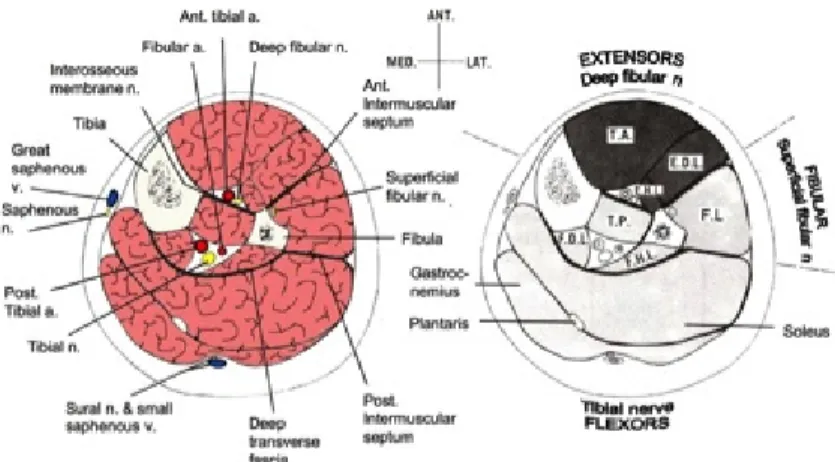
Figure 1. Cross section representation of the leg showing anatomical relationships
between neuro-vascular structures within each compartment (A), and the muscles included in each compartment (B).
Figure 2. Surgical picture showing open release of the anterior and lateral
compartments of the right leg in a case of recurrent fasciotomies for chronic exertional compartment syndrome. Note that the superficial peroneal nerve has been completely released, as it exits from the fascia. This is very important in revision cases.
L a p a to lo g ia v a sc o la re e la m e d ic in a ri a b ilit a ti va
Figure 3. Surgical picture of the right
ankle showing the Achilles tendon that has been incised longitudinally, tendon fibers have been split and degenerated tissue removed
Figure 4. Axial T1-weighted MRI of the leg
showing periostel reaction and cortical thickening of the tibial shaft. These features are consistent with tibial stress fractures.
Figure 5A. Posterior view of the left
leg of a 41-year old man showing swelling and ecchymosis of the posterior calf
Figure 5B. Posterior view of the legs of a
38-year old man with a 2-38-year history of rupture of the medial head of the right gastrocnemius of the right leg at the musculo-tendinous junction. Note the atrophy of the muscles of the posterior compartment of the leg.
L a p a to lo g ia v a sc o la re e l a m e d ic in a ri a b ili ta ti va References
[1] James SL, Bates BT, Osternig LR. Injuries to runners. Am J Sports Med. 1978;6(2):40-50.
[2] Blackman PG. A review of chronic exertional compartment syndrome. Med Sci Sports Exerc. 2000;32:4–10.
[3] Bong MR, Polatsch DB, Jazrawi LM, Rokito AS. Chronic exertional compartment syndrome: diagnosis and management. Hosp Joint Dis. 2005;62:77–84.
[4] Canale S. Campbell’s operative orthopaedics. 9th ed. Philadelphia: Elsevier; 1998. p. 405–1411.
[5] Scheltinga MR. Minimally invasive fasciotomy in chronic Exertional compartment syndrome and fascial hernias of the anterior lower leg: short and long term results. Mil Med. 2006;171:399–403.
[6] Goldfarb S, Kaeding C. Bilateral acute-on-chronic Exertional lateral compartment syndrome of the leg: a case report and review of the literature. Clin J Sports Med. 1997;7:59–62.
[7] Trease L, Every B, Bennell K, et al. A prospective blinded evaluation of exercise thallium-201 SPET in patients with suspected chronic exertional compartment syndrome of the leg. Eur J Nucl Med. 2001;28(6):688–695.
[8] Simon R, Sherman S, Koenigsknecht S. Compartment syndrome. Emergency orthopedics: the extremities. New York: McGraw Hill; 2007. p. 76–8, 441–443.
[9] Amendola A, Rorabeck CH, et al. The use of magnetic resonance imaging in exertional compartment syndromes. Am J Sports Med. 1990;18(1):29–34.
[10] Tucker AK. Chronic exertional compartment syndrome of the leg. [11] Curr Rev Musculoskelet Med. 2010 Sep 2;3(1-4):32-37.
[12] Kitajima I, Tachibana S, Hirota Y, Nakamichi K, Miura K. One-portal technique of endoscopic fasciotomy: chronic compartment syndrome of the lower leg. Arthroscopy. 2001;17(8):1–3.
[13] Gallo RA, Plakke M, Silvis ML. Common Leg Injuries of Long-Distance Runners: Anatomical and Biomechnical Approach. Sports Health. 2012;4(6):485-495.
[14] Edwards PH Jr, Wright ML, Hartman JF. A practical approach for the differential diagnosis of chronic leg pain in the athlete. Am J Sports Med. 2005;33:1241-1249.
[15] Yates B, Allen MJ, Barnes MR. Outcome of surgical treatment of medial tibial.
[16] stress syndrome. J Bone Joint Surg Am. 2003;85:1974-1980.
[17] Detmer DE. Chronic shin splints: classification and management of medial tibial stress syndrome. Sports Med. 1986;3:436-446.
L a p a to lo g ia v a sc o la re e la m e d ic in a ri a b ilit a ti va
[18] Jarvinnen M, Aho H, Niittymaki S. Results of the surgical treatment of the medial tibial syndrome in athletes. Int J Sports Med. 1989;10:55-57. [19] Komi PV. Relevance of in vivo force measurements to human biomechanics. J Biomech. 1990;23(suppl 1):23-34
[20] Komi PV, Fukashiro S, Jarvinen M. Biomechanical loading of Achilles tendon during normal locomotion. Clin Sports Med. 1992;11:521-531.
[21] Jozsa L KP. Human Tendons: Anatomy, Physiology, and Pathology. Champaign, IL: Human Kinetics; 1997.
[22] van Sterkenburg MN, van Dijk CN. Mid-portion Achilles tendinopathy: why painful? An evidence-based philosophy. Knee Surg Sports Traumatol Arthrosc. 2011;19:1367-1375.
[23] Scott A, Huisman E, Khan K. Conservative treatment of chronic Achilles tendinopathy. CMAJ. 2011;183:1159-1165.
[24] Maffulli N, Kader D. Tendinopathy of tendo achillis. J Bone Joint Surg Br. 2002;84:1-8.
[25] Wilcox DK, Bohay DR, Anderson JG. Treatment of chronic achilles tendon disorders with flexor hallucis longus tendon transfer/augmentation. Foot Ankle Int. 2000;21:1004-1010.
[26] Paavola M, Orava S, Leppilahti J, Kannus P, Jarvinen M. Chronic Achilles tendon overuse injury: complications after surgical treatment. An analysis of 432 consecutive patients. Am J Sports Med. 2000;28:77-82. [27] Maffulli N, Testa V, Capasso G, Bifulco G, Binfield PM. Results of percutaneous longitudinal tenotomy for Achilles tendinopathy in middleand long-distance runners. Am J Sports Med. 1997;25:835-840. [28] Chang PS, Harris RM. Intramedullary nailing for chronic tibial stress fractures: a review of five cases. Am J Sports Med. 1996;24:688-692.
[29] Miyamoto RG, Dhotar HS, Rose DJ, Egol K. Surgical treatment of refractory tibial stress fractures in elite dancers: a case series. Am J Sports Med. 2009;37:1150-1154.
[30] Young AJ, McAllister DR. Evaluation and treatment of tibial stress fractures. Clin Sports Med. 2006;25:117-128.
[31] Armfield DR, Kim DH, Towers JD, Bradley JP, Robertson DD. Sports-related muscle injury in the lower extremity. Clin Sports Med. 2006;25:803-842.
[32] Delgado GJ, Chung CB, Lektrakul N, et al. Tennis leg: clinical US study of 141 patients and anatomic investigation of four cadavers with MR imaging and US. Radiology. 2002;224:112-119.
[33] Miller WA. Rupture of the musculotendinous juncture of the medial head of the gastrocnemius muscle. Am J Sports Med. 1977;5:191-193.
L a p a to lo g ia v a sc o la re e l a m e d ic in a ri a b ili ta ti
va [34] Kwak HS, Han YM, Lee SY, Kim KN, Chung GH. Diagnosis and follow-up US evaluation of ruptures of the medial head of the gastrocnemius (“tennis leg”). Korean J Radiol. 2006;7:193-198.
[35] Gilligan DR, Altschule MD, and Katersky EM. Physiological intra-vascular hemolysis of exercise: hemoglobinemia and hemoglobinuria following cross-country runs. J Clin Invest. 1943;22(6):859-869.
[36] Weight LM, Byrne MJ, and Jacobs P. Haemolytic effects of exercise. Clin Sci (Lond). 1991;81(2):147-152.
[37] Telford RD, Sly GJ, Hahn AG, Cunningham RB, Bryant C, Smith JA. Footstrike is the major cause of hemolysis during running. J Appl Physiol (1985). 2003;94(1):38-42.
[38] Miller BJ, Pate RR, and Burgess W. Foot impact force and intravascular hemolysis during distance running. Int J Sports Med 1988;9: 56–60.
[39] DeLangen, C.D. Myoglobin and myoglobinuria. Acta. Med. Scand. 1946;124:214-216.
[40] Howestine JA, Boston MD. Exertion-induced myoglobinuria and hemoglobinuria. JAMA, 1960; 173:493-499.
[41] Refsum HE, Jordfald G, Strømme SB. Haematological changes following prolonged heavy exercise. Medicine Sport 1976;9:91-94. [42] Kratz A, Lewandrowski K, Siegel AJ, Chun KY, Fllod JG, Van Cott EM, Lee-Lewandrowski E. Effect of marathon running on hematologic and biochemical laboratory parameters, including cardiac markers. Am. J. Clin. Pathol., 2002;118:856-863
[43] Appell HJ, Soares JM, Duarte JA. Exercise, muscle damage and fatigue. Sports Med. 1992;13:108-115.
[44] Kanter MM. Free radicals, exercise, and antioxidant supplementation. Intern. J. Sport Nutr. Exerc. Metab. 1994;4:205-220. [45] Høieggen A, Enger E. Rhabdomyolysis and physical activity. Tidsr. Nor. Lægeforen., 1994;114:1299-1300.
[46] Bonilla FJ, Narvaez R, Chuaire L. Sports as a cause of oxidative stress and hemolysis. Colomb. Med. 2005;36:281-286.
[47] Rokitzki L, Logemann E, Huber G, Keck E, Keul J. Alphatocopherol supplementation in racing cyclists during extreme endurance training. J. Sport. Nutr. Exerc. Metab. 1994;4:253-264.
[48] Kaikkonen J, Kosonen L, Nyyssönen K, Porkkala-Sarataho E, Salonen R, Korpela H, Salonen JT. Effect of combined coenzyme Q10 and d-alpha-tocopherol on exercise-induced lipid peroxidation and muscular damage: a placebo controlled double blind study in marathon runners. Free Rad. Res. 1998;29:85-92.