Research Article
Antiproliferative Effect of Rottlerin on
Sk-Mel-28 Melanoma Cells
Elena Daveri,
1Giuseppe Valacchi,
2Roberta Romagnoli,
1Emilia Maellaro,
3and Emanuela Maioli
11Department of Life Sciences, University of Siena, Via Aldo Moro 7, 53100 Siena, Italy
2Department of Biology and Evolution, University of Ferrara, Via Luigi Borsari 46, 44100 Ferrara, Italy
3Department of Molecular and Developmental Medicine, University of Siena, Via Aldo Moro 7, 53100 Siena, Italy
Correspondence should be addressed to Emanuela Maioli; [email protected] Received 2 April 2015; Accepted 17 May 2015
Academic Editor: Olumayokun A. Olajide
Copyright © 2015 Elena Daveri et al. This is an open access article distributed under the Creative Commons Attribution License, which permits unrestricted use, distribution, and reproduction in any medium, provided the original work is properly cited. Melanoma is the most aggressive and chemoresistant form of skin cancer. Mutated, constitutively active B-RAF is believed to play a crucial role, although the selective B-RAF inhibition has shown poor clinical success, since phenomena of resistance usually occur, likely arising from additional genetic aberrations, such as loss of function of p53 and PTEN, overexpression of cyclin D1, hyperactivation of NF-𝜅B, and downregulation of p21/Cip1. Since all of them are present in the Sk-Mel-28 melanoma cells, this cell line could be an ideal, albeit hard to study, model to develop new therapeutic strategies. In the current study, we tested the cytostatic action of Rottlerin on Sk-Mel-28 melanoma cells, on the basis of the known Rottlerin effects on the main proliferative signaling pathways. We presented evidence that the drug inhibits cell growth by an Akt- and p21/Cip1-independent mechanism, involving the dual inhibition of ERK and NF-𝜅B and downregulation of cyclin D1. In addition, we found that Rottlerin increases ERK phosphorylation, but, surprisingly, this resulted in decreased ERK activity. Pull-down experiments, using Rottlerin-CNBr-conjugated Sepharose beads, revealed that Rottlerin binds to ERK, independently from its phosphorylation status. This direct interaction could in part explain the paradoxical blockage of ERK downstream signaling and growth arrest.
We would like to dedicate this paper to the memory of our friend and colleague, prematurely deceased, Claudia Torricelli, who actively contributed to this project
1. Introduction
Activating mutations of the B-RAF oncogene are present in approximately 5–10% of all human malignancies and cause aberrant cell proliferation. The most frequent mutation of B-RAF is the valine-to-glutamic acid substitution at codon 600 (V600E), which has been observed in several cancers [1]. In particular, about 50% of cutaneous melanomas harbour B-RAF V600E mutations [2] that are responsible for the increased phosphorylation of extracellular activated protein kinases (ERKs) and increased expression of cyclin D1, which is associated with progression through G1/S phase of the cell cycle, as a partner of cyclin-dependent kinases (cdks) [3].
However, B-RAF mutations alone are not sufficient to explain melanomagenesis since also benign nevi frequently
have B-RAF mutations [4, 5]. This observation implies that mutant B-RAF must combine with additional genetic aberrations and/or alterations of other signaling pathways to generate and sustain melanoma [1, 6]. In fact, despite the discovery of selective B-RAF inhibitors, their efficacy in melanoma, as single targeted therapy, was lower than hoped for because of the activation of alternative mitogen activated protein kinases (MAPK) pathways and consequent development of tumour resistance [7].
The challenge is therefore to devise strategies that could prevent therapy escape after treatment with B-RAF inhibitors. To this end, combined treatment with B-RAF and mitogen activated protein kinase kinase (MEK) inhibitors has been proposed, although resistance still develops in most patients, by not yet fully elucidated mechanisms, after
Volume 2015, Article ID 545838, 9 pages http://dx.doi.org/10.1155/2015/545838
an average of 9.4 months [8]. As the possible mecha-nisms of resistance to double B-RAF and MEK inhibition is concerned, it must be kept in mind that the activated Ras/Raf/MEK/ERK cascade can lead to opposite proliferative responses. In fact, a moderate ERK activity stimulates cell-cycle progression through cyclin D1 induction, while hyper-activated ERK can cause inhibition of growth by induction of the cdk inhibitor, p21/Cip1 [9–11]. Therefore, the possibility that the simultaneous inhibition of B-RAF and ERK rather suppresses an antiproliferative stimulus must be considered. At the same time, it is also conceivable that the constitutive activity of B-RAF V600E and the concurrent downregulation of p21/Cip1 could cooperate to confer a proliferative advan-tage, as in the case of Sk-Mel-28 melanoma cells that we used in the current study. This cell line indeed, in addition to the presence of B-RAF V600E variant, mutant p53, and PTEN [12], is characterized by the underexpression of the p21/Cip1 gene [13], likely consequent to the loss of function of p53, along with high expression of miR-106b which directly targets p21/Cip1 [14]. PTEN is a lipid phosphatase that negatively regulates the phosphoinositide kinase-3 (PI3K)/AKT axis, a pathway that plays important roles in melanoma, including resistance to MAPK inhibitors [15]. Moreover, a synergistic effect of the B-RAF mutation with the overexpression of cyclin D1 resulting from genomic amplification [16] con-tributes to explaining the high growth rate of this cell line and its intrinsic refractoriness to cell-cycle blocking drugs. Sk-Mel-28 cells are therefore a concentrate of genetic alterations that notoriously confer them a tremendous resistance to the most targeted anticancer therapies. It follows that a higher efficacy could be achieved from treatment with multitarget compounds able to affect different processes simultaneously. Such a compound might be Rottlerin, a natural polyphe-nol with a broad range of activities [17]. Importantly, despite its pleiotropic effects in vitro, Rottlerin is nontoxic in vivo [18,
19]. Among the molecules potentially inhibited by Rottlerin, relevant to this study on Sk-Mel-28 cells, are Akt [20], ERK [21, 22], and the nuclear factor 𝜅B (NF-𝜅B), which, among other transcription factors, regulates the expression of proproliferative molecules, such as cyclin D1 [23–25]. Interestingly, NF-𝜅B is constitutively active in melanoma cells harbouring mutant p53, including Sk-Mel-28 cells [26].
Against this background, the challenge was to use Rot-tlerin as a cytostatic against Sk-Mel-28 melanoma cells, focusing the study on changes in key molecules potentially involved in cell-cycle arrest, such as cyclin D1 and p21/Cip1. The major upstream signaling pathways, such as ERK, Akt, and NF-𝜅B cascades, were also investigated. We provide evidence that Rottlerin blocks Sk-Mel-28 cell proliferation through an Akt- and p21/Cip1-independent mechanism, involving the dual inhibition of NF-𝜅B nuclear migration and ERK activity, which converges on cyclin D1 downregulation and cell-cycle arrest.
2. Materials and Methods
2.1. Materials. Rottlerin with purity higher than 95% was
obtained from Calbiochem, San Diego, CA. RPMI-1640, FBS, antibiotics, DMSO, paraformaldehyde, Triton X-100,
DABCO, Ponceau S, Trypan blue, and FITC were from Sigma Aldrich, St. Louis, MO. Antibodies against total and phospho-Akt (Thr 308), total and phospho-ERK, total and phospho-p90RSK (Thr573), p21/Cip1, cyclin D1, PARP, and𝛽-actin were obtained from Cell Signaling Technology, Danvers, MA. Antibody against SQSTM1/p62 was from Santa Cruz Biotechnology, Santa Cruz, CA. Antibody against p65 NF-𝜅B was from Millipore, Temecula, CA. M-PER Mam-malian Protein Extraction Reagent and Halt Protease and Phosphatase inhibitor cocktail were from Pierce, Rockford, IL. Equipment and all reagents for protein assay and western blotting analysis were from Invitrogen, Carlsbad, CA. Nitro-cellulose, ECL Prime Western Blotting Detection Reagent, and Hyperfilm ECL were from GE Healthcare Life Sciences, Uppsala, Sweden.
2.2. Cells and Culture Conditions. Sk-Mel-28 human
melanoma cells (from ATCC) were grown and maintained in 25 cm2tissue culture flasks in a humidified atmosphere (95% air/5% CO2) at 37∘C in RPMI-1640 medium, containing 10% FBS, glutamine (2 mM), and antibiotics (100 U/mL penicillin, 100𝜇g/mL streptomycin, and 250 ng/mL amphotericin B). Rottlerin, dissolved in DMSO at a stock concentration of 20 mM, was stored in a dark coloured bottle at−20∘C. After reaching subconfluence, cells were treated with the indicated concentrations of Rottlerin or vehicle (DMSO) in complete medium containing 2.5% FBS for the indicated periods.
2.3. Cell Proliferation and Viability. Cell proliferation was
evaluated by the Sulforhodamine (SRB) colorimetric assay as previously described [27]. This assay measures cellular protein content to determine cell density. Cells were seeded in triplicate on 96-well plates, incubated 4–6 h at 37∘C to allow adherence, and treated with Rottlerin: 20𝜇M for 24–72 h or increasing concentration (0.1–100𝜇M) for 24 h. Following treatment, the medium was removed, and the cells were washed twice with PBS and fixed with 100𝜇L of cold 10% trichloroacetic acid (TCA). The plate was incubated at 4∘C for 30 min before being gently washed four times with tap water to remove TCA and dead cells. It was allowed to dry in air; then, 100𝜇L of SRB (0.4% w/v SRB dissolved in 1% acetic acid) was added. After 30 min of staining, unbound SRB was removed by four washings with 1% acetic acid. The plate was air dried again, and 200𝜇L of 10 mM aqueous Tris base (pH 10.5) was added to solubilise the cell-bound dye. The plate was mixed for 30 min by frequently pipetting up and down to dissolve the dye completely. The optical density (OD) was recorded in a microplate spectrophotometer at 550 nm.
Cell viability was assessed by Trypan blue staining. Sk-Mel-28 cells were seeded in triplicate on 6-well plates and incubated for 4–6 h at 37∘C to allow adherence. After incubation with 20𝜇M of Rottlerin for 24–72 h, cells were trypsinized, resuspended in phosphate buffer saline (PBS), and stained with 0.4% Trypan blue dye solution (v/v in PBS). The total cell number and the number of cells which excluded Trypan blue (considered viable) were counted in a Burker chamber within 5 min after staining.
C Rottlerin 20 0 60 40 80 100 C on tr ol (%) ∗ ∗ ∗ 0.1 𝜇M 1 𝜇M 10 𝜇M 20 𝜇M 100 𝜇M 24 h (a) C on tr ol (%) 100 50 0 ∗ ∗ ∗ 24 h 48 h 72 h C R C R C R (b) ∗ ∗ 24 h 48 h 72 h C R C R C R C on tr ol (%) 100 50 0 (c)
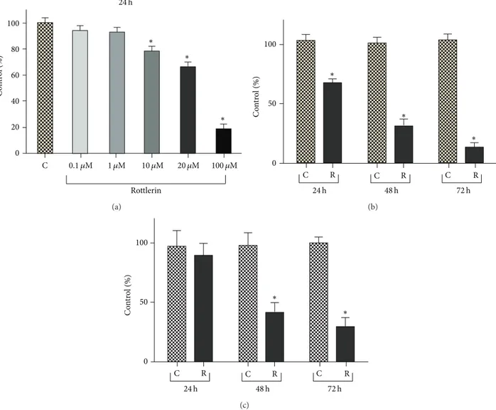
Figure 1: Rottlerin inhibits proliferation of Sk-Mel-28 melanoma cells. (a) Rottlerin treatment for 24 h reduced cell density, evaluated by the
SRB assay, in a dose-dependent manner. (b) Time-dependent decrease of cell density after 24–72 h exposure to 20𝜇M Rottlerin. Results are
the means± SD of at least three independent experiments in triplicate and expressed as % of control. (c) Cell viability, evaluated by Trypan
blue staining, after 24–72 h exposure to 20𝜇M Rottlerin. The percentage viability (live cell count/total cell count) was calculated and expressed
as % of control. Results are the means± SD of three independent experiments. Controls were exposed to DMSO alone;∗𝑃 < 0.05.
2.4. Western Blotting Analysis. Cell extracts, each
contain-ing 30–40𝜇g of total protein, were resolved on 12% SDS-polyacrylamide gel. Proteins were electrotransferred onto nitrocellulose membranes which were blocked by 5% nonfat dry milk in TBS containing 0.1% Tween 20 for 1 h at room temperature. Then, the blots were probed with primary polyclonal antibodies overnight at 4∘C.
After washing, horseradish peroxidase-conjugated IgG was added for 1.5 h at room temperature.𝛽-actin was used as a loading control. The blots were developed by the ECL and exposed on photographic film. Immunoreactive bands were quantified by Image J analysis software.
2.5. Nuclear p65 NF-𝜅B Immunostaining. Through the
canonical NF-𝜅B signaling pathway, IKK phosphorylates I𝜅B proteins, thus allowing the release of free p50/p65 NF-𝜅B dimers that translocate to the nucleus. For
immunocytochemical experiments, cells were cultured and treated on sterile glass coverslips placed in a 24-well microplate. Cells were washed twice with PBS and fixed with 4% paraformaldehyde for 40 min at room temperature (RT). After washes with PBS, 0.5% Triton X-100 (v/v in PBS) was added for 5 min at 4∘C and then washed with PBS. Cells were incubated with blocking buffer (1% BSA in PBS) for 45 min prior to incubation for 1 h at RT with primary antibody anti-p65, diluted 1 : 100 in PBS with 0.5% BSA. Cells were washed and incubated for 45 min with FITC-conjugated secondary antibody diluted in PBS containing 0.5% BSA. Cells were washed and incubated for 10 min in DAPI diluted 1 : 10.000 in PBS. Finally, the coverslips were mounted on glass slides using DABCO. Images were acquired on a Carl Zeiss Axioplan 2 imaging microscope using an AxioCam HR CCD camera and AxioVision 3.1 software (Carl Zeiss, G¨ottingen, Germany).
SQSTM1/p62 PARP 0.2 0 0.4 0.6 0.8 1.0 C 0.2 0 0.4 0.6 0.8 1.0 PARP/ 𝛽-ac tin 6 h 18 h 24 h C 6 h 18 h 24 h C 6 h 18 h 24 h SQS TM1/p62 /𝛽-ac tin Rottlerin 20 𝜇M Rottlerin 20 𝜇M Rottlerin 20 𝜇M 𝛽-actin
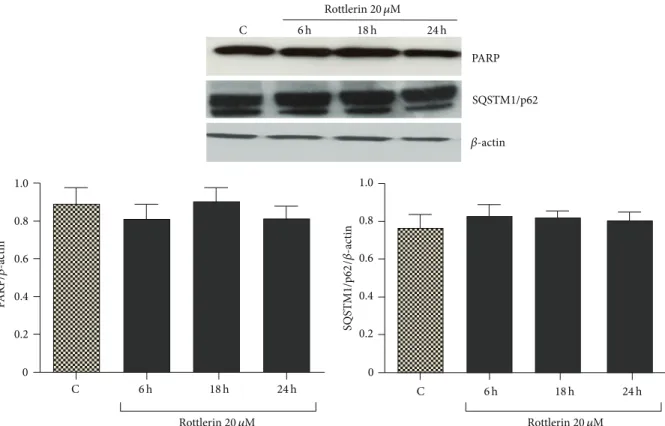
Figure 2: Effect of Rottlerin on apoptosis and autophagy. Western blotting of PARP and SQSTM1/p62 in Sk-Mel-28 cell lysate after 6–24 h
treatment with 20𝜇M Rottlerin. 𝛽-actin was used as loading control. A representative western blot of three independent experiments is
shown. Densitometry results are expressed as protein/𝛽-actin ratio.
2.6. Preparation of Rottlerin-Cyanogen Bromide- (CNBr-) Activated Sepharose 4B Complex. The procedure was
per-formed following the previously reported protocol [27]. The CNBr-activated Sepharose 4B beads (0.3 g) were swelled in 1 mM HCl for 30 min and washed in coupling buffer (0.1 M NaHCO3 (pH 8.3) and 0.5 M NaCl). Rottlerin (2 mg), dissolved in 500𝜇L of coupling buffer, was added to CNBr-Sepharose 4B beads and rotated end-over-end overnight at 4∘C. The beads were subsequently transferred to 0.1 M Tris-HCl buffer (pH 8.0) and again rotated end-over-end overnight at 4∘C. Finally, the Rottlerin-conjugated CNBr-activated Sepharose 4B was washed with three cycles of alter-nating low pH (0.1 M acetate buffer (pH 4.0) containing 0.5 M NaCl) and high pH (0.1 M Tris-HCl (pH 8.0) containing 0.5 M NaCl) buffers.
2.7. Ex Vivo Pull-Down Assay. For the ex vivo pull-down
assay, a total of 500𝜇g of Sk-Mel-28 protein extract was incu-bated with 100𝜇L (50% slurry) of Rottlerin-CNBr-conjugated Sepharose 4B (or CNBr-conjugated Sepharose 4B alone as a control) beads in reaction buffer (50 mM Tris-HCl pH 7.5, 5 mM EDTA, 150 mM NaCl, 1 mM DTT, 0.01% Nonidet P-40, 2𝜇g/mL bovine serum albumin, 0.02 mM PMSF, 1x protease inhibitor cocktail). After incubation, with gentle rocking for 3 h at room temperature, the beads were washed three times with a buffer containing 50 mM Tris-HCl (pH 7.5), 5 mM EDTA, 150 mM NaCl, 1 mM DTT, 0.01% Nonidet P-40, and 0.02 mM PMSF. The proteins of interest, bound to the beads,
were eluted with SDS loading buffer and analysed by western blotting with specific antibodies.
2.8. Statistical Analyses. Values are expressed as the
mean± SD. Student’s 𝑡-test was used to determine statistical significance with a threshold of𝑃 values less than 0.05.
3. Results
3.1. Effect of Rottlerin on Sk-Mel-28 Cells Proliferation. As
shown in Figure 1(a), the cell density, evaluated by SRB assay after 24 h of treatment, decreased progressively with increasing Rottlerin doses and became statistically significant starting from 10𝜇M Rottlerin (78%). A cell number reduction to 66% was observed at the dose of 20𝜇M and an almost total cell loss was obtained with 100𝜇M Rottlerin (18%). Therefore, the dose of 20𝜇M was used for the time course evaluation.
As shown in Figure1(b), a time-dependent decrease in cellularity was observed, with a modest cell recovery of 26% at 48 h and a very low recovery (15%) at 72 h. Therefore, the dose of 20𝜇M and the time exposure of 24 h, which yielded a mean of 68% cell recovery, were chosen for the subsequent molecular analyses on cell extracts.
No evident cell detachment was observed after 24 h but, as shown in Figure1(c), circa 13, 60 and 70% cell death were revealed by the Trypan blue exclusion test, performed on both adherent and floating cells after 24, 48, and 72 h, respectively.
C 6 h 18 h 24 h Rottlerin 20 𝜇M
Figure 3: Effect of Rottlerin on NF-𝜅B nuclear translocation. Cells were treated with 20 𝜇M Rottlerin for 6–24 h, stained with anti-p65 NF-𝜅B subunit antibody, and revealed with FITC-conjugated secondary antibody. The merge images of anti-p65 labeling (green) and nuclear staining with DAPI (blue) show that both stainings overlap in the control and after 6 h of Rottlerin treatment, while the p65 expression is restricted in the cytosol after 18–24 h (original magnification 50x). Representative images of two independent experiments.
3.2. Rottlerin Cytotoxicity on Sk-Mel-28 Cells. Sk-Mel-28
cells, exposed to 20𝜇M Rottlerin for 24 h, did not display signs of apoptosis and autophagy, as deduced by the lack of PARP cleavage (a marker of apoptosis) and SQSTM1/p62 degradation (a hallmark of autophagic flux) (Figure 2). The observed decrease in cell number is therefore mainly ascribable to inhibited proliferation.
3.3. Effect of Rottlerin on NF-𝜅B Nuclear Translocation. In
Figure3, it is possible to appreciate that in untreated cells and in cells exposed to 20𝜇M Rottlerin for 6 h, the fluorescence is distributed between the cytoplasm and the nucleus, whereas after 18–24 h of Rottlerin treatment, the fluorescence is confined to the cytoplasm. This result, in agreement with previous findings [24,25], indicates that Rottlerin interferes with the basal NF-𝜅B activation process also in Sk-Mel-28 cells.
3.4. Effect of Rottlerin on Cyclin D1 and p21 Levels. Following
Rottlerin exposure, the levels of cyclin D1 dropped to unde-tectable levels after only 6 h of treatment (Figure4), an effect already observed in other cell types [24,28,29].
In agreement with other studies on Sk-Mel-28 cells [13,
14], the basal levels of the cell-cycle inhibitor p21/Cip1 were undetectable by western blotting. The protein expression did not change during the 6–24 h of Rottlerin treatment (Figure4).
3.5. Effect of Rottlerin on ERKs and Akt Activation. As shown
in Figure5(a), phospho-ERKs levels were markedly increased after 6–24 h of 20𝜇M Rottlerin treatment. Increased ERK phosphorylation was not accompanied by enhanced down-stream signaling, as evidenced by the decrease of phospho-p90RSK1, the cytoplasmic target of ERK (Figure5(b)). Con-versely, Rottlerin did not significantly modify the phospho-Akt levels (Figure5(c)).
3.6. Rottlerin Binds to ERKs. Incubation of the Sk-Mel-28
cell lysate with 2 mg Rottlerin-coupled Sepharose 4B led to the formation of a Rottlerin/ERKs complex. As shown in
C 6 h 18 h 24 h C 6 h 18 h 24 h C Cyclin D1 𝛽-actin p21/Cip1 Sk-Mel-28 MCF-7 𝛽-actin Rottlerin 20 𝜇M Rottlerin 20 𝜇M
Figure 4: Effect of Rottlerin on cyclin D1 and p21/Cip1 levels. Western blotting of cyclin D1 and p21/Cip1 in Sk-Mel-28 cell lysate
after a 6–24 h treatment with 20𝜇M Rottlerin. Western blotting
of p21/Cip1 in MCF-7 cells was used as positive control.𝛽-actin
was used as loading control. Representative western blots of three independent experiments are shown.
Figure6(a), ERKs were pulled down by Rottlerin-Sepharose beads, but not by unconjugated Sepharose beads used as negative control. Of note, Rottlerin was able to equally bind unphosphorylated and phosphorylated proteins.
The membrane stained with Ponceau S (Figure6(b)) indi-cates that while no (visible) proteins are bound to Sepharose beads, Rottlerin can bind several proteins, thus exhibiting a not fully selective behaviour, in agreement with the notion that this compound is a multitarget drug.
4. Discussion
Recently, Rottlerin has been revealed to be a promising antitumor compound for its in vitro and in vivo activity against a wide array of cancers [30,31].
p-ERK Total ERK p-ERK/t ot al ERK 2 1 0 3 ∗ ∗ ∗ C 6 h 18 h 24 h C 6 h 18 h 24 h Rottlerin 20 𝜇M Rottlerin 20 𝜇M (a) p-p90RSK1 Total p90RSK1 1.0 0.5 0 1.5 2.0 p-p90RS K 1/t ot al p90RS K1 ∗ ∗ ∗ C 6 h 18 h 24 h C 6 h 18 h 24 h Rottlerin 20 𝜇M Rottlerin 20 𝜇M (b) p-Akt Total Akt 1.0 0.5 0 1.5 p-Ak t/t ot al Ak t C 6 h 18 h 24 h C 6 h 18 h 24 h Rottlerin 20 𝜇M Rottlerin 20 𝜇M (c)
Figure 5: Effect of Rottlerin on ERKs and Akt activation. (a) Western blotting of phospho- and total ERKs, (b) phospho- and total p90RSK,
and (c) phospho- and total Akt after 6–24 h of 20𝜇M Rottlerin treatment. The blots are representative of three independent experiments.
p-ERK Total ERK Rottlerin-CNBr Sepharose 4B
CNBr-Sepharose Sk-Mel-28 cell lysate
+ + − + + − + − − (a) 50 36 64 (kDa) M 1 2 3 (b)
Figure 6: Rottlerin binds to ERKs. (a) Western blotting of total and phospho-ERKs eluted from the Rottlerin-Sepharose beads. Unconjugated Sepharose beads were used as negative control. (b) Membrane stained with Ponceau S. Lane M: molecular weight markers; lane 1: cell lysate; lane 2: unconjugated Sepharose beads; lane 3: Rottlerin-conjugated Sepharose beads. Representative of two independent experiments.
The current study shows, for the first time, that Rottlerin has antiproliferative effects in Sk-Mel-28 melanoma cells. A 24 h treatment with 20𝜇M Rottlerin caused a reduction in cell density to 68%, a significant decrease, albeit lower than that observed in other cancer cells, such as MCF-7 breast cancer cells [32]. No signs of apoptosis (PARP cleavage) or autophagy (SQSTM1/p62 degradation) were found after 24 h exposure, although a modest cytotoxicity (circa 13%) was revealed by Trypan blue staining. Prolonged treatment, for 48–72 h, resulted in a marked cell loss, most likely due to delayed and marked cytotoxic effects.
In line with our earlier results obtained in breast can-cer cells [24], also in melanoma cells, Rottlerin potently downregulated cyclin D1 protein levels and inhibited cell proliferation.
For a mechanistic study, since the cyclin D1 gene can be regulated by different transcription factors, such as AP-1, Sp-AP-1, and NF-𝜅B [33,34], we searched for the responsible upstream signaling pathways, focusing on ERK and Akt. It is well known that AP-1 is a major target of the ERK cascade [35] and Sp-1 can be phosphorylated by ERK in two major sites, both critical for Sp-1 transcriptional activity [36]. ERK has also an indirect effect on cyclin D1 expression, based on the ability of its cytoplasmic target, p90RSK1, to activate NF-𝜅B by phosphorylation of INF-𝜅B𝛼 on Ser-32 [37]. Also, Akt can activate NF-𝜅B, through phosphorylation of IKK on Thr-23 [38,39].
Thus, the Ras/ERK and the PI3K/Akt axes are the major pathways that regulate the cyclin D1 gene expression.
In agreement with previous studies on different cells [24,
28,29], the observed decrease of cyclin D1 was accompanied (and caused) by the inhibition of NF-𝜅B nuclear migration.
Regarding the involved upstream pathways, our results do not support a role for Akt in the Rottlerin mediated effect, neither on the NF-𝜅B activation pathway nor on the
downregulation of cyclin D1, as phospho-Akt levels (Thr 308, target of PI3K) were unaffected by the drug.
Conversely and surprisingly, Rottlerin treatment resulted in a marked and early (6 h) increase in ERK phosphoryla-tion, a finding that was in complete incongruity with the decrease of p90RSK1 phosphorylation, the inhibition of NF-𝜅B, and the lack of p21/Cip1 induction. As far as p21/Cip1 is concerned, although it is basically downregulated in Sk-Mel-28 cells, the gene is functionally active and can be induced in certain circumstances by p53-independent mechanisms [40–42]. Therefore, the apparent ERK activation resulted, paradoxically, in suppressed downstream signaling.
In the search for a plausible explanation of these results and on the basis of the recent finding that Rottlerin can inhibit protein kinases, such as the mammalian target of rapamycin (mTOR), by a direct binding [27], we applied the previously employed methodology to verify a possible physical interaction between Rottlerin and ERK. We found, by ex vivo pull-down assay, that Rottlerin complexes with and likely inhibits ERK, regardless of its phosphorylation status, thus independently from upstream signaling events, just as in the case of mTOR.
In this context, this preliminary finding might help explain the observed lack of function of phospho-ERK on one hand and expands the list of the Rottlerin-modulated signaling molecules on the other hand.
In conclusion, whatever the inhibitory mechanism, the results of the current study suggest that Rottlerin may offer a promising mono- or combined-therapeutic approach for a subpopulation of melanomas with constitutively active Ras/Raf mutants, cyclin D1 overexpression, and high NF-𝜅B activity, for which a limited number of effective drugs are currently available. Studies are in progress to verify/confirm whether Rottlerin treatment longer than 24 h is also able to kill Sk-Mel-28 cells and to assess the mode of cell death.
Conflict of Interests
The authors declare that there is no conflict of interests.
Acknowledgment
This work has received financial support from the Istituto Toscano Tumori (Grant 2010 to E. Maioli).
References
[1] H. Davies, G. R. Bignell, C. Cox et al., “Mutations of the BRAF gene in human cancer,” Nature, vol. 417, no. 6892, pp. 949–954, 2002.
[2] R. Houben, J. C. Becker, A. Kappel et al., “Constitutive activation of the Ras-Raf signaling pathway in metastatic melanoma is associated with poor prognosis,” Journal of Carcinogenesis, vol. 3, article 6, 2004.
[3] J. Yang, S. Zaja-Milatovic, Y.-M. Thu, F. Lee, R. Smykla, and A. Richmond, “Molecular determinants of melanoma malignancy: selecting targets for improved efficacy of chemotherapy,” Molec-ular Cancer Therapeutics, vol. 8, no. 3, pp. 636–647, 2009. [4] R. Kumar, S. Angelini, E. Snellman, and K. Hemminki, “BRAF
mutations are common somatic events in melanocytic nevi,” Journal of Investigative Dermatology, vol. 122, no. 2, pp. 342–348, 2004.
[5] P. M. Pollock, U. L. Harper, K. S. Hansen et al., “High frequency of BRAF mutations in nevi,” Nature Genetics, vol. 33, no. 1, pp. 19–20, 2003.
[6] G. Palmieri, M. Capone, M. L. Ascierto et al., “Main roads to melanoma,” Journal of Translational Medicine, vol. 7, article 86, 2009.
[7] R. Nazarian, H. Shi, Q. Wang et al., “Melanomas acquire resistance to B-RAF(V600E) inhibition by RTK or N-RAS upregulation,” Nature, vol. 468, no. 7326, pp. 973–977, 2010. [8] N. Wagle, E. M. Van Allen, D. J. Treacy et al., “MAP kinase
pathway alterations in BRAF -mutant melanoma patients with acquired resistance to combined RAF/MEK inhibition,” Cancer Discovery, vol. 4, no. 1, pp. 61–68, 2014.
[9] A. Sewing, B. Wiseman, A. C. Lloyd, and H. Land,
“High-intensity Raf signal causes cell cycle arrest mediated by p21𝐶𝑖𝑝1,”
Molecular and Cellular Biology, vol. 17, no. 9, pp. 5588–5597, 1997. [10] S.-J. Cozzi, P. G. Parsons, S. M. Ogbourne, J. Pedley, and G. M. Boyle, “Induction of senescence in diterpene ester-treated melanoma cells via protein kinase C-dependent hyperactiva-tion of the mitogen-activated protein kinase pathway,” Cancer Research, vol. 66, no. 20, pp. 10083–10091, 2006.
[11] V. Fortino, C. Torricelli, E. Capurro, G. Sacchi, G. Valacchi, and E. Maioli, “Antiproliferative and survival properties of PMA in MCF-7 breast cancer cell,” Cancer Investigation, vol. 26, no. 1, pp. 13–21, 2008.
[12] F. G. Haluska, H. Tsao, H. Wu, F. S. Haluska, A. Lazar, and V. Goel, “Genetic alterations in signaling pathways in melanoma,” Clinical Cancer Research, vol. 12, no. 7, 2006.
[13] T. Haapaj¨arvi, K. Pitk¨anen, and M. Laiho, “Human melanoma cell line UV responses show independency of p53 function,” Cell Growth & Differentiation, vol. 10, no. 3, pp. 163–171, 1999. [14] R. Prasad and S. K. Katiyar, “Down-regulation of
miRNA-106b inhibits growth of melanoma cells by promoting G1-phase cell cycle arrest and reactivation of p21/WAF1/Cip1 protein,” Oncotarget, vol. 5, no. 21, pp. 10636–10649, 2014.
[15] H. Wu, V. Goel, and F. G. Haluska, “PTEN signaling pathways in melanoma,” Oncogene, vol. 22, no. 20, pp. 3113–3122, 2003. [16] J. Utikal, M. Udart, U. Leiter, R. U. Peter, and G. Kr¨ahn,
“Additional Cyclin D1gene copies associated with chromosome
11 aberrations in cutaneous malignant melanoma,” International Journal of Oncology, vol. 26, no. 3, pp. 597–605, 2005.
[17] E. Maioli, C. Torricelli, and G. Valacchi, “Rottlerin and cancer: novel evidence and mechanisms,” The Scientific World Journal, vol. 2012, Article ID 350826, 11 pages, 2012.
[18] D. Zhang, V. Anantharam, A. Kanthasamy, and A. G. Kan-thasamy, “Neuroprotective effect of protein kinase C𝛿 inhibitor rottlerin in cell culture and animal models of Parkinson's dis-ease,” Journal of Pharmacology and Experimental Therapeutics, vol. 322, no. 3, pp. 913–922, 2007.
[19] I. Ohno, G. Eibl, I. Odinokova et al., “Rottlerin stimulates apop-tosis in pancreatic cancer cells through interactions with pro-teins of the Bcl-2 family,” The American Journal of Physiology— Gastrointestinal and Liver Physiology, vol. 298, no. 1, pp. G63– G73, 2010.
[20] S. P. Davies, H. Reddy, M. Caivano, and P. Cohen, “Specificity and mechanism of action of some commonly used protein kinase inhibitors,” Biochemical Journal, vol. 351, no. 1, pp. 95– 105, 2000.
[21] J.-F. Liu, M. Cr´epin, J.-M. Liu, D. Barritault, and D. Ledoux, “FGF-2 and TPA induce matrix metalloproteinase-9 secretion in MCF-7 cells through PKC activation of the Ras/ERK path-way,” Biochemical and Biophysical Research Communications, vol. 293, no. 4, pp. 1174–1182, 2002.
[22] F. Gaubert, F. Escaffit, C. Bertrand et al., “Expression of the high molecular weight fibroblast growth factor-2 isoform of 210 amino acids is associated with modulation of protein kinases C delta and epsilon and ERK activation,” The Journal of Biological Chemistry, vol. 276, no. 2, pp. 1545–1554, 2001.
[23] M. Karin, “Nuclear factor-𝜅B in cancer development and progression,” Nature, vol. 441, no. 7092, pp. 431–436, 2006. [24] C. Torricelli, V. Fortino, E. Capurro et al., “Rottlerin inhibits
the nuclear factor kappaB/cyclin-D1 cascade in MCF-7 breast cancer cells,” Life Sciences, vol. 82, no. 11-12, pp. 638–643, 2008. [25] E. Maioli, L. Greci, K. Soucek et al., “Rottlerin inhibits ROS formation and prevents NF𝜅B activation in MCF-7 and HT-29 cells,” Journal of Biomedicine and Biotechnology, vol. 2009, Article ID 742936, 7 pages, 2009.
[26] Y. Ueda and A. Richmond, “NF-𝜅B activation in melanoma,” Pigment Cell Research, vol. 19, no. 2, pp. 112–124, 2006. [27] C. Torricelli, E. Daveri, S. Salvadori et al.,
“Phosphorylation-independent mTORC1 inhibition by the autophagy inducer Rottlerin,” Cancer Letters, vol. 360, no. 1, pp. 17–27, 2015. [28] G. Valacchi, A. Pecorelli, C. Sticozzi et al., “Rottlerin exhibits
antiangiogenic effects in vitro,” Chemical Biology and Drug Design, vol. 77, no. 6, pp. 460–470, 2011.
[29] G. Valacchi, A. Pecorelli, M. Mencarelli et al., “Rottlerin: a multifaced regulator of keratinocyte cell cycle,” Experimental Dermatology, vol. 18, no. 6, pp. 516–521, 2009.
[30] V. Sharma, “A polyphenolic compound rottlerin demonstrates significant in vitro cytotoxicity against human cancer cell lines: isolation and characterization from the fruits of Mallotus philippinensis,” Journal of Plant Biochemistry and Biotechnology, vol. 20, no. 2, pp. 190–195, 2011.
[31] M. Huang, S. N. Tang, G. Upadhyay et al., “Rottlerin suppresses growth of human pancreatic tumors in nude mice, and pancre-atic cancer cells isolated from Kras(G12D) mice,” Cancer Letters, vol. 353, no. 1, pp. 32–40, 2014.
[32] C. Torricelli, S. Salvadori, G. Valacchi et al., “Alternative pathways of cancer cell death by rottlerin: apoptosis versus autophagy,” Evidence-Based Complementary and Alternative Medicine, vol. 2012, Article ID 980658, 11 pages, 2012.
[33] D. C. Guttridge, C. Albanese, J. Y. Reuther, R. G. Pestell, and A. S. Baldwin Jr., “NF-𝜅B controls cell growth and differentiation through transcriptional regulation of cyclin D1,” Molecular and Cellular Biology, vol. 19, no. 8, pp. 5785–5799, 1999.
[34] D. Nagata, E. Suzuki, H. Nishimatsu et al., “Transcriptional activation of the cyclin D1 gene is mediated by multiple cis-elements, including SP1 sites and a cAMP-responsive element in vascular endothelial cells,” The Journal of Biological Chemistry, vol. 276, no. 1, pp. 662–669, 2001.
[35] K. Balmanno and S. J. Cook, “Sustained MAP kinase activation
is required for the expression of cyclin D1, p21𝐶𝑖𝑝1and a subset
of AP-1 proteins in CCL39 cells,” Oncogene, vol. 18, no. 20, pp. 3085–3097, 1999.
[36] J. Milanini-Mongiat, J. Pouyss´egur, and G. Pag`es, “Identifica-tion of two Sp1 phosphoryla“Identifica-tion sites for p42/p44 mitogen-activated protein kinases: their implication in vascular endothe-lial growth factor gene transcription,” Journal of Biological Chemistry, vol. 277, no. 23, pp. 20631–20639, 2002.
[37] G. J. Schouten, A. C. O. Vertegaal, S. T. Whiteside et al., “I𝜅B𝛼 is a target for the mitogen-activated 90 kDa ribosomal S6 kinase,” The EMBO Journal, vol. 16, no. 11, pp. 3133–3144, 1997. [38] O. N. Ozes, L. D. Mayo, J. A. Gustin, S. R. Pfeffer, L. M. Pfeffer,
and D. B. Donner, “NF-𝜅B activation by tumour necrosis factor requires tie Akt serine-threonine kinase,” Nature, vol. 401, no. 6748, pp. 82–85, 1999.
[39] W. Ouyang, J. Li, Q. Ma, and C. Huang, “Essential roles of PI-3K/Akt/IKK𝛽/NF𝜅B pathway in cyclin D1 induction by arsenite in JB6 Cl41 cells,” Carcinogenesis, vol. 27, no. 4, pp. 864–873, 2006.
[40] G. Zeng, J. Liu, H. Chen et al., “Dihydromyricetin induces cell cycle arrest and apoptosis in melanoma SK-MEL-28 cells,” Oncology Reports, vol. 31, no. 6, pp. 2713–2719, 2014.
[41] N. M. Fofaria, S.-H. Kim, and S. K. Srivastava, “Piperine causes G1 phase cell cycle arrest and apoptosis in melanoma cells through checkpoint kinase-1 activation,” PLoS ONE, vol. 9, no. 5, Article ID e94298, 2014.
[42] J. J. Wang, W. Zhang, and B. J. S. Sanderson, “Altered mRNA expression related to the apoptotic effect of three xanthones on human melanoma SK-MEL-28 Cell Line,” BioMed Research International, vol. 2013, Article ID 715603, 10 pages, 2013.
Submit your manuscripts at
http://www.hindawi.com
Stem Cells
International
Hindawi Publishing Corporationhttp://www.hindawi.com Volume 2014
Hindawi Publishing Corporation
http://www.hindawi.com Volume 2014
INFLAMMATION
Hindawi Publishing Corporation
http://www.hindawi.com Volume 2014
Behavioural
Neurology
Endocrinology
International Journal ofHindawi Publishing Corporation
http://www.hindawi.com Volume 2014
Hindawi Publishing Corporation
http://www.hindawi.com Volume 2014
Disease Markers
Hindawi Publishing Corporation
http://www.hindawi.com Volume 2014
BioMed
Research International
Oncology
Journal of Hindawi Publishing Corporationhttp://www.hindawi.com Volume 2014
Hindawi Publishing Corporation
http://www.hindawi.com Volume 2014 Oxidative Medicine and Cellular Longevity Hindawi Publishing Corporation
http://www.hindawi.com Volume 2014
PPAR Research
The Scientific
World Journal
Hindawi Publishing Corporation
http://www.hindawi.com Volume 2014
Immunology Research
Hindawi Publishing Corporation
http://www.hindawi.com Volume 2014
Journal of
Obesity
Journal ofHindawi Publishing Corporation
http://www.hindawi.com Volume 2014
Hindawi Publishing Corporation
http://www.hindawi.com Volume 2014 Computational and Mathematical Methods in Medicine
Ophthalmology
Journal ofHindawi Publishing Corporation
http://www.hindawi.com Volume 2014
Diabetes Research
Journal ofHindawi Publishing Corporation
http://www.hindawi.com Volume 2014
Hindawi Publishing Corporation
http://www.hindawi.com Volume 2014
Research and Treatment
AIDS
Hindawi Publishing Corporation
http://www.hindawi.com Volume 2014
Gastroenterology Research and Practice
Hindawi Publishing Corporation
http://www.hindawi.com Volume 2014