www.nature.com/leu
Preferential expression of the transcription coactivator HTIF1
␣ gene in acute myeloid
leukemia and MDS-related AML
D Gandini1,2, C De Angeli1,2, G Aguiari3, E Manzati3, F Lanza2, PP Pandolfi5, A Cuneo2,4, GL Castoldi2and L del Senno3,4 1Centro di Biotecnologie, Sezione di Studi Biochimici, Universita` degli Studi di Ferrara, Ferrara, Italy;2Dipartimento di Scienze Biomediche
e Terapie Avanzate, Sezione di Ematologia, Universita` degli Studi di Ferrara, Ferrara, Italy;3Dipartimento di Biochimica e Biologia
Molecolare, Universita` degli Studi di Ferrara, Ferrara, Italy;4Centro Interdipartimentale di Ricerca sul Cancro, Universita` degli Studi di
Ferrara, Ferrara, Italy; and5Molecular Biology Program, and Department of Pathology, Memorial Sloan-Kettering Cancer Center, New York,
NY, USA
HTIF1␣, a transcription coactivator which is able to mediate RAR␣activity and functionally interact with PML, is encoded by a gene on chromosome 7q32–34, which is a critical region in acute myeloid leukemias (AML). With the assumption that this gene may be related to AML, we investigated the HTIF1␣ DNA structure and RNA expression in leukemic cells from 36 M1–M5 AML patients (28 ‘de novo’ and eight ‘secondary’ to myelodysplastic syndrome (MDS)). Abnormal HTIF1␣ DNA fragments were never found, whereas loss of HTIF1␣DNA was observed in the patients with chromosome 7q32 deletion and translocation, and in one case without detectable chromosome 7 abnormality. HTIF1␣RNA was found in acute myelocytic leu-kemic blasts, and was almost undetectable in normal mono-nuclear cells. The expression varied among the patients: higher in M1 to M3 subtypes, with the highest values in M1; low levels were constantly observed in M4 and M5 AML. In addition,
HTIF1␣ was significantly overexpressed in MDS-related AML (MDR-AML), but not in MDS. We also found that HTIF1␣ expression was high in myeloid cell lines. In myeloblastic HL60 and promyelocytic NB4 cells, induced to differentiate along the monocytic–macrophage pathway by TPA or vitamin D3, HTIF1␣ expression decreased, whereas it was maintained at high levels on induction to granulocytic differentiation by RA or DMSO. In K562 cells, HTIF1␣ RNA levels did not change after hemin-induced erythroid differentiation. These results suggest that
HTIF1␣could play a role in myeloid differentiation, being dis-tinctly regulated in hematopoietic lineages.
Leukemia (2002) 16, 886–893. DOI: 10.1038/sj/leu/2402452
Keywords: TIF1alpha; gene expression; cell differentiation; AML;
MDS; myeloid cell lines
Introduction
Regulation ofgene expression is a complex multi-step process that requires the concerted action ofmany factors. Among these, the transcriptional intermediary factors (TIFs), also designated as co-activators and co-repressors, interact with sequence-specific transcription factors leading to modulation ofthe chromatin structure and/or activity ofthe transcriptional machinery.1,2The human TIF1␣ (HTIF1␣, human transcription
intermediary factor 1␣) is a nuclear protein kinase,3a TIF
fam-ily member, that has been identified via its interaction with liganded nuclear receptors, including the retinoid (RXRs and RARs) receptors.4–6 HTIF1␣ protein contains an N-terminal
RBCC (RING-B-box-coiled-coil) protein–protein interaction motif,4,7which is present in many proteins with diverse
func-tions,8,9including PML, the protein fused to RAR␣ in the APL
subtype ofmyeloid leukemia.10–13It has recently been
demon-strated that in the myeloid differentiation PML acts as a ligand-dependent coactivator ofRAR␣/RXR␣, by interacting with Correspondence: L del Senno, Dipartimento di Biochimica e Biologia Molecolare, Via L. Borsari 46, 44100 Ferrara, Italy; Fax: 39 0532202723
Received 7 November 2000; accepted 20 December 2001
TIF1␣ and CBP, in a RA-dependent transcription complex.14
In addition, HTIF1␣ like PML, is characterized by tumor and growth suppression activities,13–14 and is a partner in fusion
genes: with a truncated B-Rafgene in murine hepatocellular carcinoma,4,15 and with a truncated RET gene in thyroid
papillary carcinoma.16
Recently, the HTIF1␣ gene has been mapped to chromo-some 7q32–34,17which is a critical region ofgene loss
asso-ciated with myeloid disorders.18–21In particular, whole or
par-tial losses ofchromosome 7 are strongly associated with a dismal outcome ofacute myeloid leukemia (AML).22,23 This
is a heterogeneous disease, which is caused by a variety of pathogenetic mechanisms and characterized by variability in the degree of the commitment and differentiation of the myeloid lineage.23AML may derive from a preleukemic
con-dition, the myelodysplastic syndrome (MDS).24 MDS related
AML (MDR-AML) and true ‘de novo’ AML (TDN-AML) share morphological and clinical characteristics, but they differ in other aspects, such as incidence throughout life, hematopo-ietic lineage involvement, cytogenetic features, response to therapy and incidence ofrelapse which is higher in MDR-AML.24
The most frequent chromosomal abnormalities identified in MDS are isolated losses of5q, 20q and Y chromosome, and a variety ofchromosome 7 abnormalities (monosomy 7 and rearrangements of7q22, 7q32 and 7q36),25,26which are
asso-ciated with a poor prognosis.27Specific oncogene or a tumor
suppressor gene located on 7q could play a role in MDS25,26
and, in general, in myeloid diseases. Therefore, because of the location on chromosome 7q, and the above mentioned characteristics shared with PML, HTIF1␣ could be one ofthe genes involved in the pathogenesis ofAML. In this study the
HTIF1␣ DNA structure and RNA expression have been
inves-tigated in both MDR-AML and TDN-AML patients and in leukemia cell lines.
Materials and methods
Patients and cell lines
Samples ofbone marrow (BM, 36) and peripheral blood (PB, 13) were obtained from 36 AML patients, 28 of whom presented with de novo AML, and eight who had evolved from an antecedent MDS. This series included 16 males (ages 30–79, mean 64) and 20 females (ages 32–88, mean 61). BM and PB were analyzed in control cases (four healthy males and four females) and in 10 patients with MDS (four males and six females). Diagnosis was based on morphological, clinical and immunological criteria. Conventional cytogenetic analysis was performed in 34 AML cases showing the follow-ing clonal abnormalities: chromosome 7 abnormalities in nine
HTIF1␣ gene expression in human AML D Gandiniet al
887 cases (five with MDR-AML); t(15;17) in five;+13 in three; t(8;
21) in two;+11, 5q−, 9q−, t(6;11), i(Xq) in one patient each. There were normal karyotypes in six patients and no mitoses in the remaining four.
Cell lines (myeloblastic HL-60, promyelocytic NB4, erythroleukemic K562, B-lymphoid Raji and EBV-immor-talized lymphoblastoid) were studied. The differentiating agents used were 16 nM 12-O-tetradecanoylphorbol 13-ace-tate (TPA) and 100 nM 1,25-dihydroxyvitamin D3 (D3) (monocyte/macrophage pathway), 1.25% dimethylsulphoxide and 1Mall-trans retinoic acid (RA) (granulocytic pathway),
and 30 M hemin (erythroid pathway), as previously described.28,29Cell differentiation into neutrophils and
mono-cytes was assessed by morphology. Macrophage-like differen-tiation induced by TPA was assessed by adhesion ofthe cells to the tissue culture plastic and by the morphology ofthe adherent cells.
Figure 1 Southern blot analysis of HTIF1␣ DNA in AML. (a) Schematic representation of HTIF1␣ cDNA. E: EcoRI restriction sites. The a and b arrows indicate the previously reported breakpoint sites.15,16 The HPRR-1 probe was produced by RT-PCR using primers HPRR-F2:
5⬘-AAGACCACACTGTCAGACAG-3⬘ and HPRR-R2: 5⬘-ATTCTGTTCCACGACAGGAT-3⬘ on reverse-transcribed HL60 cell total RNA. HPRR-2 is a cDNA probe.7(b) DNA from PB (asterisk) and BM samples of leukemic (AML) and non-leukemic (C) cases were restricted with BamHI or
EcoRI enzymes. Patient numbers correspond to cases indicated in Figure 2a and Table 1, with the exception ofNo. 34, an AML M1 patient showing a –7 chromosome abnormality. After gel fractionation and blotting of the DNA, filters were hybridized with HPRR probes and rehy-bridized with the bcl-1 pB probe for the major cluster region, as previously described.32The amount of HPRR DNA was related to that oftotal
DNA and bcl-1 DNA. Reduction in HPRR-specific bands was found in cases 34, 11, 8 and 32. In the latter case a marked reduction in HTIF␣ DNA was detected with the HPRR-1, and not with the HPRR-2 probe. Similar restriction patterns were observed in PB and BM samples when available, as shown for case 11.
DNA and RNA extraction and analysis
Experiments were carried out on cultured cells and patient blast cells obtained by Ficoll separation. Nucleic acid extrac-tion, Southern and Northern blotting were performed as pre-viously described.30–32Filters were hybridized with the probes
shown in Figure 1a: HPRR-1 (a 750 bp RT-PCR-derived probe) and HPRR-2 (a 1.5 kb EcoRI-EcoRI cDNA fragment).7 The
amount ofRNA was assessed by hybridization with a GAPDH gene probe,32 and level ofgene expression was determined
by densitometric analysis. TIF1␣ expression values were defined by the TIF1␣/GAPDH ratio.
Statistical analysis
Data are expressed as the mean ± s.e.m. Statistical analysis was performed using the unpaired and paired Student’s t-test, as applicable.
888
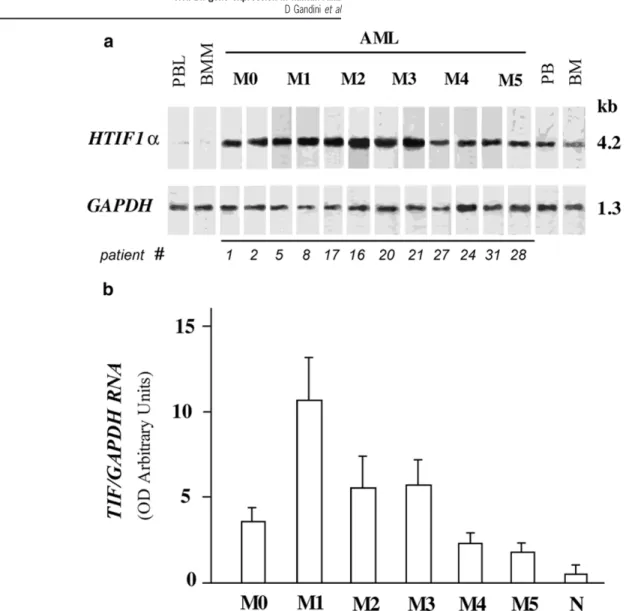
Figure 2 HTIF1␣ gene expression in AML patients. (a) Northern blot of HTIF1␣ RNA in BM samples from AML patients. Total RNA from two cases for each FAB subtype are shown. Patient numbers correspond to those indicated in Figure 1b and Table 1. PBL, normal peripheral blood lymphocytes; BMM, normal bone marrow mononuclear cells; BM, whole normal bone marrow; PB, whole normal peripheral blood. After hybridization with the32P-labelled HPRR-2 probe, filters were rehybridized with the32P-labelled GAPDH probe (see Methods). (b) Mean
± s.e.m. of HTIF1␣ expression level in each AML FAB subtype. Only BM samples were included in this analysis.
Results
Loss of HTIF1
␣
DNA in AML with abnormal chromosome 7qThe HTIF1␣ gene was analyzed in 23 AML cases by Southern blotting, after restriction with BamHI and EcoRI, and hybridiz-ation with the HPRR-1 and HPRR-2 c-DNA probes (Figure 1a). The probes cover 75% ofthe HTIF1␣ coding sequences including the two breakpoints associated with previously reported TIF␣ gene rearrangements.4,15,16No rearrangements
were detected within the 50 kb region ofthe HTIF1␣ ana-lyzed. However, weaker bands were found in patients with monosomy 7 or deletion of7q32 consistent with loss ofone allele as expected, but also in cases with translocation involv-ing 7q32 band (cases 34, 11 and 8, respectively, see Figure 1b). No abnormality was observed in the case with a translo-cation involving 7q22 (patient 12). In case 32, an AML M5 patient with no detectable abnormality ofchromosome 7, a marked reduction in TIF␣ DNA was detected with the
HPRR-1 probe, and not with the HPRR-2, suggesting a partial loss
ofthe gene.
Variable HTIF1
␣
RNA expression in AML patients Considering the possibility that small mutations, which are undetectable by Southern blotting, could lead to abnormal transcripts, HTIF1␣ expression was analyzed by Northern blotting in 33 AML cases and in a series ofhemopoietic cell lines. Only the expected transcript ofapproximately 4.2 kb in size was detected with both probes in all the AML cases (Figure 2a). Surprisingly, the HTIF1␣-specific band was almost undetectable in mononuclear cells obtained by separation over a Ficoll gradient ofnormal peripheral blood (PB) and bone marrow (BM) samples. However, it was present in nor-mal whole (PB) and (BM) samples containing a majority of neutrophilic cells.In the AML patients, HTIF1␣ expression was specifically related to the FAB subtype (Figure 2b and Table 1): the mean
HTIF1␣ gene expression in human AML D Gandiniet al
889
Table 1 Comparison of HTIF1␣ RNA expression in TDN-AML and MDR-AML
AML MDS Nor (1–10) (2–8) M0 M1 M2 M3 M4 M5 (1–4) (5–10) (11–18) (19–23) (24–27) (28–33) TDN-AML 1.7 2.5 1.7d 2.2 1.1 0.7 0.0 0.0 (25) 2.2 10.4 2.2t 3.8 2.2 1.3 0.3 0.0 4.3 11.3 2.5 6.6 3.3 2.6 0.5 0.0 2.6d 6.8 2.8 2.6 0.5 0.5 6.0 10.6 0.7 0.5 6.5 1.0 1.1 1.4 1.5 1.6 1.8 2 MDR-AML 6.0m,d,t 6.1t 5.5 1.2 (8) 16.5m 18t 3d 23 Total 3.5± 0.8 11.6± 2.6 5.6± 1.8 6.0± 1.4 2.3± 0.5 1.9± 0.3 (P= 0.004) 0.98 ± 0.22 0.51 ± 0.2 (Mean± s.e.m.) Total TDN-AML 2.7± 0.6 8± 2.2 3.5± 0.7 6.0± 1.4 2.3± 0.5 1.8± 0.4 4± 0.6 (Mean± s.e.m.) MDR-AML 6.0 15.2± 3.9 11.7± 4.3 2.1± 0.6 9.9± 2.6 (Mean± s.e.m.) TDN-AMLvs 0.4 MDR-AML (P= 0.002)
Values represent the amount ofHTIF1␣ RNA in mononuclear cells from patients (AML, MDS) and control subjects (Nor) that are indicated by numbers in italics (see Methods for technical details). M1–M5 indicate FAB AML subtypes. Patient cases 3, 6–12, 15, 17, 18, 20, 22– 24, 28, 30, 32, have been analyzed by Southern blotting.m,−7;d, del(7q),t, translocations involving 7q. MDS patients include one RAEBt
(1) and two CMML (2, 3).
value ofexpression was higher in M1 (11.6± 2.6), five times lower in M5 subtype (1.9 ± 0.3) (P = 0.003), and 20 times lower in normal samples. In BM and PB samples from 11 patients, HTIF1␣ displayed a comparable expression. No association with patient age or sex was observed. The data indicate a decrease in HTIF1␣ expression with monocytic dif-ferentiation, in agreement with the undetectability of HTIF1␣ expression in normal mononuclear cells, including monocytes and lymphocytes.
Different HTIF1
␣
RNA expression in myeloid and lymphoid cell linesThe detection of HTIF1␣ RNA in myeloid cells led us to inves-tigate whether it was related to a specific cell lineage, and we examined the gene expression in myeloid and lymphoid cell lines. A marked HTIF1␣ expression was observed in myeloid HL-60, NB4 and K562 cells, a clearly lower level in B lymph-oid Raji cells and an almost undetectable level in two B-lym-phoblastoid cell lines (LCL 1 and 2) (Figure 3). This result con-firms a prevalence of HTIF1␣ expression in myeloid vs B-lymphoid cells.
HTIF1
␣
RNA is markedly down-regulated in TPA-induced macrophage differentiationThe possible relation between HTIF1␣ expression and cell dif-ferentiation was investigated in cells treated with differen-tiating agents (Figure 4a and b).
Figure 3 HTIF1␣ gene expression in hematopoietic cell lines. Northern blot of HTIF1␣ RNA in various hematopoietic cell lines. Acute promyelocytic leukemia, NB4; acute myeloblastic leukemia, HL60; chronic myeloid erythro-leukemia, K562; B lymphoid cells: Raji; B-lymphoblastoid cell line: LCL1 and LCL2. See Methods and legend ofthe previous figure for technical details.
Treatment ofHL60 cells for 48 h with 16 nMTPA, that com-mits the cells to macrophage-like lineage, markedly reduced
HTIF1␣ mRNA levels (Figure 4a). Compared to untreated
cells, HTIF1␣ RNA values found in adherent and non-adher-ent cells were 10 and three times lower, respectively (a and
n a ofFigure 4a); similar, but less pronounced results were
obtained in NB4 cells (Figure 4b).
Treatment ofHL60 and NB4 cells for 5 days with 100 nM
D3, that is reported to commit HL60 cells to monocytic differ-entiation,28,33,34 induced only minimal changes in
890
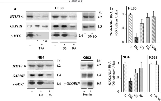
Figure 4 Decrease in HTIF1␣ expression upon monocytoid–macrophage differentiation of HL60 and NB4 cells. (a) Autoradiograms of a Northern blot oftotal RNA from HL60, NB4 and K562 cells, untreated (−) and treated (+) with differentiating agents (TPA, D3, RA, DMSO, Hemin, as described in Materials and methods). Filters were hybridized with the HTIF1␣ HPRR-2 probe and subsequently re-hybridized with GAPDH probe, c-myc or␥-globin probes, as previously described.32,29a and n a indicate adherent and non-adherent cells. (b) Mean values
(± s.e.m.) of HTIF1␣/GAPDH RNA ratios in treated and untreated cells, from at least three different experiments.
control values). c-myc RNA levels, that were down-regulated by TPA, remained unchanged after treatment with D3, as shown in Figure 4a. Nevertheless, in both cell types, a reduction in HTIF1␣ RNA levels was observed in treated cells. Treatment ofHL60 and NB4 cells for 4 days with 1MRA, that commits cells to granulocyte-like differentiation,28,35,36
produced significant morphological changes (about 50–70% ofthe cells aggregated to form clusters), a reduction in cell proliferation (55 and 45% of control) and disappearence of c-myc RNA levels. However, a significant reduction in HTIF1␣ RNA levels, relative to GAPDH RNA levels, was not observed when compared with untreated control cells (Figure 4b). Treatment ofHL60 cells for 6 days with DMSO inducing gra-nulocyte differentiation, did not produce a significant reduction in HTIF1␣ RNA levels (Figure 4a and b). In K562 cells induced to erythroid differentiation by treatment with hemin for 5 days, as previously reported,29HTIF1␣ RNA levels
remained unchanged while a marked increase in ␥-globin mRNA levels was observed (Figure 4a).
High HTIF1
␣
RNA expression in MDR-AMLIn order to evaluate whether the different level of expression in AML subtypes could correlate with the clinical history of the patients, we compared HTIF1␣ expression in TDN-AML
vs MDR-AML cases (Table 1). A total mean value of4.0± 0.6
was found in TDN-AML, while a markedly higher mean value of9.9± 2.6 was found in MDR-AML, with a statistically sig-nificant difference between the subgroups (P= 0.002). Thus, overexpression of HTIF1␣ appears to correlate mostly with the MDR-AML. In the 10 cases ofMDS analyzed, levels ofHTIF1␣
expression were comparable to those ofcontrol cases (Nor, Table 1).
Since the HTIF1␣ gene has been localized on chromosome 7q32–34, we considered the possible association between
HTIF1␣ expression and chromosome 7 abnormalities,
analyz-ing eight ofthe nine patients presentanalyz-ing the involvement of chromosome 7 (superscript letters in Table 1), including five MDR-AML. The abnormalities were prevalent in MDR-AML cases, but also present in TDN-AML cases with low levels of
HTIF1␣ RNA expression (Table 1). Thus, high levels of HTIF1␣
expression seem to be a feature of the MDR-AML population, regardless ofthe presence of7q abnormality.
Discussion
TIF1␣ is a chromatin-associated factor with a conserved struc-tural motif, the RBCC domain,4,8present in fusion
oncoprote-ins like PML.9 Because ofthis structural similarity and the
ability ofTIF1␣ to interact with RAR␣ and PML,14that have
been demonstrated to be critical proteins in growth inhibition and myeloid differentiation,37–38we investigated the possible
involvement of HTIF1␣ in AML.
Southern analysis in 23 AML cases revealed no rearrange-ments of HTIF1␣, at least in the regions previously found rearranged in a papillary thyroid carcinoma16and
hepatocel-lular carcinoma.15 Nevertheless, the group ofinvestigated
cases was not large enough to exclude the occurrence of
TIF1␣ rearrangements in AML, considering that well known rearrangements, such as those involving DEK-CAN, occur at a frequency lower than 4%.39 The reduction in the level of
HTIF1␣ gene expression in human AML D Gandiniet al
891 chromosome 7q abnormalities, including loss of7q, deletions
and translocation at 7q32 and not at 7q22, in accordance with the mapping of HTIF1␣ gene to the 7q 32–34 region.17 The
partial loss ofthe HTIF1␣ DNA 5⬘ region observed in a MDR-AML case with no apparent chromosome 7 abnormalities, would indicate the occurrence ofa submicroscopic chromo-some 7 lesion involving the gene. This is in accordance with results previously reported in AML and MDS patients, demon-strating loss of7q by PCR-based techniques19,26and hidden
translocations of7q by spectral karyotyping,25 otherwise not
revealed by conventional cytogenetics. The case presented here remains to be analyzed in more detail. In spite ofthis sample, disruption of HTIF␣ seems to be a rare event in AML patients.
Although DNA analysis did not implicate HTIF1␣ gene rearrangement as a mechanism associated with the develop-ment ofAML, the relative expression ofHTIF1␣ RNA in leu-kemic cells ofAML patients and in myeloid cell lines, suggests an important role ofthis gene in the myeloid lineage.
Previous studies have shown that TIF1␣ is expressed in vari-ous human tissues, including whole peripheral blood.17In the
mouse,40 during embryogenesis it is ubiquitously expressed
until mid-gestation, then, it remains highly expressed in developing nervous system and within the proliferating regions ofthe kidneys and teeth, whereas, in the adult, it is predominantly expressed in both the male and female gonads. No data are available on TIF1␣ expression in hematopoietic cells. Our data show HTIF1␣ to be specifically expressed in granulocytic cells, both in the PB and in the BM (Figure 2a). In our patients HTIF1␣ was found to be predominantly expressed in those leukemias reflecting an early and inter-mediate stage of myeloid differentiation (M1–M3 of the FAB classification), in M2-derived myeloblastic HL60, in M3-pro-myelocytic NB4 and chronic K562 myeloid cell lines. On the contrary, low levels ofexpression were found in Raji B-lymph-oid and EBV-transformed B-lymphoblastB-lymph-oid cell lines (see Figure 3).
The low levels of HTIF1␣ RNA expression in leukemias with predominant monocytic differentiation seem to be in accord-ance with the reduction in HTIF1␣ RNA expression found in HL60 and NB4 cells induced to differentiate down the mono-cyte-macrophage pathway. The less pronounced reduction in
HTIF1␣ RNA after D3 treatment, in comparison with the TPA
treatment, might be explained by the previously observed weak effectiveness of D3 on both leukemia cell lines;35
fur-thermore, it has been shown that D3 or TPA in NB4 cells treated individually, result in only a partial or incomplete dif-ferentiation along the monocyte–macrophage pathway, and that both are required to achieve optimal macrophage func-tion.33Accordingly, the down-regulation ofthe c-myc
proto-oncogene which parallels terminal differentiation,33,36was not
observed in D3-treated cells (Figure 4a). HTIF1␣ RNA expression was not significantly decreased when HL60 and NB4 cell lines differentiated along the granulocytic pathway after treatment with DMSO or RA. However, in NB4 cells treated with RA, down-regulation ofthe c-myc was observed, associated with the decrease in cellular proliferation and pro-gression of maturation and differentiation.
Therefore, the downregulation of HTIF1␣ expression in macrophage and not in granulocytic maturation, obtained after treatment of different cell lines with agents acting at dif-ferent molecular levels, supports the hypothesis of a differen-tial regulation of HTIF1␣ gene according to a different cell lineage.
In myeloid differentiation PML acts as a ligand-dependent
coactivator ofRAR␣/RXR␣, by interacting with TIF1␣ and CBP, in a RA-dependent transcription complex.14 The
pres-ence of PML, therefore, is crucial for the growth-inhibitory activity of RA, as well as for RA induction of myeloid differen-tiation.41Intriguingly, it has been reported that, in HL60 cells,
RA increases the level ofPML.42It is, therefore, possible that
RA could also sustain the HTIF1␣ expression to maintain the cellular homeostasis ofthe two co-receptors.
The hypothesis ofPML and HTIF1␣ co-expression is also supported by the sustained levels of HTIF1␣ RNA detected in K562 cells induced to erythroid differentiation (Figure 4). In fact, PML which has been found to functionally interact with nuclear proteins in K562 cells,43,44 appears to be highly
expressed through the erythroid pathway, as observed in differentiating hematopoietic progenitor cells.44
Thus, HTIF1␣ expression appears to be not only associated with immature proliferating cells, but also with cells deriving from precursors with specific differentiation commitments. Further studies of HTIF1␣ expression in GEMM- and GM-CFU could confirm this hypothesis.
The distinction of‘de novo’ AML from MDS-derived AML is extremely useful because, despite the fact that the two con-ditions share a number of characteristics, they differ in other aspects such as sensitivity to conventional therapy.23,24
Mor-phological, clinical and genetic differences are valuable tools for better differentiation of these two conditions. The obser-vation ofa significant correlation between MDS-derived AML and a higher level of HTIF1␣ expression, in comparison to the lower level detected in TDN-AML and MDS, is ofinterest, suggesting that HTIF1␣ expression could be a typical feature ofMDS progressing to AML and that the MDS transition to AML could be mediated by transcriptional mechanisms requiring high levels of HTIF1␣ and/or its interacting proteins. On the other hand, deregulation of HTIF1␣ expression could be associated with the abnormal DNA methylation frequently observed in AML.45–47For instance, methylation ofp15 gene
promoter has frequently been reported in AML at presen-tation,48 and in MDS it is highly associated with leukemic
transformation.49 However, it becomes difficult to reconcile
the overexpression of HTIF1␣ with DNA hypermethylation which is generally associated with gene expression silencing, unless it occurs on a hypothetical gene encoding a repressor of HTIF1␣ gene expression.
On the other hand, it has been shown that the activation of the HOX11 proto-oncogene in T-ALL occurs in the absence oftranslocation, but in association with extensive demethyl-ation ofthe proximal HOX11 promoter.50Examination ofthe
methylation status ofthe HTIF1␣ promoter sequence, when available, will be necessary to verify its methylation status in AML cases with high HTIF1␣ expression.
The association ofmyeloid leukemia with chromosomes 7−/7q−,18–22 and their involvement in MDS as a well known
negative prognostic factor,22–27supports the concept that these
chromosomal regions contain novel tumor suppression genes. In the AML patients examined here, chromosome 7 abnor-malities, although prevalent in the MDR-AML cases, did not show an invariable association with the high expression of
HTIF1␣ gene, located at 7q32–34.
In conclusion, the absence ofabnormal HTIF1␣ DNA frag-ments or transcripts suggests that gross genomic recombi-nations of HTIF1␣ do not occur frequently in AML. Neverthe-less, the differential expression in myeloid and B-lymphoid cells, and its overexpression in some AML subtypes, strongly indicate that the HTIF1␣ gene could play an important role in myeloid differentiation. Therefore, at variance with the
892
housekeeping expression previously reported,17we found that
HTIF1␣ gene shows a cell- and differentiation-specific
expression.
Acknowledgements
We would like to thank Dr Valeria Bertagnolo for kindly pro-viding us with HL60 and NB4 cells. The work is supported by MURST Cofin, AIRC coordinated grant to AC, and ex-60%. DG is a recipient ofa Cofin fellowship, CDA is a recipient of a FIRC fellowship.
References
1 Le Douarin B, vom Baur E, Zechel C, Heery D, Heine M, Vivat V, Gronemeyer H, Losson R, Chambon P. Ligand-dependent action ofnuclear receptors with potential transcriptional inter-mediary factors (mediators). Philos Trans R Soc Lond B Biol Sci 1996; 351: 569–578.
2 Remboutzika E, Lutz Y, Gansmuller A, Vonesch JL, Losson R, Chambon P. The putative nuclear receptor mediator TIF1 ␣ is tightly associated with euchromatin. J Cell Sci 1999; 112: 1671–1683.
3 Fraser R A, Heard DJ, Adam S, Lavigne AC, Le Douarin B, Tora L, Losson R, Rochette-Egly C, Chambon P. The putative cofactor TIF1alpha is a protein kinase that is hyperphosphorylated upon interaction with liganded nuclear receptors. J Biol Chem 1998;
273: 16199–16204.
4 Le Douarin B, Zechel C, Garnier JM, Lutz Y, Tora L, Pierrat P, Heery D, Gronemeyer H, Chambon P, Losson R. The N-terminal part of TIF1, a putative mediator ofthe ligand-dependent acti-vation function (AF-2) of nuclear receptors, is fused to B-raf in the oncogenic protein T18. EMBO J 1995; 14: 2020–2033. 5 Thenot S, Henriquet C, Rochefort H, Cavailles V. Differential
inter-action ofnuclear receptors with the putative human transcriptional coactivator hTIF1. J Biol Chem 1997; 272: 12062–12068. 6 Le Douarin B, You J, Nielsen AL, Chambon P, Losson R.
TIF1al-pha: a possible link between KRAB zinc finger proteins and nuclear receptors. J Steroid Biochem Mol Biol 1998; 65: 43–50. 7 Gandini D, Pandolfi PP. Cloning and characterization ofHPRR: a
gene related to PML. Blood 1994; 84 (Suppl. 1): 439a.
8 Saurin AJ, Borden KL, Boddy MN, Freemont PS. Does this have a familiar RING? Trends Biochem Sci 1996; 21: 208–214. 9 Borden KL, Boddy MN, Lally J, O’Reilly NJ, Martin S, Howe K,
Salomon E, Freemont PS. The solution structure ofthe RING finger domain from the acute promyelocytic leukaemia proto-oncogene PML. EMBO J 1995; 14: 1532–1541.
10 Longo L, Pandolfi PP, Biondi A, Rambaldi A, Mencarelli A, Lo Coco F, Diverio D, Pegoraro L, Avanzi G, Tabilio A, Zangrilli D, Alcalay M, Donti E, Grignani F, Pelicci PG. Rearrangements and aberrant expression ofthe retinoic acid receptor alpha gene in acute promyelocytic leukemias. J Exp Med 1990; 172: 1571– 1575.
11 Borrow J, Goddard AD, Sheer D, Solomon E. Molecular analysis ofacute promyelocytic leukemia breakpoint cluster region on chromosome17. Science 1990; 249: 1577–1580.
12 de The H, Chomienne C, Lanotte M, Degos L, Dejean A. The t(15;17) translocation ofacute promyelocytic leukemia fuses the retinoic acid receptor alpha gene to a novel transcribed locus. Nature 1990; 347: 558–561.
13 Pandolfi PP. Oncogenes and tumor suppressors in the molecular pathogenesis ofacute promyelocytic leukemia. Hum Mol Genet 2001; 10: 769–775.
14 Zhong S, Delva L, Rachez C, Cenciarelli C, Gandini D, Zhang H, Kalantry S, Freedman LP, Pandolfi PP. A RA-dependent, tumour-growth suppressive transcription complex is the target ofthe PML-RARalpha and T18 oncoproteins. Nat Genet 1999; 23: 287–295. 15 Miki T, Fleming TP, Crescenzi M, Molloy CJ, Blam SB, Reynolds SH, Aaronson SA. Development ofa highly efficient expression cDNA cloning system: application to oncogene isolation. Proc Natl Acad Sci USA 1991; 88: 5167–5171.
16 Klugbauer S, Rabes HM. The transcription coactivator HTIF1 and a related protein are fused to the RET receptor tyrosine kinase in childhood papillary thyroid carcinomas. Oncogene 1999; 18: 4388–4393.
17 Venturini L, You J, Stadler M, Galien R, Lallemand V, Koken MH, Mattei MG, Ganser A, Chambon P, Losson R, De The H. TIF1-gamma, a novel member ofthe transcriptional intermediary factor 1 family. Oncogene 1999; 18: 1209–1217.
18 Bernstein R, Philip P, Ueshima Y. Fourth International Workshop on Chromosomes in Leukemia 1982: abnormalities ofchromo-some 7 resulting in monosomy 7 or in deletion ofthe long arm (7q−): review oftranslocations, breakpoints, and associated abnor-malities. Cancer Genet Cytogenet 1984; 11: 300–303.
19 Koike M, Tasaka T, Spira S, Tsuruoka N, Koeffler HP. Allelotyping ofacute myelogenous leukemia: loss ofheterozygosity at 7q31.1 (D7S486) and q33–34 (D7S498, D7S505). Leuk Res 1999; 23: 307–310.
20 Specchia G, Cuneo A, Liso V, Contino R, Pastore D, Gentile R, Rocchi M, Castoldi GL. A novel translocation t(1;7)(p36;q34) in three patients with acute myeloid leukemia. Br J Haematol 1999;
105: 208–214.
21 Tosi S, Scherer SW, Giudici G, Czepulkowski B, Biondi A, Kear-ney L. Delineation ofmultiple deleted regions in 7q in myeloid disorders. Genes Chromosomes Cancer 1999; 25: 384–392. 22 Velloso ER, Michaux L, Ferrant A, Hernandez JM, Meeus P,
Dier-lamm J, Criel A, Louwagie A, VerhoefG, Boogaerts M, Michaux JL, Bosly A, Mecucci C, Van den Berghe H. Deletions ofthe long arm ofchromosome 7 in myeloid disorders: loss ofband 7q32 implies worst prognosis. Br J Haematol 1996; 92: 574–581. 23 Lowenberg B, Downing JR, Burnett A. Acute myeloid leukemia.
N Engl J Med 1999; 341: 1051–1062.
24 Heaney ML, Golde DW. Myelodysplasia. N Engl J Med 1999; 340: 1649–1660.
25 Mohr B, Bornhauser M, Thiede C, Schakel U, Schaich M, Illmer T, Pascheberg U, Ehninger G. Comparison ofspectral karyotyping and conventional cytogenetics in 39 patients with acute myeloid leukemia and myelodysplastic syndrome. Leukemia 2000; 14: 1031–1038.
26 Xie D, Hofmann WK, Mori N, Miller CW, Hoelzer D, Koeffler HP. Allelotype analysis ofthe myelodysplastic syndrome. Leukemia 2000; 14: 805–810.
27 Mauritzson N, Johansson B, Albin M, Rylander L, Billstrom R, Ahlgren T, Mikoczy Z, Stromberg U, Mitelman F, Hagmar L, Nils-son PG. Survival time in a population-based consecutive series of adult acute myeloid leukemia – the prognostic impact ofkary-otype during the time period 1976–1993. Leukemia 2000; 14: 1039–1043.
28 Trayner ID, Bustorff T, Etches AE, Mufti GJ, Foss Y, Farzaneh F. Changes in antigen expression on differentiating HL60 cells treated with dimethylsulphoxide, all-trans retinoic acid, alpha1, 25-dihydroxyvitamin D3 or 12-O-tetradecanoyl
phorbol-13-acetate. Leuk Res 1998; 22: 537–547.
29 Aguiari G, Piva R, Manzati E, Mazzoni E, Augello G, Chiari E, Moretti S, Neri LM, del Senno L. K562 erythroid and HL60 macro-phage differentiation down regulates polycystin, a large mem-brane-associated protein. Exp Cell Res 1998; 244: 259–267. 30 Gandini D, Moretti S, Latorraca A, De Angeli C, Lanza F, Cuneo
A, Castoldi GL, del Senno L. p53 exon 5 mutations in two cases ofleukemic mantle cell lymphoma. Cancer Genet Cytogenet 1996; 86: 120–123.
31 De Angeli C, Cuneo A, Aguiari G, Roberti MG, Piva N, Moretti S, Cavazzini P, Castoldi GL, del Senno L. 5⬘ region and exon 7 mutations ofthe TP53 gene in two cases ofB-cell prolymphocytic leukemia. Cancer Genet Cytogenet 1998; 107: 137–143. 32 De Angeli C, Gandini D, Cuneo A, Moretti S, Roberti MG, Bigoni
R, Bardi A, Castoldi G, del Senno L. Bcl-1 rearrangements and p53 5’ mutations in atypical chronic lymphocytic leukemia with t(11;14)(q13;q32). Haematologica 2000; 85: 913–921.
33 Bhatia M, Kirkland JB, Meckling-Gill KA. M-CSF and 1,25-dihyd-roxy vitamin D3 synergize with 12-O-tetradecanoylphorbol-13-acetate to induce macrophage differentiation in acute promyelo-cytic leukemia NB4 cells. Leukemia 1994; 8: 1744–1749. 34 Miyaura C, Abe E, Kuribayashi T, Tanaka H, Konno K, Nishii Y,
HTIF1␣ gene expression in human AML D Gandiniet al
893 human myeloid leukemia cells. Biochem Biophys Res Commun
1981; 102: 937–943.
35 Hu ZB, Ma W, Uphoff CC, Lanotte M, Drexler HG. Modulation ofgene expression in the acute promyelocytic leukemia cell line NB4. Leukemia 1993; 7: 1817–1823.
36 Khanna-Gupta A, Kolibaba K, Zibello TA, Berliner N. NB4 cells show bilineage potential and an aberrant pattern ofneutrophil sec-ondary granule protein gene expression. Blood 1994; 84: 294– 302.
37 Pandolfi PP, PML, PLZF and NPM in the pathogenesis ofacute promyelocytic leukemia. Haematologica 1996; 81: 472–482. 38 Grignani F, Valtieri M, Gabbianelli M, Gelmetti V, Botta R,
Luch-etti L, Masella B, Morsilli O, Pelosi E, Samoggia, P Pelicci PG, Peschle C. PML/RAR alpha fusion protein expression in normal human hematopoietic progenitors dictates myeloid commitment and the promyelocytic phenotype. Blood 2000; 96: 1531–1537. 39 Soekarman D, von Lindern M, van der Plas DC, Selleri L, Bartram
CRI, Martiat P, Culligan D, Padua RA, Hasper-Voogt KP, Hageme-ijer A, Grosveld G. Dek-Can rearrangement in translocation (6;9)(p23;q34). Leukemia 1992; 6: 489–494.
40 Niederreither K, Remboutsika E, Gansmuller A, Losson R, Dolle P. Expression ofthe transcritional intermediary factor TIF1alpha during mouse development and in the reproductive organs. Mech Dev 1999; 88: 111–117.
41 Rego E, Wang Z, Peruzzi D, He L, Cordon-Cardo C, Pandolfi PP. Role ofpromyelocytic leukemia (pml) protein in tumor sup-pression. J Exp Med 2001; 193: 521–530.
42 Pelicano L, Li F, Schindler CF, Chelbi-Alix MK. Retinoic acid enhances the expression ofinterferon-induced proteins: evidence for multiple mechanisms of action. Oncogene 1997; 15: 2349– 2359.
43 Topcu Z, Mack DL, Hromas RA, Borden KL. The promyelocytic leukemia protein PML interacts with proline-rich homeodomain protein PRH: a RING may link hematopoiesis and growth control. Oncogene 1999; 18: 7091–7100.
44 Labbaye C, Valtieri M, Grignani F, Puglisi R, Luchetti L, Masella B, Alcalay M, Testa U, Peschle C. Expression and role ofPML gene in normal adult hematopoiesis: functional interaction between PML and Rb proteins in erythropoiesis. Oncogene 1999;
18: 3529–3540.
45 Fukuhara T, Hooper WC, Baylin SB, Benson J, Pruckler J, Olson AC, Evatt BL, Vogler WR. Use ofthe polymerase chain reaction to detect hypermethylation in the calcitonin gene. A new, sensitive approach to monitor tumor cells in acute myelogenous leukemia. Leuk Res 1992; 16: 1031–1040.
46 Melki JR, Vincent PC, Clark SJ. Concurrent DNA hypermethylation ofmultiple genes in acute myeloid leukemia. Cancer Res 1999;
59: 3730–3740.
47 Toyota M, Kopecky KJ, Toyota MO, Jair KW, Willman CL, Issa JP. Methylation profiling in acute myeloid leukemia. Blood 2001; 97: 2823–2829.
48 Chim CS, Tam CY, Liang R, Kwong YL. Methylation ofp15 and p16 genes in adult acute leukemia: lack ofprognostic significance. Cancer 2001; 91: 2222–2229.
49 Tien HF, Tang JH, Tsay W, Liu MC, Lee FY, Wang CH, Chen YC, Shen MC. Methylation ofthe p15(INK4B) gene in myelodysplastic syndrome: it can be detected early at diagnosis or during disease progression and is highly associated with leukaemic transform-ation. Br J Haematol 2001; 112: 148–154.
50 Watt PM, Kumar R, Kees UR. Promoter demethylation accompanies reactivation ofthe HOX11 proto-oncogene in leuke-mia. Genes Chromosomes Cancer 2000; 29: 371–377.