UNIVERSITÀ DEGLI STUDI DI ROMA
"TOR VERGATA"
FACOLTA' DI MEDICINA E CHIRURGIA
DOTTORATO DI RICERCA IN
BIOCHIMICA E BIOLOGIA
MOLECOLARE
XXI CICLO
STRUCTURAL ASPECTS OF DYNAMIC AND DNA RECOGNITION
OF HPV-16 E2C PROTEIN
Dr. Riccardo Melis
A.A. 2008/2009
Docente Guida/Tutor: Prof. Daniel Oscar Cicero
Coordinatore: Prof. Daniel Oscar Cicero
ABSTRACT 4
OBIECTIVES OF THE RESEARCH 5
INTRODUCTION 6
NUCLEAR MAGNETIC RESONANCE IN STRUCTURAL BIOLOGY 31
MATERIALS AND METHODS 38
RESULTS 43 DISCUSSION 66 CONCLUSIONS 73 BIBLIOGRAPHY 76 CURRICULUMVITAE 85 LIST OF PUBBLICATIONS 87
ABSTRACT
The Human Papillomavirus (HPV) infection is linked to cervical cancer and represents a serious problem for women health worldwide .The existence of effective vaccines will not affect the course of infection , in particular those in developing countries and the tremendous prophylactic effect of the vaccine is counterbalanced by the lack of accessibility to most of the population. In view of these considerations, there is a need to develop specific antiviral drugs to prevent HPV infections. My PhD work was focused on the particular properties and structure-functional aspects of the E2 HPV16 DNA binding domain (E2C) E2C is the only transcription factor encoded by the viral genome and plays a central role in controlling the expression of all Papilomavirus genes and in regulating the virus life cycle .
OBIECTIVES OF THE RESEARCH
Our objective was to learn about the structure and its connection with the function of thie E2C protein using NMR spectroscopy . In particular we focused our research on
• Structural characterization of the recognition helix of E2C to explore the differences with the whole protein.
• Study about the protein dynamics and the connection with the DNA recognition mechanism.
• Characterization of the role that key residues play in determining the interaction of the protein with DNA
• The study of a monomerized version of the protein to infer the role of dimer-monomer equilibrium in the structural properties of the protein.
Human papillomavirus (HPV) is the name of a large group of viruses that includes more than 100 different strains or types. More than 30 strains of these viruses are responsible of a a sexually transmitted disease (STD) that infect the skin and mucosal areas of the body of men and women. Today approximately 20 million people are affected by genital Papilloma virus in the world. At least 50% of sexually active men and women acquire genital HPV infection at some point in their lives and, by age 50, at least 80% of women will have acquired genital HPV infection. In the U.S. country, for example, about 6.2 million Americans get a new genital HPV infection each year. The mucosal HPV types are recognized as the major cause of cervical cancer., the second most common cancer in women, and are also a causative agent of vaginal, anal, penile and head and neck cancer (Bosch et al. 2006; D'Souza et al. 2007). Moreover, the mucosal HPV types can infect also the genital areas of men, including the skin on and around the penis or anus. According to epidemiological evidence and oncogenic potential, the mucosal HPV are classified as “high-risk” and “low-risk” types (Munoz et al. 2003). To the “high-risk” group belong all oncogenic or carcinogenic HPVs types that are involved in malign tumors insorgents while HPVs belog to “low-risk” group may cause warts, or skin papillomas, which are benign (noncancerous) tumors. Actually are knowed 19 HPVs types classified as “high-risk” (types 16, 18, 26, 31, 33, 35, 39, 45, 51, 52, 53, 56, 58, 59, 66, 68a, 73, 82, 82subtype) and 13 as “low-risk” (types 6, 6a, 6b, 11, 40, 42, 43, 44, 54, 61, 70, 72 and 81) .
HPV diagnosis
Unfortunately, most HPV infections have a problematic diagnostic because the virus that lives in the skin or mucous membranes usually causes no syntomatic evidence except in some cause of visible genital warts, or insurgence of visible genital pre-cancerous changes . Therefore, most infected persons are unaware they are infected, yet they can transmit the virus to a sex partner. Rarely, a pregnant woman can also pass HPV to her baby during vaginal delivery, furthermore a baby that is exposed to HPV very rarely develops warts in the throat or voice box. Some people can present genital warts which usually appear as soft, moist, pink, or flesh-colored swellings, especially in the genital area. They can be raised or flat, single or multiple, small or large, and sometimes cauliflower shaped. They can appear on the vulva, in or around the vagina or anus, on the cervix, and on the penis, scrotum, groin, or thigh. After sexual contact with an infected person, warts may appear within weeks or months, or not at all. In these case Genital warts are diagnosed by visual inspection. Visible genital warts can be removed by medications the patient applies, or by treatments performed by a health care provider. Some individuals choose to forego treatment to see if the warts will disappear on their own. No
treatment regimen for genital warts is better than another, and no one treatment regimen is ideal for all cases. Most women are diagnosed with HPV on the basis of abnormal Pap tests. The Pap test is the primary cancer-screening tool for cervical cancer or pre-cancerous changes in the cervix, many of which are related to HPV. Also, a specific test is available to detect HPV DNA in women. The Pap test involves the collection of cells from the cervix for examination under the microscope. and may eventually lead to cancer if left untreated. Pap test results may also be described using an older set of categories called the “dysplasia scale.” Dysplasia is a term used to describe abnormal cells. Although dysplasia is not cancer, it may develop into very early cancer of the cervix. The cells look abnormal under the microscope, but they do not invade nearby healthy tissue. There are four degrees of dysplasia: mild, moderate, severe, and carcinoma in situ. Carcinoma in situ is a precancerous condition that involves only the layer of cells on the surface of the cervix, and has not spread to nearby tissues. Cervical intraepithelial neoplasia (CIN) is another term that is sometimes used to describe abnormal tissue findings. Neoplasia means an abnormal growth of cells. The term CIN along with a number (1, 2, or 3) describes how much of the thickness of the lining of the cervix contains abnormal cells. CIN–3 is considered to be a precancerous condition that includes carcinoma in situ.The results of HPV DNA testing can help health care providers decide if further tests or treatment are necessary. No HPV tests are available for men. Actually there is no "cure" for HPV infection, although in most women the infection goes away on its own. The treatments provided are directed to the changes in the skin or mucous membrane caused by HPV infection, such as warts and pre-cancerous changes in the cervix. All types of HPV can cause mild Pap test abnormalities which do not have serious consequences. Approximately 10 of the 30 identified genital HPV types can lead, in rare cases, to development of cervical cancer. Research has shown that for about 90% of women, cervical HPV infection becomes undetectable within two years. Although only a small proportion of women have persistent infection, persistent infection with "high-risk" types of HPV is the main risk factor for cervical cancer. A Pap test can detect pre-cancerous and cancerous cells on the cervix. Regular Pap testing and careful medical follow-up, with treatment if necessary, can help ensure that pre-cancerous changes in the cervix caused by HPV infection do not develop into life threatening cervical cancer. The Pap test used in U.S. cervical cancer screening programs is responsible for greatly reducing deaths from cervical cancer. For 2004, the American Cancer Society estimates that about 10,520 women will develop invasive cervical cancer and about 3,900 women will die from this disease. Most women who develop invasive cervical cancer have not had regular cervical cancer screening. The surest way to eliminate risk for genital HPV infection is to refrain from any genital contact with another individual. For those who choose to be sexually active, a long-term, mutually monogamous relationship with an uninfected partner is the strategy most likely to prevent future genital HPV infections. However, it is difficult to determine whether a partner who has been sexually active in the past is currently
infected. For those choosing to be sexually active and who are not in long-term mutually monogamous relationships, reducing the number of sexual partners and choosing a partner less likely to be infected may reduce the risk of genital HPV infection. Partners less likely to be infected include those who have had no or few prior sex partners. HPV infection can occur in both male and female genital areas that are covered or protected by a latex condom, as well as in areas that are not covered. While the effect of condoms in preventing HPV infection is unknown, condom use has been associated with a lower rate of cervical cancer, an HPV-associated disease.
HPV vaccine prospective
No HPV vaccines has been proven to provide complete protection against persistent infection with other HPV types. Therefore, about 30 % of cervical cancers and 10 % of genital warts will not be prevented by these vaccines. In addition because the vaccines will not protect against all infections that cause cervical cancer, it is important for vaccinated women to continue to undergo cervical cancer screening as is recommended for women who have not been vaccinated. Briefly the HPV vaccines work like other immunizations that guard against viral infection. The advanced hypothesis is that the HPVs unique surface components might create an antibody response that is capable of protecting the body against infection, and these components could be used to form the basis of a vaccine. These surface components can interact with one another to form virus-like particles (VLP) that are noninfectious and stimulate the immune system to produce antibodies that can prevent the complete papillomavirus from infecting cells. They are thought to protect primarily by causing the production of antibodies that prevent infection and the development of those cervical cell changes seen on Pap tests that may lead to cancer. Although these vaccines can help prevent HPV infection, it is not known if they can help eliminate existing cervical cell changes due to HPVs. Very recently, a recombinant vaccine produced by Merck & Co., Inc. (Merck), the Gardasil, was approved as prophylatic HPV vaccine. It is called quadrivalent vaccine because it protects against four HPV types (6, 11, 16, and 18). Another promising vaccine, Cervarix™, which targets HPV types 16 and 18, produced by GlaxoSmithKline (GSK), was approved by the FDA in 2007. However, they are unlikely to be introduced in the short term in developing countries, which account for 80% of the deaths due to cervical cancer (Cohen 2008). Moreover, they do not protect against infection with all high-risk types and cannot cure the millions of people that are already infected. Therefore, there is still a need for understanding the oncogenicity of papillomaviruses in more details
The E2 protein
The E2 protein is one of the eight protein expressed by HPV genome (Figure 1). E2 is required for efficient HPV replication and is generally acknowledged to play an important role in viral gene expression. However, it is important to note that despite years of intensive study, many of the biological functions of E2 are still poorly understood or indeed remain elusive. This is due in large part to difficulties associated with studying the HPV life cycle in vitro. Viral replication requires differentiation of the host epithelial cell and HPV DNA must therefore persist over an extensive period with many generations of host cell division (Doorbar et al , 2006). During this period, HPV must evade the host immune response and in fact HPV infections can persist for several months or even years before viral clearance. The small size of the papillomavirus genome means that the virus is absolutely dependent on host cell proteins in order to replicate and complete its life cycle. Viral proteins recruit a wide variety of host proteins and subvert many host cell pathways in order to achieve these ends. The expression of viral proteins must be controlled and varied as the host cell differentiates, culminating in the large-scale production of infectious viral particles. The E2 protein plays crucial roles in all of these processes. Furthermore, although a multitude of host cell transcription factors are used by the virus to control viral gene expression, the E2 protein also regulates viral transcription. Thus, E2 is a multifunctional protein that plays diverse roles in the HPV life cycle. Mutations in the viral genome that prevent the expression of the E2 protein block viral replication in cells and E2 is essential for efficient HPV DNA replication in vitro replication assays. E2 binds to the HPV origin of replication and acts to recruit the viral E1 helicase (Abbate et al , 2004). Subsequent steps in viral replication depend on the host cell DNA replication machinery and E2 has been shown to interact with several host cell proteins involved in DNA replication including Topoisomerase I (Clower et al 2006). E2 also binds to proteins involved in DNA repair including the tumour suppressor protein p53 and TopBP1 and E2 may play a role in directing the repair of damaged viral genome ( Massimi et al, 1999; Boner et al, 2002) . Recent work has shown that E2 is also important for long-term persistence of viral DNA in infected cells (You et al, 2004; Parish et al, 2006). E2 proteins bind simultaneously to the cellular proteins Brd4 and ChlR1 that attach to mitotic chromosomes and to the viral origin of replication (You et al, 2004; Parish et al, 2006). The E2 protein is thus thought to act as a tether that links HPV genomes to the host chromosomes, thereby ensuring equal segregation of viral DNA during host cell division. The host proteins that enable E2 to perform this role appear to be specific to different viral strains and/or functionally redundant. E2 functions as a transcription factor to regulate the expression of viral genes and very likely to regulate the expression of some host genes. The HPV genome contains four binding sites for E2 within a region of around 1kb known as the upstream regulatory region (URR) or long control region (LCR) (Figure 2).
The LCR controls transcription of the viral genes and contains the origin of replication. When the LCR is placed upstream of a reporter gene E2 represses transcription and the mechanisms whereby E2 can repress transcription in this context have been described in elegant detail ( Cripe et al, 1987; Tan et al, 1994; Bouvard et al, 1994; Thierry et al, 1987) the binding of E2 to two sites proximal to the transcription start site blocks the binding of cellular transcription factors to adjacent DNA sequences and thereby represses transcription initiation. More recent work has shown that the Brd4 protein also plays a role in transcriptional repression by E2 as well as viral genome segregation ( Wu et al, 2006). However, it has been claimed that E2 has no effect on viral transcription in the context of the intact viral genome (Bechtold et al, 2003). When E2 sites are placed upstream of a reporter gene and a non-cognate promoter sequence E2 is generally reported to activate transcription, hence, the description of the N-terminal domain of E2 as a transcription activation domain. However, the ability to activate transcription is not essential for HPV replication (Stubenrauch et al, 1998). Interestingly, the full-length BPV 1 E2 protein binds co-operatively to DNA fragments with multiple E2 binding sites and can mediate the formation of DNA loops (Knight et al, 1991). The E2C domain does not form these DNA loops suggesting that the N-terminal transcription/replication domains of distantly bound E2 proteins interact to form DNA loops. Splice variants of E2 protein that lack the N-terminal domain have been characterised in BPV and HPV. These E2 splice variants can repress viral transcription or at least block the functions of the full-length E2 proteins ( Barsoum et al, 1992; Stubenrauch et al, 1998 ). However, the role that these splice variants play in the HPV life cycle is not well understood. The HPV E6 and E7 genes encode oncoproteins that interact with the cellular tumour suppressor proteins p53 and pRB, respectively (Hausen et al, 2002). These interactions modulate the activities of these growth control proteins and allow completion of the HPV life cycle. During the process of viral tumourigenesis, HPV DNA from a sub-set of viral types including HPV 16 and 18 and collectively known as the high-risk HPV types, often become integrated into the host genome ( Baker et al, 1987). Integration most often results in disruption of the E2 gene but leaves the E6 and E7 genes intact (Figure 2). The outcome of integration is therefore thought to be the derepression of E6 and E7 by removal of E2. This leads to uncontrolled cell proliferation (Jeon et al, 1995), cell transformation and in due course, tumorigenesis. DNA from highrisk HPV types is found in virtually all human cervical carcinomas (Walboomers et al, 1999). In contrast, while low risk HPV types such as HPV 6 and 11 may well integrate into their host cell genomes at rates equivalent to those of high-risk HPV types, these viruses do not bring about tumourigenesis. Re-introduction of E2 can have dramatic effects on the proliferation and survival of HPV-transformed cells. BPV and HPV E2 proteins have been shown to repress transcription of the E6 and E7 oncogenes in HPV-transformed cells leading to the induction of cell senescence and apoptosis. E2 can also influence cell proliferation via direct interactions with p53 (Parish et al , 2006) and interactions with the E6
and E7 proteins (Gammoh et al, 2006; Grm et al 2005). However, the roles that these interactions play in the viral life cycle have yet to be elucidated. Finally, it is worth noting that E2 may also be important in viral clearance. Host immune responses to E2 are seen at the time of viral clearance (Bontkes et al, 1999). Furthermore, vaccination of rabbits with a recombinant adenovirus expressing the cotton tail rabbit papillomavirus E2 protein has been shown to reduce the number and size of papillomas (Brandsma et al, 2004). This suggests that E2 proteins might be useful in vaccines that target HPV infections. More broadly, E2 also represents a promising target for drug development. Drugs that prevent E2 from interacting with E1 have the potential to block HPV replication (Kasukawa et al , 1998). Similarly, drugs that prevent the DNA binding activity of E2 would also have the potential to block viral replication.
The DNA binding activity of E2
The E2 proteins bind with high affinity to specific DNA sequences found in the viral LCR. In early studies immunoprecipitation of protein-DNA complexes and DNase I foot printing were used to identify E2 binding sites within the BPV and HPV genomes (Androphy et al, 1987; Moskaluk et al, 1987). Later, electrophoretic mobility shift assays (EMSA) and DNAse I foot printing showed that the BPV E2 protein binds to many sites within the BPV genome ( Knight et al. 1989). The consensus E2 binding site is an inverted repeat tract with the sequence 5’ ACCG NNNN CGGT 3’, where the four Ns represent a 4 base pair spacer of conserved length that varies in sequence (Knight et al, 1989). The HPV E2 protein binds to just 4 sites within the HPV genome and these have an extended version of this consensus sequence: 5 AACCG NNNN CGGTT 3’ (Thain et al, 1997). Like the full-length E2 proteins, the E2C proteins recognise sequences that correspond to the consensus shown above with KDs in the region of 1-10nM (Alexander et al, 1996; Sanders et al 1994). The specificity of DNA binding thus appears to be unaffected by removal of the N-terminal domain and hinge region although the affinity for DNA is reduced (Thain et al , 1997; Pepinsky et al 1994; Hou et al 2002). The nature of the bases in the spacer region has a profound effect on the binding of some E2 proteins (Hines et al, 1998; Bochkarev et al, 1996) and this will be discussed in detail below. Whether E2 represses or activates viral transcription is thought to depend on the affinity of E2 for each of its four sites within the LCR. Binding studies have shown that in HPV 16 the E2 protein binds tightly to the most distal promoter E2 site and less tightly to the promoter proximal E2 sites (Thain et al, 1997; Sanders et al, 1994 ). This work and the earlier work described above, resulted in models in which at low protein expression levels E2 binds to the promoter distal site and activates viral transcription whereas at higher expression levels E2 binds to the promoter proximal sites to repress viral transcription. However, the hierarchy of binding site affinity is different between
HPV 16 and HPV 11 and HPV 6 suggesting that different virus types may not share this regulatory mechanism (Thain et al, 1997; Sanders et al, 1994, Dell et al, 2003). Furthermore, due to the cooperative binding of E2, the presence of diverse cellular transcription factors and the precise conditions within the cell, the occupancy of the E2 sites seen in vivo may differ from that predicted from the hierarchy of affinities measured in vitro.
The structure of E2C: defining a unique fold
Fifteen years have passed since the earliest work describing the structure of an E2C-DNA complex belonging to BPV-1 ( Hedge et al, 1998). In their pioneering paper, Hedge, Sigler and coworkers described a previously unobserved dimeric antiparallel β-barrel. Contacts with the DNA bases were exclusively observed for two symmetrically disposed α-helices, the so-called recognition helices, positioned inside two successive major grooves of the DNA. The DNA double helix is severely bent in the structure as a consequence of these interactions. Each monomer displays a β1-α1-β2-β3-α2-β4 topology, with a long loop connecting the two repeated motifs (Figure 1A). The dimeric beta-barrel is closed by intermonomer ß2-ß2 and ß4-ß4 hydrogen bonds. Remarkably, before the structure of an E2C from any other papillomaviruses was solved, a second example of the dimeric β-barrel was found in the DNA-binding domain of the Epstein-Barr virus origin-binding protein, EBNA1 (Bochkarev et al, 1996). As Figure 1 shows, the two proteins present a high degree of structural similarity. Unexpectedly, the crystal structure of the EBNA1-DNA complex showed significant differences in the way the two protein interact with the DNA (Bochkarev et al, 1996). In fact, the homologues of the E2C recognition helices appear not to be used by EBNA1 to establish contacts with the DNA. Instead, sequence-specific contacts are made by an extended chain inserted into the minor groove and a helix reaching into the major groove ( Bochkarev et al, 1996 ). Since then, both x-ray crystallography and NMR have contributed to unraveling the structural features of E2C and details of the DNA interaction mechanism. Analysis of the data contained in some of these structures has already been accomplished exhaustively in a recent review (Hegde: et al, 2002).To date, structures of E2C proteins belonging to BPV-1 (Hegde et al, 1998) high risk strains HPV-16 (Hegde et al ,1998; Nadra et al 2004 ) , HPV-31 (Liang et al, 1996; Bussiere et al 1998), HPV-18 (Kim et al, 2000) and the low risk HPV-6 (Dell et al, 2003) have been determined. In addition to the already cited BPV-1-DNA complex (Hegde et al 1992), crystal structures of HPV-18 (Kim et al, 2000) and HPV-6 (Hooley et al 2006) complexes are also available. Also, an NMR study was conducted on the interaction of HPV-16 with one of the four DNA binding sites from the HPV 16 genome (Cicero et al, 2006), although no high-resolution structure is yet available in this case.
Crystal structures of E2C domains
A common feature of all structures determined so far is that the core of the barrel is intricate, suggesting that any rearrangement of the subunits upon interaction with DNA must be subject to a large energetic penalty. A number of hydrophobic residues occupy almost completely the inner space of the barrel, including a couple of tryptophan residues that are stacked in parallel. In the case of HPV-16 and HPV-31 a molecule of water or sulfate, respectively, was observed in a crystal structure, making a bridge between the two histidine residues (Hegde et al, 1998; Bussiere et al 1998)]. HPV-6 shows a longer C-terminus after ß4, with the inclusion of an extra hydrophobic residue, Leu365, participating in the formation of the core (see Figure 1A for the structural elements). A region of the protein that shows variable behaviour is the ß2-ß3 loop. It was not observed in the crystal structures of BPV-1, HPV-16 and HPV-18 E2C free proteins, suggesting that this region is flexible, whereas it was observed for HPV-31 and HPV-6. Evidences from NMR studies indicate that the loop is highly flexible in solution for BPV-1, HPV-31 and HPV-16. For HPV-6, two proline residues and a charge interaction from Lys323 in the loop to residues Asp311 and His336 in the other subunit confer a particular rigidity to the loop, and its presence in the crystal structure reflects an ordered situation that probably is maintained in solution. One possible criterion to classify the different structures available so far is to superimpose one of the two monomers and observe the relative position of the other monomer ( Hegde et al 2002). The five known crystal structures of E2C proteins were superimposed in this way and are shown in Figure 3A. By inspection of the non-superimposed subunits, the E2 proteins can be divided into two classes: HPV-16 and HPV-31 belong to one group and HPV-18, BPV-1 and HPV-6 belong to another. There is a difference of about 4 Å in the relative position of the recognition helix of the non-superimposed subunit of HPV-16 E2 and that of BPV-1. The reason behind this differentiation is a shift in the β4-β4 hydrogen bond register, which creates the observed difference in quaternary structure . It is important to note that the E2Cs from high-risk strains HPV-16 and HPV-18 are classified as members of two distinct structural groups, suggesting that there is no direct connection between the quaternary structure of E2C. An alternative way of classifying the available structures of E2C proteins is to look at the relative orientation of the two recognition helices, rather than the two monomers. This is a sensitive way to detect changes in the relative position of the two helices, and is performed by superimposing a single recognition helix and looking for changes in the position of the other helix (Hegde et al, 1998; Hooley et al 2006). This is particularly relevant because this helix contains the only residues that make contacts with the DNA bases, and which is conserved in all known E2 proteins. According to this alignment, a much larger spread in helix position is observed for the five known free E2C proteins (Figure 3B), with a continuum disposition of the non-aligned recognition helix ranging form the most internal helix of HPV-16
until the most external HPV-18. Table 1 summarizes the differences among the structures, all referring to HPV-18 E2C. The monomers show very similar conformations, as the RMSD for backbone atoms lies between 0.8 and 1.2 Å. When comparing the overall dimer and monomer RMSDs, the division into two families becomes clear, as HPV-16 and HPV-31 E2C proteins show a significantly higher value for the entire dimer as compared to the single monomer. When looking at the position of the non-aligned recognition helix, while superimposing the other, it is clear that we have significantly different situations, with RMSDs for Cα atoms ranging form 4.7 to 11.0 A, and with interhelical angles from 8.9 to 13.9°, all referred to the non-aligned helix of the HPV-18 E2 protein. The principal conclusion from this alignment is that the proteins appear to display the two recognition helices in ways that are specific for each viral strain.
Solution structures of E2C domains
Together with crystallography, NMR has contributed to the structure determination of E2C domains. However, from a technical point of view, the description of a structure like that of the dimeric ß-barrel is a major challenge for NMR. This is so mainly because solution structure by NMR was classically derived using only short-range interproton distances, along with dihedral angles from coupling constants. Small deviations in the relative position of residues in the central core can be translated into large deviations for the two recognition helices that are positioned at the two sides of the barrel. Nevertheless, the first evidence of a shift in the hydrogen bond register in the ß4-ß4 sheet and the consequent variation of the quaternary structure came from the solution structure of HPV-31 (Liang et al, 1996). In addition to the structure description, this work also showed that both the recognition helix and the ß2-ß3 loop presented a dynamical character in solution. Although the flexibility of the latter was later corroborated by the absence of electron density in the crystal structures of 18 and HPV-16, the observation of a dynamical behavior for the recognition helix remains an exclusive contribution of NMR, as the helix shows a very defined conformation in all crystal structures. The E2C protein of BPV-1 was also studied by NMR (Veeraraghavan, et al, 1999). The difference with the crystal structure of the same protein lies in the inclusion of a stretch of 16 additional N-terminal residues. This extended version of the E2C protein is significantly more stable than the minimal domain comprising the C-terminal 85 amino acid residues. The extra N-terminal 16 residues were found to form a flap that covers a cavity at the dimer interface and may play a role in DNA binding. In recent years, the potentiality of NMR to obtain more accurate structures was significantly improved by the introduction of residual dipolar couplings (RDCs) ( Bryce Dl et al, 2005) , in which informational content from NOEs is not local but
rather can be considered long-range structural constraints. They were first used in the E2C structural field for the calculation of the HPV-16 solution structure (Nadra et al 2004). The introduction of these additional constraints improved the agreement between solution and crystal structures, with an observed RMSD of 1.2 Å for the superposition of the entire dimer. This number is substantially lower than those obtained for the same comparison of HPV-31 (1.8 Å) and BPV-1 (2.2 Å).
The structure of the DNA binding sites
The viral genome contains a number of DNA binding sites (E2-BS) for E2, ranging from four in the human strains, up to seventeen in the bovine counterpart (Figure 2). All of the E2-BS have two features in common: an inverted repeat consensus sequence of the form 5’ ACCG NNNN CGGT 3’, and the presence of a central “spacer” region of conserved length but variable nucleotide composition. The discrimination between binding sites is specific for the different virus strains; spacers rich in A/T are preferred by all the human strains. On the other hand, BPV-1 E2 protein displays no apparent spacer sequence preference (Hines et al, 1998) . This difference is reflected in the corresponding viral genomes: the HPV genomes have E2-BS with A/T-rich spacers, whereas the genomes of non-primate animal viruses (including BPV-1) have no such predominance of A/T-rich spacers ( Hedge et al 1998-2002). The affinity of the protein for a given DNA sequence can be modulated by direct contacts between amino acids and DNA base pairs (direct readout), and/or by the recognition of the intrinsic three-dimensional shape or flexibility of the DNA binding sequence (indirect readout). In order to establish the importance of the latter, it is necessary to know the starting conformational state(s) that different DNA targets adopt in solution in the absence the protein. In this respect, X-ray crystallography was used to study the structures of dodecamers representing a bovine E2-BS (Rozenberg et al, 1998) and a human E2-BS (Hizver et al, 2001) revealing significant differences between the two. In the first case, DNA bearing an ACGT central spacer shows an α-helix continuously bent toward the major groove, which for the central spacer differs markedly with the situation in the protein bound state that shows bending towards the minor groove. On the contrary, the E2-BS with an AATT spacer was found to be already bent toward the central minor groove by 9° (Hizver et al, 2001), constituting a favorable prebending of the E2-BS in the direction of the necessary deformation to bind the protein. Additional intrinsic curvature in the flanking major grooves gives rise to an overall helix axis deflection of ~10°. In contrast, the spacers ACGT and GTAC are straight ( Rozenberg et al, 1998). These features were recently reproduced by Monte Carlo simulations (Rhos et al, 2005) and suggest a model in which the intrinsic DNA shape and/or flexibility of each spacer creates a distinct energy cost for converting the intrinsic DNA
conformation to the protein-bound conformation, and thus modulates the E2 binding affinity. Experimental data derived from high-resolution crystal structures provide detailed structural information only on static conformations of E2-BS. Other techniques that estimate the curvature of the DNA in solution, like gel electrophoretic phasing ( Zimmerman et al, 2003) and cyclization kinetics measurements ( Zhang et al 2004) can provide measurements on averaged helical parameters. Using an electrophoretic phasing assay, it was possible to estimate that in solution an E2-BS with a central AATT spacer shows 18° of net curvature, whereas a sequence with the sequence ACGT shows only 1° (Zimmerman et al, 2003). With respect to the crystal structure, this technique agrees in the direction of the curvature, which goes in the expected direction to form a high-affinity complex, but the estimated value is almost doubled. As the authors stressed, however, this technique cannot distinguish between static curvature and anisotropic flexibility, which can partially accounts for the difference with the quantitative estimation of the curvature between their results and the static X-ray structure. Another attempt to correlate the intrinsic shape and mechanical properties of E2-BS with the observed affinity was carried out using a DNA cyclization method, coupled with a statistical mechanical theory ( Zhang et al, 2004). Using this approach, a number of structural parameters like roll, twist, bending flexibility and twisting flexibility were used to estimate the binding affinity of 16 sites containing different spacer sequences (Zhang et al, 2004). For 15 of these sites, the variation in affinity was predicted within a factor of 3. No attempt has been made so far to obtain a high-resolution structure of one of the E2-BS by NMR. In principle, determination of the curvature by classical NMR methods, as was already discussed for the intermonomer orientation, is difficult, and theoretical studies have demonstrated that without the aid of residual dipolar couplings, the local information content of NOEs and dihedral angles are not sufficient to accurately define the curvature of a short DNA sequence ( Vermeulen et al, 2000). The DNA oligomer best characterized in solution by NMR is, however, very similar to that of a high affinity E2-BS: the so-called Dickerson dodecamer (CGCGAATTCGCG)2 (Wu et al 2003), which differs only by the two first and two last nucleotides with one of the HPV E2-BS (ACCGAATTCGGT) studied by X-ray crystallography . By an extensive use of residual dipolar couplings and 31P chemical shift anisotropy, the final structure obtained represented a quite regular B-form helix with a global bending of ~10°, coincident to that observed for the E2-BS analogue. This work demonstrated that with this new approach DNA curvature is now accessible to high resolution NMR, and also pointed to the existence of flexibility in certain regions of the molecule, particularly the pyrimidines, by detecting the presence of rapid equilibria between C2’-endo and C3’-endo deoxyribose conformations.
The structure of E2C-DNA complexes
As already mentioned, HPV and BPV E2C proteins show distinctive cognate sequence discrimination (Ferreiro et al, 2000). One of the main sources for this differential selectivity appears to be of electrostatic nature. BPV-1 E2 displays positive charges located near the C-terminus of the recognition helices, which can help bending even of non-predeformed DNA, explaining the lower selectivity. On the other hand, HPV16 lacks this accumulation of positive charges, and this explains the greater affinity toward sequences with A/T-rich spacers, considered as prebent DNA molecules (Shatzky-Schwartz et al 1997; Stefl et al, 2004). In the case of the HPV-18 E2C protein, there is an accumulation of positive charges in the center of its DNA-interaction surface, which results in electrostatic complementary to the negative minor groove of A/T-rich sequences, contributing to its enhanced selectivity. However, this is less apparent in the HPV 6 E2C DNA-interaction surface, despite the fact that this protein is more selective towards A/T-rich spacers than the HPV 16 E2C protein. When comparing the available structures of E2C-DNA complexes using the criterion of superimposing just one of the two recognition helices, the observed spread in helix disposition is reduced with respect to the situation observed for the free proteins (Figure 3C and Table 1). Using the HPV-18 E2C complex as a reference, the non-aligned helices are disposed in a similar way (Figure 3B). This suggests that, at least for proteins belonging to the HPV-18 family, binding to DNA makes the conformations more uniform. By analyzing the differences between free and bound states it has been concluded that HPV-6 E2C experiences only slight changes upon binding a 16mer DNA fragment (with Cα displacements and interhelical angle changes of 2.5 Å and 5.6 °, respectively). This reduced adaptability of the protein can be related to the higher selectivity of this protein BPV-1 and HPV-18 E2C proteins experience slightly larger changes upon DNA binding (3.4 Å and 9.3° for HP18 E2C and 2.9 Å and 3.2° for BPV-1 E2C). Interestingly, HPV-6 bound to a 18mer DNA experiences a larger change (4.4 Å and 6.1°), leading to the highest displacement of the not-aligned recognition helix between free and bound states. The minor groove width is also different, as a wider groove was observed for BPV-1 (4.0 Å) compared to HPV-18 (2.8 Å) and HPV-6 (2.7 Å). These data indicate that slight differences have a large effect on the bending and minor groove dimensions of the DNA. One of these differences is the fact that the β2-β3 loop becomes ordered in the BPV-1 complex, establishing electrostatic interactions with the phosphate groups in the minor groove. The HPV-6 E2C complexes also show an ordered β2-β3 loop, but its conformation is very different to that observed for the BPV-1 structure, and no interaction with the DNA was detected . HPV-18 E2C shows a disordered loop in the bound state. NMR evidence was also presented about the flexibility of the β2-β3 loop of the HPV-16 E2C in the DNA complex , however, mutagenesis analysis suggests at least temporary contacts between positively charged residues of this loop
and the DNA (Ferreiro et al, 2005). To date, no high-resolution structure of the complex is available for HPV-16 E2C. However, a study was presented showing that HPV-16 displays differences in the interaction with DNA of varying lengths. A large enthalpy difference (∆∆H) of 10.0 kcal mol-1 was observed for the interaction of E2C and a 18mer or a 14mer site. This large difference may reflect a conformational change in the DNA in the two complexes, resembling the diversity found for HPV-6 E2C complexed to 16mer and 18mer DNAs. In addition, a charged residue located outside the recognition helix, K349, shows chemical shift perturbation only when a 18mer DNA is used for the interaction, but not with a 14mer duplex. The mutant K349A shows also a decreased affinity to the 18mer duplex by 1.4 kcal mol-1 per symmetric interaction. This value is similar to the effect of shortening the duplex to 14 bases, which indicates the uncovering of an additional contact between the E2C protein and the DNA outside the recognition helix and the ß2-ß3 loop, not previously observed. Figure 4 shows a model for the complex between HPV-16 E2C and an 18mer DNA, and the position of K349 very close to the C5’ of the sugar in the 5’ end of the nucleic acid. Clearly, a shorter DNA cannot exploit this contact and this will translate into lower affinity. K349 is replaced by an alanine in E2C-18, a mutation that weakens the DNA binding of E2C-16 domain, and by a proline in E2C-BPV1, but is conserved in HPV-6 and in the more frequently found high- and low-risk viral strains. This finding adds to the above-mentioned model of electrostatic aid that E2C uses to bend the DNA molecule. As well as the electrostatic contribution to DNA bending, additional protein-base contacts may also facilitate DNA bending. EMSA suggests that the HPV 6 E2C protein makes additional base-specific contacts with the base pairs flanking the core recognition site when the central spacer region is AATT as opposed to CCGG . The main question that still remains after all these studies is the adaptability of the E2C proteins of HPV-16 and HPV-31 strains during the interaction with their E2BS, for which there is no high resolution structure of the DNA complex. In a first analysis, it was suggested that a necessary event to change significantly the orientation of the recognition helices was a modification in the β4-β4 interaction. There are however indications against the occurrence of such an event during the interaction with DNA for HPV-16 E2C: chemical shift of residues belonging to the β4 strand do not change significantly between the free and bound proteins. Moreover, RDCs were used to calculate a model for the bound conformation, and although the pattern of hydrogen bond were not included during the calculations, two independent models starting either from free HPV-16 E2C or HPV-18 E2C converged to a similar structure, presenting the same features of the starting HPV-16 E2C protein (Cicero et al, 2006). Measurements of DNA conformation when bound to HPV-16 E2C gave contrasting results. On one hand, experimental data on the bending induced by HPV-6 and HPV-16 E2 proteins showed very similar results, indicating that the two proteins induce a similar distortion to the DNA. On the other hand, CD measurements showed that the conformation of the same DNA oligomer bound to HPV-16 E2C
and BPV-1 displays a very different conformation . Moreover, the data were interpreted in terms of partial unwinding and base unstacking of the E2-BS when bound to HPV-16 E2 as distinct from the change in winding angle and base pair twist seen in the CD spectrum of a BPV-1 E2/E2-BS complex. A high-resolution structure of the complex between HPV-16 or HPV-31 E2C and DNA is still required to answer these remaining structural questions.
The E2 protein folding of a dimeric ß-barrel domain
The minimal DNA binding of E2 (E2C) was defined based on sequence alignments and this was confirmed by later structures (see above). The HPV16 E2C 80 amino acid (residues 286-365) domain was recombinantly expressed and initially characterised in solution, before a structure of this HPV type was available (Mok et al, 1996). The existing BPV1 E2C structure anticipated an intertwined domain , where the dimeric interface clearly appears to stabilise the whole fold. A two-state transition is observed at pH 5.6, where tertiary, secondary and quaternary structure change concomitantly to yield unfolded monomers. Chemical unfolding spectroscopic and sedimentation analysis coincide in unfolding constants (Ku) of 3.0-6.5 x10⁻⁸ M, where Ku is indistinguishable from the dissociation constant of the dimer (KD), since both processes are coupled at equilibrium, with a ~10.0 kcal mol-1 free energy of unfolding/dissociation. At pH 7.0, the ∆∆GU/D is increased by 3.0 kcal mol-1 and the use of a phosphate buffer further increases the stability by 6.2 kcal mol1. With KDs of 0.5 nM and 1.4 pM, the latter in phosphate, in the in vitro binding analyses, E2C can be considered as a highly stable dimeric species. Efforts to uncouple dissociation of the dimer from unfolding have failed so far, indicating how cooperative secondary, tertiary and quaternary structure are. Dissociation without unfolding may eventually be observed in experiments at extreme dilutions, but these are out of the reach of standard methods, including fluorescence, due to the sensitivity required to quantify monomeric and dimeric species in equilibrium. Besides, a close inspection of the structure suggests that any folded monomer most likely will not display a structure as observed in the intertwined native dimer. Non covalent interactions can be mildly and reversibly perturbed using high hydrostatic pressure, a physical as opposed to chemical denaturation method, that leaves no residues of chemical denaturants, and is noninvasive and often reversible (Silva et al, 2003). This approach allows the thermodynamic characterization of protein interactions in oligomers and protein folding. The pressure induced dissociation of HPV16 E2C is a fully reversible process with a KD of 60 nM at pH 5.5, 10-fold lower than that obtained from urea denaturation (Foguel et al, 1998). The pressure denaturation yields an at least partly folded and rather compact monomer, not accessible to detailed structural studies, but not fully extended or unfolded. Thus, at pH 5.5 and in the absence of salts, the KD obtained
from high pressure may well be a better estimation for an eventual “folded” monomer in solution. At pH 7.0, however, the dissociation process is incomplete (i.e., tighter KD), indicating a similar pH stabilisation to that observed in urea unfolding experiments , most likely related to a yet unidentified histidine residue. Pressure dissociation of HPV16 E2C is accompanied by a volume change of 76 ml/mol corresponding to an estimated increase in exposed area upon dissociation of 2775 Å2, in agreement with the expected exposed surface from the crystal structures . Nevertheless, it is difficult to imagine how the halfbarrel interface will remain unaltered in a monomer, and the partial burial of tryptophans in the pressure induced monomer suggest that a native monomer is not viable. In addition, the stability of this species is marginal and it is converted into dimers immediately upon release of pressure (Foguel et al, 1998). Sodium chloride and phosphate show a substantial stabilisation of the domain to urea denaturation, of 5.0 and 4.5 kcal mol-1, respectively. Heparin and DNA showed a stabilisation too large to be measured accurately, which suggests that the complex, once formed is extremely stable and may required other proteins or degradation to be disassembled (Lima et al, 1997). The kinetic folding mechanism of HPV16 E2C reveals the formation of a monomeric intermediate species that precedes a concomitant dimerisation-folding reaction leading to the final folded dimer. This species was shown to involve substantial secondary structure and the ability to bind an increased amount of the hydrophobic patch mapping dye ANS, with a negative heat capacity change (∆Cp) component in its transition state, and was proposed to be of non-native nature precisely because of the fact that half of the barrel cannot remained exposed to the solvent ( Mok et al, 1996). In any case the intermediate converts rapidly to the dimeric natively folded form. Further analysis of the kinetic folding and unfolding pathway revealed that the monomeric intermediate is compact and cooperative indicating tertiary structure with a ∆G of unfolding of 3.5 kcal mol-1, compared to 11.0 kcal mol-1 for the overall dimer unfolding transition, that represents 31% of the stability of the native dimer in identical buffer conditions (Prat-Gay et al, 2005). Reconstruction of its fluorescence spectrum at 100 ms by stopped flow experiments shows its tryptophan residues fairly exposed in the monomeric intermediate, but becoming buried as the native dimer interface is formed. Most of the burial of surface area takes place in the last rearrangement step leading to the consolidated native dimer. It is tempting to suggest that the transient monomeric intermediate could be similar to a monomer, product of spontaneous dissociation of E2C in non-denaturaing conditions. However, as the protein concentration increases there is a dimerisation process of the intermediate that leads to a parallel unimolecular folding route (Prat-Gay et al, 2005). The KD of this association is estimated between 1 and 5 µM, much weaker than the minimal expected for a spontaneous association-dissociation in the absence of denaturants. The unfolding reaction consists of a major phase with a half-life of ~1 min, with secondary and tertiary (and thus, quaternary) structures disappearing in parallel, indicative of no intermediated being populated.
Using the koff/kon approximation the equilibrium dissociation constant obtained is 0.5 nM, identical to that obtained by pressure denaturation. All the accumulated evidence strongly suggests that a spontaneous monomer will have to be obtained by a very sensitive dilution method, with the impossibility of structural characterisation, mutagenesis or solvent modification. In addition, the bimolecular folding rate constant of HPV 16 E2C is 105 M-1 s-1, substantially lower than the theoretical rate expected from the collision of two spheres in solution, and much slower than that of trp or arc repressors (Waldburger et al 1996). If the monomeric intermediate was native-like, the reaction would not present such a barrier and would be much faster. This species, however, has compact tertiary structure that must undergo a rearrangement or unfolding, coupled to the concomitant dimerisation and native folding step. We believe that the slower folding association process is related to the complexity of formation of the interface barrel, in particular because ß-sheet structure is more dependent on long range interactions than α-helix formation, as in the case of the repressors. A similar picture emerges from the comparison of the folding of fragments of two paradigmatic proteins, CI2 and Barnase (Prat-Gay et al, 2005). Further studies with other HPV types will establish the possible existence of an isolatable monomeric species. Refolding from urea or acid unfolded states yield identical pathways involving the monomeric intermediate and a subsequent dimerisation-folding step. This indicates equivalent pathways and that the unfolded state ensembles are similar in structure and/or energetically. NMR characterisation of the urea unfolded state ensemble (UFSE) revealed two regions with clusters of residual structures, at the DNA binding helix and in the second ß-strand, although there appear to be no persistent long-range interactions (Mok et al 2000). These regions most likely act as nuclei for the formation of early events in the folding pathway, i.e., the formation of the monomeric intermediate. Both equilibrium and kinetic folding depends on protein concentration, but this dependence is lost at 20 µM, which supports the two parallel routes, at high and low protein concentration, respectively . This is because of the formation of a weak dimer product either by a weak association (KD) of the UFSE or the monomeric intermediate (see model in Figure 5).
Detailed mechanism of DNA site recognition in solution
The first approach to in vitro DNA binding studies with purified components and spectroscopic methods in solution using a fluorophore was described for the HPV 11 E2C protein ( Alexander et al 1996). The dissociation constant for the four sites in the HPV 11 genome was determined spectroscopicaly and an approximate hierarchy of affinity for these sites was established, where the closer to the replication origin, the higher the affinity. The equilibrium DNA binding analysis of HPV 16 E2C, showed that at low ionic strength, the domain has a tendency to form aggregates in excess of protein over DNA, using a 18 bp duplex containing one of the E2 binding sites (site 35, based on the nucleotide position) . The domain has a 2:1 protein:DNA stoichiometry at pH 7.0 and a stoichiometry of 1:1 at pH 5.5, confirmed by different methods. A KD of 0.2 nM was determined for the high affinity 1:1 binding event, and a 10-fold lower equilibrium binding constant for the lower affinity binding event. The homologous BPV1 E2C domain binds to the same site with 350-fold lower affinity, and binds 7-fold less tightly even for a cognate bovine E2 DNA site. The HPV 16 E2C has 50-fold higher capacity than the bovine counterpart for discriminating cognate from noncognate DNA, and is 180-fold better at discriminating specific from non-specific sequences. In addition, both domains impose a different conformation to the bound DNA, as indicated by circular dichroism, which could be related to the more pronounced bent or differences in base stacking. The kinetic binding mechanism of HPV 16 E2C to the specific site 35 DNA duplex was investigated using stopped-flow techniques (Ferreiro et al 2003 ). Two parallel routes were observed. One, a multi-step pathway, initiated by the diffusion controlled formation of an encounter complex (kon 109 M-1 s-1), which completely lacks sequence specificity and is weakly affected by a single charge mutation at the DNA binding helix. This step is followed by a conformational first-order rearrangement (k2 8.0 s-1), and finally, a slow solvent exclusion event (k3 0.04 s-1) where the consolidated complex, including all the precise protein-DNA interactions, is formed. This last phase involves the major burial of surface area from the protein-DNA interface, leading to the consolidated direct readout of the DNA bases. The parallel pathway consists of a “fast-track” where the final complex is formed at the rate of collision, strongly suggesting a highly favourable conformational state of the interacting partners. Presumably this is means a protein conformation favourable for interaction and possibly a pre-bent DNA partner. The koff/kon ratio of the fast route yields a KD of 0.15 nM, in astonishing agreement with that from equilibrium in identical conditions (0.2 nM). The ratio of constants of the multistep pathway, the product of three forward and three reverse rate constants, yields a KD of 0.04 nM, still in excellent agreement with equilibrium data, considering the number of steps involved . In the multi-step pathway, the encounter complex is stabilized by electrostatic interactions and even “accelerated” because of electrostatic steering, and no difference in rate was observed for any of
the specific and non-specific DNA duplexes tested. Thus at this stage, “sliding” along the DNA could be occurring over this isopotential surface until the specific site is found, where conformational rearrangement and finally solvent exclusion takes place, strengthening the binding, and giving place of the precise direct readout of bases and side chains. The burial of surface, indicated by changes in heat capacity change (∆Cp) in the transition state, appears minimal in the initial stages and most of the burial takes place during the final solvent exclusion, something not unexpected but measured for the first time for a protein-DNA complex . Initial studies on the kinetic binding of HV6 E2C suggest minor rearrangements upon DNA binding, similar on rates and faster off-rates (Figure 6) (Hooley et al 2006). This is consistent with the proposed lack of conformational freedom in the ß2-ß3 loop of the HPV 6 E2C protein. The DNA binding and dimerisation domain of the EBNA1 protein, where the only connection to HPV E2C is their dimeric ß-barrel fold and a shared function as DNA replication origin binding proteins, displays very similar binding affinity, a multi-step DNA binding mechanism, and parallel routes originated by conformers, except that a fast one-step binding route was not found ( Oddo et al 2006).
HPV 16 E2C and a quasi-spontaneous amyloid route
Although the recombinant HPV 16 E2C is a stable and readily purified domain, along the years, we have observed that the sample storing conditions, repetitive freeze and thaw cycles, or heating, lead to different amounts of aggregated conformers that affected some determinations and had to be eliminated, and we even observed gelified species in NMR samples (unpublished results from GPG and DC). These conformers show an increase in ß-sheet content by circular dichroism. The addition of small amounts of the solvent trifluoroethanol (12% TFE) at pH 5.6, and most importantly, at low protein concentrations (1 µM, and probably lower extending the elapsed time) and at room temperature, lead to the formation of an oligomeric species with increased ß-sheet content, and an expanded nature (Wetzler et al, 2007). The oligomer is slowly transformed into insoluble short “curly type” amyloid fibres as visualised by electron microscopy, with typical dye binding properties of amyloid structures. Addition of stoichiometric amounts of specific DNA completely prevents the formation of the oligomeric ß-sheet species and the amyloid route, suggesting a role for the local unfolding of the major DNA binding helix, something also suggested by the slowing of the reaction at pH 7.0 over pH 5.6. The mild perturbation required to initiate this change is indicative of a pre-existing equilibrium, which is shifted by addition of small amounts of TFE. However, addition of over 30% TFE leads to species with non-native increased helical content. An important observation is that the formation of neither the ß-sheet oligomer intermediate nor that of the fibres can be achieved from unfolded protein, suggesting that partly folded/structured species are required, in line with general observations on amyloid forming proteins .
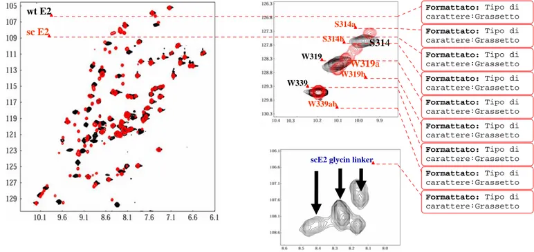
The single-chain E2 variant
The E2C binding to its target DNA takes place in its native, dimeric state. Thus, stability and function of this domain are coupled, as in many transcription factors (Beckett et al 2001). Several groups have engineered monomerized variants of dimeric transcription factors in order to decouple stability and DNA binding and, in most cases, was dimostrated that the stability at physiological protein concentrations was improved, while retaining wild type binding affinity (Jana et al, 1998; Moran et al, 1999; Liang et al, 1993; Robinson et al, 1996; Sieber et al, 1998). In order to better understand and caracterize the folding and DNA binding mechanisms of the E2 protein model domain, three single-chain variants of HPV16 scE2C were constructed. wiht engineered linkers of 6, 9, and 12 residues, respectively ( Dellarolle et al, 2008). The length of the linker was constrained by the need to join approximately 17 Å, the distance between the N-
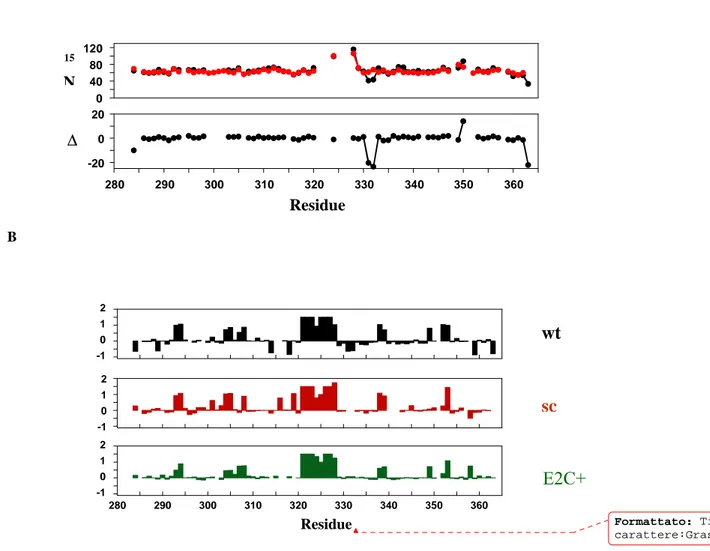
and C-termini of two different subunits in the E2C dimer. A glycine-rich linker sequences (GGGTGGGSGGGS, SGGSGGTGG, and GGTGGS) were chosen in an attempt to maintain maximum flexibility and reasonable solubility, on the basis of previous studies on monomerized transcription factors (Jana et al, 1998; Moran et al, 1999; Liang et al, 1993; Robinson et al, 1996; Sieber et al, 1998).The scE2C-12 variant showed the best expression level and purity and so was chosen as monomeric model of native of E2C .Preliminary spectroscopic investigation on this single chain variants of E2C suggest that monomerization of dimeric transcription factors using flexible linkers does not lead to substantial changes in the structure. The affinity of scE2C-12 for binding to its target DNA increases upon monomerization evenly along the tested phosphate concentration range, while the affinity for nonspecific DNA decreases to a larger extent, resulting in a 10-fold increase in specificity . Specific binding of E2C to DNA induces changes in the stability and dynamics of the protein , and nonspecific binding is known to induce larger changes, at least in one protein . We interpret that monomerization of E2C restricts the structural and dynamic changes that take place upon nonspecific DNA binding but favors the changes that take place upon specific DNA binding. Because the native E2C is known to be highly dynamic in solution as seen in molecular dynamic simulations (Falconi et al , 2007 ) , NMR spectroscopy ( (Cicero et al., 2006), and binding to DNA (Hegde et al, 2002; Cicero et al, 2006) an ulterior purpose of this work is the characterization of the solution structure and dynamics of the scE2C-12 variant in its free form and in the complex with its target site.
NUCLEAR MAGNETIC RESONANCE IN STRUCTURAL
BIOLOGY
NMR spectroscopy application in structural biology is today a field of great interest in basic research and pharmaceutical applications. Since the first experimental observation of a protein NMR spectrum was achieved in 1957), tremendous technological developments and fundamental methodological advances occurred, both in spectrometer development, sample preparation, specific pulse seqeunce and computational analysis . Proteins are the molecules for which the greatest effort has been spent in biomolecular NMR, followed by nucleic acids and their complexes with binding proteins (oligosaccharides conformational characterization and membrane structures are nowadays promising subjects of investigation). With respect to X-Ray crystallography NMR spectroscopy presents advantages and drawbacks. The major advantage is the study of the system in solution (and in membranes), that is in an environment much more similar to the native state of biological molecules. The major drawback is the limit in molecular size imposed to structural investigation by NMR. Transverse magnetization constitutes the observable signal in NMR experiments. The profound effect of the molecular dimensions on the transverse magnetization relaxation rates precludes the high-resolution determination of macromolecular systems larger than 25-30 kD, although recent advances have brought further this frontier (Kay et al, 1997). Moreover NMR is very well suitable for rapid study of interactions of small and intermediate biologically active molecules with proteins and membranes and is the unique technique capable to obtain exhaustive information on the dynamics properties at the atomic level.
The assignment problem
The assignment is the first, necessary phase that constitutes a prerequisite in every NMR structure study . It consists in labeling the atomic sites of the molecule by the corresponding resonance frequency and in the successive extraction of structural information by quantifying the interatomic magnetic interactions . A protein contains a remarkable number of protons with similar magnetic characteristics, that is protons situated in residues of the same type present in multiple copy within the sequence. The traditional assignment method, developed by Wuthrich and coworkers, is based on homonuclear two-dimensional spectroscopy (Wuthrich, 1986; Roberts, 1993). The first step in this assignment strategy is to identify proton spins that belong to a particular amino acid using through-bond correlation spectroscopy (prevalently COSY and TOCSY experiments). This result is achieved by correlating the J-coupling network of aliphatic side chain protons to the respective amide proton which normally display greater C.S. (chemical shift) dispersion. The resonance pattern is then examined in comparison with the residue-dependent chemical shifts. The identification of the residue location within the sequence
is accomplished by analyzing NOESY spectra to individuate inter-residue cross-peaks generated by amide protons belonging to sequential adjacent amino acids. When the molecular size increases, the spectra appear too crowded of signals. Moreover, homonuclear J-scalar correlation experiments for proton spins fails for systems with large molecular dimensions. The time needed to build-up the scalar correlation is not adequate because of the large relaxation rate of proton spins that dramatically reduces the efficiency of magnetization transfer. Resolution limits prevent the possibility to carry out a correct and unambiguous assignment. The heteronuclear one-bond couplings, 1J
CH (125-160 Hz), 1JCN (12 Hz) and 1JNH (about 92 Hz), are
sufficiently large and relatively uniform, depending weakly on conformation. On the other hand, transverse relaxation rates for heteronuclei in the globular proteins below the 25-30 k size still permit to transfer magnetization from 15N and 13C nuclei to scalar-coupled nuclei with high
efficiency. Thus, it’s possible and convenient to resolve very close proton resonances using an additional heteronuclear dimension which doesn’t suffer in serious relaxation limits. In addition, from the late 1980 sensitive three-dimensional experiments were developed specifically for uniformly or fractionally 13C- and 15N-labeled proteins (Bax et al., 1993). Three-dimensional
NMR spectroscopy is conceptually identical to two-dimensional one and makes use of the same pulse schemes, that are concatenated in such a way to extend the dimensionality of the experiment. The success of 3D and 4D spectra rapidly led to revolutionate the assignment procedure for proteins. In the traditional two-dimensional approach the critical step in the assignment is the individuation of NOE correlations between sequential residues. Errors in assignment of NOE connectivities at this stage propagate into the successive phase of the NMR structure determination. On the other hand, in a dipeptide unit of 15N- and 13C- labeled protein
the 13C carbonyl atom of the first amino acid is scalar-coupled to the 15N amide atom of the
second amino acid. This one-bond and other two-bond correlations between two sequential residues offer the opportunity to establish the connectivity avoiding the use of NOE crosspeaks, which could introduce ambiguities in the assignment process. Transverse magnetization is generated for the proton nucleus, then is transferred to 15N nucleus. In this way only
magnetization of protons that are correlated to nitrogen atoms survives at the end of the pulse scheme. Successively, magnetization can be transferred to 13C nucleus of the same residue
(residue i) or to the C nucleus of the preceding residue (residue i-1) because scalar interaction magnitude for the two nuclei is similar (11 and 7 Hz). The uniqueness of amino acid identification is achieved by comparison of this sequential connectivity with the chemical shift values that are characteristic of amino acid type. Once the resonances are assigned to the corresponding amino acid type, these sequential stretches of chemical shifts are mapped onto the protein sequence to find the aminoacidic segment that matches those chemical shifts. Side chain spins are successively correlated to backbone spins of their specific residue by 15N-edited
combined use of three-dimensional and, if needed, four-dimensional heteronuclear experiments allows to obtain the sequential connectivity and to internally verify the assignment relying only on J-scalar pathways (Ikura et al., 1990; Bax and Grzesiek, 1993). Advantages arise from the heteronuclear multidimensional techniques also in the assignment process of nucleic acids. A difference exists with respect to proteins: there is no way to date to connect by scalar couplings sequential nucleotide residues. Anyway, isotopic enrichment can be useful to connect the base spin system to the sugar spin system and resolve such ambiguities in the assignment.
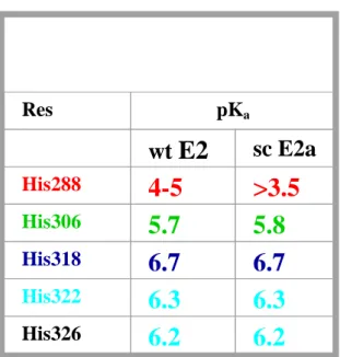
NMR determination of histidine protonation state
Histidine is frequently involved in the function of proteins, mainly because of the chemical versatility of its imidazole ring, which includes protonated and deprotonated forms as well as the tautomeric states To determine the protonated and deprotonated forms and the tautomeric states of histidine residues in solution, NMR spectroscopy has been extensively used with good success. Traditionally, the pKa of histidines has been determined by measuring chemical shift
changes during pH titration experiments. The protonated or low pH form of the imidazole side chain 1 exists according to acid-base equilibrium described by constant Ka with two
unprotonated species. These unprotonated or highpH forms of imidazole, the more common N
2-H tautomer 2 and therare N1-H tautomer 3 exist in equilibrium described by constant
KT.
NMR, particularly 15N NMR, has made important contributions to the elucidation of the
chemical behavior of histidine in -lytic protease, a member of the trypsin superfamily (Bachovchin et al, 2001). 15N NMR is exquisitelysensitive to the local magnetic environment,
and thus capableof revealing the presence of H-bonding on both sides of theimidazole ring as well as pKa values and tautomeric states. 1H NMR, when correctly applied, can also reveal the
presenceof histidine C 1-H-donated H-bonds in serine protease activesites, as we first presented
in 1996 (E.L. Ash, M.P. Vincent,J.L. Sudmeier, and W.W. Bachovchin, unpubl.; Ash et al. 2000),in addition to the often reported determination of histidinepKa values in proteins by 1H
NMR. If the ionization of a monobasic acid HA is rapid on the NMR time scale, the observable chemical shift δobs of an adjacent nucleus is averaged over the individual chemical shifts of the
species HA (δHA) and A− (δA−), the weighting coefficients (xHA and xA−) being the pH dependent
molar fractions:
However, this method cannot be applied to proteins that are unstable over a wide pH range, and it does not provide information about the tautomeric states. To circumvent this problem, several NMR techniques have been developed, based on HMQC (Pelton et al , 1993; Van Dijk et al 1992 ) HMBC (Bax et al, 1986; Schmidt et al 1991), and HSMQC (Zuiderweg, et al 1990; Xia et al 1995) experiments, which discriminate each state from the ratio of two- and three-bond remote couplings, such as 2
JN 2-H 2 and 3JN 1-H 2 in the imidazole ring. Unfortunately, the small
value of these couplings necessitates long coherence transfer times, making this method not applicable to large proteins. The method can, however, be extended to proteins of higher molecular weight if larger one-bond couplings centered at the C1 and C2 carbons in the
imidazole ring are used .
. However, so far the C-N coupling constants of the histidine residues have only been used for a qualitative analysis to identify the state with the highest population. Today NMR titration has become a routine means of pK determination , even for individual groups of large biopolymers combining the advantages of the potentiometric and NMR spectroscopic approach over potentiometry alone.
Nuclear spin relaxation and protein dynamics
It’s well known that proteins are not static systems in solution but undergo extensive fluctuations and motions on a broad range of time scales. Distinct NMR techniques exist that can report on these motional properties. Each technique displays characteristic sensitivity to a particular range of motional frequencies. Nuclear spin relaxation rates can accurately describe the global protein mobility due to the overall molecular tumbling and can individuate local flexibility on the picoseconds time scale. In addition, relaxation data analysis allows characterizing slower conformational motions such as exchange processes. A great advantage of NMR studies of protein mobility is its high-resolution nature that is the ability to examine the local motions with site-resolved specificity. After a spin system is perturbed by one or more radiofrequency pulses (spectroscopists say that it is excited), it relaxes to the original equilibrium state following different pathways that depend on the particular excited state, on the magnetic environment surrounding the nuclei and on the molecular geometry. In this way, it’s possible to extract structural and dynamical information by appropriate perturbation of the equilibrium state and by monitoring the rate at which the spin system recovers to the original state. For the relaxation of diamagnetic protein spins, two mechanisms of interaction with the