UNIVERSITY OF MOLISE
DEPARTMENT OF MEDICINE AND HEALTH SCIENCES
“Vincenzo Tiberio”
PhD course in Translational and Clinical Medicine
XXXI cycle
CD8 T cells with constitutively active PI3Kγ induce
hypertension in mice and increase myogenic tone of resistance
arteries
Coordinator
Prof COSTAGLIOLA Ciro
S.S.D. MED/30
Supervisor PhD Student
Prof LEMBO Giuseppe VINCIGUERRA Iolanda
S.S.D. MED/50
Identification number 155908
TABLE OF CONTENTS
SOMMARIO ... 1 ABSTRACT ... 3 1. INTRODUCTION ... 5 1.1 Hypertension: an overview ... 5 1.2 Myogenic tone ... 81.3 Immune system in hypertension: the early studies ... 12
1.4 Immune cells and hypertension ... 13
1.4.1 Adaptative immunity in hypertension ... 14
1.4.2 Innate immunity involved in hypertension ... 19
1.5 The spleen: an important lymphoid organ involved in hypertension ... 21
1.6 PI3K family ... 25
1.7 PI3K in immune system ... 28
1.7.1 PI3K in inflammatory diseases ... 32
1.8 PI3Kγ and cardiovascular diseases ... 35
1.8.1 PI3K in hypertension ... 36
2. AIM OF THE STUDY ... 40
3. MATERIALS AND METHODS ... 42
3.1 Murine Models ... 42
3.2 Blood pressure measurements ... 42
3.3 Isolation and preparation of single cell suspensions from spleen and kidney ... 43
3.4 Flow Cytometry Analysis ... 43
3.5 Adoptive transfer of purified CD8+ T cells ... 44
3.6 Immunohistochemistry ... 44
3.7 Set up of a long-term vessel culture system ... 45
3.7.1 Vessel Culture Medium ... 46
3.8 Myogenic tone experiments ... 48
3.9 Statistical analysis ... 48
4. RESULTS ... 50
4.1 PI3K KO mice are protected from AngII-induced infiltration of T cells in kidneys 50 4.2 Mice with constitutive expression of PI3K showed a hypertensive phenotype ... 52
4.3 PI3KCX/CX mice show a significant infiltrate of activated CD8+ T cells in kidney ... 54
4.4 The hypertensive phenotype depends on T lymphocytes ... 55
4.5 Innovative experimental approach development: culture myograph system ... 58
4.6 Activated CD8+ by hypertensive stimuli are capable to increase peripheral vascular resistance ... 60
5. CONCLUSIONS AND DISCUSSION ... 63
6. ABBREVIATIONS ... 66
7. APPENDIX ... 68
8. REFERENCES ... 69
1
SOMMARIO
L’ipertensione è una condizione clinica caratterizzata da elevati valori di pressione arteriosa ed associata a danno cardiaco, renale e vascolare. Eziologicamente è definita come essenziale, se la causa non è ben identificabile, o come secondaria, se l’incremento di pressione ha un’origine nota. Il trattamento consiste nella modificazione dello stile di vita e nella somministrazione di trattamenti antipertensivi; nonostante ciò sono riscontrabili alcune forme di ipertensione farmaco-resistente. Ricercare i meccanismi alla base della patologia risulta, quindi, importante al fine di individuare nuovi bersagli farmacologici. Numerosi articoli scientifici hanno evidenziato la correlazione tra ipertensione ed attivazione del sistema immune, ma i meccanismi attraverso cui l’immunità agisce incrementando la pressione arteriosa sono sconosciuti. Questa tesi si pone l’obiettivo di chiarire come le cellule immunitarie sono implicate nell’insorgenza del fenotipo ipertensivo. È stato precedentemente studiato il coinvolgimento della fosfatidilinositolo-3-chinasi (PI3K) nell’ipertensione ed è stato evidenziato che il modello murino non esprimente questo enzima (PI3K) era protetto dall’incremento di pressione indotto dall’angiotensina II; inoltre, PI3Kè espressa nei linfociti regolandone la maturazione e la migrazione. Sulla base di queste premesse, mediante analisi FACS, è stato evidenziato che topi PI3KangIImostravano un ridotto infiltrato immunitario renale rispetto a topiWT angII Successivamente è stato osservato che il modello murino con espressione costitutiva di PI3K (PI3KCX/CX) era caratterizzato da ipertensione spontanea e l’analisi istologica
effettuata sui reni di tali topi evidenziava i danni tipici del fenotipo ipertensivo, quali fibrosi, incremento delle dimensioni dei corpuscoli renali e della capsula di Bowman. In base a queste evidenze è stato valutato se i meccanismi immunitari sono coinvolti nel link tra PI3K ed elevata pressione arteriosa. Analisi FACS hanno evidenziato che topi PI3KCX/CX presentavano un incrementato infiltrato immune renale caratterizzato da cellule
CD8+CD69+CD44+. Esperimenti di adoptive transfer hanno mostrato la capacità delle
cellule CD8+ spleniche isolate da topi PI3KCX/CX nel determinare l’incremento dei valori
2
capacità di esplicare una risposta contrattile in funzione all’aumento della pressione intravascolare indipendentemente dalla regolazione neurormonale, risulta incrementato in diversi modelli animali di ipertensione, è stato studiato se le cellule CD8+ sono determinanti
di questa incrementata risposta miogenica. A tal fine è stato sviluppato un innovativo sistema sperimentale che permette la co-coltura di vasi di resistenza e cellule CD8+. Grazie
a ciò è stato dimostrato che cellule CD8+ attivate da stimoli ipertensivi inducevano un
significativo aumento della risposta miogenica in arteriole mesenteriche isolate da topi normotesi. Complessivamente questi dati suggeriscono che la segnalazione di PI3Kγ nelle cellule CD8+ è cruciale nello sviluppo dell’ipertensione, nella migrazione di queste cellule
verso i reni, dove contribuiscono al danno d’organo, e nella modulazione del tono miogenico che rappresenta un meccanismo chiave nella regolazione della pressione arteriosa.
Parole chiave: Ipertensione, sistema immune, fosfoinoside-3-chinasi , tono miogenico, sistema in coltura di vasi
3
ABSTRACT
Hypertension is a clinical condition characterized by elevated arterial pressure values associated with cardiac, renal and vascular damage. It can be defined etiologically as essential, whether the cause is not known, or secondary, whether the cause is well identifiable. The treatment consists in the lifestyle modification and in the antihypertensive therapy administration; despite this, some forms of drug-resistant hypertension can be found. Therefore, the research to understand the mechanisms underlying the pathology is important to identify new pharmacological targets. Numerous scientific articles showed a correlation between hypertension and immune system activation, but the mechanisms by which immunity acts to increase arterial pressure are unknown. This thesis aims to clarify how immune cells are involved in the onset of hypertensive phenotype. The involvement of phosphatidylinositol-3-kinase (PI3K) in hypertension has been previously investigated showing that the murine model depleted of this enzyme (PI3KKO) is protected from angiotensin II-induced hypertension; furthermore, PI3K is expressed in lymphocytes regulating their maturation and migration. Based on these assumptions, trough FACS analysis, it is shown that PI3KKO ang II mice are featured by reduced renal immune infiltrate compared to PI3KWT angII mice. Subsequently, it is observed that the murine model with constitutive expression of PI3K (PI3KCX/CX) was characterized by
spontaneous hypertension and histological analysis performed on kidneys of these mice highlighted the typical damages of the hypertensive phenotype, as fibrosis, increased dimensions of renal corpuscles and Bowman’s capsule. Based on these evidences, it is investigated whether immune mechanisms are involved in link between PI3K and high blood pressure. FACS analysis highlighted that PI3KCX/CX mice showed an increased renal
immune infiltrate characterized by the presence of CD8+CD69+CD44+ cells. Using adoptive
transfer experiments, it is demonstrated the ability of CD8+ splenic cells isolated from
PI3KCX/CX mice to determine the increase in blood pressure in normotensive mice that was
infused with these cells. Since the myogenic tone, ie the ability to perform a contractile response to counteract the increase in intravascular pressure independently of the
4
neurormonal regulation, is enhanced in different animal models of hypertension, it is investigated whether the immune cells were determinants of this increased myogenic response. To this end, it is developed an innovative experimental system that allowed the co-culture of resistance vessels with CD8+ cells. Thanks to this system it was shown that
CD8+ cells activated by hypertensive stimuli induced a significant increase in myogenic
response in mesenteric arterioles isolated from normotensive mice. Altogether these data suggest that the signaling of PI3Kγ in CD8+ T cells is crucial in hypertension development,
in the migration of these cells to the kidneys, where they contribute to organ damage, and in the modulation of myogenic tone which represents a key mechanism in regulation of arterial pressure.
Key words: Hypertension, immune system, phosphoinositide-3-kinase , myogenic tone, vessel culture system
5
1. INTRODUCTION
1.1 Hypertension: an overview
Hypertension (HTN or HT), also known as high blood pressure (HBP), is traditionally defined as a long-term medical condition characterized by the presence of elevated systemic arterial pressure above a threshold value.
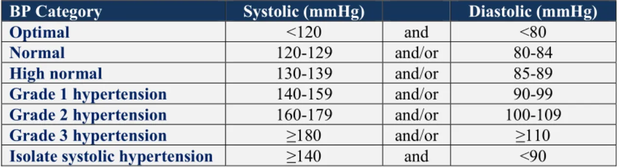
The latest guidelines published by the European Society of Cardiology (ESC) and the European Society of Hypertension (ESH) defined that hypertension is characterized by office systolic BP (SBP) values at least 140 mmHg and/or diastolic BP (DBP) values at least 90 mmHg (Williams B et al., 2018). For the American population, the 2017 American College of Cardiology/American Heart Association (ACC/AHA) guidelines delineated the new categories of BP, according to which patients with blood pressure greater than 130/80 mmHg are considered hypertensive; this new classification makes almost half of the adult population hypertensive (Table 1; Whelton PK et al., 2017).
Worldwide trends in blood pressure highlightedthat in 2015 1.13 billion of people suffered of high BP, with a prevalence of over 150 million in central and Eastern Europe (figure1). The overall prevalence of hypertension in adults is around 30 - 45%, with a global age-standardized prevalence of 24 and 20% in men and women, respectively. At the global level, the increase of hypertensive populations is attributable to population growth and ageing; moreover, in the high-income region the absolute number of people with high blood pressure is decreased compared to the past steadily, conversely in the low-income and middle-income region the number of people with raised blood pressure is still increasing (Zhou B et al., 2017).
In the light of the most recent discoveries, Giles T D and colleagues refined and updated the definition and classification of hypertension. This pathological condition can be defined as “a progressive cardiovascular syndrome arising from complex and interrelated etiologies” and its progression is associated with functional and structural injury to the heart, kidneys, brain and vasculature.
6
Many factors contribute to the raised blood pressure (BP), the most intensively studied are: salt intake, smoking, sedentary lifestyle, obesity and insulin resistance, elevated level of total cholesterol, genetic factors, endothelial dysfunction, low birth weight, early-onset menopause, intrauterine nutrition, neurovascular anomalies, renin-angiotensin system and sympathetic nervous system (Giles T D et al., 2009).
Etiologically, HTN can be divided in two main groups: primary (or essential) and secondary hypertension. The majority of patients are affected by primary HTN in which HBP is not associated with any identifiable pathological causes. On the contrary, a minority of patients present secondary hypertension characterized by a specific cause of HBP, as obstructive sleep apnea, renal parenchymal disease, renal artery stenosis, primary aldosteronism, thyroid disease, Cushing's syndrome, pheochromocytoma, coarctation of the aorta (Carretero OA and Oparil S, 2000; Rimoldi SF et al., 2014).
Since HBP is the main factor risk for cardiovascular disease the treatment of this pathology is fundamental to limit the negative consequences induced in the short and long term. There are two established strategies to fight hypertension: lifestyle modifications and drug treatment. The lifestyle recommendations that have been shown helpful to reduce BP are: salt intake restriction, moderation of alcohol consumption, tobacco smoking cessation, diet rich in fruits and vegetables, maintaining an ideal body weight, and regular physical activity. Although healthy lifestyle contributes to lower BP, in many cases the pharmacological intervention is necessary. Major classes of antihypertensive drugs are represented by blockers of the renin-angiotensin system (angiotensin converting enzyme inhibitors and angiotensin receptor blockers), calcium channel blockers, thiazides and thiazide-like diuretics and beta-blockers. Furthermore, other drags for the treatment of this condition exist and are used in the drug-resistant hypertension where all other pharmacological options are not efficacy (Williams B et al., 2018).
People that suffer of HBP likely develop other pathologies as atherosclerosis, stroke, myocardial infarction, heart failure, chronic kidney disease and dementia. Since uncontrolled blood pressure remains a major threat to cardiovascular health, the prevention and management of hypertension are global public health issues (Lionakis N et al., 2012; WHO, 2013).
7
BP Category Systolic (mmHg) Diastolic (mmHg)
Optimal <120 and <80
Normal 120-129 and/or 80-84
High normal 130-139 and/or 85-89
Grade 1 hypertension 140-159 and/or 90-99
Grade 2 hypertension 160-179 and/or 100-109
Grade 3 hypertension ≥180 and/or ≥110
Isolate systolic hypertension ≥140 and <90
Table 1. Categories of BP in Adults. Systolic and diastolic blood pressure values identify optimal, normal, high normal blood pressure conditions, grade 1, 2 and 3 of hypertension (Williams B et al., 2018).
Figure 1. Trends in hypertension prevalence, 1975 - 2015. Trends by region in the number of adults aged 18 years and older with raised blood pressure.The estimated number of adults with raised blood pressure increased from 594 million in 1975 to 1·13 billion in 2015 (Zhou B et al., 2017).
8
1.2 Myogenic tone
Myogenic response is a smooth muscle cells (SMCs) hallmark of resistance arteries and arterioles to counteract to blood pressure changes. The first description of this vascular response is attributed to William Maddock Bayliss that described this phenomenon in 1902; the pressure-induced response has been demonstrated to occur independently of the endothelium and it does not require neurohumoral input (Bayliss WM, 1902; Schubert R et al., 1999). In particular, when an increase in transmural pressure occurs resistance arteries react by vasoconstriction; conversely, when a decrease in pressure takes place resistance vessels respond by vasodilatation (Davis MJ et al., 2012); therefore, vascular tone of resistance vessels is due to blood pressure within these vessels and it depends on the balance between vasoconstrictor and vasodilator signals (Davis MJ et al., 2011). There is an extensive literature showing that in experimental animal models of hypertension and in patients with high blood pressure occur an increase in peripheral vascular resistances that in turn contributes to the maintenance of chronic hypertensive status (Carnevale D et al., 2018; Goulopoulou S and Webb RC, 2014; Guzik T et al., 2007; Tang KM et al., 2003; Huang PL et al., 1995). The pressure-induced mechanosensory effects involve membrane depolarization, Ca2+ signaling, activation of contractile proteins via a myosin light chain
kinase (MLCK)-mediated mechanism and remodeling of the cytoskeleton structure. SMCs depolarization can be ascribable mainly to voltage-gated Ca2+ channels (VGCC) opening,
but also to closure of K+ channels or opening of a Cl2 channel. Electrophysiological
approaches showed that SMC membrane deformation (e.g. due to stretch) causes activation of a cation current that presumably leads to membrane depolarization and a subsequent opening of VGCC. Stretch-activated channels (SACs), for which gating is modulated by physiological levels of stretch, participate in the membrane potential control. The opening of these channels results in a predominantly Na+ current that causes the membrane
depolarization. In addition, membrane stretch activates BKCa channels, producing a
hyperpolarizing current that limits the extent of depolarization and, hence, myogenic constriction; this can be considered an important negative-feedback mechanism to limit the effects of additional myogenic contraction owing to pressure-induced vasoconstriction of
9
downstream arterioles. Others important cationic channels involved in myogenic signaling are represented by TRP (transient receptor potential) channels, that have been characterized as nonselective cation and Ca2+- selective channels. In particular, it was demonstrated that
the reduced expression of TRPC6 and TRPM4 resulted in marked attenuation of both pressure-induced depolarization and myogenic constriction. An alternate mechanosensory mechanism is represented by integrins, transmembrane receptors that facilitate cell-extracellular matrix (ECM) adhesion. It is demonstrated that the block of either v3 or
51 integrins abolishes myogenic constriction to step increases in intravascular pressure.
According with the fact that integrin activation and cytoskeletal remodeling are coordinated processes, it is showed that myogenic activation of arterioles is associated with cytoskeletal rearrangements including globular to filamentous actin transformations, demonstrating the importance of the actin cytoskeleton in myogenic response. Myogenic contraction is also modulated by second messengers; in fact, the increase in transmural pressure leads to the generation of specific factors such as 20-HETE, sphingosine-1-phosphate (S1P) and diacylglycerol (DAG). Pressure activation also causes an alteration in Gq/G11 activity in a receptor-ligation-independent manner where activation of the trimeric
G protein subsequently stimulates PLC and TRPC6 to cause membrane depolarization. Myogenic contraction is dependent on a global intracellular Ca2+- calmodulin - MLCK
regulated mechanism, as well as on Ca2+ sensitization. Ca2+ sensitization refers to
processes that inhibit myosin phosphatase, thus blocking the dephosphorylation of myosin regulatory light chain. Candidate mechanisms for inhibition of the phosphatase include Rho-kinase-mediated phosphorylation of MYPT1 (myosin phosphatase target subunit 1) and PKC (protein kinase C)-mediated CPI-17 (C-kinase-activated protein phosphatase-1 (PP1) inhibitor, 17kDa) activation. As mentioned earlier, the opening of L-type Ca2+
channels is the principal event that follows the pressure stimulation. Moreover, there is a minor contribution from Ca2+ entry via non-voltage-gated Ca2+-entry pathways. However,
Ca2+ through these pathways may participate in the regulation of ion channels and SR
(sarcoplasmic reticulum) Ca2+ dynamics. Despite the role of Ca2+ release from the SR in
myogenic signaling is difficult to understand because of the technical limitations that do not allow a detailed study of this aspect, it is known that the entry of extracellular Ca2+ causes
10
the activation of RyR (ryanodine receptor) and, therefore, the release of calcium from the SR to the cytoplasm contributing to myogenic response. To the other hand the SR provides an inhibitory action trough the stimulation of 1 subunit of BKCa channels, giving rise to
spontaneously transient outward currents (STOCs) and acting as a negative feedback mechanism to prevent excessive depolarization as pressure-induced constriction occurs. Moreover, the Ca2+ release from intracellular compartment can occurs by the binding of the
second messenger inositol 1, 4, 5-trisphosphate (IP3) to IP3R/Ca2+ channels (Figure 2; Hill
MA and Meininger GA, 2012). In summary, in the context of the myogenic response it is possible to identify three different phases (Osol G et al., 2002). The first phase, in which there is the development of myogenic tone or basal tone, is characterized by a significant elevation of intracellular calcium influx through the L-type voltage gated calcium channel (LTTC; Hill MA et al., 2001; Tajada S et al., 2013), cellular depolarization and deformation, followed by a reduction in vessel diameter. The second phase, named myogenic reactivity, is characterized by minor changes in membrane potential and intracellular calcium level but is present an intracellular calcium sensitization (Schubert et al., 2008). In this phase there is a further constriction in response to an intraluminal pressure increase. In the last phase, named dilatative force, is observable a complete loss of tone together with vasodilatation in response to high transmural pressure (Hill MA et al., 2006; Carnevale D and Lembo G, 2012).
11
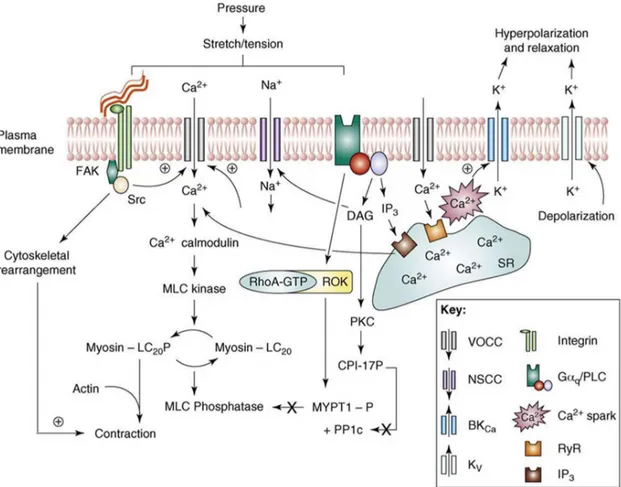
Figure 2. Schematic illustration of specific signaling mechanisms that mediate arteriolar myogenic vasoconstriction. Mechanical stimulus leads to membrane depolarization mainly mediated by VGCCs, increases in cyctosolic Ca2+ and contraction
through calmodulin-mediated activation of MLCK. Phosphorylation of MLC20 (MLC20P)
allows acto-myosin interaction, cross-bridge cycling and contraction. The exact molecular events underlying this membrane depolarization are unclear but important candidates include direct activation of cation channels (for example, SACs, ENaC -Epithelial sodium channels- or TRP channels), second messenger-mediated (for example, DAG, PKC) activation of cation channels and integrin-mediated facilitation of VGCC opening. The mechanical stimulus activates cytoskeletal remodeling through mechanisms likely to be dependent on integrin-mediated activation of focal adhesions. A role of the SR in myogenic contraction has the potential for both stimulatory (through Ca2+ release to the bulk
cytoplasm) and inhibitory actions (through both generation of Ca2+ sparks and activation of
BKCa-mediated STOCs, and through its ability to sequester Ca2+ from the cytoplasm).
Increased intraluminal pressure and cell stretch also activate tyrosine-kinase-mediated mechanisms (for example, involving FAK -focal adhesion kinase-, Src kinase and p42/44 MAPK -mitogen-activated protein kinase).
12
1.3 Immune system in hypertension: the early studies
Hypertension is generally attributed to perturbations of vasculature, kidney, and central nervous system (CNS). During the past several years, studies have shown consistent association between this disease, proinflammatory cytokines and cells of the innate and adaptive immune systems and have focused on defining the mechanisms linking the immune system to the hypertensive disease state.
Link between immune system and hypertension emerged in 1964 with the pioneering studies conducted by White and Grollman that described the importance of immunosuppressive therapy on regulation of BP levels in rats with partial kidney infarction (White FN and Grollman A, 1964). Subsequently, other studies demonstrated that the transfer of immune cells isolated from lymph nodes or spleen of hypertensive animals induced hypertension in normal recipient rats (Okuda T and Grollman A, 1967; Olsen F, 1980). In 1972 Olsen demonstrated that in arterioles and small arteries of hypertensive humans is observable an inflammatory mononuclear cellular infiltration (Olsen F, 1972). Studies performed by Svendsen revealed that in athymic nude mice hypertension did not maintain after renal infarction (Svendsen UG, 1976). Moreover, thymectomy performed in hypertensive mice, treated with deoxycorticosterone acetate (DOCA)-salt (Svendsen UG, 1976), or in genetically hypertensive rat model (Bataillard A et al.,1986) attenuated experimental HT. A further work demonstrated that in T cell-depressed spontaneously hypertensive rats (SHR) the thymus grafts reduced the blood pressure. Moreover, transplantation of compatible thymus tissues into neonatal SHR produced long-lasting recovery of immune functions and the complete immunologic restoration caused the suppression of HBP (Ba D et al., 1982). These first studies about the involvement of immune system and hypertension provided key elements for subsequent studies in this research field.
13
1.4 Immune cells and hypertension
The described discovers aroused much interest and the researchers focused on the investigation of immune cells subtypes that contribute to the modulation of hypertensive phenotype. To date, the involvement of adaptive and innate immunity has been demonstrated in the onset of hypertension (figure 3). The aim of subsequent paragraphs is that to provide a description of different types of immune cells involved in the development of HBP.
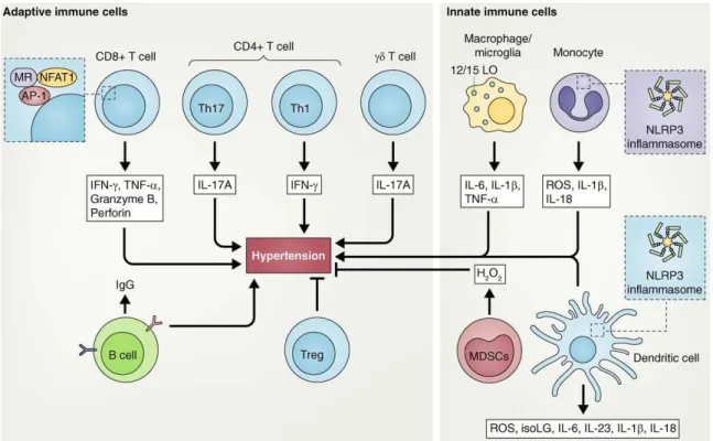
Figure 3. Innate and adaptive immune cells involved in hypertension. Adaptive immune cells that have been shown to play a role in high blood pressure are: CD8+, CD4+ T
cells (Th1, Th17, and T reg cells) γδ T cells and B cells. These populations produce factors that promote or inhibit hypertension. Innate immune cells involved in hypertension are: macrophages, microglia, monocytes, DCs, and MDSCs that also produce cytokines and ROS, which promote or inhibit this pathological condition. The NLRP3 inflammasome in monocytes and DCs plays a key role in hypertension (Norlander AE et al., 2018).
14
1.4.1 Adaptative immunity in hypertension
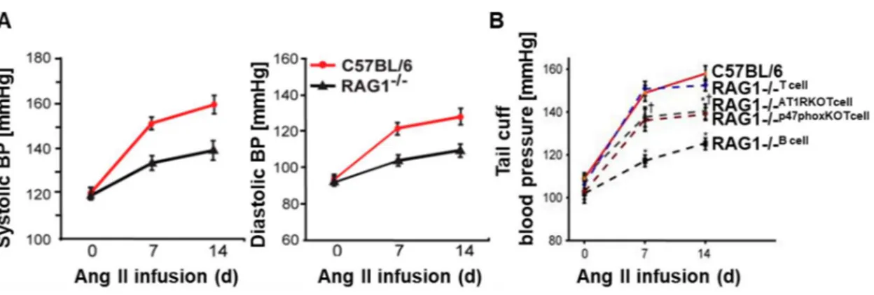
Many researches highlighted the involvement of T cells in the onset of hypertension. It is described that pharmacological approaches that act with different mechanisms on immune cells are effective in the modulation of blood pressure. In particular, it is demonstrated that rats treated with mycophenolate mofetil (MMF), a selective T lymphocyte immunosuppressive agent, are protected to development of salt-dependent hypertension induced from angiotensin (Ang II) infusion (Rodríguez-Iturbe B et al., 2001). Additionally, it is showed that abatacept, a T cells costimulation inhibitor, is able to prevent angiotensin II and (DOCA)–salt induced hypertension (Vinh A et al., 2010). Important advances derived from the study of RAG1 knock out (KO) murine model. This model is characterized by a deletion of Rag1 coding for recombinase protein involved in V(D)J recombination, a typically process that occurs in developing lymphocytes during the early stages of T and B cells maturation; therefore, this gene deletion causes the absence of mature B and T cells. It is observed that AngII or DOCA-induced hypertension is blunted in RAG1 KO mice (Figure 4) and that this phenotype is independent from angiotensin II receptors (AT1R and AT2R) expression. Moreover, many of the negative vascular consequences induced by angiotensin II, such as ROS production and impairment of endothelium-dependent vasodilatation, were prevented in these mice. Adoptive transfer of T lymphocytes, but not of B lymphocytes, restored the hypertensive phenotype, demonstrating that T cells play a critical role in the onset of hypertension. Moreover, the adoptive transfer of T lymphocytes isolated from AT1a KO mice partially restored hypertension, indicating that some hypertensive effects mediated by angiotensin II result from its action on T cells. Similarly, adoptive transfer of T cells that lack of NADPH oxidase p47 phox subunit, partially restored the hypertensive response to angiotensin II in
RAG1 KO mice, suggesting a function of the T cell NADPH oxidase in hypertension (Figure 4; Guzik TJ et al., 2007).
Subsequent study highlighted that genetic deletion of Rag1 gene in Dahl sensitive salt rats reduced the degree of hypertension and renal damage (Mattson DL et al., 2013). Further studies investigated the role of deletion of CD3 ζ chain (CD247), a gene involved in T-cell
15
signaling, in Dahl salt-sensitive rats and found that the mean arterial blood pressure and kidney injury were reduced in CD247–/–, showing a similar phenotype to that observed in
the rats lacking RAG1 (Rudemiller N et al., 2014). Moreover, other research group using a SCID (severe combined immunodeficiency) mice, that lack of lymphocytes activity, demonstrated the importance of lymphocyte responses in AngII-induced hypertension (Crowley S D et al., 2010).
Figure 4. Blood pressure measurements in C57BL/6 and RAG1-/- mice. A. Invasive
measurements of systolic and diastolic blood pressure at baseline and during angiotensin II infusion in C57BL/6 (red line) and RAG1-/- mice (black line). B. Blood pressures
measurements at baseline and during angiotensin II infusion in C57BL/6 (red line), RAG1
-/-that received T cells by adoptive transfer (RAG1-/- T cell; dotted blue line), RAG1-/- that
received T cells from AT1a-/- mice (RAG1-/- AT1RKOT cell; dotted gray line), RAG1-/- that
received T cells from mice lacking the oxidase subunit p47phox (RAG1-/- p47phoxKO T cell;
dotted burgundy line), RAG1-/- that received B cells (RAG1-/- B cell; dotted black line - Guzik
TJ et al., 2007).
Subsequently the subtypes of T cells involved in hypertension were analyzed: CD8, CD4, Treg, T cells.
Different scientific articles highlighted the role of CD8+ T cells in the onset and
development of hypertensive disease. Trott D W and colleagues analyzed the region V of T cell receptor (TCR) as powerful indicator of clonal expansion of T cells that provide insight into adaptive immune responses. In this study, spectratyping profiles of TCRs revealed that in the kidney of angII-treated mice there was an increase in Vβ chain clonality
16
of CD8+ cells, but no clonal skewing of CD4+ T cells was observable. In the same way no
clonal skewing of CD4+ and CD8+ T cells was present in mesenteric arteries and spleen of
the hypertensive mice. The importance of CD8+ T cell in the hypertensive contest was
supported by experiments showing that CD8-/- mice were protected from hypertension
induced from angII or DOCA salt, but this protection was missing in CD4-/- and MHCII
-/-(major histocompatibility complex II-/-, an alternate model of CD4+ deficiency) mice.
Moreover, adoptive transfer of CD8+ cells from angiotensin II treated mice into RAG-1−/−
mice caused an increase in blood pressure; this elevation was absent when RAG-1−/− mice
was infused with CD4+/CD25− cells. It is also observed that CD8+ T cells exert an
important role in endothelial disfunction, antidiuretic and antinatriuretic responses to angiotensin II (Trott D W et al., 2014).
It was also showed that CD8+ T cells are crucial mediator of salt sensitive hypertension. In
fact, these cells are able to up-regulate and activate the thiazide-sensitive sodium-chloride-co-transporter (NCC) in distal convoluted tubules (DCTs) of the kidney, which results in sodium retention and development of salt-sensitive hypertension. In particular, these immune cells stimulate NCC by upregulating the potassium channel Kir4.1 and subsequently the chloride channel ClC-K on the plasma membrane, thereby decreasing intracellular chloride. This last event leads to NCC activation and development of salt-sensitive hypertension (Liu Y et al., 2017).
An interesting human prospective study documented the pathogenic role of T cells in hypertension. Hypertensive patients showed a significant increase of senescent CD28
-CD57+CD8+ T cells compared with normotensive subjects. In HTN patients these
immunosenescent cells was characterized by cytotoxic and proinflammatory phenotype since they produced high levels of granzyme B, INFand TNF. In the same study, the hypertensive condition is associated with renal infiltration of CD4+ and CD8+ T cells and
increased circulating levels of CXCR3 chemokines, a tissue-homing chemokine for proinflammatory T cells (Youn J C et al., 2013).
There are evidences that also CD4+ T cells are involved in hypertension. It is known that
interleukin 17-A, mainly produced by a subset of CD4+ T cells (Th17 cells), plays a critical
17
condition, as well as pre-eclampsia and systemic lupus erythematosus. Recent study demonstrated that IL-17 increases Rho-kinase-mediated eNOS phosphorylation leading impaired endothelium-dependent vasodilatation and hypertension in mice (Nguyen H et al., 2013).
Experiments performed in humanized mice model, in which the murine immune system is replaced by the human immune system, showed that after AngII infusion there was a significant increase of CD4+ T cells in the kidney and lymph nodes. In these cells were
observable an increase of the memory cell marker CD45RO but there were few cells expressing the activation marker CD69. Moreover, analysis on human blood revealed that hypertensive patients showed an increase of CD4+ and CD8+ circulating T cells than
normotensive controls. It is found that production of IL-17A is increased in CD4+ T cells
from hypertensive subjects; in the same way production of IFN- and TNF is augmented in CD4+ and CD8+ cells isolated from patients with high blood pressure (Itani HA et al.,
2016).
It is known that a regulatory T lymphocytes (Tregs), expressing CD4, CD25 and Foxp3, are able to suppress innate and adaptive immune responses, as well as proinflammatory effects of other lymphocytes, macrophages, dendritic cells, and neutrophils. It is demonstrated that adoptive transfer of Tregs prevents AngII-induced hypertension. Furthermore, AngII treatment impaired the vasodilatory response to ACh and increased wall stiffness of mesenteric arteries, increased NADPH oxidase activity in both the aorta and the heart, plasma levels of cytokines and T-cell infiltration in the aortic adventitia and periadventitial fat. It is verified that adoptive transfer of Tregs prevent all these events induced by AngII (Barhoumi T et al., 2011).
Recently, it was demonstrated that complement receptors are necessary to prevent AngII-induced hypertension. In fact, C3aR and C5aR double deficiency enhances the functions of Foxp3+ Tregs cells that have an immunosuppressive role and, consequently, are able to
attenuate Ang II-induced inflammatory factors expression, target organ damage, and so BP elevation (Chen XH et al., 2018).
A recent evidence suggests that another subset of T cells involved in hypertension is represented by T cells. It is known that T cells can be divided in two families based on
18
TCR chain constitution: those expressing and TCR chains and those expressing andTCR chains. This second family is much smaller than the first one and represented only from 1 to 4% of total T cells. CD4+ and CD8+ αβ T cells recognize antigens presented
by APC cells by their MHC complex-II and -I, respectively. On the contrary, γδ T cells recognize antigens without MHC restriction and without help from APC; for this reason, are unconventional T lymphocytes, mostly CD4 and CD8 double negative, with rapid innate like-responses that act in the initiation phase of the immune reaction. It was already observed that the number of aortic-infiltrating double negative T cells and the percentage of peripheral blood double negative T cells were increased in Ang II-infused mice (Guzik TJ et al., 2007). Caillon and colleagues confirmed that AngII-induced hypertension determines an increase of γδ T cells and showed that these cells are CD69 positive and, so, activated. Tcrδ−/− mice, deficient in γδ T cells, are protected from AngII-induced hypertension and
vascular dysfunction and showed a blunted spleen T-cell activation. Moreover, the importance of these cells in human hypertension is highlighted by an association between γδ T-cell frequency in the blood and systolic blood pressure (Caillon A et al., 2017). The mechanism by which these cells behave in order to support hypertension need to be further deepen.
There are also suggestions that B cells and the antibodies that they produce participate to hypertension. In fact, BAFF-R−/− mice, deficient in B cells, showed attenuated pressor
response to Ang II compared to wild-type mice. The magnitude of cardiac hypertrophy, that is a consequence of sustained elevations in BP, was lower in BAFF-R−/− mice than that in
wild-type mice. The relevance of B cells in the hypertensive context is confirmed by adoptive transfer of these cells from WT in BAFF-R−/− mice; B lymphocytes introduction
in mice deficient of these cells restored the pressor response to Ang II. It is demonstrated that AngII infusion causes activation of B cells, indicated as increase of CD86+ expression
in peripheral lymphoid organs and induces in the spleen a major differentiation of B cells in antibody-producing plasma cells and plasmablasts. In agreement with this, there was elevated level of IgG in serum and aortic wall of mice infused with AngII compared with saline-infused animals. The lack of mature B cells in BAFF-R−/− mice eliminated the
19
1.4.2 Innate immunity involved in hypertension
There are ample evidences that innate immune system plays a role in hypertensive disease. The earliest demonstration of this became from the study of op/op mice, defective in production of functional colony-stimulating factor-1 (CSF-1), that have reduced macrophages number. It is showed that this condition is associated with blunted hypertension in response to chronic Ang II infusion, preserved vascular morphology, attenuated endothelial dysfunction, O2- generation, NAD(P)H oxidase activation, and
vascular inflammation compared with WT littermates (De Ciuceis C et al., 2005).
The role of inflammatory myelomonocytic cells is evaluated using selective ablation of lysozyme M-positive myelomonocytic cells by low-dose diphtheria toxin in mice with inducible expression of the diphtheria toxin receptor (LysMiDTR mice). In this murine model
the reduction of monocytes number in the circulation is correlated with prevention of hypertension, reduced vascular dysfunction and ROS formation. Adoptive transfer of wild-type CD11b+Gr-1+ monocytes into depleted LysMiDTR mice reestablished the effects
induced by AngII (Wenzel P et al., 2011).
The involvement of macrophagic component in the hypertension (Justin Rucker A and Crowley SD, 2017) is highlighted studying Alox15-/- mice lacking 12/15 lipoxygenase.
This experimental animal model results protected from hypertension induced by DOCA-salt or NO synthase inhibitor nitro l-arginine methyl ester (L-NAME). A rescue experiment that restore the macrophagic functions by transfusing WT peritoneal macrophages into Alox15-/- mice showed a restoration of increased blood pressure after 5 days of L-NAME
treatment (Kriska T et al., 2012).
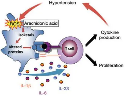
In 2014 Kirabo and colleagues described a new pathway linked to hypertension disease in which dendritic cells play a critical role. It was showed that in DCs from angiotensin II-infused mice there was an increased production of ROS, isoketals accumulation, cytokines release, such as IL-6, IL-18, IL-1β, and IL-23 and increase in costimulatory proteins CD80 and CD86. These activated DCs promoted CD8+ T cell proliferation, IFN-γ and IL-17A
20
Other cells involved in hypertension are the myeloid-derived suppressor cells (MDSCs). In different hypertensive models, these cells exert their role in the spleen where they interact with T cells. In particular, the role of MDSCs is to suppress inflammation, through hydrogen peroxide production, and to limit blood pressure increase (Shah KH et al., 2015).
Figure 5. Hypothesized pathway for dendritic cells activation. Hypertensive stimuli increase ROS production, isoketals formation, cytokine release from DCs. Activated DCs promote T cell proliferation and cytokine production causing hypertension (Kirabo A et al., 2014).
21
1.5 The spleen: an important lymphoid organ involved in hypertension
The spleen is a secondary lymphoid organ surrounded by a capsule of connective tissue from which depart trabeculae. The splenic vessels are distributed in the thickness of the trabeculae and leave these structures to go through organ parenchyma. The central arterioles depart from the splenic artery branch and arrive in the white pulp (WP) area, which contains B cells in the follicles and T cells. The blood flows through the marginal zone (MZ), surrounding the WP, toward the red pulp (RP) area, where blood is directed to the venous sinuses. MZ represents a connection between circulating blood and immune cells and contains B cells together with subsets of neutrophils, DCs, and macrophages. RP is characterized for antibodies production and blood filtration process during which resident macrophages phagocyte old or damaged erythrocytes. In RP blood flows are conveyed to the splenic vein that converges in the portal vein and, in this way, blood leave the splenic organ (Mebius RE and Kraal G, 2005).
Recent papers highlight a central role of this organ in mediating hypertensive phenotype through a modulation of immune responses activated by nervous stimulation (Carnevale D et al., 2014; Lori A et al., 2017; Perrotta M et al., 2018).
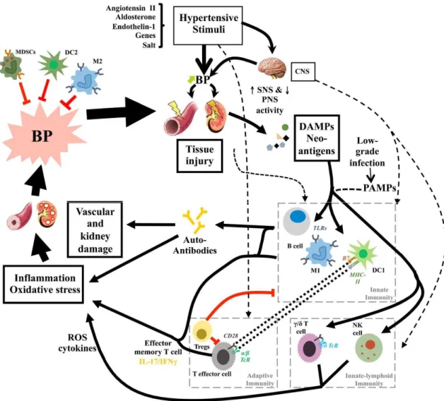
As described in the previous paragraphs, different subpopulations of immune cells are involved in the development of high blood pressure. The figure 6 represents as these cells interact in order to elicit hypertensive phenotype. Vasoactive agents (for example Ang II, endothelin I -ET1-, aldosterone), genetic susceptibility, and high salt intake are important contributors in the hypertension. These mediators act increasing sympathetic nervous system (SNS) activity that is responsible of initial blood pressure elevation with subsequent renal and vascular damage. This tissue injury leads to DAMPs (damage-associated molecular patterns) and neoantigen formation that are responsible to innate immune activation through TLRs (toll like receptor) expressed on B cells, DCs and macrophages. Innate immunity contributes to inflammation and oxidative stress directly or through adaptive immune activation. All together these mediators promote progression of disease and cause end-organ damage (Caillon A and Schiffrin EL, 2016).
22
It is hypothesized that neoantigens are formed from modified endogenous proteins in the prehypertensive phase. These altered proteins activate T cells that infiltrate the vasculature and kidney, promote endothelial dysfunction, vasoconstriction and salt and water retention causing ultimately severe hypertension (Harrison DG et al., 2011).
It is described that the fibers of SNS innerve the spleen and that the synaptic endings are close to immune cells that populate this organ (Nance DM and Sanders VM, 2007). The neurotransmitters release from synaptic vesicles is able to regulate the immune responses through modulation of cytokines production. This represents the key point of neuro-immune communication, that is characterized by a bidirectional connection in which also the produced cytokines can affect neurons activity (Straub RH, 2004).
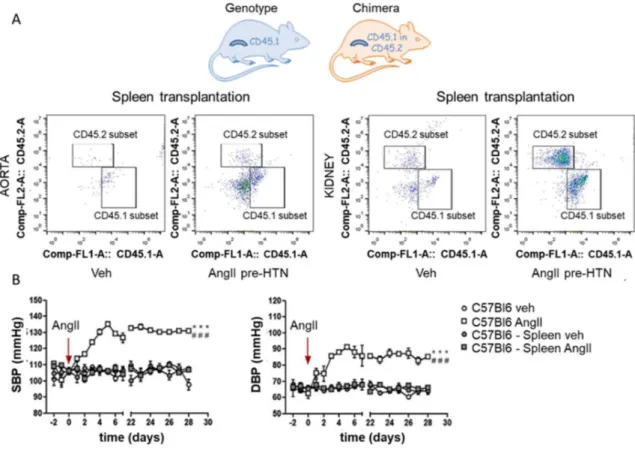
An important goal in the study of inflammation in the hypertension came from the discovery, made by the research group of Carnevale D and colleagues, that the spleen is a key regulator in the pathogenesis of hypertension. In particular, they showed for the first time the pivotal role of PlGF (placental growth factor), belonging to vascular endothelial growth factor (VEGF) family, as molecular pathway involved in the onset of hypertension by mediating immune response to the SNS activated by the AngII-induced hypertensive stimulus. Using adoptive transfer experiments, performed on mice expressing different allelic variants of CD45 (CD45.1 or CD45.2), it was demonstrated the splenic origin of T cells that infiltrate the target organs of hypertension. In particular, CD45.2 mice were splenectomized, transplanted with CD45.1 spleen and then infused with AngII to induce hypertension. The cytofluorimetric analysis revealed an increased number of donor (CD45.1) T cells in aortas and kidneys of AngII pre-HTN animals. Moreover, they showed the importance of splenic immune reservoir for the establishment of a hypertensive response to AngII. In fact, splenectomized mice infused with AngII were protected from BP elevation and T cells target organ infiltration (Figure 7; Carnevale D et al., 2104). Subsequent study showed that also in hypertension resulting from (DOCA)-salt PlGF exerts a pivotal role in immune response originating in the spleen (Perrotta ML et al., 2018).
23
Figure 6. Schematic inflammatory mechanism in hypertension. Hypertensive stimuli may cause an initial elevation in BP due, in part, to increased SNS activation. This leads to mild tissue injury, formation of damage associated molecular patterns (DAMPs), and neo-antigens, promoting activation of innate immunity. Innate immune cells contribute to inflammation and oxidative stress directly or via the activation of adaptive immunity. The effects exercised by activated immune system lead to vascular and kidney injuries (Caillon A and Schiffrin EL, 2016).
24
Figure 7. The spleen has a crucial role in deployment of T cell upon AngII and in hypertensive response. Accumulation of T cells originating exclusively from the donor spleen (CD45.1) as measured by flow cytometry (A) and BP response to chronic AngII, in splenectomized WT versus sham mice (B; Carnevale D et al., 2014).
25
1.6 PI3K family
The phosphoinositide 3‑kinases (PI3K) is an enzyme able to phosphorylate the 3‑hydroxyl group of the inositol ring of three species of phosphatidylinositol (PtdIns) lipid substrates, namely PtdIns, PtdIns4P and PtdIns(4,5)P2. PI3K signaling plays a key role in many processes, including cell cycle progression, cell growth, survival and migration, and intracellular vesicular transport, also if the specific contribution of several PI3K isoforms remains in part unknown.
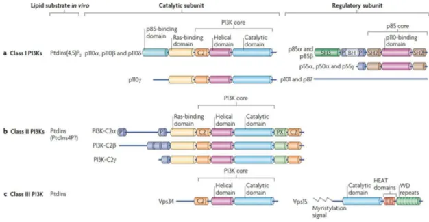
These kinases have been assigned to three different classes (class I, class II and class III) based on structural features and lipid substrate preferences (figure 8).
Class I PI3Ks use phosphatidylinositol-4,5-bisphosphate (PtdIns(4,5)P2) as substrate to generate phosphatidylinositol (3,4,5)-trisphosphate (PIP3) and they consist of catalytic
subunit in complex with a regulatory subunit. In all PI3Ks the catalytic subunit includes a core structure that consists of a C2 domain (protein-kinase-C homology-2), a helical domain and a catalytic domain. Moreover, the catalytic subunit of class I and II PI3Ks also comprehends the Ras‑binding domain (RBD); a subgroup of class I PI3Ks also presents the p85 binding domain. Regulatory subunit can include p85 isoform (for p110α, p110β and p110δ) or p101 or p87 (for p110γ). All p85 isoforms have two Src homology 2 (SH2) domains and are encoded by either PIK3R1 (which, through differential promoter usage, encodes p85α, p55α and p50α), PIK3R2 (which encodes p85β) and PIK3R3 (which encodes p55γ). Roles of the individual p85 subunits are unknown. p101 and p87 lack SH2 domains, do not have homology to other proteins and have no identifiable domains. Relative to this, p110 subunits were historically divided into class IA, which binds the p85 type of regulatory subunit, and class IB, which do not. The SH2 domains bind phosphorylated tyrosine (pTyr) and the subdivision into IA or IB classes was correlated with the capacity to be activated through Tyr kinases or G protein coupled receptors (GPCRs), respectively. Most class I PI3K subunits might be activated by GPCRs directly through Gβγ protein subunits or indirectly, through a small GTPasi Ras. Ras can be activated by Tyr kinases and GPCRs, and it might engage all class I PI3Ks through their Ras‑binding domain (RBD).
26
Therefore, class IA PI3Ks may be more responsive to GPCR stimuli than initially supposed (figure 9).
Class II PI3Ks use PtdIns as a substrate but might also use PtdIns-4-phosphate (PtdIns4P) under certain conditions. They lack regulatory subunits but have amino- and carboxy-terminal extensions to the PI3K core structure, which could mediate protein–protein interactions.
Class III PI3K has one catalytic member, vacuolar protein sorting 34 (Vps34; also known as PIK3C3 in mammals), which uses PtdIns as a substrate and binds Vps15 (also known as PIK3R4 in mammals). Vps15 consists of a catalytic domain (which is thought to be inactive), HEAT domains (which probably mediate protein–protein interactions) and WD (tryptophan-aspartic acid) repeats, which have structural and functional characteristics like a Gβ subunit. The WD repeats are essential for interaction with RAB5–GTP, the yeast guanine nucleotide-binding protein 1a (Gpa1; the homologue of the mammalian Gα of heterotrimeric G proteins) and autophagy-related protein 14 (Atg14; a potential Gγ protein; Vanhaesebroeck B et al., 2010).
It is noted the importance of aberrations in PI3K signaling in the contribution to a broad spectrum of human diseases, such as cancer, immunological and neurological disorders, diabetes, localized tissue overgrowth, and cardiovascular disease (Fruman DA et al., 2017). In following paragraphs of this dissertation, the role of PI3K isoform will be explored as key point that connects immune system and the pathogenesis of hypertension.
27
Figure 8. Classification and domain structure of phosphoinositide 3-kinases (PI3Ks). Three different classes of PI3Ks. A) Class I, B) Class II, C) Class III. BH, BCR homology domain; P, Pro-rich region; SH3, Src-homology 3 domain; SH2, Src-homology 2 domains; PX, phox homology domain (Vanhaesebroeck B et al., 2010).
Figure 9. Activation mechanisms of class I PI3K. SH2 domains of the p85 protein bind with phospho-tyrosine generated by Tyr kinases in Tyr kinase receptors, resulting in the activation of p110α and p110δ, and probably also p110β (left). Tyr kinase pathways and GPCRs can also activate Ras, which then activates class I PI3Ks (middle). Direct interaction of PI3Ks with Gα or Gβγ subunits, which are downstream of GPCRs, can engage class I PI3Ks. p110α might be inhibited by Gα, whereas p110β and p110γ are activated by Gβγ subunits (right; Vanhaesebroeck B et al., 2010).
28
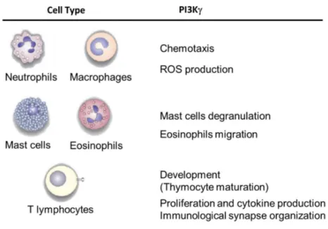
1.7 PI3K in immune system
Several studies described that PI3Kγ is selectively enriched in leukocytes. This enzyme exerts a specific role in distinct leukocyte populations (figure 10) and it is involved in inflammatory diseases (Costa C et al., 2011). Neutrophils and macrophages represent the cellular populations that act as first defense line against an inflammatory insult. In order to effectively exert their function these cells need to migrate to the inflammatory sites through a process called chemotaxis. It was showed that PI3K has an important role in chemotaxis process of neutrophils and macrophages, in fact PI3Kdeficient mice presented an impairment of migration of these cells in response to GPCR-dependent stimuli (Li Z et al., 2000; Jones GE et al., 2003). It is demonstrated that the altered migration observed in PI3Kneutrophils is due to the regulation of cell movement directionality exerted by
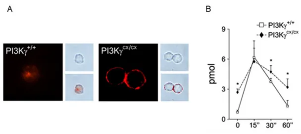
PI3KHannigan M et al., 2002). To study PIP3 function in leukocyte chemotaxis, Costa and coworkers generated a murine model characterized by constitutively expression of PI3K. To generate these knock-in animals, the locus encoding the catalytic subunit of PI3K was replaced with a chimeric minigene containing a mutant form of the human cDNA, in which the CAAX motif, derived from small GTPasi K-Ras, was added at its 5’ end and followed by a neomycin resistance gene cassette sandwiched between loxP sequences. PI3K fused to the specific sequences CAAX-box causes constitutive PIP3 production and its association with membrane fraction (figure 11). Mice with this mutation were viable, fertile and did not show any overt phenotype or alteration of life span. Furthermore, bone marrow-derived macrophages (BMDMs) homozygous for the mutation (PI3KCX/CX) compared with those PI3K presented the same pick of PIP3 production
upon GPCR stimulation but, with the time, the amount of this phosphatidylinositol resulted higher in PI3KCX/CX cells. It is observed that in PI3KCX/CX mice leukocyte proliferation
and survival are enhanced. The Authors showed that the constitutive expression of PI3Kinduced a chemotactic defect in neutrophils and macrophages that fail to efficiently follow the chemotactic gradient because of a less persistent orientation toward the gradient,
29
indicating that modulation of PIP3 levels is crucial for efficient persistent directional movement (Costa C et al., 2007).
An important role of PI3Kis this to mediate the endothelial cells (EC)-neutrophils interaction in order to support the action of these cells of innate immunity in the inflamed vessel wall. In PI3Kendothelial cells there was observable the decrease of
selectin-mediated neutrophil attachment, an important event in the multistep process that enables neutrophils to migrate into inflamed tissues, and the increase of rolling speed of these cells in response to TNF (Puri KD et al., 2005). Moreover, it is showed that the absence of PI3Kmade the neutrophils unable to stably adhere to the endothelial wall in response to
chemokine stimulation (Smith DF et al., 2006).
To fight the inflammatory process, neutrophils and macrophages exert phagocytic action and participate to the “oxidative burst” in order to generate ROS using the NADPH oxidase complex; ROS play a key role in microbial killing via a combination of direct ROS toxicity and indirect activation of proteases. It is demonstrated that PI3K activity is important in regulating NADPH oxidase assembly and activation. In particular, NADPH oxidase is composed by transmembrane proteins and soluble proteins, among the latter the small GTPase Rac. When Rac is activated it migrates from the cytosol to the membrane in order to form an active complex. PI3Ks act in this last process activating Rac protein, via regulation of guanine nucleotide exchange on Rac, and providing lipid anchoring sites for other complex components (Hawkins PT et al., 2007). It was showed that in mouse neutrophils ROS production is regulated only by PI3K, whereas in human neutrophils, ROS generation has a biphasic regulation in which the first phase is dependent on PI3K and the second phase is mediated by p110Condliffe AM et al., 2005).
The analysis of hematopoietic lineage performed on peripheral blood and bone marrow showed that PI3Kmice presented an increase in monocytes, neutrophils and basophils
populations. In the spleen appeared an increase of myeloid cells whereas a decrease in thymocytes were observable. Thymocytes undergo different development stages in which the thymocytes precursors are represented by CD4- and CD8- cells (DN; double negative
30
(CD44+CD25−), DN2 (CD44+CD25+), DN3E (CD44−CD25+FSClow), DN3L
(CD44−CD25lowFSChigh), and finally DN4 (CD44−CD25−) cells. DN4 cells give rise to
immature thymocytes that are represented by CD4+ and CD8+ cells (DP; double positive
cells) that finally mature in CD4+ or CD8+ single positive cells (SP). The first checkpoint in
T cell development occurs at the DN3 to DN4 transition and is called β-selection because only cells that express a functional pre-TCR pass this checkpoint (Michie AM et al., 2002). In PI3Kmice the proportion of SP cells did not result changed, only a slight decrease of
DP cells and an increase DN cells was observable; also the maturation of DN and DP cells did not result altered, suggesting that PI3Kis not required for early thymocytes development. However, it was found that PI3K plays an important role in the thymus homeostasis, controlling TCR and GPCR-induced thymocytes apoptosis. Moreover, in PI3Kmice proliferation and cytokines production of T cells were impaired therefore,
this enzyme exerts an important role in controlling these mechanisms and operates through secondary pathways resulting from TCR stimulation (Sasaki T et al., 2000).
A subsequent study analyzing the role of PI3Kand PI3Kin T cell development showed that it depends on the combined function of p110γ and p110δ. Analysis of DN thymocytes in p110γ/δ−/− mice showed that there was a reduction in the percentage of DN4 thymocytes,
with normal numbers of DN3E and DN3L cells, providing evidences that T lymphocytes have a profound block of their development that occurs at the β-selection checkpoint. p110γ/δ−/− thymocytes presented a reduced pre-TCR signaling with a proliferative defect
and increased apoptosis (Webb LM et al., 2005).
According with studies previously described it was determined the involvement of PI3Kin TCR induced-T cell activation. It was showed that p110γ is activated by the TCR and is involved in PIP3 localization at the immunological synapse (IS). Activated p110γ regulates in turn Rac activation and actin polymerization, which is fundamental for the stability of the IS. Therefore, p110γ affects the interaction between T cells and APCs, which could explain the defective activation of p110γ−/− T cells (Alcázar I et al., 2007).
Martin AL and coworkers demonstrated that CD8 effector T cells lacking p110γ have an impaired ability to migrate into an inflammatory site. This migration flaw is due to an
31
intrinsic defect of these cells since the adoptive transfer of this population in WT mice showed an impairment of p110γ-/- CD8+ migration into inflamed peritoneum. Moreover,
these cells exhibit defective migration in response to a classic inflammatory chemokine, CCL5 (RANTES), as well as the proinflammatory lipid leukotriene B4 (LTB4). According to the cellular defects that occur in the absence of p110-/-,the PI3K KO mice are more
susceptible to infection than control mice (Martin AL et al., 2008).
Figure 10. PI3K function in inflammation. Role of PI3K in different cell types of innate and adaptive immune system (Ghigo A et al., 2010).
32
Figure 11. Effects of PI3Kconstitutive expression. Immunofluorescence analysis revealed plasma membrane distribution of PIP3 in PI3KCX/CX bone marrow-derived
macrophages (BMDMs) but not in PI3K+/+ controls (left, A). Phase contrast images (right
upper, A) merged with confocal images (right lower, A). ELISA measurement of intracellular PIP3 in PI3KCX/CX BMDM showed significantly increased PIP3 levels in
resting conditions, before and after C5a stimulation (B; Costa C et al., 2007). 1.7.1 PI3K in inflammatory diseases
Since p110 orchestrates different functions in leukocyte populations, it has been investigated its potential involvement in various immune disorders. The results from these researchers indicate that this enzyme represents a pharmacological target for autoimmune diseases; for this reason, current studies are focusing on the identification of target molecules for PI3Kγ.
It was demonstrated that the block of this enzyme is able to reduce incidence and severity of pathological condition such as systemic lupus erythematosus (SLE) and rheumatoid arthritis (RA).
SLE is a chronic inflammatory condition characterized by deregulation of T cell-mediated B-cell activation, which causes glomerulonephritis and renal failure. In particular, it occurs an abnormal activation of CD4+ T cells that accumulate as memory cells and that contribute
33
with immunosuppressants, cytostatic agents and corticoids that involve numerous side effects. The abnormal activation of T cells and the consequent B cells expansion can be due to different mechanisms, for example cell-death defects such as Fas-Fas ligand deficiency in MRL-lpr mice and excess of lymphocyte activation signals including class I phosphoinositide-3 kinase (PI3K). Using mice homozygous for the lymphoproliferation spontaneous mutation (Faslpr) it was showed that intraperitoneal administration of a PI3Kγ
selective inhibitor, AS605240 [5-(quinoxalin-6-ylmethylidene)-1,3-thiazolidine-2,4-dione], decreased pathogenic CD4+ memory cells, reduced glomerulonephritis and increased
lifespan in this mouse model of SLE limiting the adverse effects typical of classical treatments (Barber DF et al., 2005).
RA is a chronic systemic inflammatory disorder that affect mainly the joints that appear inflamed with infiltration of different population of leukocytes, including macrophages, neutrophils, mast cells and T cells that migrate to these sites thanks to chemokines and other chemoattractants. Since PI3K is important in mediating mast cell degranulation, leukocyte chemotaxis and activation, pharmacological blockade of this molecule was proposed as a new therapeutic strategy for RA. It was demonstrated that oral administration of AS605240 inhibitor suppressed neutrophil chemotaxis and, in turn, the progression of joint inflammation and cartilage erosion in collagen-induced arthritis (CIA; Camps M et al., 2005).
It was also showed the therapeutic potential of PI3K inhibitors in respiratory diseases. The dual PI3Kγ inhibition is showed to be effective in the allergic asthma and obstructive pulmonary disease (COPD) treatment. Intranasal administration of an aerosolized form of the double-selective compound TG100-115 to OVA-immunized mice results in a marked decrease of lung eosinophilia, accompanied by reduced production of IL-13, diminished perivascular and peribronchiolar leukocyte accumulation, and impaired mucin production. The same compound significantly reduces neutrophilia and TNF- production in LPS or cigarette smoke exposure-induced murine model of CODP (Doukas J et al., 2009).
Since the role of PI3Kγ as an amplifier of mast cell activation, it takes a central role in the modulation of inflammation and allergy. In fact, PI3Kγ-derived PIP3 was a key element for external Ca2+ influx and release of histamine-containing granules from mast cells.
34
According to this, the absence of PI3Kγ disrupts mast cell degranulation (figure 10) and protects animals from edema formation induced by passive systemic anaphylaxis (Laffargue M et al., 2002). Finally, PI3Kγ exerts its role also in eosinophils, an important component of the inflammatory response in allergic asthma. It was showed that in chronic OVA-challenged PI3Kγ-deficient mice the airway responsiveness, the number of bronchoalveolar lavage (BAL), peribronchial eosinophils and peribronchial fibrosis were reduced than chronic OVA-challenged WT mice. It was proposed that the reduction in eosinophils number into the airway is likely due to reduced chemokine-induced migration of eosinophils (figure 10). On the other hand, the reduced peribronchial fibrosis can be attributed to reduced numbers of TGF-β1+ cells and reduced Smad 2/3 signaling, that are
35
1.8 PI3Kγ and cardiovascular diseases
There are studies that explored the role of PI3Kγ in the cardiovascular diseases in which there is an involvement of the immune reactions. Among these pathological conditions atherosclerosis and myocardial infarction (MI) were found.
Atherosclerosis is a chronic disease characterized by lesions formation in the arteries, known as plaques, with immune infiltration, lipid accumulation, cell death and fibrosis. These plaques, besides to cause stenosis, can break determining thrombotic occlusion of the artery. In the heart, it can occur myocardial infarction and heart failure, whereas in the brain atherosclerosis can cause ischemic stroke. If the pathology affects other arterial branches, they can occur renal impairment, hypertension and abdominal aortic aneurysms (Hansson GK and Libby P, 2006). The role of oxidized LDL (low density lipoprotein) and atherogenic cytokines/chemokines in trigger PI3K signaling and in Akt activation in inflammatory cells was already known (Biwa T et al., 2000). A paper of 2007 demonstrated that PI3Kγ is the specific isoform involved in this pathway and it is required for activation of Akt in macrophages in response to oxidized LDL, atherogenic cytokines, and angiotensin II in vitro and in atherosclerotic lesions of hypercholesterolemic mice in vivo. To study this pathological condition was used apolipoprotein E-deficient (ApoE-/-) mice
that develop spontaneous atherosclerotic lesions and are well characterized as a model for the early stage of atherosclerosis. Genetic deletion of PI3K in the ApoE-/- mice correlated
with the persistent reduction of plaque size. For this reason, p110γ was proposed as target for prevention of atherosclerotic disease (Chang JD et al., 2007). It was also demonstrated that AS605240-mediated PI3Kinhibition reduced lesion formation in ApoE-/- mice. This
datum is also confirmed using LDLR-/- (low density lipoprotein receptors) mice, another
model of atherosclerosis characterized by development of advanced lesion, in which the inhibition of PI3Kattenuated advanced atherosclerosis. The generation of mice lacking PI3K exclusively in immune cells by bone marrow transplants from PI3K-/- mice to
LDLR-/- recipient mice demonstrated that loss of PI3K in immune cells is sufficient to
reduce atherosclerosis. Therefore, PI3Kexpression in immune cells drives the formation of atherosclerotic plaques. Moreover, in this model was observable a decrease of