Tagging of potentiated
synapses with a locally
translated optogenetic
reporter
abstract
Increasing evidence points to the importance of dendritic spines in the formation and allocation of memories, and alterations of spine number and physiology are associated with memory and cognitive disorders. Modifications of the activity of subsets of synapses are believed to be crucial for memory establishment. Indeed, treatments or conditions that affect synaptic potentiation almost inevitably lead to an impairment in the acquisition of memories. Thus, the potentiation of synaptic transmission is a likely correlate of the acquisition of associative memories. This suggests that, in the brain, memories are formed and stored as a persistent modification of the strength of a subset of synapses within a neural circuit. To test this hypothesis, it would be necessary to re-excite such a subset of synapses and observe a behavioural response coherent with the encoded memory. A precondition for this experimental test is the ability to detect potentiated synapses during the encoding phase. However, the development of a method to directly test this hypothesis is currently lacking. Here I developed a hybrid RNA/protein approach to selectively express proteins at potentiated synapses (SynActive). To open the possibility for recalling synaptic activity, SynActive can be used to express a light-sensitive membrane channel of the Channelrhodopsin2
family (SA-Ch). After the characterization of SA-Ch using primary neuronal culture SA-Ch was expressed in the hippocampus of living mice. By controlling the timing of its expression with doxycycline, I was able to tag and identify potentiated synapses during the exposure of mice to a novel context. This allowed the identification of a candidate synaptic engram encoding the representation of the explored context. Furthermore, SA-Ch was used to map the relative distribution of potentiated synapses within and across neurons. In both the hippocampal CA1 and dentate gyrus regions, I demonstrate the presence of clusters of potentiated synapses, whose dimension (i.e. number of synapses) is increased by the behavioural task. I also highlight differences between CA1 and DG in terms of distribution of potentiated synapses between different neurons. The results provide strong experimental evidence in support of proposed models for memory acquisition and activity flow in the hippocampal circuit. The SynActive approach can then be used to map potentiated synapses in the brain and will make it possible to re-activate neurons only at previously activated synapses, testing if the activity of the identified synaptic trace is sufficient to recall the memory of the encoded representation. Furthermore, it will provide an experimental way to expand the current neuron-tagging technologies in the investigation of memory processes to the synaptic level.
‟ Memory is the steamstress, and a capricious one at that. […] Thus, the most ordinary movement in the world, such as sitting down at a table and pulling the inkstand towards one, may agitate a thousand odd, disconnected fragments, now bright, now dim, hanging and bobbing and dipping and flaunting, like the undelinen of a family of fourteen on a line in a gale of wind. ”
Table of Contents
Introduction ... 1
Memory... 3
The engram ... 4
The law of Engraphy ... 4
The law of Ecphory... 5
Where are memories stored? ... 6
The hippocampus ... 6 Hippocampus anatomy ... 8 Other areas ... 10 Cell assemblies ... 10 Activity mapping ... 11 Engram cells ... 15
Creating new memories ... 21
From engram cells to connections ... 24
Synapses and memory ... 26
LTP ... 27
LTP mechanisms... 30
Plasticity related transcripts ... 36
LTP and memory ... 42
Synaptic engrams ... 49
Synaptic vs cellular engrams ... 50
Aim of the thesis ... 56
Materials and Methods... 59
Constructs ... 61 Cell culture. ... 64 FRET... 66 FLIM-FRET ... 67 Neuron treatments. ... 68 Immunofluorescence. ... 69 Microscopy. ... 70 Two-photon uncaging. ... 71 Calcium imaging. ... 72 Culture optogenetics. ... 73
In utero electroporation and animal experiments. ... 75
Data quantification. ... 78
Statistics. ... 82
SynActive Generation and Validation ... 85
The SynActive approach ... 87
RNA candidates ... 89
Cooperative protein/RNA sequences ... 94
BDNF sequences ... 104
SA-Ch localizes to postsynaptic densities ... 109
SA-Ch marks potentiated spines ... 112
Focal LTP induction drives SA-Ch expression ... 116
Synaptic light-evoked calcium transients ... 120
Optogenetic activation of SA-Ch neurons ... 125
Discussion ... 129
A marker for potentiated synapses ... 129
Protein permanence after translation ... 131
Synaptic translation... 132
SA-Ch functionality ... 134
In vivo mapping of potentiated synapses ... 137
In vivo delivery for synaptic tagging ... 139
SA-Ch expression pattern in vivo ... 140
In vivo SA-Ch expression does not alter neuron physiology ... 143
Mapping synapses potentiated by context exploration 147 Potentiated synapses form clusters in vivo... 149
Discussion - Clustering in LTP and memory encoding ... 159
Uneven distribution of spines in DG dendrites ... 169
Uneven distribution of potentiated spines across neurons ... 175
Discussion – Uneven distribution of potentiated synapses
... 177
Concluding remarks ... 179
Tagging potentiated synapses ... 181
A tool for synapse reactivation ... 189
A SynActive approach to study the proteome ... 195
Final conclusions ... 200 References ... 203 Appendix A ... 235 Appendix B ... 253 Appendix C ... 261 Publications ... 273 Acknowledgments ... 275
Memory
Memory is the ability to store mental representations of past events, and enables to respond accordingly when a new situation is faced. Memories shape our interpretation of reality, and have a unifying role in the representation of our mental life (Kandel et al., 2014). There are two main forms of memory: implicit memory regulates our perceptual and motor skills (“memory of ways”), and declarative memory is involved in the representation of facts and events, people and objects (“memory of things”). The latter has such a prominent and pervasive role in our life, making sense of the fragments of reality that reach our sensation, that the loss of memory in aging and pathology shatters the life of patients.
Memory is often perceived as a collection of pieces of information and stories, which are stored in the mind, in Plato’s metaphor, as if it were a block of wax, and memories were imprinted on it (Plato). Aristotle then argues that memory is a state of imagination relating as a copy to its original and that memory rests in sense-perception rather than imagination (Aristoteles). These ideas would then have a profound effect on the philosophers of memory, and to scientists embarking in the search of the mechanisms of memory.
The engram
The concept of “engram” was first introduced by early 20th
century scientist Richard Wolfgang Semon to describe “... the enduring though primarily latent modification in the irritable substance produced by a stimulus...” (Semon, 1921). An intrinsically linked concept to engram in Semon’s theory of mind is ecphory, which he defines as “... the influences which awaken the mnemic trace or engram out of its latent state into one of manifested activity...”.
The law of Engraphy
Semon postulated that all simultaneously concurring excitations form a connected complex in the organism (Semon does not explicitly name the brain as the physical substrate for memories) leaving behind a connected trace. Such trace is what Semon calls the engram (Semon, 1921). An important tract of Semon’s engram theory is its unitary nature in the formation of the excitation-complex. According to his postulate, sensations would be represented by the emergence of a unitary representation in the “active substance”, which would then be maintained as if this representation is inscribed onto the substance in the form of a dormant trace (Schacter et al., 1978).
The law of Ecphory
Intrinsic in the engram definition is the ability of the memory trace to be retrieved, being reactivated from a state where the engram is present, although latent (Semon, 1921). Only part of the engram needs to be present to recall it in its entirety (Schacter et al., 1978): “resemblance, that is to say, partial coincidence between the components of an actual group of excitations and those of any previous engram-complex, causes ecphory of the latter through the former”. The cuing of a stimulus, and the resulting ecphory, “does not strengthen an already existing engram, but generates a new engram, and the mnemic excitations resulting from any subsequent ecphory are in homophony” (Semon, 1921). That is, anytime an engram is reactivated, it is subject to changes and it is engraphycally stored in a modified version. The old (original) and the new engrams become associated by contiguity, in an interplay described as homophony.
One last notion that Semon introduced is the competition between engrams for ecphory. When two engram-complexes are related equally to a particular ecphoric cue, Semon argued that, in most situations, serially processing the two complexes would happen (Semon, 1923).
Where are memories stored?
“To follow [the engram] into the molecular field seems to me […] a hopeless undertaking at the present stage of our knowledge; and for my part I renounce the task”
Semon (1923) The search for the physical location of engrams in the brain was pioneered by Karl Lashley, who systematically lesioned portions of cerebral cortex in the attempt to drive a correlation with the deterioration of the performance of the animal in solving a memory maze task (Lashley, 1950). Although establishing a cornerstone criterion to assess the involvement of a brain area in the memory, he found that no lesion could impair the memory task, and only large-scale lesions did (Josselyn et al., 2017).
The hippocampus
In search of epileptic foci in his patients, Wilder Penfield found that stimulation of the lateral temporal lobe elicited the recall of past memories in a fraction of subjects (Tonegawa et al., 2015). Brenda Milner, analysing amnesia in Penfield’s patients with resected epileptic lobes, including famous patient HM, found that the hippocampal region, including the hippocampus proper and adjacent temporal
lobe structures (Figure 1), was critical for the formation of long-term memories (Scoville and Milner, 1957). Patients with surgically removed hippocampal regions could form short-term memories; however, they were not able to keep them in mind for more than a few minutes. At the same time, they lost memories formed up to 11 years before the hippocampal damage (Andersen et al., 2007). Decades of experiments have then confirmed and demonstrated the involvement of the hippocampus in the formation of new memories and, in particular, of episodic memories (Neves et al., 2008). As in the case of patient HM, lesions to the hippocampus produce severe anterograde amnesia (i.e. the impairment in forming new memories) and time-dependent retrograde amnesia (i.e. the inability to recall past
Figure 1 Coronal magnetic resonance of patient HM compared to a control
who did not undergo surgery. Note the bilateral resection of the hippocampal formation (H) and damage of the enthorhynal (EC) and perirhynal cortex (PR). Reproduced from Andersen et al. (2007).
experiences) (Anagnostaras et al., 2001; Langston et al., 2010). Consistently, rodents with experimentally damaged hippocampal displayed sever deficits in learning tasks involving more complex mazes than a Y-turn (Andersen et al., 2007). Olga Vinogradova, among the first scientists performing single unit registrations in the hippocampus, later demonstrated that hippocampal neurons responded to the presentation of a novel environment. Later work also showed that the hippocampus was involved in the spatial representation of the animal location (Andersen et al., 2007).
Hippocampus anatomy
The hippocampus is a conserved structure in vertebrates characterized by a peculiar ultrastructure, with neurons prevalently disposed in single layers, and a prominent laminated segregation of inputs (Figure 2) (Andersen et al., 2007). Anatomically, the hippocampus is composed by the dentate gyrus (DG) and the cornu ammonis, which comprises areas CA1, CA2, CA3 and CA4. The main input comes from the entorhinal cortex (EC), in the parahippocampal gyrus (Figure 2), via the perforant path that synapses the dendrites of granule cells in the molecular layer (sm in figure 2b). The EC also forms synapses with apical
CA1 dendrites in the stratum lacunosum moleculare (slm), distally to the soma layer. DG then forms excitatory synapses with CA3 pyramidal neurons, which then project to CA1 pyramidal neurons, particularly in the stratum radiatum (sr) containing the proximal portion of apical dendrites (Andersen et al., 2007). Pyramidal neurons in CA3 and CA1 form a single cellular layer and display both apical (in the sr and slm) and basal dendrites (in the stratum oriens, so) (Figure 2c). CA1 basal dendrites receive prevalently CA3 inputs, including a significant proportion of CA3 axons from the hippocampus in the other hemisphere (Shipton et al., 2014), as well as less characterized inputs from the other hemisphere CA1.
Figure 2 a 1911 drawing by Ramon y Cajal showing a rabbit hippocampus
stained with the Golgi impregnation method. b Nissl staining of mouse hippocampus. so: stratum oriens, sr: stratum radiatum, slm: stratum lacunosum moleculare, sm: stratum moluculare. Modified from Paxinos and Franklin (2001) c Typical DG, CA3 and CA1 neurons showing the differences in the dendritic arbours shape.
Other areas
The hippocampus is not the sole area involved in the formation and storage of memories. The amygdala plays a fundamental role in the encoding of the emotional valence of a memory, and some associations like the tone-shock conditioning involve inputs from the auditory cortex in a hippocampus-independent manner. Cortical areas like the prefrontal cortex are also recruited in contextual memories (Tse et al., 2007; Ben-Yakov et al., 2015), which have the tendency to become progressively independent of hippocampus (Genzel et al., 2017; Nadel and Moscovitch, 1997; Squire, 1986). However, the hippocampus remains indispensible for the formation of contextual memories, and lesions to such area cause severe and permanent retrograde amnesia (Nadel and Moscovitch, 1997).
Cell assemblies
Imagining a physical counterpart of engrams, Donald Hebb postulated groups of reciprocally interconnected cells that are simultaneously active during an event. Their interconnection produces a recurrent activity which in turn, if sustained for a long enough time, can induce metabolic changes that strengthen the interconnections between
assembly cells (neurons that fire together wire together), allowing the event to be represented in long-term memory. Because of their mutual connections, the assembly can be activated by the activity of any subset of cells (Josselyn et al., 2017).
Activity mapping
A first step towards the engram is the identification of neurons that are active during the encoding phase of a memory. Immediate Early Genes (IEGs) are a class of genes whose expression is induced as a result of neuron activation, and comprises genes like Arc (also known as activity regulated gene 3.1, arg3.1), c-fos, zif268, Homer1a and
cox-2 (Guzowski et al., cox-2001; Kubik et al., cox-2007). Making use of
this property, Guzowski et al. (1999) pioneered the use of IEGs to map neurons activated by a specific experience. Active neurons can be therefore identified looking at either IEG transcription or translation by in situ hybridization or immunodetection of the resulting protein. For example, by detecting Arc transcript in the nucleus (2-15 minutes from stimulation) or in the cytoplasm (15-30 minutes) (Figure 3), Guzowski et al. (1999) were able to identify neurons activated by the exploration of a novel context. In addition, they could compare the sets of active neurons in two
different contexts separated by 20 minutes by looking at the subcellular localization of Arc signal (i.e., nuclear or cytoplasmic).
Transcription of reporters from IEG promoters – and their translation – has been since used to mark and recognize active neurons, FosGFP mice being one of the first examples (Barth et al., 2004). A major advance came through when
Figure 3 (a-h) Arc (green) and zif268 (red) expression induced by electroconvulsive shock in CA1 neurons. a caged control b 2 min c 5 min d 15 min e 30 min and f 60 min after stimulation. g RNAse A treatment control and h sense riboprobe control. i Arc expression in hippocampal regions DG, CA3 and CA1, and in the parietal cortex in home caged animals (left column) and after exploration of a novel environment (central and right comuns). Modified from Guzowski et al. (1999).
Reijmers et al. (2007) combined the use of IEG c-fos promoter with the tetracycline system to tag active neurons in a defined time window given by the removal of doxycycline repression of tTA activity. c-fos+ neurons can
be permanently tagged because the initial activation of tTA transcription factor is converted in a self-sustaining expression loop by a bidirectional promoter driving the expression of lacZ and doxycycline-insensitive tTA* (Figure 4). These TetTAG mice were removed from a doxycycline diet before auditory fear conditioning or contextual fear conditioning, and active neurons were tagged by LacZ expression, then mice were put back on doxycycline diet. A few days later, mice were tested for memory recall, and active neurons during this second
Figure 4 a scheme of TetTAG
system. Transactivator tTA under the c-fos promoter is expressed by neuron activation. If doxycyline is removed, tTA binds bidirectional TetO promoter, which expresses lacZ and doxycycline-insensitive tTA*, allowing for the autonomous lacZ expression. b examples of home caged and seizure animals. Modified from Reijmers et al. (2007).
exposure were identified by Zif268 expression. This demonstrated that neurons that were active during the encoding of the memory were preferentially reactivated during memory recall (Reijmers et al., 2007).
The use of the Tetracycline system is just one of the possible ways to implement the activity tagging (Gore et al., 2015). For example, activity tagging has been implemented using tamoxifen-sensitive Cre (CreER)-mediated recombination. CreER expression is driven by Arc promoter, and the floxed reporter is only expressed when tamoxifen is administered to the animal (Denny et al., 2014; Guenthner et al., 2013). Notably, also synthetic versions of IEG promoter have been recently started to emerge and to be implemented in the activity tagging, usually modifying existing IEG transcriptional regulatory elements to increase fold induction (Kawashima et al., 2014; Sørensen et al., 2016). A variant of this system expresses via the c-fos promoter a membrane protein that acts as a receptor to a modified envelope protein of a viral vector, enabling only active cells to be transduced (Sakurai et al., 2016).
An ingenious way to restrict the timing of activity tagging has been recently described, and allows tagging active neurons by combining light and calcium influx (Lee
et al., 2017; Wang et al., 2017). Making use of the M2 peptide, that binds CaMKII in presence of calcium, and a light sensitive LOV domain, both groups were able to transduce light signal in coincidence with activity-related calcium influx into the expression of a reporter. They showed the usefulness of this approach by labelling active neurons in the motor cortex during rewarded tasks, although it not exactly clear how this set is related to c-fos+ neurons.
If proven successful, this could restrict the tagging time window with a precision that currently available activity-tagging technology cannot achieve, although more limited by the spatial dimension.
Engram cells
If tagged neurons constitute a putative engram, it is expected that manipulating their activity has a behaviour counterpart in the associated memory task. The role of specific cells in the encoding of memory was first suggested by experiments from the Mayford group: Garner et al. (2012) tagged active cells during the presentation of context A with chemogenetic, CNO-responsive activator hM3Dq. This
ensemble was then reactivated during fear conditioning in context B by CNO administration; this formed a mixed representation A/B, as upon testing the animal in context A
alone, no freezing was observed (Figure 5b) – however, animals in context B froze significantly less if no CNO was administered (Figure 5c). The ensemble tagged by hM3Dq
represents a neutral (not feared) engram indeed, since CNO administration during retrieval of a conditioned context competed with memory recall blocking freezing, suggesting memory occlusion (Figure 5e).
The experiment described above generated a mixed, artificial engram that had a complex crosstalk with the physiological circuit, which is assumed to exist rather than proved. If an engram exists, it should recruit a number of cells which are tagged and, after the stimulation has passed,
Figure 5 Generation of a synthetic memory trace a Outline of experiment.
‘Context A’ engram is labeled with DREADD expression and later reactivated by CNO in concomitance to Context B conditioning with a footshock. b,c Retrieval in context A (b) or B (c) for hM3Dq and control mice shows the formation of a mixed A/B memory trace. d,e Reactivation of ‘context A’ engram competes with the recall of conditioned context B during retrieval. f Scheme of construct expression in engram cells. Modified from Garner et al. (2012).
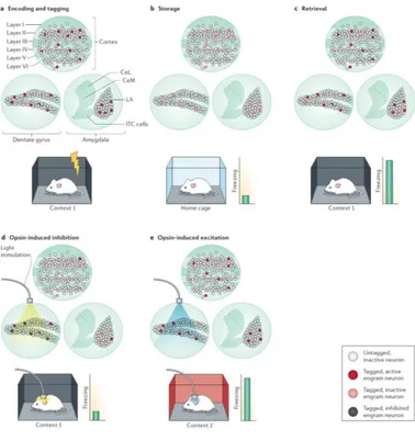
Figure 6 a During fear conditioning, a mouse is placed in context 1 and
given a footshock, activating neuronal ensembles in the dentate gyrus, cortex and lateral amygdala. Neurons are tagged during the first epoch of stimulus presentation, or encoding. b In the home cage, the engram is consolidated, and tagged engram neurons are inactive. c Re-exposure to context 1 reactivates the ensemble, causing the animal to freeze. d If tagged neurons in the dentate gyrus are optogenetically silenced when the mouse is returned to context 1, mice show reduced conditioned fear, indicating impaired memory retrieval, which is reflected in a reduced activation of downstream areas. e Optogenetic activation of tagged engram neurons in the dentate gyrus alone is sufficient to act as a memory retrieval cue such that mice now freeze in a third unrelated context (context 2). Reproduced from Josselyn et al. (2015).
they become part of a silent trace, only to be reactivated by the later retrieval induced by a stimulus (or part of it) sufficiently close to the original one (Figure 6a-c). If a subset
Figure 7 Optogenetic engram labeling and reactivation a Schema of AAV
activity Tet-tagging b Experimental outline (c-g) expression of ChR2-EYFP on dox (c), d home caged animals e fear conditioned, f exposed to context, non shocked animals, g seizure, demonstrating dox- and activity-dependence of ChR2-EYFP expression. h Quantification of the fraction of ChR2-EYFP cells in c-g. i expression of ChR2-EYFP (i1) in DG cells strongly correlate with c-fos (i2) expression. l Reactivation of tagged DG cells with blue light illumination induced significant freezing in fear conditioned (l) but not in non-shocked (m) animals, demonstrating the reactivation of the encoded memory trace. Modified from Liu et al. (2012).
of cells makes up an engram, then, it is expected that silencing their activity during the recall should block the animal response and, conversely, reactivating their activity in the absence of the original natural context should elicit that same response (Figure 6d, e).
Using Tet-tag mice, Liu et al. (2012) tagged c-fos+
neurons during a fear conditioning epoch while in Context B, and demonstrated the existence of such a population engram in the hippocampus DG (Figure 7): tagged cells expressed ChR2-EYFP, and their reactivation with blue-light illumination recalled the fear memory, causing the animal to freeze. This was paralleled by experiments performed by Denny et al. (2014), who confirmed the existence of such engram population in the DG (as well as in CA3) by tagging active cells in an analogous manner with inhibitory opsin Arch-GFP. Green-light illumination significantly reduced freezing with respect to control animals when exposed in a fear-conditioned context. Together, these two works demonstrate that a subset of active neurons in the DG is part of an engram representing the association of a specific context with a noxious stimulus (the shock), and that their activity is both necessary (Denny
et al., 2014) and sufficient (Liu et al., 2012) to at least partially recall the original experience.
The cell tagging-and-capture described above has been since found use in many brain areas, including the amygdala (Redondo et al., 2014), ventral CA1 (Okuyama et al., 2016), the retrosplenial (Cowansage et al., 2014) and prefrontal cortex (Kitamura et al., 2017), each encoding different portions of engrams in various forms of memory (e.g. contextual, social) (Tonegawa et al., 2015). A non-exhaustive list of brain areas where engrams have been identified is presented in Figure 8. Engrams can be identified
Figure 8 Non-exhaustive list of regions where engram cells have been
identified. Three criteria may be used to define them: observational (e.g. calcium imaging during activity) provide the less strong evidence of the three – loss-of-function, i.e. tag-and-inactivation experiments – gain-of-function implies that cells have been reactivate to recall existing memories or create new ones. Reproduced from Tonegawa et al. (2015).
by either of three criteria, that is (i) observational: the activity of a defined set of neurons is correlated with a specific memory task or memory retrieval, for example by means of calcium imaging (Kitamura et al., 2015; Roy et al., 2017a) or IEG expression (Deng et al., 2013; Tayler et al., 2013); (ii) loss-of function: inhibiting the activity of engram cells should result in memory impairment, that is engram cells activity is necessary for the recall (Figure 6d), for example in Denny et al. (2014); (iii) gain-of-function: stimulating their activity is sufficient to instate the memory recall (Figure 6e), as in Liu et al. (2012).
Creating new memories
Given the specialization of brain areas, a complete engram is likely to be composed by multiple components, which may be localized in distinct regions (Tonegawa et al., 2015). Each component then could convey defined information of the complete engram. It would be possible, then, to create false memories by combining multiple subtraces in a new, artificial engram. Ramirez et al. (2013) tried to associate a neutral, contextual engram with a noxious stimulus by means of optogenetic tagging-and capture technology. The active population of cells was first tagged with ChR2-Cherry during the presentation of context A (Figure 9), and after 24
hours the animal was first conditioned in context B while putative ‘context A’ engram was activated by blue light. The next day, animals were put back in context A, where they never experienced a shock, and tested for freezing. While DG engram reactivation could form an association of context A to an unpleasant experience, the (putative) CA1 engram could not (Figure 9). This could mean that (i) CA1 does not allocate part of the engram, (ii) CA1 engram does not represent contextual information, (iii) CA1 has no causal role in providing such information, or (iv) the putatively identified engram deviates too much from the actual one.
Figure 9 (a-d) c-fos::tTA mice injected with AAV9-TRE-ChR2-mCherry in the
DG were exposed to context A while off Dox, then put back on Dox and exposed to the same context A (a,c) or a novel context C (b,d); activated cells express c-fos (green). (f,g) (Top) Training and testing scheme of animals injected with AAV9-TRE-ChR2-mCherry or AAV9-TRE- mCherry. (Bottom) Animals’ freezing levels in context A before fear conditioning and in context A and C after fear conditioning (h-n) Same as in (a-g) except the virus injection was targeted to CA1. The lack of freezing in the test session (A’) shows that the identified CA1 ensemble could not be associated with the aversive stimulus. Reproduced from Ramirez et al. (2013).
Explanation (i) can be excluded by a series of evidences that imply the presence of engram cells: first, memory-specific cells have been identified by means of IEG expression (Tayler et al., 2013). Second, optogenetic CA1 inhibition prevents contextual fear memory recall (Goshen et al., 2011; Sakaguchi et al., 2015) and third, selective silencing of c-fos+ cells in CA1 impaired memory retrieval
in fear conditioning (Tanaka et al., 2014) and novel object recognition (Nomoto et al., 2016). Fourth, CA1 is the main area sending outputs from the hippocampus, while DG only projects to the downstream CA3 area (Basu and Siegelbaum, 2015; Cenquizca and Swanson, 2007; Paxinos, 2015). CA3 then massively projects to CA1, so CA1 is, by definition, part of the engram. Explanation (ii) is also unlikely, given the overwhelming amount of data implying CA1 as a major area processing spatial and contextual information (Barrientos and Tiznado, 2016; Jeffery, 2007; Ocampo et al., 2017). Similarly, explanation (iii) can be also excluded for the same reasons as (i); it is also difficult to imagine how CA1 contextual (sub)engram could not have a causal role in the building of the memory engram, even if it is role was only to relay the activity generated in the DG via the trisynaptic circuit. We are therefore left with explanation (iv). Indeed, a high proportion of total cells in CA1 is tagged
in Ramirez et al. (2013) and, most importantly, there is significant overlap in the engram representing two different contexts (Figure 9l). Indeed, optogenetically silencing CA1 ‘context A´’ engram inhibits freezing during the recall of an animal trained in a similar context A (which activates an overlapping population of neurons to A´), but has little of no effect on an animal trained in a distinct context B (Tanaka et al., 2014). The vast majority of tagged cells are also expressing c-fos (Figure 7i) (Liu et al., 2012), so it must be concluded that, if an engram exists in CA1, it is either a subset of this population, or it otherwise represented within it.
From engram cells to connections
A similar proportion of cells is activity-labelled in CA1 when the animal is exposed to a new context when this is coupled to a shock and when it is not (Figure 10), further suggesting that a contextual engram is formed in the hippocampus independently from an association (Ohkawa et al., 2015). Accordingly, presentation of context B alone activity-tagged a population of cells in CA1; later, animals were shocked in a different context without giving them the time to explore it (Ohkawa et al., 2015). This was not enough
Figure 10 Artificial association of pre-stored memories a expression of
ChR2-EYFP by context exposure or immediate shock when off-dox b experiment and outcome of optogenetic association of the two memories. The concomitant activation with light of the tagged amygdala and CA1 ensembles could form a meaningful association. c The association between the two engrams is dependent on NMDAR activation and de novo translation. Modified from Ohkawa et al. (2015).
OFF Dox + Home Cage OFF Dox + “Unpaired” CA1 BLA a b c
to induce an association between the shock and context B the next day. However, optogenetically co-activating the CA1 ‘context B’ engram and BLA ‘foot shock/Ouch ouch ouch!’ engram was sufficient to form an association between the two engrams in a unitary representation: when tested in context B, but not in the unrelated context C, animals froze significantly. This association was blocked if BLA and CA1 were injected with NMDAR blocker D-AP5 or translation inhibitor anisomycin, suggesting a Hebbian-like form of plasticity (Figure 10c).
Synapses and memory
Synapses, the physical connections between neuron, have always been ascribed a critical role in learning and memory processes (Rudy, 2008; Yuste, 2010). Virtually all excitatory synapses are associated with postsynaptic structures called spines. Due to their structure, spines have the ability to compartmentalize secondary signals, and to modify their responsiveness as a consequence of their past activity (Yuste, 2010). For its properties, synaptic plasticity has been long regarded as a candidate for sustaining changes that occur during memory encoding.
LTP
First described by Bliss and Lømo (1973) in the rabbit hippocampus, LTP is the long-lasting change in synaptic efficacy observed after a strong initial stimulation. LTP is usually measured as change in field potential, but is also detected in whole-cell (Nicoll, 2017). While some forms of LTP act at the presynaptic level (Castillo, 2012), most mechanisms are a modification of the postsynaptic response (Kandel et al., 2016).
A broad classification distinguish E-LTP from L-LTP, plus a series of changes in synaptic efficiency on shorter time scales (e.g. facilitation, depression…) which have more to do with biophysics than with biological regulation. Typically, LTP is induced by tetanic or theta-burst electrical stimulation, or by stimulation of the presynaptic neuron while depolarizing the postsynaptic neuron (Wigstrӧm et al., 1986). Although E- and L-LTP can be dissociated (Kelleher III et al., 2004; Park et al., 2014), generally E-LTP precedes and L-LTP is only observed if the stimulation is sufficiently strong. Weak tetanisation (one train) of the Schaffer collateral causes an increase in synaptic response that decays to pre-induction level in 1 or 2 hours. This E-LTP is not sensitive to transcription or translation inhibitors (Kelleher III et al., 2004), and is due to the
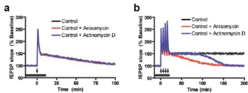
exposure of more AMPA-type glutamate receptors (AMPAR) which are not, generally, permeable to calcium (Kandel et al., 2016). L-LTP is induced by stronger stimulation (e.g. four trains of tetanisation) and lasts much longer, typically hours and (possibly) days (Kandel, 2012). Unlike E-LTP, L-LTP is affected by translation inhibitors like anisomycin and emetine, as well as transcription inhibitors (whose effect, however, acts on a longer timescale than translation inhibitors) (Figure 11) (Kelleher III et al., 2004).
When translation is blocked, only E-LTP is observed; thus, E-LTP and L-LTP are generally viewed as two distinct mechanisms that function in parallel from a common induction signal. Indeed, a slow rising form of L-LTP is
Figure 11 a Time course of E-LTP induced by a single tetanus at CA1
synapses. b Time course of L-LTP induced by four trains of tetanization. In red and blue, treatment with anisomycin or actinomycin D during stimulation. Reproduced from Kelleher III et al. (2004).
induced by BDNF without the induction of E-LTP (Kelleher III et al., 2004; Reymann and Frey, 2007). This is also consistent with the tag-and-capture phenomenon: single tetanisation of a pathway (S2) – which normally induces transient LTP – can be converted into a long lasting form if it is coupled by the strong tetanisation of another pathway (S1) (Figure 12). This suggests that, upon induction, a tag is created in S2 synapses that can capture effectors of LTP generated by S1 tetanisation, collectively called Plasticity Related Products (PRPs) (Frey and Morris, 1997).
Figure 12 Synaptic capture and tag at CA3 to CA1 connections a Scheme
of setup and possible mechanism b Anisomycin blocks L-LTP in 4x tetanized S1 and S2 input pathways c Prior 4x tetanization of S1 prevents anisomycin block of S2 L-LTP. d a weak tetanization of S2 cannot be converted into L-LTP by prior 4x tetanization of S1. e 1x strong tetanization alone produces a decaying LTP. However, if it is preceded by 4x tetanization of S1, also 1x strong tentanization of S2 produces a long lasting form of L-LTP. Modified from Frey and Morris (1997)
LTP mechanisms
The main form of L-LTP is NMDAR-dependent LTP. NMDAR are calcium-permeant glutamate receptors found predominantly on the postsynaptic sites. Unlike AMPAR, NMDAR are normally blocked by extracellular Mg2+ ions
that occlude the channel pore (Kandel et al., 2013). The depolarization of the postsynaptic neuron removes the Mg2+
block; therefore, NMDAR are detectors of coincident activity of the pre- and postsynaptic neurons (Lüscher and Malenka, 2012). Calcium influx through NMDARs activates a series of events including the recruitment of kinases like CaMKII, PKC and PKA (Mayford et al., 2012).
Selective deletion of NMDAR1 (grin1) in the CA1 (Tsien et al., 1996), CA3 (Nakazawa et al., 2002) and DG (McHugh et al., 2007) regions abolishes LTP induction in the corresponding pathway (respectively, EC-DG via perforant path, DG-CA3 via Mossy fibers, and CA3-CA1 via the Schaffer collateral). Downstream, αCaMKII phosphorylation and activation is also necessary for LTP induction. Inducing LTP causes a spine-specific αCaMKII activation (Lee et al., 2009) and recruitment from the surrounding regions (Otmakhov et al., 2004). LTP is abolished in acute slices in αCaMKII knock-out mice (Silva et al., 1992a). In addition, knock-in mice with
non-phosphorylable αCaMKII T286A lack LTP; similarly, phosphomimetic T286D have occluded LTP (Kandel et al., 2016; Lisman et al., 2012). NMDAR activation activates CaMKII holoenzyme via calcium/calmodulin (calcium chelators EGTA and EDTA block LTP induction), which can mediate LTP by phosphorylating a number of downstream proteins, including αCaMKII itself (Lisman et al., 2012).
LTP and local translation
As seen above (Figure 11), blocking protein synthesis with anisomycin, cycloheximide or emetine blocks L-LTP induction (Fonseca et al., 2006a). Given the temporal profile of L-LTP in presence of translation or transcription inhibitors (Figure 11), and that the sensitive period for inhibition is around the time of induction, the treatment is likely to interfere with translation from pre-existing mRNAs. Indeed, LTP can be induced even when dendrites are physically isolated from the cell body (Kang and Schuman, 1996) (Figure 13). Even so, LTP is sensitive to D-AP5 and cycloheximide; in addition, LTP is accompanied by
35S-methionine incorporation, indicating novel protein
translation (Vickers et al., 2005). 3H-leucine is incorporated
Figure 13 Translation of dendritically localized tales place in dendrites. a
BDNF-stimulated dendrites translate a CaMKII reporter even when severed from the cell body. Reproduced from Aakalu et al. (2001). b Event map showing translation sites of a PSD95 reporter. c Translation events are predominantly confined to dendritic spines, as confirmed by association with presynaptic marker synapsin. Modified from Ifrim et al. (2015). d Identification of the newly translated PSD95 with TimeSTAMP. Modified from Butko et al. (2012).
classes of RNAs are present within dendrites (Torre and Steward, 1992).
Dendrites contain a whole set of translation and regulation machinery, including initiation and elongation
Figure 14 a A set of RNAs are present in dendrites, like αCaMKII and MAP2;
for comparison, the ISH signal of soma-localized NF-68 marks the cell body and the most proximal part of the apical dendrites. Modified from Paradies and Steward (1997). b Ribosomes (arrowheads) are associated with dendritic spines (s) c Most spines have associated ribosomes in the spine head or at the dendritic junction. Furthermore, internal membrane stores known as Golgi apparatus are present in some spines. Modified from Steward and Reeves (1988).
factors, ribosomes, tRNA, ncRNA and an internal membrane system (Figure 14); it is also possible that a subclass of ribosome exists, with different isoform/protein composition from somatic ribosomes (Bramham and Wells, 2007; Holt and Schuman, 2013; Muslimov et al., 1998; Sutton and Schuman, 2006). RNA translation is stimulated by neuron activity via NMDAR, TrkB or mGluR signalling, which regulate the various steps of the translation process: RNA distribution, polyadenylation, ribosome assembly, initiation and elongation complexes formation (Jung et al., 2014; Kindler and Kreienkamp, 2012; Klann and Dever, 2004).
Dendrites contain several mRNAs, which are translated locally, contributing to protein homeostasis or producing effectors to respond in a spatially confined way to incoming stimulation. Although the whole picture is still incomplete, different stimuli can induce the expression of different transcripts (or transcript classes) (Bramham and Wells, 2007). mRNAs are generally transported in a translationally repressed state either singly (Batish et al., 2012; Mikl et al., 2011) or in complexes containing multiple transcripts (Gao et al., 2008). Translation is prevented by RNA binding proteins (RBPs) like CPEB, FMRP and hRNPA2 (Kindler and Kreienkamp, 2012; Wells, 2006),
forming large (0.4-0.8μm) macromolecular complexes that travel along the cytoskeleton, known as RiboNucleic Particles (RNPs) or granules (Kindler et al., 2005). RNP assembly starts co-transcriptionally (Giorgi and Moore, 2007) and their protein content may change during transcription, granule assembly and transport progression (Fritzsche et al., 2013; Hachet and Ephrussi, 2004; Lewis and Mowry, 2007). For example, intron sequences are found in dendritically localized transcripts (Buckley et al., 2011), and dendrites have a minimal set of spliceosomal proteins to remove introns independently of the soma (Glanzer et al., 2005). Thus, for some RNAs, introns can influence mRNA transport either directly (Hachet and Ephrussi, 2004) or indirectly (Giorgi et al., 2007).
The transport of RNAs is mediated by dendritic (or axonal) targeting elements (DTEs or ATEs), instructive portions of the transcript that bind RBPs and molecular motors (Blichenberg et al., 2001). Most DTEs form secondary structures comprising one or more stem loops (Muslimov et al., 2006) or G-quadruplexes (Subramanian et al., 2011), but also linear DTEs have been described (Raju et al., 2011). Most sequences, however, are poorly characterised, and multiple DTEs generally exist in a single transcript, and could act in a cooperative or redundant
fashion; for example in the 1.6 kb αCaMKII 3’UTR, probably the first identified RNA element to confer dendritic targeting (Mayford et al., 1996), at least four distinct DTEs have been identified (Blichenberg et al., 2001; Mori et al., 2000; Raju et al., 2011; Subramanian et al., 2011). While most DTEs are localized in the 3’UTR of transcripts, DTEs and instructive sequences that can influence targeting have also been reported in the 5’UTR as well as in the coding sequence (Baj et al., 2011; Chiaruttini et al., 2009).
Plasticity related transcripts
While some dendritic transcripts may predominantly contribute to protein homeostasis (including ribosomal and mitochondrial proteins), some, if not most of the more than 2,500 transcripts encode proteins involved in synaptic plasticity. Among them there can be found kinases like αCaMKII (Figure 15a) and PKMζ, a constitutively active form of PKC (Muslimov et al., 2004), scaffold proteins like PSD95 and Shank3, and AMPAR and NMDAR subunits (Figure 15b) (Cajigas et al., 2012). For instance, abolishing αCaMKII dendritic translation in the αCaMKII 3’UTR knockout mice (while sparing somatic translation) causes impairments in LTP induction, and memory deficits in contextual and cued responses in associative fear
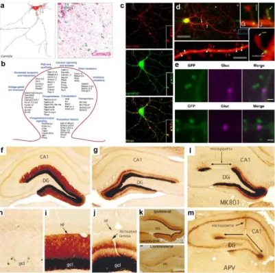
Figure 15 a αCaMKII gene is involved in synaptic plasticity and is one of the
most abundant dendritic RNAs. αCaMKII smFISH in cultured neurons and hippocampal CA1 (nuclei are in green). b Dendritically localized RNAs include a large number of synaptic proteins including signaling, scaffold, and membrane receptor proteins. Modified from Cajigas et al. (2012) and Will et al. (2013). c Arc RNA is a dendritically localized RNA that predominantly exists in granules, associated with RBPs. Modified from Gao et al. (2008) d Live imaging of MS2-tagged Arc RNA shows that it predominantly associated with dendritic spines either at the base or with the spine head. Modified from Dynes and Steward (2012). e Gluc-fused Arc is translated at dendritic spines (marked with PSD95-GFP) after glutamate application. From Na et al. (2016).
f,g (magnified in i,j) Dendritic localization of Arc RNA after electroconvulsive
seizure in anestethized rat. Perforant path stimulation of one hemisphere (g) induced selective re localization of the RNA. h Arc expression in control animal. k High-frequency stimulation of the medial perforant path also induces lamina-specific ribosomal RPS6 phosphorylation. Arc expression and localization is blocked by NMDAR blockers MK-801 (l) and APV (m). Modified from Pirbhoy et al. (2016) and Steward and Worley (2001b).
conditioning, as well as in the Morris water maze (Miller et al., 2002).
Among plasticity-related transcripts, Arc (also known as Arg3.1) is particularly interesting, being at the same time an IEG and a dendritically localized transcript (Steward and Worley, 2001a). Under resting conditions, Arc RNA levels are low, and mainly present in dendrites in granules associated to RBPs that prevent translation (Figure 15c). Live imaging of the transcripts shows that Arc RNA localizes in correspondence to spines, either in the head or at the spine-dendrite junction (Figure 15d), where it is translated after glutamate stimulation in a NMDAR-dependent way (Figure 15e) (Dynes and Steward, 2012; Na et al., 2016). Furthermore, Arc translation has been reported in synaptoneurosome preparations, indicating that translation can occur, at least in part, also in spines (Yin et al., 2002). Arc 5’UTR, then, has one of the highest IRES (internal ribosome entry site) activity among dendritic mRNAs (Pinkstaff et al., 2001), a process involved in spine potentiation (Barco et al., 2008; Kindler et al., 2005)
Arc is rapidly expressed after neuron and synaptic activation by electroconvulsive seizures and perforant path (PP) high frequency stimulation that induce LTP (Figure
15f-k); the administration of the NMDAR blocker MK-801 blocks both LTP and Arc expression induction (Lyford et al., 1995). Arc RNA rapidly translocates in the dendritic layer of stimulated DG; concomitant PP stimulation induces Arc RNA accumulation to the activated lamina (Steward and Worley, 2001b). Notably, in an unbiased screen, Arc was found to be the mRNA with the greatest fold induction change in the dendritic layer after PP-HFS stimulation of the DG (de Solis et al., 2017). Dendritic targeting of Arc RNA is due to sequences in the 3’UTR, where a DTE has been identified (Kobayashi et al., 2005), although other sequences involved in targeting have been identified (Bramham et al., 2010; Gao et al., 2008).
ARC protein associates with PSD95 and synaptic proteins (Fernández et al., 2017), and has been reported to associate with AMPAR vesicles, enhancing their mobilization (Bramham et al., 2008). Novel Arc synthesis is necessary for induction and consolidation of LTP (Plath et al., 2006). Arc KO mice have impaired L-LTP, and blocking activity-dependent Arc expression with antisense oligonucleotides in rat hippocampus inhibits LTP consolidation. Furthermore, injected animals have a severe impairment in recalling the platform position in the Morris water maze (Guzowski et al., 2000). Indeed, ARC associates
with the cytoskeleton stabilizing F-actin, suggesting a possible positive feedback loop that docks novel transcribed mRNAs during the consolidation phase (Bramham et al., 2010). Consistently, F-actin stabilization with jasplakinolide prevents LTP loss due to Arc antisense oligo administration (Messaoudi et al., 2007).
Although ARC protein has been ascribed a complementary role in LTD and homeostatic scaling, in vivo stimulations at frequencies that induce LTD (e.g. 1 Hz) do not initiate Arc expression (Steward and Worley, 2001b), and LTD induced by NMDA-only application does not induce Arc expression and does not require ARC protein (Bramham et al., 2010). Most data come from pharmacological treatments in culture or slices, and acute Arc overexpression in vivo does not induce LTD (Steward et al., 2015). Recently, it has been reported that ARC may have a priming effect on synapses rather than causing LTD (Jakkamsetti et al., 2013). An intriguing possibility is that these could be synapses surrounding potentiated ones, which could explain the accumulation of ARC protein in inactive synapses following LTP-inducing BDNF administration, as reported in Okuno et al. (2012). Although ARC may play additional roles, then, most data imply a role in LTP induction and consolidation at the spine level: after the
induction of LTP, Arc mRNA is transcribed and accumulates in stimulated zones, Arc associates with synapses, and blocking Arc expression impairs LTP consolidation and memory formation.
In addition to a structural role (Zhang et al., 2015b), ARC has been recently reported to be capable of forming oligomeric particles and to transfer its own RNA to other cells, where it is competent for activity-dependent translation (Pastuzin et al., 2018). The authors ascribe this effect to the ability of ARC protein to form virus-like particles, based on the established retrotransposon origin of
Arc gene, and the structural homology with retroviral gag
protein (Zhang et al., 2015b). If observed in vivo in physiological situations, this would provide an interesting mechanism of neuron-to-neuron information transfer that could affect the plasticity of close cells. Also, it would be necessary to further assess the mechanism of RNA transfer observed by Pastuzin et al. (2018) to exclude the contribution of mechanisms other than the proposed encapsidation of ARC particles (e.g. exosomes, microsomes), and to evaluate whether the employed concentration in which ARC particles are formed in vitro are compatible with actual concentration expected in neurons.
LTP and memory
For what has been said above, LTP is an attractive candidate mechanism to be the substrate for memory. First, electrical LTP can be induced in brain structures implicated in learning and memory. It is found in amygdala preparations, and L-LTP has been described at all synapses in the hippocampus circuit (Kandel et al., 2016). Notably, NMDAR conditional deletion in postsynaptic populations DG, CA3 or CA1 selectively impairs L-LTP in the corresponding station of the trisynaptic circuit (Figure 16). Second, it is associative in nature, as it requires coincident pre- and postsynaptic activity (section “LTP mechanisms”). Third, its long-lasting nature has the right temporal scale of memory retention (section “LTP”). Fourth, it is synapse-specific (Bliss and Collingridge, 1993), and potentiation can be induced even in single spines (Bosch et al., 2014; Hill and Zito, 2013). Fifth, potentiation is observed in vivo in parallel to memory formation. Recordings from contextual fear conditioned rats show increased AMPAR currents in response to test stimuli in the amygdala (McKernan and Shinnick-Gallagher, 1997); similarly, training rats in contextual fear conditioning, or letting animals experience a novel context, increased fEPSP slope of CA1 response to Schaffer collateral (SC)
stimulation in ex vivo preparations (Sacchetti et al., 2001) (Figure 17a,b). Whitlock et al. (2006) recorded potentiated
Figure 16 a Deletion of NMDA receptor NR1 subunit in specific subregions
of the hippocampus can be achieved crossing floxed nr1 mice with Cre mice under a region-specific promoter, as confirmed by ISH (b). c,f,i region-restricted deletion of the NR1 subunit impairs LTP in the corresponding pathway (c CA1: Schaffer collateral, f CA3: commissural-associative synapses, i DG-Perforant path). This is paralled by memory impairments: d,e Mice lacking NMDAR CA1 are impaired in the spatial version of the MWM. g,h although the training in MWM in CA3 nr1 knockout, their performance is reduced in probe trials. j,k DG nr1 KO have reduced discrimination ability between different contexts. Reproduced from Havekes and Abel (2009)
Figure 17 a Amygdala slices from fear-conditioned rats have higher
EPSCs and input-output curves. From McKernan and Shinnick-Gallagher (1997). b Slices from fear-conditioned or context-exposed rats display higher input-output response in the Schaffer collagteral-CA1 synapses than control animals. From Sacchetti et al. (2001). c Potentiation of stimulus-response is observed in CA1 of rats after inhibitory avoidance learning in some recording sites. Potentiates sites display occlusion, and electrical LTP produces smaller changes in potentiated vs. non potentiated sites (d). Modified from Whitlock et al. (2006). e Arc -/-performance in MWM is lower than control mice, and freezing during recall of fear-conditioned Arc-/- mice is dramatically reduced. f Arc-/- have enhanced E-LTP but severly impaired L-LTP. Modified from Plath et al. (2006). g In hippocampal slices, a single tetanus, that normally induces E-LTP, induces L-LTP in presence of phosphodiesterase inhibitor rolipram. Rolipram enhances memory maintenance in cued contextual conditioning with a mild footshock. Modified from Barad et al. (1998) h Blocking NMDAR with D,L-AP5 during training impairs (continue on next page)
responses in some electrodes in a multi-electrode array implanted in CA1 following training in the inhibitory place avoidance task (Figure 17c); importantly, occlusion of experimentally induced LTP was more prominent at potentiated electrode positions (Figure 17d). Thus, the synaptic plasticity and memory hypothesis (which posits a causal role of synaptic plasticity in the formation of memories) satisfies the principle of detectability (Martin and Morris, 2002). Sixth, pharmacological and genetic manipulations that affect LTP also affect memory, and vice versa. Arc knock-out mice have a worse performance in memory tasks like the Morris Water Maze and the contextual fear conditioning than wild type mice (Figure 17e). Analogously, L-LTP is impaired in these animals, although E-LTP is enhanced (Figure 17f) (Plath et al., 2006). This seems the rule rather than the exception: for example, mutant mice knock-out for αCaMKII have severe L-LTP deficits (Silva et al., 1992a), and spatial learning is heavily impaired in MWM and plus maze (Silva et al., 1992b). Similar results (continued from previous page) learning of platform location in MWM. From Morris et al. (1986). i Complete LTP electrical saturation (<10%) in PP-DG synapses prevents learning of the platform location in MWM. From Moser et al. (1998). j LTP saturation of PP-DG synapses after learning impairs the retrieval of platform location in MWM. From Brun et al. (2001).
are observed in non-phosphorylable T286A CaMKII mice (Lisman et al., 2002). Conversely, mutant mice with enhanced or facilitated L-LTP generally have a better performance in learning and memory tasks (Lee and Silva, 2009). These correlative observations suggest that synaptic plasticity and memory formation share underlying mechanisms of induction.
Experimental interventions that block L-LTP also impair learning: NMDAR inhibition during training with hippocampal infusion of D,L-AP5 blocks memory formation (Morris et al., 1986). Inhibition of protein synthesis has been often reported to impair both L-LTP and memory formation (Barrientos et al., 2002; Fonseca et al., 2006b), although the generalized block of translation can have confounding effects (Fonseca et al., 2004; Gold, 2008). Immediate pre- and peristimulation inhibition of αCaMKII activation with a light-sensitive inhibitor blocked L-LTP induction. Light inhibition of αCaMKII in the amygdala hindered learning in the place avoidance task (Murakoshi et al., 2017). Inhibition of actin polymerization with latrunculin or cytochalasin D impairs L-LTP induction (Krucker et al., 2000) and memory acquisition (Mantzur et al., 2009); thus, interfering with at least four of the main components implied in L-LTP formation (NMDAR,
CaMKII, novel protein synthesis and actin cytoskeleton reorganization) also blocks memory formation.
Saturating LTP with implanted electrodes in the DG prevents the acquisition of the platform location in rats trained in the MWM (Figure 17i). The effect was specific to LTP, since it was not observed when the DG was stimulated with a lower frequency paradigm (which does not induce LTP) (Moser et al., 1998). The same holds for pharmacological treatments that enhance memory function: for example, phosphodiesterase inhibitor rolipram facilitates L-LTP, which can be induced by a single tetanus, a stimulation that produces E-LTP only when cAMP levels are not altered. Rolipram administration to mice trained in a weak contextual fear conditioning task enhances memory and increases memory performance during recall (Barad et al., 1998).
If the memory relies on (relative) changes of synaptic strength in a subset of synapses, then inducing LTP in the complementary set of synapses should result in impairment in the ability to recall the original memory. Indeed, the artificial tetanisation of PP-DG synapses impairs memory recall in rats trained to find the exit tunnel in the Barnes maze (McNaughton et al., 1986) and the platform in the Morris
Water Maze (Brun et al., 2001) (Figure 17j). Consistently, overexpression of dominant negative αCaMKII*, or constitutively active αCaMKII impair in CA1 impair memory retention in a place-avoidance task (Rossetti et al., 2017).
While the data presented so far (and more) advocate a role for LTP as the underlying mechanism for the formation and storage of memories, they still do not provide a definitive demonstration. Most manipulations in fact affect LTP formation as well as other cellular mechanisms (Gold, 2008). The most direct evidence of causal involvement of plasticity in memory is probably the manipulations performed by Nabavi et al. (2014). First, they showed that the conditioned stimulus (tone) in cued fear conditioning could be substituted by optogenetic stimulation of axons from the auditory cortex when paired with a foot shock; the association was dependent on NMDAR and resulted in an increase AMPAR/NMDAR current ratio compared to control mice. The learned tone-shock association, then, could be reversed by optical LTD and afterwards reinstated with optical LTP. However, optical LTP could not induce any association that resulted in freezing in response to the auditory cue.
Synaptic engrams
When learning occurs, patterns of neural activity representing the occurrence of events cause changes in the strength of synaptic connections within the brain. Going back to Semon’s definition, active synapses during the presentation of a stimulus apparently meet the requirements to be an engram. It is generally assumed that the reactivation of these altered connections constitutes the experience of memory for these events and for other events with which they may be associated. If this were true, then, potentiated synapses would have all the properties of an engram: (1) they are activated by the occurrence of a stimulus, (2) they undergo modifications that change their response as a consequence of stimulus presentation and (3) their reactivation should start the memory recall. However, a direct way to test this last point is still missing. Indeed, there are evidences that specific synapses are the stable representation of a given stimulus. For instance, single spines in pyramidal neurons of the auditory cortex can be repeatedly activated by a sound of their preferred tone frequency (Chen et al., 2011). Analogously, some spines in the barrel cortex were activated uniquely by single whisker stimulation (Varga et al., 2011). In the visual cortex, synapses maintain their orientation preference over at least
27 days of imaging (Chen et al., 2013). This suggests that indeed a given stimulus can be represented at the synaptic level. Furthermore, new synapses formed during motor learning are preferentially stabilized, and at least a subset of them is still present after four months (Xu et al., 2009; Yang et al., 2009). Notably, there appears to be a correlation between the performance and the persistence of spines formed upon learning (Yang et al., 2009). The stabilization of new synapses has also been reported for the amygdala-auditory cortex connections during cued fear conditioning (Yang et al., 2016). Therefore, possible candidates for synaptic engrams exist, i.e. synapses with a constant information content over time, and task-related synapses that are stable over time.
Synaptic vs cellular engrams
Cellular engram theory regards neurons as the unitary element constituting engrams (Figure 19a). Synaptic engram theory posits that instead of whole cells, engrams are stored in the stabilized modifications that spines undergo during memory encoding (Figure 19b) (Papoutsi et al., 2014). It is expected that a relationship between cellular and synaptic engram exists (Kaczmarek, 1992), as c-fos expression correlates with potentiation of synaptic inputs. However,
considering the c-fos+ cells ensemble as the engram neglects
the different weights that presynaptic inputs have (Figure 19c,d). Cell firing is a rather downstream manifestation of efficient synaptic transmission (Grienberger et al., 2014). This could be the reason why CA1 c-fos+ cells activation
does not provide a contextual representation that could be associated to a foot shock (Ramirez et al., 2013) (although, apparently, CA1 c-fos+ neurons activation could induce
freezing when they were already part of a naturally formed contextual fear memory (Ryan et al., 2015)). The pattern of activity generated by the natural recall and by the optogenetic reactivation of the c-fos+ neuron ensemble could
have been too different from each other, with the result of recalling a mixed context having some features of the original one. Indeed, silencing a putative CA1 c-fos+ cellular
engram impaired the physiological recall of a similar, but not of a distinct, context (Tanaka et al., 2014).
← Figure 18 Synaptic correlates of candidate engrams a Frequency-tuned
response of synapses in the auditory cortex (Chen et al., 2011). b Orientation tuned synapses in the visual cortex. Their response is stable over a long period of time (Chen et al., 2013). c-e Fear conditioning (c) and motor learning (d,e) induce the formation and selective stabilization of new spines during the formation of the memory in the auditory and motor cortex, respectively. Modified from Yang et al. (2016), Yang et al. (2009) and Xu et al. (2009).
Figure 19 a cellular engrams are generally identified as c-fos+ neurons active during the encoding of a given representation. b a putative synaptic engram is expected to be the subset of synapses undergoing LTP during the encoding. c a set of synapses is activated during an event. This will lead to (1) potentiation of a set of active synapses and (2) c-fos activation if the activation is strong enough. A relationship between the number of potentiated synapses and c-fos activation is expected. If tagged neurons are reactivated with ChR2 and an engram-coherent response is elicited, neurons are assigned the role of engram cells. d If two different representations converge onto the same neuron (continue on next page)
Importantly, engram cells display many features that accompany LTP. For instance, engram cells in the DG have a higher AMPA/NMDA current ratio, and a higher spine density (Kitamura et al., 2017; Ryan et al., 2015), suggesting that they undergo synaptic plasticity upon recruitment into an engram trace. An unexpected result of the c-fos-based optogenetic tag-and-reactivate experiments is the ability of light stimulation to reinstate the engram even when the formation of the natural engram is apparently blocked by anisomycin infusion (Ryan et al., 2015) or by the pathological condition of Alzheimer’s disease model (Roy et al., 2016). Not only was a consistent behaviour response elicited, but also the reactivation of a coherent subset of cells in the downstream circuit with the originally activated one was observed (Ryan et al., 2015).
While the recall observed despite anisomycin infusion apparently contrasts with a role of potentiated synapses in (continued from previous page) population, two sets of synapses will become potentiated (let us assume there are no shared synapses); consistently, some neurons will express c-fos only after context A presentation, some after context B, and some will be activated by both. Having two distinct sets of synapses, natural recall will activate neurons differently, yielding different activity profile. ChR2 stimulation, however, will activate all c-fos+ neurons to the same extent, giving largely overlapping activity for context A and context B.