FACULTY OF MEDICINE AND SURGERY
PhD Course in Experimental and Regenerative Medicine
XXX Cicle
Multi-integrated approach based on HPT-JT
families for the identification of a set
of biomarkers of Parathyroid Carcinoma
Tutor PhDStudent
Prof. V.M. Fazio Luigia Cinque
Supervisor
Dr. Vito Guarnieri
TABLE OF CONTENTS
ABSTRACT pg.1
CHAPTER 1: PRIMARY HYPERPARATHYROIDISM pg.6
1.1 PRIMARY HYPERPARATHYROIDISM (PHPT) pg.6 1.2 MULTIPLE ENDOCRINE NEOPLASIA TYPE 1 (MEN1) pg.10 1.3 HYPERPATHYROIDISM-JAW TUMOR SYNDROME (HPT-JT) pg.13
1.4 PARAFIBROMIN AND WNT pg.16 1.5 HEDGEHOG PATHWAY (Hh) pg.18 1.5.1 MENIN AND Hh pg.19 1.5.2 PARAFIBROMIN AND Hh pg.21 1.6 INTEGRINS pg.23 CHAPTER 2: OBJECTIVES pg.27
2.1 AIM OF THE STUDY pg.27
2.1.1 FIRST TASK: IDENTIFICATION OF GENES/PROTEINS/ pg.28 MOLECULAR PATHWAYS ASSOCIATED TO KP BY WES AND
EXPRESSION PROFILING
2.1.2 SECOND TASK: DRUG TEST ON HEK293 CELL LINES pg.28 2.1.3 THIRD TASK: VALIDATION OF THE VARIANTS IDENTIFIED, pg.29 IMMUNOHISTOCHEMISTRY (ON GOING)
2.2 PATIENTS pg.30
2.2.1 PHENOTYPIC/GENETIC DESCRIPTION OF THE pg.30 5 HPT-JT FAMILIES UNDER STUDY
2.2.2 SPECIAL CLINICAL CASE: FAMILI VI-MEN+KP pg.43
2.2.3 INTRAFAMILIAL ANALYSIS pg.46
3.1 FIRST TASK: IDENTIFICATION OF GENES/PROTEINS/ pg.49 MOLECULAR PATHWAYS ASSOCIATED TO KP BY WES AND
EXPRESSION PROFILING
3.1.1 WES ANALYSIS pg.49
3.1.2 EXPRESSION PROFILING pg.53
3.1.3 RT-qPCR pg.59
3.2 SECOND TASK: DRUG TEST ON HEK293 CELL LINES pg.64
3.2.1 FUNCTIONAL ASSAY ON THE 5 CDC73 GENE MUTATIONS pg.64 3.2.2 MEN1 WT AND EXPRESSION OF THE MUTANT PROTEINS pg.68 3.2.3 ESTABLISHMENT OF PRIMARY PARATHYROID TUMOR pg.69 CELL LINES
3.3 THIRD TASK: VALIDATION OF THE VARIANTS IDENTIFIED, pg.71 IMMUNOHISTOCHEMISTRY (ONGOING)
CHAPTER 4: DISCUSSION pg.72
4.1 WES ANALYSIS pg.74
4.2 DISCUSSION ON MEN1 FAMILY pg.76
4.3 EXPRESSION PROFILING pg.79
4.4 DRUG TEST pg.80
4.5 IHC AND SEARCH ON URINE-SERUM pg.82
CHAPTER 5: CONCLUSION pg.83
APPENDIX pg.84
METHODS pg.84
1. DNA AND RNA EXTRACTION pg.84
2. WHOLE EXOME SEQUENCING (WES) pg.84
3. EXPRESSION PROFILING pg.86
4. RT-qPCR pg.87
4.1 cDNA SYNTHESIS pg.87
4.2 REAL TIME QUANTITATIVE POLYMERASE CHAIN REACTION pg.87 (RT-qPCR)
5. FUNCTIONAL ASSAYS pg.89
5.1 cDNA EXPRESSION VECTORS pg.89
5.2 CELL CULTURE pg.89
5.3 PROLIFERATION ASSAY pg.89
5.4 WESTERN BLOT pg.90
5.5 PROTEIN DEGRADATION ASSESSMENT pg.90
REFERENCES pg.91
1
ABSTRACT
Introduzione. Nella sindrome da iperparatiroidismo associato a tumore della
mandibola (HPT-JT), il carcinoma paratiroideo (PC) è causato da mutazioni del gene oncosoppressore CDC73 codificante per parafibromina, un componente del complesso PAF1 coinvolto nel rimodellamento della cromatina e nella regolazione del ciclo cellulare.
Le mutazioni del gene MEN1 causano la sindrome omonima (MEN1), le cui lesioni paratiroidee sono benigne nel 99% dei casi ma, negli ultimi 40 anni, sono stati riportati 15 casi di associazione insolita fra PC e la sindrome MEN1 nel mondo.
Il PC è un tumore raro e aggressivo per il quale le attuali terapie sono risultate inefficaci e l'asportazione chirurgica della lesione rimane l'unico approccio curativo. Tuttavia, la distinzione in prima diagnosi fra una lesione paratiroidea maligna e un’ iperplasia indolente o un’adenoma benigno rappresenta ancora oggi una sfida, in assenza di segni patognomici, come metastasi a distanza o recidive locali. Finora, sono stati compiuti sforzi infruttuosi per la ricerca di biomarcatorimolecolari che potessero indirizzare verso il (migliore) trattamento chirurgico (conservativo/demolitivo) al fine di ridurre il rischio di recidiva e di estendere la sopravvivenza libera da malattia.
Scopo del progetto e metodi. Questo progetto mira a identificare le
cause genetiche che portano allo sviluppo di un carcinoma paratiroideo (PC) e a individuare una serie di biomarcatori utili per una inequivocabile e precoce diagnosi di PC. A tal fine sono state applicate strategie di Next Generation Sequencing, quali il sequenziamento dell’ intero esoma e studi di espressione genica, a casi familiari (costituiti da uno o più soggetti affetti, portatori non affetti e controlli sani con lo stesso background genetico) piuttosto che a casi sporadici al fine di ridurre, per quanto possibile, la variabilità intrinseca causata dai diversi background genetici. La nostra rara coorte consta di 5 famiglie con HPT-JT e lesione paratiroidea maligna e mutazione costituzionale del gene CDC73 e una famiglia molto rara con mutazione MEN1 associata a PC.
2
Risultati. Il sequenziamento dell’intero esoma ha mostrato che solo i
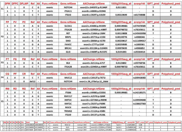
soggetti affetti di 4 (delle 5) famiglie con HPT-JT reclutate per lo studio, condividono varianti rare (MAF <0,004) in geni codificanti per le integrine (ITGA3, ITGA2B, ITGA11, ITGAB6, ITGA9), recettori di superficie coinvolti nell'adesione cellulare alla matrice extracellulare (ECM) ed essenziali per la proliferazione, la sopravvivenza, l'adesione e la migrazione delle cellule. Inoltre sono state identificate ulteriori varianti nei geni che codificano per proteine coinvolte nel riparo del DNA come FANCC e BRIP1; NOTCH4; RET; BRCA1; BLK; MUC12; KMT2C; geni target della pathway di Hedgehog quali SMO, GLI3, mentre una variante del gene GLI2 è stata identificata solamente nei soggetti affetti della famiglia MEN1 associata a PC.
Lo studio dell’ espressione genica è stato condotto confrontando i pazienti affetti vs controlli fra le famiglie con HPT-JT e l'unica famiglia MEN1-PC. L’ analisi effettuata ha mostrato un'espressione differenziale dei geni del sistema immunitario e inoltre ha evidenziato che i pazienti MEN1 e HPT-JT utilizzano diversi set di geni per controllare la mobilizzazione del calcio.
Saggi funzionali. Cellule HEK293 sono state trasfettate con vettori di
espressione codificanti la forma WT o mutata del gene CDC73: sono state testate 5 diverse mutazioni in presenza/assenza di Bortezomib, un farmaco già utilizzato in clinica per la terapia del mieloma multiplo. Questo inibitore del proteasoma sembra essere in grado di recuperare parzialmente l'espressione di proteine CDC73 mutate a causa di mutazioni missenso, in-frame e persino frameshift.
Conclusioni e prospettive. Questo progetto, seppur preliminare, può
aiutarci ad identificare biomarker utili per una diagnosi precoce e immediata di PC al fine di indirizzareverso il miglior approccio chirurgico e identificare i portatori asintomatici nelle famiglie affette per un intervento precoce ed efficace.
Ipotizziamo che l'insorgenza/progressione/aggressività del PC possa essere dovuta alla deregolazione di proteine coinvolte nell'adesione cellula-cellula (come le integrine); allo squilibrio nell’ attivazione del sistema immunitario; alla deregolazione della pathway di Hedgehog e alla perdita di
3
fattori scatenanti, come le proteine implicate nel mantenimento dell'integrità del DNA (FANCC, BRIP1, BRCA1).
Infine, considerando che la ricerca di un farmaco efficace come approccio alternativo al trattamento chirurgico è risultata fino ad oggi vana, per la prima volta, riportiamo il possibile utilizzo di un farmaco chemioterapico ben conosciuto, il Bortezomib, per la terapia del PC dovuto a mutazioni del gene CDC73.
4
ABSTRACT
Background. As part of the Hyperparathyroidism with Jaw Tumor
syndrome (HPT-JT), parathyroid carcinoma (KP) is caused by mutations of the CDC73 tumor suppressor gene, encoding the parafibromin, a PAF1 co-transcription factor, involved in chromatin remodelling and cell cycle regulation. Mutations of MEN1 gene cause the namesake syndrome (MEN1), in whose parathyroid lesions are benign in 99% of cases: instead, in the last 40 ys, only 15 cases worldwide have been reported with the unusual association of KP in MEN1 syndrome.
KP is a rare, aggressive life-threatening tumor for whose at the present current therapies resulted ineffective and the surgical removal of the lesion remains the only curative approach. However, to recognize in first diagnosis a malignant parathyroid lesion from an indolent hyperplasia or benign adenoma still represents a challenge, in absence of pathognomic signs, such as distant metastasis or local recurrences. So far, unsuccessful efforts have been made in search of clinical biomarkers that could address to the (best) surgery option (conservative/demolitive) in order to reduce the risk of recurrence and to extend the disease free survival.
Purpose and methods. The present project aims to identify the genetic
causes leading to the development of a parathyroid carcinoma (KP) and to detect a set of biomarkers for a possible unequivocal, early first diagnosis. We decided to apply high throughput strategies such Whole Exome Sequencing (WES) and Expression Profiling (EP) to familial cases (consisting of one or more affected subjects, non-affected carriers and healthy controls with the same genetic background) rather than to sporadic ones in order to, possibly, reduce the intrinsic variability caused by different genetic backgrounds. Our rare cohort consisted of 5 HPT-JT families with malignant parathyroid lesion and constitutional mutation of the CDC73 gene and a very rare family with MEN1 mutation associated with recurrent and familial KP.
Results.WES analysis revealed that in 4 (out of 5) HPT-TJ families,
5
genes encoding the integrins (ITGA3, ITGA2B, ITGA11, ITGAB6, ITGA9), cell surface receptors involved in cell adhesion to the extracellular matrix (ECM) and essential for proliferation, survival, adhesion and migration of cells. Moreover, other variants in genes encoding proteins involved in DNA repair such as FANCC and BRIP1; NOTCH4; RET; BRCA1; BLK; MUC12; KMT2C; Hedgehog target genes such as SMO, GLI3 were identified. Finally a variant in GLI2 gene was found inaffected subjects of MEN1- KP family.
The EP analysis compared the affecteds vs controls between HPT-JT families and the unique MEN1-KP family. The analysis showed a differential expression of genes of immune system. The EP also evidenced that MEN1 and HPT-JT patients use different set of genes to control the calcium mobilization.
Functional assay. Human embryonic kidney (HEK293) cell lines were
transfected with eukaryotic expression vectors carrying WT or mutated CDC73 gene: 5 different mutations weretested in presence/absence of Bortezomib, a drug already used in clinic for the therapy of multiple mieloma. We proved that this proteasome inhibitor was able to partially rescue the expression of missense, in-frame and even frameshift CDC73 gene deletions.
Conclusions and perspectives. This preliminary project may help to find a
biomarkers set for a prompt early diagnosis of KP, in order to suggest the best surgical approach and identify asymptomatic carriers in affected families for an efficient early intervention.
We suppose that the onset/progression/aggressivity of KP may be due to the deregulation of proteins involved in cell-cell adhesion (such integrins); the derangement of immune system; the deregulation of the Hedgehog pathway and the lack of trigger factors, such as the proteins assigned to DNA integrity (FANCC, BRIP1, BRCA1).
Finally we reported, for the first time, the possible use of a well known chemotherapy drug, the Bortezomib, for the therapy of KP CDC73 induced, taking into account that, so far, all the attempts to find an efficient drugs as alternative approach to the surgery, resulted ineffective.
6
CHAPTER 1: PRIMARY HYPERPARATHYROIDISM (PHPT)
1.1 PHPT
Primary hyperparathyroidism (PHPT) is the third more frequent endocrine disorder, after diabetes and thyroid disease. Although mostly sporadic, familial forms of HPT exist among 2-5% of total cases. The most common genetic disorders associated with HPT are Multiple Endocrine Neoplasia (MEN) type 1 and HPT-Jaw Tumor (HPT-JT) syndrome caused by germline mutations of MEN1 and CDC73 gene respectively (1, 2).
PHPT is characterized by hypercalcemia accompanied by an inappropriate secretion of parathyroid hormone (PTH) from one or more neoplastic parathyroid glands (3).
Usually, serum calcium level is mantained within a physiologic narrow range by the action of PTH secreted from parathyroid glands. Parathyroid chief cells express the G protein-coupled calcium-sensing receptor (CaSR) on their surface membrane, which, in response to small changes in the ionized calcium level, negatively regulates PTH secretion. Instead the CaSR is able to inihibit the secretion of pre-formed PTH that, in turn, reduces the proliferation of parathyroid cells and the transcription of the pre-pro PTH gene (by a loop negative feedback). If normocalcemia is not restored, parathyroid glands enhance their secretory capacity: PTH acts on bone, kidney and gut to regulate blood calcium concentration, modulating movement of ions in/out of the bone and the renal tubular reabsorption in order to keep the serum calcium concentration within a narrow range.
When a benign or malignant parathyroid lesion develops, an over secretion of PTH causing hypecalcemia can be observed (4, 1). Parathyroid lesions are benign in up of 99% of cases consisting of parathyroid adenomas (85%) and hyperplasia in the remaining (15%) of cases. Parathyroid carcinomas (KP) is a rare aggressive life-threatening tumor, accounting for less than 1% of all PHPT cases, for whom at the present, current therapies resulted ineffective since the surgical removal of the lesion remains the only curative approach (5).
7
Distinguishing between a parathyroid benign adenoma from a malignant lesion is challenging and it is based only upon histopathological criteria. Indeed this rare malignant carcinoma is not recognized before surgery and often is not conclusively identified during the operation or at histological examination (6).
In absence of invasion of surrounding structures and/ or metastasis, histopathologic features including fibrous bands and mitotic figures are common and could be suggestive but not pathognomic of malignancy. Instead some of these features are not specific but shared among parathyroid adenomas. So an unequivocal diagnosis of KP is based on the findings of regional or distant metastases, invasion of adjacent soft tissues, thyroid gland and vascular invasion of perineural spaces. These features should alert the surgeon of the possibility of malignancy and should lead to consideration of an en bloc resection still being the only chance of cure (6, 7).
In addition to parathyroid adenoma and the rare KP, subsets of tumors with an uncertain malignant potential are referred as to “atypical adenomas” which include those lesions that share some of the histophatological features of KP such as fibrosis, mitoses, capsular invasion but lacking evidences of invasive growth (8, 9).
To recognize in first diagnosis a parathyroid malignant tumor from its benign counterparts still represents a challenge. The immunohistochemistry seems to be a useful but not definitive tool.
Parafibromin protein is encoded by the CDC73 gene (1q32), identified as the master TSG of KP, in 2002 (see chapter 1.3, for a detailed description). Parafibromin immunohistochemistry was first reported as a molecular marker for parathyroid tumor classification with parafibromin-positive cases having a low risk of malignancy while cases with reduced protein expression representing either carcinomas or adenomas with CDC73 genemutations (8, 10). Nevertheless, it was found that some parathyroid carcinomas have had positive staining. This result would suggest the need of an additional standardized protocol, to test the validity of this approach and to determine the roles of other genes in the development of these tumors.
8
Recently Adenomatous Polyposis Coli (APC) has become an additional screening marker for atypical adenoma and KP (9). APC immunoreactivity was shown to be completely lost in the majority of malignant parathyroid tumors while APC expression was retained in all the adenomas under study. The authors concluded that loss of APC and partial loss of parafibromin might co-occur in KP and atypical adenoma allowing to distinguish parathyroid malignant tumors from benign ones.
Fernandez-Ranvier et al, asked whether parathyroid tumors could have a distinctive molecular profile for differentiating benign tumors from malignant ones. They indicated that no single diagnostic marker currently determines whether a parathyroid tumor is an actual carcinoma, but loss of parafibromin and Rb expression and overexpression of galectin-3, a member of the lectin family, generally is able to distinguish KP from other parathyroid tumors (11).
Taking into account the difficulties linked to the histological interpretation of various forms of parathyroid cancer, genetic screening of CDC73 and MEN1 genes along with the immunohistochemistry (IHC) of the corresponding proteins, parafibromin and menin, represents important clinical tools for improving diagnostic accuracy. However these strategies are not always pursued or follow the surgery, thus not fulfilling the need of an unequivocal/prompt diagnosis for the choice of surgery.
So far, many efforts have been made in search of clinical biomarkers that could address to the best surgery option (conservative/demolitive) that, however, resulted unsuccessful. Our preliminary study, based on a metanalysis of all published and personal (Muscarella…Cinque et al, accepted on Oncotarget) available biochemical, clinical, histology, molecular data, seems to indicate that early onset of the disease (<40 ys) and high serum calcium levels (>14 mg/dL) may predict the presence of a CDC73 gene mutation. However, novel prognostic biomarkers would be desirable, since prevention and early diagnosis make the difference in this rare cancer disease.
CDC73 and MEN1 genes are the major players involved in the development of parathyroid carcinoma, encoding for parafibromin and menin proteins, respectively, both transcription factors implicated in the regulation of
9
cell cycle genes and/or chromatin remodelling. On the other hand, both represent two oncosoppressor genes whose the biallelic inactivating mutation is frequent in DNA derived from parathyroid tumors, according to the so-called Knudson “Two hit hypothesis”. Two “hits” (i.e. mutations, germline/somatic or somatic/somatic, or germline-somatic/LOH) are required for a tumor to develop a selective growth advantage in an affected cell and resulting in its clonal expansion. While for sporadic tumors both the mutations mostly occur somatically, in hereditary tumors at least one mutation is germline, while the second hit might present as somatic or as loss of heterozygosity (LOH) (12).
10
1.2 MULTIPLE ENDOCRINE NEOPLASIA TYPE 1 (MEN1)
Multiple Endocrine Neoplasia 1 Syndrome (MEN1) is an autosomal dominant endocrine disorder, primarily characterized by familial hyperparathyroidism (PHPT) and by the occurence of parathyroid, gastro-entero-pancreatic (in approximately 40% of patients) and pituitary tumors (in approximately 30% of patients). Less frequently MEN1 patients may develop adrenocortical, thyroid, carcinoid tumors, lipomas and leiomyomas (13, 14).
Clinical association of familial PHPT associated with lesions in at least 2 of the 3 “ P ” glands (pancreatic, pituitary, parathyroid) is paradigmatic for a MEN1 clinical diagnosis: “ Familial MEN1 ” refers to a family in which one individual and one or more first-degree relatives have at least 2 of the 3 MEN1 – related tumors (15).
Parathyroid tumors are the most common feature of MEN1 and occur in about 95% of patients. Pancreatic islet cell tumors, which consist of gastrinomas, insulinomas and glucagonomas, occur in about 40% of patients and anterior pituitary tumors, which consist of prolactinomas, somatotrophinomas, corticotrophinomas, or nonfunctioning adenomas, occur in about 30% of patients (14, 16-18). The clinical manifestations of MEN1 are generally related to their products of secretion and less frequently to their primary sites or metastasis. In the absence of treatment, MEN1 tumors result in an earlier mortality (19). Unlike HPT-JT, many MEN1 patients with parathyroid tumors have multiglandular disease (12).
Although MEN1 is inherited as an autosomal dominant disorder, sporadic forms may develop in 8-14% of patients due to de novo germline mutations of the gene in approximately the 10% of patients (14, 17).
Moreover, KP is a rare occurrence in MEN1 patients, being the parathyroid lesions almost exclusively benign (adenoma or hyperplasia) up to the 99% of cases. So far, only 15 well-documented cases of KP and 1 atypical adenoma (AA) are known to be associated with MEN1 mutation and of these only in 7, an inactivating mutation of the MEN1 gene was identified (20-22).
11
The MEN1 gene was primarily identified in 1997 and localized to chromosome 11q13. MEN1 gene consists of 10 exons with a 1.83 Kb coding region. The region ranging from exon 2 to exon 10 is translated while exon 1, the 5’ part of exon 2 and the 3’ part of exon 10 are untranslated. The main transcript of MEN1 gene is a 2.8 kb mRNA (23, 15) of whose 1821 bp encode for the corresponding protein.
Menin is a 610 amino-acid intracellular protein, ubiquitously expressed but primarily localized in the nucleus in non-dividing cells. Menin has three nuclear localization signals (NLSs) at codons 479-497 (NLS1), 546-572 (NLSa) and 588-608 (NLS2) and, being a nuclear protein, menin acts as a co-transcription factor involved in transcriptional regulation, genome stability, cell cycle regulation and proliferation interacting with a plethora of transcription factors (13, 15).
As transcriptional regulator, menin seems to be a component of histone methyltransferase complexes containing members from the mixed-lineage leukemia (MLL) and trithorax protein family (24). These complexes are responsible of the activation of transcription by methylating the lysine 4 residue of histone H3 (H3K4) of Hox Homeobox genes and the genes for cyclin-dependent Kinase inhibitors, p27 and p18 (25, 26).
Morover, it was found that menin interacts with: 1) members of the nuclear factor-KappaB (NF-Kb) family (p50, p52, p65) repressing NF-kb-mediated transcriptional activation; 2) Smad3 and the Smad1/5 complex, to inhibit the transforming growth factor-b (TGF-b); 3) bone morfogenetic protein 2 (BMP-2) signaling pathways; 4) transcription factor checkpoint suppressor 1 (CHES1) in response to DNA damage (27).
Menin also interacts with the activating protein-1 (AP1), transcription factors JunD and C- Jun to suppress Jun-mediated transcriptional activation: Kim et al, reported that menin interacts with JunD via recruitment of a mSin3A-histone deacetylase (HDAC) complex to repress JunD transcriptional activity (28). Indipendently of the histone methyltransferase complex, menin seems to be able to bind a broad range of gene promoters, functioning as a general transcription regulator that helps to maintain stable gene expression (15).
12
As far as the role in controlling genome stablity, menin has also been shown to interact with a subunit of replication protein (RPA2) and Fanconi anemia complementation group D2 protein (FANCD2) necessary for DNA recombination and replication and for DNA repair respectively (29, 30). Through the interaction with nonmuscle myosin II-A heavy chain (NMHCII-A), fibrillary acidic protein (GFAP) and vimentin, menin plays the role as a regulator of cell division (13). Menin also participates in cell cycle control interacting with the tumor metastases suppressor (NM23H1) and the activator of S-phase Kinase (ASK), a component of Cdc7/ASK Kinase complex involved in cell proliferation. It seems that menin completely represses ASK-induced cell proliferation (31). Furthermore, it has been reported that menin regulates itself by a feedback mechanism (13, 32, 33).
To date, more than 1800 mutations have been reported and the majority (>70%) of these are frameshift and nonsense mutations leading to truncated form with a reduced ability of tumor suppressor thus leading to a derangement of the cell cycle and proliferation.
Indeed, 20% of these MEN1 mutations are missense resulting in inactivated menin that may impair the interactions with other proteins or may reduce menin stability by enhancing proteolytic degradation (13). The mutations are scattered throughout the coding region and the splice-sites of gene, without evidence for clustering. However, no phenotype-genotype correlation between MEN1 gene mutations and clinical manifestation of the disease has so far been reported.
MEN1 germline mutations have been reported in patients with hereditary and sporadic MEN1 syndrome while somatic mutations are detected in approximately 20% of sporadic parathyroid tumors (15). However, 5-10% of MEN1 cases do not harbour mutations in the MEN1 gene (13). More than 90% of MEN1 tumors consist of loss of heterozygosity (LOH) at the MEN1 locus, which is consistent with a tumor suppressor role of MEN1, according to Knudson’s two-hit hypothesis. The remaining 10% of the MEN1 tumors, showed other mechanisms for the inactivation of the second allele, for example: intragenic deletions and point mutations by which the second “hit” may occur (13, 34).
13
1.3 HYPERPATHYROIDISM-JAW TUMOR SYNDROME (HPT-JT)
The HPT-JT syndrome is an autosomal dominant disorder characterized by the co-presence of parathyroid tumors in association with ossifying fibromas of the maxilla and mandible. Parathyroid neoplasm is the first manifestation of the disease and occurs in > 95 % of patients; in particularly the frequency of parathyroid carcinoma is high in patients with HPT-JT, up to the 15%.
Patients may develop other related disease manifestations such as renal abnormalities, which affect about 15% of patients and uterine tumors, up to75% (35).
The Cell Division Cycle 73 (CDC73) gene, firstly identified in 2002 as gene responsible for HPT-JT, consists of 17 exons encoding an ubiquitously expressed and evolutionary conserved 531 amino-acid protein known as parafibromin. CDC73 inactivating mutations identified in HPT-JT patients are scattered throughout the coding region and splice sites of the gene. To date, a total of 118 different coding mutations were identified and the majority of these reported CDC73 mutations are frameshift and nonsense (36), large or whole-gene deletions have also been described (37, 38). Missense mutations are rare and generally affect residues located in the N-terminus of parafibromin (39). Germline coding mutations of CDC73 gene have been identified in 75% of the reported HPT-JT families while 25% of these families, who do not harbour CDC73 mutations or deletions, may have defects involving the promoter or untranslated regions, uncharacterized alternative transcripts or epigenetic modifications (40-42). Moreover, the occurrence of the same germline CDC73 mutation in patients with different diseases such as HPT-JT, sporadic KP or Familial Isolated Hyperparathyroidism (FIHP), demonstrates the lack of a genotype-phenotype correlation while biallelic inactivation of CDC73 gene may be seen in sporadic KP (43).
Parafibromin is identified as a component of the PAF1 complex, a key transcriptional regulatory complex that directly interacts with RNA polymerase II subunit A. As part of the human PAF1 complex that binds at the residues 223-415, parafibromin is required for expression of several cellular genes involved in
14
cell cycle regulation, cell growth, protein synthesis, lipid and nucleic acid metabolism. Functions attributed to parafibromin are so many: behind the regulation of genetic transcription, parafibromin interacts with the histone methyltransferase complex for histone modifications and chromatine remodeling (44, 45); it regulates cell growth via the downregulation of cyclin D1 expression and Wnt signaling (46, 47) and inhibition of the c-myc proto-oncogene (12, 48).
Parafibromin is also implicated in embryonic development directly regulating genes involving in cell growth and survival like H19, insulin-like growth factor 1 and 2 (IGF1 and IGF2), insulin-like growth factor binding protein 4 (IGFBP4), high mobility AT-hook 1 and 2 (HMGA1 and HMGA2), 3-hydroxy-3-methylglutaryl-coenzyme A synthase 2 (HMGCS2) (49). Parafibromin was shown to have a dual role as a tumor suppressor and oncoprotein depending on the cellular enviroment. As a tumor suppressor, parafibromin over expression seems to result in inhibition of NIH3T3 and HEK293 cells proliferation; increase in G1 arrest and apoptosis in Hela cells; and in downregulation of the cell cycle regulator ciclyn D1.
As oncogene, over expression of parafibromin was proved to enhance growth in cells expressing the SV40 large T-antigen, as the vice-versa: it inhibits growth in cells that do not express the SV40 large T-antigen (50).
The functional role of parafibromin in the nucleus is consistent with the identification of highly conserved nuclear localization signal (NLS) at residues 125-139. Parafibromin localizes into the nucleolar compartment, thereby three nucleolar localitation signals (NoLSs) at residues 76-92, 192-194, 393-409 has been reported (51, 52). Absence of parafibromin nucleolar expression was found in sporadic KP with CDC73 mutations even showing a nuclear expression. So the disruption of nucleolar localization might cause parathyroid tumorigenesis independent of nuclear parafibromin expression (53).
Although parafibromin is predominantly a nuclear protein, a lot of evidence suggest the interaction of parafibromin with the actin-binding proteins, actinin-2 and actinin-3 in the cytoplasm, that are involved in organization of the cytoskeletal structure (54).
15
Parafibromin shares no homologies to known protein domains. However, the 200 aminoacids of the parafibromin C-terminal domain share 27% sequence identity with the yeast Cdc73 protein, a component of the yeast polymerase-associated factor 1 (PAF1) (40). The PAF1 complex interacts with RNA Polymerase II at both the promoter and the coding regions of transcriptionally active genes regulating key transcriptional events of histone modification, chromatin remodeling, initiation and elongation. The yeast Paf1 complex is composed by 5 subunits: Paf1, Ctr9, Leo1, Cdc73 and Rtf1. Deletion of Rtf1 or Cdc73 results in the loss of association of the remaining Paf1 complex members with chromatin and a significant reduction in binding of the complex to RNA pol II (55, 44).
The human PAF1 complex consists of 5 subunits (hPaf1, hCtr9, hLeo1, hCdc73, shared with yeast Paf1 complex, and hSki8, a higher eukaryotic-specific subunit) and it has been reported to have several functions (35):
• The PAF1 complex plays role in both initiation and elongation of the transcription, associating with nonphosphorylated-Ser2 and phosphorylated- Ser5 on the major subunit of RNA polymerase II.
• In the process of transcriptional initiation, the PAF1 complex is required for histone H2B monoubiquitination mediated by Rad6/Bre1, a prerequisite for both H3K4 and H3K79 methylation mediated by Set1 and Dot1 respectively.
• At initiation, the PAF1 complex associates with the Set1-like HMTase complex required for H3K4 methylation.
• The PAF1 complex controls RNA polymerase II C-terminal domain (CTD) serine 2 phosphorilation.
• During elongation, the PAF1 complex has been shown to associate with elongation factors in conjunction with the histone chaperon FACT.
Furthermore, a recent work revealed parafibromin as a transcriptional scaffold whose distinct morphogen intracellular signals converge on, coordinating the expression of specific target genes and generating appropriate cellular responses to each morphogens. Parafibromin competitively interacts with the Wnt and Hedgehog signal effectors, β-catenin and GLI1 respectively,
16
and also binds Notch intracellular domain (NICD), promoting downstream target genes activation.
The platform function of parafibromin is regulated by its phosphorylation and dephosphorylation status on Y290/293/315, mediated by PTK6 and SHP2 respectively.
The tyrosine-dephosphorilated form of parafibromin at Y290/293/315 is required to bind β-catenin, GLI1 and NICD and seems to enhance the Wnt, Hh and Notch signaling activation. Because of β-catenin and GLI1 binding sites overlap in the N-terminal domain of parafibromin, this two effectors compete,intracellularly, for the same parafibromin region and interact with the PAF1 complex to increase the expression of their target genes, while the interaction between NICD and parafibromin requires the C-terminal region (56). It has been observed a reciprocal inhibition of the Wnt and Hh signals, also reported in human gastric cancer cells (57).
1.4 PARAFIBROMIN AND WNT
Among the most relevant parafibromin functions, there is the activation of the Wnt signaling pathway (46, 47, 58, 59). The Wnt signaling pathway is involved in the development of multicellular organism, in cell proliferation, differentiation, survival, cell motility and apoptosis via it is central component β-catenin (60).
In absence of the Wnt ligand, most of endogenous β- catenin is located at the cell membrane, where it associated with α-catenin and E-cadherin in epithelial cell adherent junctions. The cytoplasmatic counterparts is marked for proteosomal degradation by a multisubunit destruction complex, consisting of Axin, APC, protein phosphatase 2 A (PP2A) and GSK3b.
When the Wnt ligand binds the receptor complex, composed by Frizzled and LRP5/ 6, the inhibitory complex is dissociated and active unphosphorylated β- catenin enters the nucleus and interacts with TCF/LEF DNA binding proteins to initiate transcription of Wnt target genes (60, 61).
Studies in Drosophila shows that Hyrax, a component of the Wnt/Wg signaling, is a homolog of parafibromin. Parafibromin/Hyrax have been proved to
17
be a positive regulator in the Wnt pathway and it is required for nuclear transduction of the WNT/Wg signaling in HEK293 cells. Parafibromin binds directly to the C-terminal region of nuclear β- catenin/Armadillo via its conserved N-terminal domain and recruits the other components of the PAF1 complex, in order to regulate the transcription of Wnt target genes encoding for example the c-myc oncoprotein and the cell cycle protein cyclin D1 (47, 62).
Disregulation of the Wnt signaling pathway causes many different human tumor types, including colon, hepatocellular carcinoma, leukemia and melanoma (63) and aberrant activation of Wnt/β-catenin signaling pathway was observed in a subset of parathyroid carcinomas due to epigenetic APC loss of expression (64). Furthermore, it has also been identified in primary hyperparathyroidism and in parathyroid tumors associated with chronic renal failure (65).
18
1.5 HEDGEHOG PATHWAY
The Hedgehog pathway (Hh) is an evolutionary conserved regulator of tissue homeostasis and development in both vertebrate and invertebrate embryons, and it is involved in proliferation and self-renewal in adult stem cells. Hh signaling converges to the activation of DNA-binding GLI transcription factors that regulates the expression of Hh target genes (66).
In vertebrates, the canonical Hh pathway is activated by three ligands: Indian (IHH), Desert (DHH) and the most abundant Sonic Hh (SHH).
Secreted Hh ligands induce signalling trasduction by binding the transmembrane Patched 1 (PTCH1) receptor. Consequently, attenuating its inhibitory effect, PTCH1 promotesthe release of repressed Smoothened (Smo) (67). It has been suggested that activated Smo inhibits Suppressor of Fused (Sufu), Fused and Cos2 complex that sequesters the GLI proteins in the cytoplasm and represses transcription by recruiting the histone deacetylase complex SAP18-MSin3 (68). Once activated and translocated into the nucleus, GLI proteins promote the transcription of genes involved in cell cycle processes, survival and metabolism.
In absence of Hh ligands, GLI1 is transcriptionally repressed while GLI2 and GLI3, once phophorilated by Dyrk2, are recognized by the F-box protein β-TrCP for proteasome-dependent degradation to generate the truncated repressor forms (69, 70). In presence of Hh ligands, the transcription of GLI1 is activated and the GLI2-GLI3 proteolytic processing is inhibiting, leading to the activation of specific GLI target genes (66, 67).
The members of the GLI family are zinc finger transcription factors that possess different domains: an N-terminal domain of transcriptional repression, lacking in GLI1; central five zinc-finger DNA binding domains and a C-terminal domain of trans activation. These proteins can act as activators or transcriptional repressors of target genes of Hh: GLI1 acts as a transcriptional activator while GLI2 and GLI3 act both by activators and by transcriptional repressors even if evidences supported the hypothesis that GLI3 primarily plays the role of SHH pathway repressor (71, 72).
19
An aberrant function of Hh pathway occurs in many human tumors such as basal cell carcinoma and medulloblastoma (73).
Among the various candidate genes emerging from the arbitrary filtering of WES data, we focused our attention on the GLI2 gene as there are evidences showing strong implications of menin and parafibromin with the Hh pathway.
Human GLI2 gene consists of 13 exons located on chromosome 2q14. This gene encodes a 1586 amino acid protein belonged to the C2H2-type zinc finger protein subclass of the GLI family that bind DNA through zinc finger motifs.
The functional domains of the human GLI2 protein are not yet well understood. The murine Gli2 ortholog is characterized by a central DNA-binding domain consisting of 5 zinc fingers, a N-terminally repressor domain and a C-terminally transactivation domain (74). Accordingly, a human variant lacking the N-terminal repressor domain (ΔN) in vitro shows a transcriptional activity 30 times higher compared with the full-length protein (75).
1.5.1 MENIN AND HH
Data reported in literature show that:
1) The Hedgehog pathway plays a key role in pituitary development and seem to be involved in pituitary alterations typical of MEN1 syndrome (76). In chicken, zebrafish and rodents the morphogen Sonic Hedgehog (Shh) signals is required during the development of the pituitary gland in early stages (77).
In humans, defects of Shh secretion and signaling during early stages of development are responsabile for holoprosencephaly (HPE) with pituitary defects. Several GLI2 genetic variants were reported in patients with pituitary and extra pituitary malformations, craniofacial anomalies, hypopituitarism and Multiple Pituitary Hormone Deficiency (MPHD) and are associated with anterior pituitary hypoplasia. Mutations in GLI2 gene are mainly nonsense or frameshift introducing a premature codon stop, thus resulting in a protein truncated of the functional C-terminal transactivation domain (76).
2) It was demonstrated that a link between menin and Hh signaling exists in menin-mediated tumors. Menin is able to epigenetically repress the Hedgehog pathway in dependent MEN1 tumors and mutations in the MEN1 gene induce an
20
over activation of the Hh pathway (78). As a component of histone methyltransferase complex, menin can posistively or negatively affect gene expression. So mutations in MEN1 gene cause a dysregulation of signaling pathway related to menin transcription function.
As previously described, the Hedgehog signaling pathway is activated by the binding of ligands to the cell surface receptor Patched (PTCH1), thanks to accessory proteins, such as GAS1, BOC or CDO. Upon activated, SMO triggers a signal transduction cascade leading to the activation of GLI transcription factors (GLI1, 2, 3) that allow the transcription of target genes including regulating a variety of processes such as embryonic development, cell cycle and tumorigenesis (67, 79, 80).
Gurung et al, reported that menin normally prevents the over-activation of the canonical Hh signaling pathway in pancreatic islets, repressing expression of GAS 1, a crucial co-factor for Hh ligands binding to PTCH1 receptor (78).
Menin potently suppresses Hh signaling binding the GAS 1 promoter and recruiting protein arginine methyltransferase 5 (PRMT5), a negative transcriptional regulator which dimethylates protein arginine residues, such as histone H4 arginine 3 and histone H3 arginine 8 at this target gene.
MEN1 mutations reduce the interaction between menin and PRMT5 and the repressive dymethylation at GAS 1 promoter both in vitro and in beta cells in vivo. Gurung et al showed that loss of menin enhances Hedgehog (Hh) signaling in murine pancreatic islets and may contributes to increasing beta cell proliferation (78).
Recently Gurung et al. showed that menin can directly repress expression of GLI1 epigenetically via the histone H4 arginine 3 methyltransferase protein PRMT5, indipendently of the canonical Hedgehog Signaling pathway. Menin binds to the GLI1 promoter and recruits PRMT5 and its histone methylation negatively affects binding of the active GLI1-HDAC complex to the GLI1 promoter with a consequent transcriptional repression (81). Loss of menin lead to increase GLI1 transcriptional activity: upon MEN1 mutations, GLI1 and its target genes, including PTCH1 and c-Myc, resulted elevated in absence of a Hedgehog pathway activating ligand.
21
The direct non canonical GLI1 activation, indipendent of mutations in Hh pathway components, may play a role in tumorigenesis of endocrine organs (82). To date, target-based therapy against MEN1 syndrome is currently lacking, but considering the crosstalk between menin and the Hh effectors, Hh antagonists may provide an effective therapy for treating neuroendocrine tumors with MEN1 mutation.
1.5.2 PARAFIBROMIN AND HH
Parafibromin appears to be involved in the activation of Hedgehog pathway and it is required for the expression of specific Hh target genes.
The detailed way of how nuclear GLIs co-factors are recruited and functionally cooperate to regulate transcription and mediate direct chromatin remodeling of all the Hh target genes remain doubtful: the C-terminal activation domain of GLI3 and Ci, the single Drosophila Gli, has been reported to interact with CREB binding protein (CBP) which has HAT activities involved in transcription control (82, 83). Moreover, the C-terminal GLI/Ci domain contains a motif comparable to a consensus binding element for the TATA-box recognition component TAFII31 (84).
Mosimann et al. provided evidences for the involvement of parafibromin/Hyrax as a novel GLI/Ci binding partner and as a positive component in Hh signaling. Impairment of Hyrax function decreases Hh signaling activity in Drosophila cell culture and lead to a decrease of Hh target gene expression in vivo, while RNAi-mediated knockdown of parafibromin decreased the transcriptional activity of GLI1 and GLI2 in human cell culture (85).
Like β-catenin, GLIs/Ci directly bind the N-terminus of parafibromin/Hyrax via the domain of interaction with Su (fu) known as Region 1, a highly conserved recruitment site in the N-terminal portion of all the GLIs/Ci factors. The parafibromin region involved in GLI proteins interaction includes amino acids 200-343.
These evidences suggests the role of parafibromin as a GLI/Ci auxiliary factor and the involvement of the PAF1 complex to control Hh target gene expression in human cells.
22
The N-terminal parafibromin domain also contains residues for binding to β-catenin: GLI and β-catenin proteins seem to use a common site for parafibromin binding, perhaps interacting with the PAF1 complex to increase the expression of their target genes (85).
23
1.6 INTEGRINS
Integrins are heterodimeric cell surface receptors that mediate cell adhesion to the extracellular matrix (ECM) and are essential for embryonic development, proliferation, survival, adhesion, differentiation, migration of cells. The integrins family comprises 18 α and 8 β subunits that, if combined each other, may formed at least 24 distint αβ-heterodimers (86, 87). Specific integrin heterodimers recognize own ECM proteins like collagen, fibronectin and laminin. For instance, integrins αv and α5β1 preferentially bind the RGD sequence on their respective ligand (88).
Integrin receptor is composed by a large extracellular ligand-binding domain, a short transmembrane domain and a small intracellular domain that lacks kinase activity. Upon ligand binding, integrins undergo a conformational changes and by clustering, they recruit and activate kinases and scaffolding molecoles to focal adhesions (FA) in order to initiate a trasduction signaling pathway (89). FA are a distinct areas at the cytoplasmic face of the cell membrane and include a variety of intracellular kinases and adaptor molecules regulating cellular motility, survival, proliferation and differentiation such as: Focal adhesion kinase (FAKs), Src family kinases (SFKs), Integrin- linked kinase (ILK), Particularly interesting new cysteine-histidine rich protein (PINCH), Non- catalytic tyrosine kinase adaptor protein 2 (Nck2) and p130 CRK-associated substrate (p130, CAS or BCAR1). Moreover, FA mediate the connection of integrins to the cellular actin cytoskeleton by recruiting proteins such as talin, paxillin, parvins, α-actinin and vinculin (90-92).
The integrin family is crucial for the initiation, progression and metastasis of solid tumors. In several tumor cells, the expression of particular integrins can influence the malignant potential and the progression of neoplasia (89).
Integrins seem to have a dual role in enhancing cell survival or inducing apoptosis.
ECM ligated integrins trasduce positive survival signals thorught increasing expression of BCL-2 or FLIP and activation of PI3K-AKT pathway or nuclear factor –kB (NF-kB) signalling (93-95). Unligated integrins can promote
24
pro-apoptotic cascade in a process termed integrin-mediated death (IMD) by recruiting and activating caspase 8. However, some tumors resistent to IMD, can survive in an anchorage-indipendent manner, resulting in the ability to metastatize (96).
Recent studies reported that some growth factors receptors and oncogenes require specific integrins to enhance tumorigenesis and metastasis. The cooperation between integrin α6β4 and EGF receptors family member ERBB2, induces tumor formation and invasion in patients with breast cancers (97).
Moreover, integrins are also expressed on the surface of tumor-associated host cells such as endothelial cells, fibroblasts, pericytes, bone marrow-derived cells, platelets where are involved in cancer progression by mediating angiogenesis and inflammation. The integrins αIIbβ3 on platelets and αvβ3 on tumors cells interact throught the binding of the same ECM protein fibronectin. This interaction induces tumor cells arrest in tumor-associated blood vessels that show structurally and biologically compromised characteristics, facilitating tumor cells intravasation and metastasis in various sites (98, 99).
ITGA2B, also known as platelet glycoprotein αIIbβ3, is the most abundant receptor on circulating platelets surface where it is usually inactive. If αIIbβ3 were in an active state, platelets would aggregate leading to thrombosis. So ITGA2B takes a very important role in homeostasis (100): in fact, mice lacking αIIb or β3 subunit shows a bleeding disorder that mimic the human genetic disease Glanzmann thrombasthenia (GT) (101). ITGA2B plays an important role in the hematopoietic stem cell and megakaryocite/platelet functions (102).
αIIbβ3 is dependent on its membrane-proximal cytoplasmic domain to maintain an inactive state. Deletion of the αIIb or β3 cytoplasmic domains renders the αIIbβ3 integrin constitutively active. Partial truncations of the β3 cytoplasmic domain maintain αIIbβ3 in a low affinity state (103).
Following platelets activation, αIIbβ3 is activated within the cell, so it can bind fibrinogen, von Willebrand factor and fibronectin to form a bridge for the aggregation with other platelets (104).
25
Studies have revealed a tight link between integrins espressed on tumor cells and tumor microenviroment platelets in the cancer progression. Both elevated number and activated platelets have been found in cancer patients (105). Moreover, several data proved that platelets may contribute to the spread of cancer from a primary site to a different secondary site: recent findings revealedhow ITGA2B is crucial for hematogenous metastasis of human breast carcinoma MDA- MB-231 cells (106).
Xiaolin Lu et al. reported that prognosis of cancers like glioblastoma, gastric cancer, ovarian and melanoma may be correlated with integrin expression. They identified ITGA2B as indipendent prognostic factor of overall survival in patient affected by clear cell renal cell carcinoma (ccRCC). Expression profiling analysis was performed among the 525 patients with ccRCC collected for the study. It emerged a high expression level of ITGA2B correlated with a poor prognosis (107).
ITGA3 or α3β1 integrin is a receptor for the extracellular matrix (ECM) and belongs to β1 family of integrins. It is encoded as a preproprotein and proteolytically processed to generate light and heavy chains that comprise the α3 subunit. This subunit joins with a β1 subunit to form an integrin that interacts with extracellular matrix proteins including members of the laminin family. It has been reported that α3β1 preferentially binds laminins 5, 10, and 11 (108-110). Moreover,expression of gene encoding α3β1 may be correlated with breast cancer metastasis.
Integrins have been reported to have more complex functions than the traditional ability to bind the ECM components.
Studies on α3β1 integrin in keratinocytes suggested a role as a transdominant inhibitor of other integrins. In this case, it was observed an increased adhesion to fibronectin and type IV collagen mediated by other integrins, if cells are deficient for α3β1 (111).
Recently it was found a novel role for α3β1 integrin as a component of a cell–cell adhesion complex in epithelial cells. α3β1 integrin interacts with the tetraspanin CD151, a cell surface molecule with four transmembrane domains, to directly stimulate cadherin-mediated adhesion in epithelial cells.
26
In vivo, α3β1 integrin and CD151 are coexpressed in a variety of epithelial cells, including basal keratinocytes of the skin and glomerular epithelial cells of the kidney (112). So in addition to α3β1 binding laminin integrins involved in cell–matrix adhesion, another pool of α3β1 integrins exists in epithelial cells that are located along baso-lateral membranes and associated with the cadherin-catenin complex (113).
Integrins inhibitors have been approved and are currently used in cardiovascular disease, Crohn’s disease, multple sclerosis. Several integrins inhibitors have also been tested in clinical trilas for cancer therapy including monoclonal antibodies and RGD peptide mimetics (111). Preclinical studies showed that integrins antagonists inhibit tumor growth by affecting both tumors cells and tumor-associated host cells (114).
27
CHAPTER 2: OBJECTIVES
2.1 AIM OF THE STUDY
The project aims to identify the genetic causes leading to the development of a KP and to detect a set of biomarkers useful for a possible unequivocal, early first diagnosis of KP.
NextGen strategy was applied to different HPT-JT families consisting of one or more affected (and carriers of a CDC73 mutation) subjects, non-affected carriers and healthy controls (not carriers). We decided to apply the NGS analysis to familial cases rather than sporadic ones in order to, possibly, reduce the intrinsic variability caused by different genetic backgrounds.
Comparing the data obtained from Whole Exome Sequencing and Expression Profiling of recruited families, it could be possible to infer data about possible biomarkers detectable within the blood (serum/ urine) to:
- better understand the molecular events leading to tumor development; - to address the best surgical choice in order to reduce the risk of recurrence and to extend the disease free survival.
This approach has been applied to 5 families (out of the less 90) families worldwide identified (https://cdc73.css-mendel.it/) with malignant parathyroid lesion and constitutional mutation of the CDC73 gene and a very rare family with MEN1 mutation associated to KP (rather than to benign lesion).
At least one carrier, a proband and a controlwere selected for each family. Clinically, probands had hyperparathyroidism due to malignant parathyroid lesion. From a histologic point of view, in all the 5 cases, the parathyroid lesion showed clear and unequivocal indications of malignancy, such as: i) invasion of the capsule; ii) a high number of mitotic figures; iii) adherence to peripheral tissues and vascular or perineural invasion. For each family, it was also possible to characterize several relatives with pHPT and/or maxillary or parathyroid lesions.
In MEN1 family, the proband, the brother and the daughter were affected by hyperparathyroidism with pancreatic lesion, lipoma and melanoma. The proband and the brother were also operated for malignant parathyroid
28
lesion, histologically diagnosed as KP. Screening of the MEN1 coding sequence identified a mutation in exon 9, c.1252G> A /p.D418N, shared by all the affecteds and also by a clinically health nephew.
Themulti-faceted research approach based on 5 HPT-JT families, basically consists of three major tasks to be developed sequentially.
2.1.1 FIRST TASK: IDENTIFICATION OF GENES/PROTEINS/
MOLECULAR PATHWAYS ASSOCIATED TO KP BY WES AND
EXPRESSION PROFILING
1) Whole Exome Sequencing (WES) to be applied on: germline DNA extracted from blood of the proband, and on germline DNA of an unaffected (carrier) and of a not carrier (control) relative;
2) the Expression Profiling (EP) assay to beperformed on: RNA extracted from the same specimens.
2.1.2 SECOND TASK: DRUG TEST ON HEK293 CELL LINES
Human embryonic kidney (HEK293) cell lines were transfected with eukaryotic expression vectors carrying WT or mutated CDC73 gene: 5 different mutations were tested (1 out of the 5 previously identified in one of the HPT-JT families under study).
Transfected cell lines were treated with the Bortezomib (10 µM), a well-known proteasome inhibitor, yet used in clinics for the treatment of tumor diseases such as multiple myeloma (115).
The following assays were performed:
- cell vitality, in order to investigate whether the Bortezomib is able to influence the tumor growth;
- Western Blot, in order to verify whether the Bortezomib is able to restore the expression levels of mutated parafibromin.
- Colony agar formation assay, in order to verify whether the drug is able to reduce the cellular proliferation (this latter assay is still ongoing).
29
2.1.3 THIRD TASK: VALIDATION OF THE VARIANTS IDENTIFIED,
IMMUNOHISTOCHEMISTRY (ONGOING)
1) After prioritization, some of the variants resulted from the WES will be searched by Sanger sequencing on a selected validation cohort consisting of sporadic parathyroid, atypical and classic, adenoma tissues.
2) proteins resulted as more suggestive from WES will be assayed by IHC on FFPE tissues of the validation cohort. As other possible option, proteins yet reported in literature as differentially expressed in KP or atypical or classic adenoma (116) will be assayed by IHC as well on the validation cohort.
30
2.2 PATIENTS
2.2.1 PHENOTYPIC/GENETIC DESCRIPTION OF THE 5 HPT-JT
FAMILIES UNDER STUDY
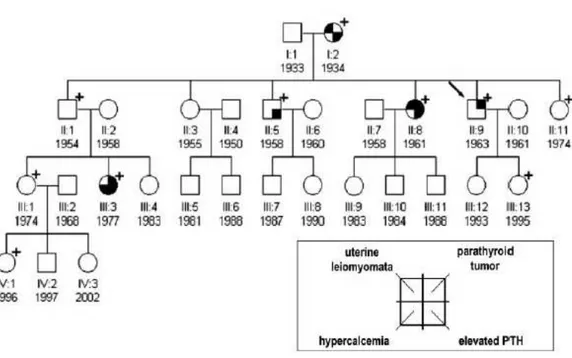
Family I (Figure 1a)
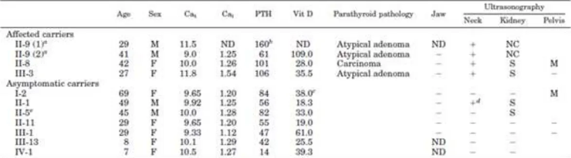
The index case first came to our attention at 29 yr, for a hypertensive crisis and acute heart failure. He had nephrocalcinosis with chronic renal failure and a serum calcium of 10.2 mg/dl (reference interval, 8.2–10.4 mg/dl). The family history was negative for parathyroid disease, but renal stones were reported in siblings and more distant paternal relatives. After 6 months, serum calcium was 11.5 mg/dl, creatinine = 2.1 mg/dl, phosphate = 2.45 mg/dl, intact PTH = 160 pg/ml (reference interval, < 72 pg/ml). Ultrasonography of the neck showed a 15-mm nodule behind the left lobe of the thyroid and parathyroid scintigraphy with 99mTc-SestaMIBI showed uptake at the base of the left thyroid lobe. The patient underwent left hemithyroidectomy and excision of a 1.5-cm hard parathyroid nodule.
Microscopy revealed oxyphil cells arranged in a trabecular or nesting pattern and nonoxyphil cells of the usual type with nondescript diffuse architecture and fibrous banding. Mitotic figures and capsular invasion were observed, without evidence for metastatic spread suggesting a diagnosis of atypical parathyroid adenoma. Moreover, another normal parathyroid gland was identified and biopsied. Two weeks postoperatively, serum calcium decreased to 8.32 mg/dl while PTH decreased to 42 pg/ml.
The 69-year old mother of index case was apparently clinically unaffected, despite her carrier status while, there was a strong paternal family history for renal stones. With identification of the CDC73 gene mutation, more extensive monitoring was initiated. Jaw pantomograms and renal ultrasonograms were performed in carriers, but were uniformly negative for tumors or cysts, although a number of carriers and noncarriers showed renal stones (Table 1). By transabdominal ultrasonography, uterine leiomyomata were found in 2 out of 5 women. Serum calcium concentrations in first-degree relatives of the index case
31
were normal, although the serum PTH was modestly elevated in 3 of 6. Ultrasonography of the neck with attention to the thyroid gland and surrounding structures was performed and was positive in 2 of these relatives, initiating a diagnostic work-up. In a sister (II:8), a 10-mm nodule was observed below the left lobe of the thyroid, consistent with a parathyroid gland (Figure 1b). Serum calcium was 10.0 mg/dl, and the ionized fraction, 1.26 mmol/liter, but serum PTH was 101 pg/ml (reference interval, < 72 pg/ml). The patient underwent excision en bloc of the left thyroid lobe, isthmus, and parathyroid nodule and removal of the lymph nodes surrounding the laryngeal recurrent nerve. A hard 1.0-cm parathyroid nodule was found and histopathologic examination revealed chief cells with intratumoral dense fibrous banding, mitotic figures, and full-thickness capsular invasion extending into the surrounding tissues but not the adjacent lymph nodes, warranting a diagnosis of parathyroid carcinoma. Three normal parathyroids were identified, and the ipsilateral gland was excised with the left lobe of the thyroid. The two controlateral glands were biopsied and found to be histologically normal. Postoperatively, serum PTH and calcium decreased to 66 pg/ml and 8.88 mg/dl, respectively. In a brother (II:1), an extrathyroidal nodule below the right lobe was observed on ultrasonography and SestaMIBI scan, but serum calcium (9.92 mg/dl), blood ionized calcium (1.25 mmol/liter), and PTH (56 pg/ml) were normal. The patient elected to undergo excision en bloc of the right thyroid lobe, isthmus, and suspected parathyroid nodule. Histologically, the nodule was a 1-cm extrathyroidal goiter nodule, not a parathyroid tumor. Among the more distant relatives of the index case, a 27-yr-old carrier niece (III:3) was found to have an increased serum calcium (11.8 mg/dl), ionized calcium (1.54 mmol/liter), and serum PTH (106 pg/ml). Neck ultrasonography showed a spindle nodule in the posterior aspect of right thyroid lobe, which was also positive on SestaMIBI scan. Aspirate fluid contained high PTH (1000 pg/ml) and low thyroglobulin (0.5 ng/ml) concentrations. Excision en bloc of the thyroid lobe with nodule, isthmus, and lymph nodes surrounding the laryngeal recurrent nerve revealed a 2-cm atypical parathyroid adenoma with capsular invasion. Two months postoperatively, serum calcium and PTH were 8.72 mg/dl and 55 pg/ml, respectively. As part of a second annual screening of all
32
affected individuals and asymptomatic carriers, the index case (now age 41) underwent repeat neck ultrasonography, and an extrathyroidal nodule below the remaining right lobe was noted. Serum calcium (9 mg/dl), blood ionized calcium (1.25 mmol/liter), and PTH (61 pg/ml) were in the normal range. Aspirate fluid from ultrasonographically guided fine-needle aspiration showed a high PTH (1000 pg/ml) and low thyroglobulin (0.2 ng/ml) and consistent with a parathyroid origin. The patient underwent excision en bloc of the thyroid lobe with the nodule and the lymph nodes surrounding the laryngeal recurrent nerve. Histologically, neoplastic parathyroid tissue was found throughout the 1.2-cm encapsulated nodule. Capsular invasion was apparent, but without invasion of surrounding tissues, suggesting the diagnosis of a second primary atypical parathyroid adenoma. Another parathyroid gland was also identified at surgery and excised, but was histopathologically normal.
Figure 1a. Family pedigree of Family I. Clinical status is indicated by open symbols (unaffected or unknown) and filled symbols (affected). Filled quadrants indicate a diagnosis as indicated in the inset legend. Proband is indicated by the arrow. The presence (+) or absence (-) of a mutation in tested family members is shown.
33
Table1. Clinical and biochemical data of proband and relatives.
Figure 1b. Ultrasonography of the neck in patient II-8 shows an extrathyroidal 10-mm diameter nodule below the left thyroid lobe (white arrow) consistent with a parathyroid mass.
Further considerations on this family.
In this family, we report a phenotype restricted to parathyroid neoplasms. Although the penetrance is clearly reduced, as evidenced by the unaffected status of the carrier mother of the proband, the predisposition to carcinoma or atypical adenoma, the latter potentially malignant, rather than
34
adenoma, is high. Such a variable penetrance underscores the importance of appropriate surveillance.
Moreover, in our index case, the appearance of a second parathyroid neoplasm 12 yr after the first had been completely resected indicates that more aggressive surgical intervention with ipsilateral hemithyroidectomy and total parathyroidectomy may be warranted in some. On the other hand, the ability to detect the tumor at an early stage may mean that close surveillance is sufficient, given the clinical burden and costs that lifelong replacement therapy with calcium and vitamin (or, in the future, recombinant human PTH) can represent. Experience with a single family cannot define which surgical approach is indicated in others, but our observations can contribute to the development of better guidelines.
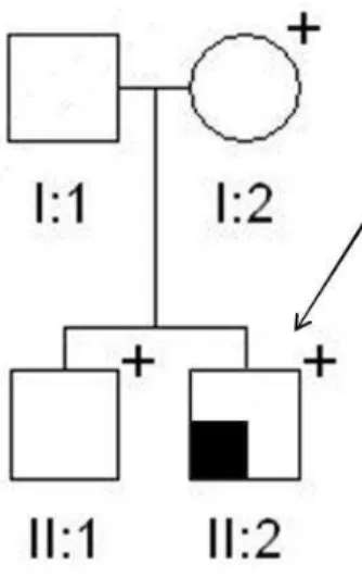
Family II (Figure 2)
A 28-yr-old female presented with a slowly growing mass for two years in her left mandible. Laboratory data revealed severe hypercalcemia (total serum calcium 16 mg/dl) and elevated levels of parathyroid hormone (660 pg/ml). Phosphorus and alkaline phosphatase levels were 1.9 mg/dL and 948 IU/L, respectively, and renal functions were normal. The patient also suffered from severe osteoporosis, renal colics and a lytic lesion in the right tibia. Ultrasound of the neck suggested a right inferior parathyroid tumor (20 mm) that was confirmed by a 99mTC sestamibi scan. The parathyroid tumor was excised under general anaesthesia and histologically diagnosed as a parathyroid carcinoma. The removal of the parathyroid tumor resulted in a severe hungry bone syndrome, which required administration of calcium and vitamin D. Two months after surgery the serum calcium and parathyroid hormone levels reversed to the normal range. Six months later the left mandible tumor was removed and histological examination confirmed the diagnosis of ossifying fibroma. The total bone mineral density increased 6 months after parathyroidectomy. Two years after treatment the patient is well with no evidence of tumor recurrence and a complete recovery of facial deformity. Anamnestic analysis of all available relatives from the paternal side revealed that subject II:1 was operated in the
35
past for a parathyroid adenoma, while subject II:2 was found to be operated for uterine leiomyomata. No other clinical data were available from the other relatives at the time of this report.
Molecular screening of germline DNA of the proband revealed a novel germline mutation of the exon 15 of the CDC73 gene, c.1379delT, that is predicted to introduce a premature stop codon at position 478 (p.L460Lfs*18). Subsequent screening of available relatives revealed that also the proband’s father (II:4), uncle (II:6) and aunt (II:1) (both from the paternal side) are carriers of the same deletion (Figure2). Since subject II:2was found to be negative upon the screening, the uterine leiomyomata in this subject are not considered to be part of the classical hyperparathyroidism-jaw tumors (HPT-JT) syndrome. The c.1379delT deletion is novel, since it has not been reported either in the
Mutation Discovery Database
(http://www.mutationdiscovery.com/md/MD.com/home_page.jsp, last access: June 2014), nor in the 1,000 genomes database (http://www.ncbi.nlm.nih.gov/variation/tools/1000genomes, last access: June 2014).
Figure2. Family pedigree of Family II. Clinical status is indicated by open symbols (unaffected or unknown) and filled symbols (affected). Filled quadrants indicate a diagnosis as indicated in the inset legend. Diagonal slash mark through a symbol indicates deceased. Proband is indicated by the arrow. The presence (+) or absence (-) of a mutation in tested family members is shown.