Alma Mater Studiorum
Alma Mater Studiorum –– Università di Bologna
Università di Bologna
DOTTORATO DI RICERCA IN
Biologia Cellulare e Molecolare
Ciclo XXXII
Settore Concorsuale: 05/E2
Settore Scientifico Disciplinare: BIO/11
Characterization of the heat-shock circuit in Campylobacter jejuni
Presentata da:
Marta Palombo
Coordinatore Dottorato
Supervisore
Prof. Vincenzo Scarlato
Prof. Giovanni Capranico
Co-supervisore
Prof. Stefano L. Ciurli
Abstract
This thesis consists of two different works developed on two distinct research themes, both of them included in the broader issue of bacterial virulence.
The first part concerns the molecular characterization of the heat-shock regulatory circuit in the food-borne pathogen Campylobacter jejuni. The heat-shock response, being cellular damage protection mechanism, is involved in the establishment of successful infections in different bacteria. Moreover, in C. jejuni, this regulatory circuit emerged as a crucial pathway for the shift between commensalism and pathogenicity in different hosts, tanks to the peculiarity to modulate host-pathogen interactions. This evidence makes the heat-shock circuit characterization indispensable to elucidate the molecular basis of C. jejuni pathogenicity and virulence.
The second section is inherent to urease activity in the presence of gold-based compounds. The urease enzyme is a virulence factor for different pathogenic bacteria, of which Helicobacter pylori, Mycobacterium tuberculosis, Cryptococcus
neoformans, Yersinia pestis, and Proteus mirabilis. This peculiarity makes the urease
an interesting target for potential new specific-drugs against ureolytic pathogens.
Characterization of the heat-shock regulatory circuit in C. jejuni
The heat-shock response is a mechanism of cellular protection that triggers a sudden increase in the cellular concentration of different proteins, including molecular chaperones and proteases. This response entails protection against protein folding that is damaged by the stress conditions. In the human pathogen C.jejuni, the response to thermic stress is controlled by a regulatory circuit, which acts
at the transcriptional level and involves the repressors HspR and HrcA. To characterize the molecular mechanism underpinning HspR and HrcA regulatory function, we investigated in detail the HspR and HrcA interactions with their operator sites by DNase I footprinting assays. These analyses allowed the identification of their binding sites, and highlight a complex architecture resulting from protein-DNA interactions. More in detail, the binding architecture is composed
of multiple HspR-recognition sites located in a singular promoter, added to the HrcA-binding element on co-regulated promoters. Moreover, our results indicate that HspR interacts cooperatively with high and low-affinity DNA binding sites mapping on each promoter. To elucidate the role of this complex DNA-binding, we tested the HspR binding ability to several DNA probes harbouring mutations within the target sequences. We also explored the DNA-binding properties of HspR and HrcA competitively on their common targets and observed for the first time that each regulator has a positive effect on DNA-binding ability of the regulatory partner. This mutual cooperative effect, of HspR and HrcA, on DNA binding could explain the synergic repressive impact of the two repressors observed in vivo on co-regulated promoters.
Urease inactivation by gold-based compounds
Urease is an enzyme that plays a crucial role as virulence factor in the pathogenesis of different microorganisms. In this work, we analysed the inhibition ability of different gold-based compounds on urease, investigating the potentiality of these compounds as future antimicrobials in ureolytic bacteria. We implemented different enzymatic assays allowed to kinetically characterize the inhibitory activity of the Au(III)-complexes, and revealing their ability to inactivate the enzyme in the low nanomolar concentration range. Moreover, our results permitted to elucidate the inhibition modality of these compounds, which resulted based on a slow-binding mechanism requiring the two-isomerization steps.
Contents
Characterization of the heat-shock circuit in C.jejuni ... 7 Introduction ... 8 1.1 Campylobacter jejuni ... 8 1.1.1 Epidemiology ... 8 1.1.2 Pathogenicity ... 9 1.1.3 C. jejuni genome ... 11 1.2 Regulatory systems ... 13 1.2.1 The heat-shock circuit ... 14 Aim of the project ... 19 Results ... 20 3.1 Investigation of the CjHspR heat-shock promoters target interaction ... 20 3.1.1 CjHspR interacts with multiple sites of the heat-shock promoters ... 21 3.1.2 Analysis of the HAIR-like motif sequence in C. jejuni ... 23 3.1.3 Elucidation of the repressor binding architecture and the binding-site hierarchy ... 25 3.2 Characterization of the CjHrcA binding on heat-shock promoters ... 31 3.2.1 CjHrcA specifically binds to Pgro and Phrc promoters ... 32 3.3 Characterization of the CjHspR/CjHrcA binding on the co-regulated promoters ... 34 3.3.1 CjHspR and CjHrcA mutual cooperative effect on DNA binding to specific operators ... 35 3.4 Investigation of the CjHspR/CjHspR and CjHspR/CjHrcA interactions ... 37 3.4.1 CjHspR and CjHrcA interaction ... 37 Discussion ... 41 Conclusion ... 44 Materials and methods ... 45 6.1.1 Bacterial strains and media ... 45 6.1.2 DNA manipulation ... 45 6.1.3 RNA isolation ... 51 6.1.4 Primer extension assays ... 51 6.1.5 Overexpression and purification of recombinant proteins ... 52 6.1.6 DNase I footprinting ... 53 6.1.7 Cross-linking experiments ... 54 6.1.8 GST pull-down experiments ... 54 6.1.9 Bacterial Adenylate Cyclase Two-Hybrid System (BACTH) assays ... 55 Urease inactivation by gold-based compounds ... 56 Introduction ... 57 1.1 Urease ... 57 1.1.1 Urease as virulence factor ... 57 1.1.2 Urease as antibacterials target ... 59 1.1.3 Urease structure ... 59 1.1.4 Urease inhibition strategy ... 62 1.1.5 Urease inhibitors as potential drugs ... 62
1.2 Gold-based drugs ... 63 Aim of the project ... 64 Results ... 65 3.1 Au(III)-based compounds’ inhibition on urease activity ... 65 3.1.1 Introduction to fast and slow binding inhibition ... 65 3.1.2 Au(III)-based compounds inhibit urease with a slow-binding mechanism ... 66 3.1.3 Evaluation of the Ki and Ki* ... 69 Discussion ... 71 Conclusions ... 72 Materials and methods ... 73 6.1.1 Enzyme and inhibitor sources ... 73 6.1.2 Kinetics measurements ... 73 6.1.2.1 Progress-curves assays ... 73 References ... 75
Characterization of the heat-shock
circuit in C.jejuni
Introduction
1.1 Campylobacter jejuni
Campylobacter jejuni (Fig.1) is a Gram-negative, microaerophilic, spiral, and
flagellated bacterium belonging to the delta-epsilon group of proteobacteria, in the order Campylobacteriales, including also the genera Helicobacter and Wolinella.
Figure 1: Scanning electron micrograph of C. jejuni clinical strain M129 with an INT 407 epithelial cell. Adapted from WSU School of molecular biosciences Konkel & colleagues.
1.1.1 Epidemiology
C. jejuni is the leading cause of bacterial food-borne diarrhoeal disease worldwide
(Blaser, 1997), accounting for more infection than Escherichia coli O157:H7,
Salmonella spp. and Shigella spp. (Apel, Ellermeier, Pryjma, DiRita, & Gaynor, 2012).
While most cases of gastroenteritis associated with C. jejuni infection are generally self-limiting, it is also linked to severe post-infection complications, such as Gullian-Barré syndrome, a demyelinating polyneuropathy causing bilateral paralysis (Nyati & Nyati, 2013). Although C. jejuni is a human pathogen, it can colonize in an asymptomatic manner the gastrointestinal tract of different animals, including chicken and other avian species. This ability is at the root of its contamination cycle, where contaminated water and unpasteurized milk or meat, especially poultry,
serve as C. jejuni reservoirs, and it is a source for human infection (Young, Davis, & DiRita, 2007).
1.1.2 Pathogenicity
Throughout the establishment of an infection, bacteria must overtake the mechanical and immunological barriers of the gastro-intestinal tract. C. jejuni succeeds in bypassing the mucus layer of the gastro-intestinal epithelium, the first defence line, because of its motility, corkscrew morphology, and of an unusual lipooligosaccharide. Indeed, C. jejuni lipooligosaccharide is made up of relatively short O-sidechain that may reduce nonspecific binding to the mucin glycoproteins (McSweegan & Walker, 1986). After passing the mucus layer, C. jejuni interacts with the epithelial host cells and elicits an immune response that differs between human and chicken. In humans, C. jejuni can interact with the epithelial cells both through external bond and internalization by the cells. These interactions cause interleukin (IL)-8 production and induce the recruitment of dendritic cells and the subsequent interaction of macrophages and neutrophils with the bacterial cells. The C. jejuni interactions with human cells trigger a massive pro-inflammatory response associated with an increase of the cytokines (Young et al., 2007). Dissimilarly, in chicken C. jejuni stimulates the production of IL-1β, IL-6, and intracellular nitric oxide synthase by epithelial cells and macrophages, without leading the pathological inflammatory response. This peculiarity allows colonizing chicken in high numbers. Various factors may reduce or redirect the chicken immune response towards tolerance; heterophils and macrophages might also have a role, whereas epithelial cell invasion is not reported (Young et al., 2007).
1.1.2.1 Flagella and chemotaxis in C. jejuni pathogenicity
Among the different C. jejuni’s elements that allow it to establish a successful infection, an essential role is exerted by the flagella and flagellar motility, which are involved in host colonization, virulence, host-cell invasion and secretion (Young et al., 2007). The chemotaxis seems to play a crucial role both in commensalism and pathogenicity in C. jejuni. Indeed, mutations of the chemotactic genes affect chicken colonization, as well as virulence in pathogenic hosts (Hendrixson & DiRita, 2004)
(Yao, Burr, & Guerry, 1997). Besides, C. jejuni displays chemotactic motility towards components of the mucus and amino acid found in the chick gastrointestinal tract (Hugdahl, Beery, & Doyle, 1988).
1.1.2.2 Cytolethal distending toxin
C. jejuni, like other bacterial species, produces cytolethal distending toxin that is
linked with the double-strand DNA brakes and the arrest of the cell cycle (Lara-Tejero & Galan, 2000)(Hassane, Lee, & Pickett, 2003). The cytolethal distending toxin is expressed booth in chicken and in human, but in chicken were not found neutralizing antibody agaist the toxin, highlighting a variance in the recognition way by the two hosts (Young et al., 2007).
1.1.2.3 Heat-shock response in C. jejuni pathogenicity
An interesting difference between chicken and mammals, including humans, is the body temperature, 42 °C in chicken, and 37 °C in human. This difference could make the temperature a potential signal for host-specific infection, and in turn the heat-shock response a mechanism able to promote the switch between commensalism and pathogenicity (Apel et al., 2012) (Young et al., 2007). Generally, the heat shock response plays a central role in host-pathogen interaction of different bacteria species as a mechanism of cellular protection able to preserve the protein folding, which is damaged by the stress conditions encountered during the establishment of an infection. This makes different heat-shock proteins virulence factors or elements that contribute to pathogenesis in an indirected manner (Roncarati & Scarlato, 2017). In C. jejuni, the heat-shock response is mediated by the two transcriptional repressors, HspR and HrcA (Andersen et al., 2005) (Holmes, Penn, & Lund, 2010), which repress transcription of the heat-shock genes at low temperature, 37 °C. Another regulatory system that might be involved in the survival at high temperature is the RacR/RacS two-component system. This system also responds to different environmental conditions encountered during infection, but independent of the temperature switch (Apel et al., 2012). Interestingly, the deregulation of the heat-shock proteins, the transcriptional repressors HspR and HrcA, and the RacRS system is linked to an altered bacterial length causing an elongation of the cell. Thisphenotype is also connected to motility defects, and consequently, to a deficiency in epithelial cell invasion. This motility deficiency is caused by filamentation of the cells and does not involve defects in the flagellar apparatus (Andersen et al., 2005) (Apel et al., 2012).
1.1.2.4 Genetic variation and natural transformation
Other C. jejuni’s elements relevant to its pathogenicity are the extensive genetic variation and the natural transformation. Intriguingly, the majority of the hypervariable sequences are in regions encoding proteins associated with the production and modification of the surface structure, and involved in immune avoidance, virulence, secretion and invasion of host cells. This class includes capsule, surface polysaccharide, lipooligosaccharide, and flagellar genes (Parkhill et al., 2000)(Linton et al., 2000)(Young et al., 2007). Moreover, C. jejuni is naturally competent and can take up DNA from the environment. That leads to recombination between strains, allowing the generation of high genetic diversity. The horizontal transfer of both plasmid and chromosomal DNA occurs both in vitro and during chick colonization, indicating that natural transformation could play an important role in genome plasticity and in the spread of new genetic tracts such as antibiotic resistance, even in the absence of selective pressure (De Boer et al., 2002)(Avrain, Vernozy-Rozand, & Kempf, 2004)(Young et al., 2007).
1.1.3 C. jejuni genome
C. jejuni has a genome of approximately 1600 kb in size. The genome analysis of C. jejuni NCTC11168 strain predicted the existence of 1654 protein-coding genes and 54 stable RNA species (Parkhill et al., 2000). Its genome compared to the genome of the closely phylogenetic related microorganism H. pylori shows strong similarity, which, however, is exclusively restricted to housekeeping functions. Indeed, just 55.4% of C. jejuni genes show an orthologous in H. pylori (Parkhill et al., 2000).Figure 2: Coding sequences transcribed in the clockwise direction (dark
green), and coding sequences in the anticlockwise direction (pale green). It is marked the putative origin of replication. In black the positions of hypervariable sequences are highlight. The genes involved in the production of surface structures are marked (clockwise in dark red and anticlockwise in pale red). The similarity of each gene to its H. pylori orthologue is indicated by the height of the bar and the intensity of the colour. Adapted from Springer Nature, License Number: 4698690911335 (Parkhill et al., 2000). As H. pylori, the C. jejuni genome sequence shows a low G+C content of 30.6% with two large regions including the portion for the lipooligosaccharide and the extracellular polysaccharide biosynthesis cluster, where the G+C content drops down to 25.4% and 26.5%, respectively (Parkhill et al., 2000). The higher A+T content maps in the proximity of promoter regions. Analysis of these regions carried out in different C. jejuni strains showed an extended -10 box with an upstream periodic A/T-rich pattern as a consensus motif in 89% of the promoters. The relationship between this A/T-rich region and their relevance in transcription initiation is also confirmed by the identification of several examples of disruptive single-nucleotide-polymorphisms (SNPs) in the A/T-rich region (Dugar et al., 2013). An A/T-rich sequence followed by the AAGGA-motif is also detected as the consensus sequence for the ribosome binding site. Highlighting the relevance for the A/T-rich pattern even in the mRNA translation (Dugar et al., 2013).
C. jejuni is predicted to code for a higher number of regulatory proteins than H. pylori, although a similar genome size. Likely, this may be due to a higher number of
ecological niches colonized by C. jejuni (Parkhill et al., 2000). As H. pylori, also C.
jejuni displays only three sigma factors, the housekeeping σ70, and two alternative σ54 and σ28 sigma factors, which regulate the expression of many flagellar genes (Carrillo et al., 2004) (Dugar et al., 2013). In contrast, the more significant portion of regulatory factors belongs to the two-component regulatory systems (Parkhill et al., 2000). A peculiar feature of C. jejuni is represented by a high frequency of genome variation, provided by the high number of hypervariable sequences. Intriguingly, this variability may be linked with slipped-strand mispairing in the proximity of homopolymeric tracts during the replication, and probably linked to the absence of several E. coli DNA-repair genes homologues. The high levels of variation observed in the shotgun sequences denote that it is not possible to build a single definitive sequence for the C. jejuni genome. As such, it possesses some of the properties of quasi-species; a phenomenon that is well described in RNA viruses (Young et al., 2007) (Parkhill et al., 2000). Another peculiarity of the C. jejuni genome is the absence of functional insertion sequence (IS) elements, retrons, transposons, and prophages, except for the Cj0752 insertion sequence that is similar to IS605 tnpB from H. pylori (Parkhill et al., 2000). The absence of these elements seems to correlate with the presence of a type-II CRISPR/Cas system. Indeed, strains that display degenerated CRISPR loci frequently show plasmids and integrated elements (Dugar et al., 2013).
1.2 Regulatory systems
Generally, bacteria react to changes in environmental conditions or the intercellular state, re-modulating the expression of genes that code for proteins involved in the adaptation to the new state. This response is commonly mediated by transcriptional regulatory circuits, which are switched on by sensors upon detection of the signals. The active sensors are able to transduce the signals to transcriptional factors. This produces a change in the regulators ability to directly interact with DNA sequence on target promoters to activate or repress genes transcription (Perez & Groisman, 2009). These regulatory patterns are bundled in densely connected networks able to modulate the cellular response by interconnecting several stimuli and producing anaccurate output suitable for the specific condition (Seshasayee, Bertone, Fraser, & Luscombe, 2006).
1.2.1 The heat-shock circuit
The heat shock response is a mechanism of cellular protection that implicates the production of different heat-shock proteins, as chaperone and proteases, able to prevent the accumulation and the consequent aggregation of denatured proteins, which occurs during stress conditions, and avoid a toxic effect on the cell (Roncarati & Scarlato, 2017). Although the heat-shock response is a universal phenomenon observed in all studied bacterial species, microorganisms do not use a unique regulatory strategy. Indeed, they have evolved various mechanisms based on both transcriptional and posttranscriptional regulation, and involving both positive and negative mechanisms of regulation (Roncarati & Scarlato, 2017).
A classic example of heat-shock circuit is the Escherichia coli positive mechanism that combines a transcriptional and translational regulation. E. coli uses two alternative sigma factors, one of them, RpoH, is able to interact with RNA polymerase core enzyme and direct the proteins complex to the promoter region of the heat-shock genes, recognizing specific DNA sequences located in these promoters (Sharp et al., 1999) (Kumar et al., 1995). In this system, the heat-shock response is activated by the increment in the amount of the RpoH alternative sigma factor in response to temperature increase. Indeed, the rpoH mRNA region that includes the ribosome-binding site and the AUG start codon acquires a complex secondary structure with high stability at low temperatures. This peculiar mRNA conformation hinders the recruitment of the translation machinery and the consequent translation of the rpoH messanger. At 42 °C, a partial misfolding of the
rpoH mRNA secondary structure occurs and allows the assembling of the ribosome
and the consequent efficient translation of the sigma factor (Morita et al., 1999). Moreover, also a post-translational regulatory mechanism monitors the amount of the available RpoH protein to enhance transcription of the heat-shock genes. Indeed, in the absence of denatured protein, different chaperons can interact with the alternative sigma factor and sequester it. Whereas, when the amount of misfolded
proteins increase, the chaperones are recruited by denatured polypeptides, and release the sigma factor (Tomoyasu, Ogura, Tatsuta, & Bukau, 1998). In addition to this chaperon’s sponge effect towards the available RpoH, chaperon systems seem to play a crucial role in promoting protease-mediated degradation, fundamental to decrease the alternative sigma factor stability and re-establish the steady-state level (Roncarati & Scarlato, 2017). Although most gram-negative bacteria exploit an alternative sigma factor, other bacteria, among which C. jejuni and H. pylori, evolved an opposite strategy to control chaperone genes’ expression, which is based on a dedicated repressor system. So at 37 °C, the transcription of the heat-shock genes is inhibited by the repressor binding to promoter regions. In contrast, the increase in temperature reduces the repressor affinity for the target DNA and allows the transcription to start (Roncarati & Scarlato, 2017). It is known that the heat-shock circuit in C. jejuni is based on two repressors, HrcA and HspR (Andersen et al., 2005)(Holmes et al., 2010). Although a molecular characterization of their DNA-binding activity in C. jejuni is not available, there is a variety of information that came from the HrcA and HspR investigation in other bacterial species. In H. pylori, the HrcA/HspR circuit was previously characterized, showing a complex regulatory network composed of the master regulator HspR, which directly regulates another regulator (HrcA) and a target gene (groESL), which in turn is regulated by both HrcA and the master regulator (HspR). Moreover, considering that all the regulatory interactions are repressive, these circuits seem to represent rare examples of incoherent type-2 feed-forward loops (Danielli, Amore, & Scarlato, 2010)
1.2.1.1 HrcA
The HrcA repressor protein was first identified and characterized in Bacillus subtilis, where it was also identified the cis-element CIRCE (controlling inverted repeat of chaperons expression), an inverted repeat (IR) necessary for the binding of HrcA to the target genes promoters (A. Schmidt, Schiesswohl, Volker, Hecker, & Schumann, 1992) (Schumann, Homuth, & Mogk, 1998). Afterward, different studies tried to obtain the biochemical identification of HrcA-CIRCE interaction at a molecular level. Unfortunately, this characterization has been possible only in H. pylori and
aggregate in insoluble bodies (Roncarati, Danielli, Spohn, Delany, & Scarlato, 2007) (Roncarati, Spohn, et al., 2007) (Wilson & Tan, 2004). The available information about the HrcA interaction with promoters shows the repressor-binding region mapping close the transcription start sites. This evidence indicates that HrcA exerts its repressive function by interfering with the transcriptional machinery, occluding the RNA polymerase binding site on the heat-shock promoters at 37 °C (Roncarati & Scarlato, 2017). HrcA loses DNA-binding affinity upon a temperature increase, and releases the operator allowing the binding of the RNA polymerase and transcription. The mechanism by which HrcA can sense heat-shock conditions is based on the ability of the chaperon GroE to enhance the repressor binding ability (Schumann et al., 1998). Indeed, this GroE positive effect can take place only at normal grow temperature, when the amount of denatured proteins is relatively low. By contrast, the increment of damaged proteins, as a consequence of the stress condition, produces chaperon recruitment, including GroE, with the consequent stop of its HrcA stimulating activity (Roncarati & Scarlato, 2017). Besides, in H. pylori, it was demonstrated that HrcA is also an intrinsic heat-sensing protein. Indeed, this protein showed thermal stress high sensitive structure, which allows the activity of the folded repressor only under heat-shock temperature (Roncarati, Danielli, & Scarlato, 2014). Interestingly, in Rhodobacter capsulatus, it was observed that the CIRCE element could also exert an extra function, affecting transcript stability at low temperature, through the formation of a secondary structure promoted by the inverted repeat (Jäger, Jäger, & Klug, 2004). Although HrcA is found in various bacteria, the sequence similarity among proteins from different species is low, generally less than 30%. However, the degree of similarity increases in highly related microorganisms. Interestingly, the similarity is restricted to three short amino acid regions, likely suggesting their essential role to the repressor activity (Morimoto, 1998). The high HrcA instability compromised its structural determination; therefore, limited information is available nowadays. The only protein architecture characterized comes from the Thermatoga maritima HrcA crystal structure. Unfortunately, T. maritima expresses an inactive form of this protein, unable to bind to the DNA. However, this structure reveals a dimeric form, where each monomer is composed of a helix-turn-helix (HTH) N-terminal DNA-binding domains (DBD), a central domain probably involved in dimerization, and a
C-terminal inserted dimerizing domain (Liu et al., 2005).
1.2.1.2 HspR
HspR is a transcriptional repressor showing amino acid similarities to members of the MerR family of protein regulators. HspR was first identified in Streptomyces
coelicolor, where it was demonstrated its ability to bind three inverted repeats,
named HAIR from HspR associated inverted repeats (Bucca, Hindle, & Smith, 1997) (Bucca et al., 1997). Then, the HspR-HAIR system was found and characterized in diverse bacterial species. In several cases, the HAIR sequences were identified in proximity to the core promoter region (Grandvalet, De Crécy-Lagard, & Mazodier, 1999) (Stewart et al., 2002), a position compatible with a mechanism of repression exerted by a steric hindrance of the RNA polymerase to the promoter region (Spohn et al., 2004) (A. Schmidt et al., 1992). In H. pylori, HspR represses transcription of some promoters in conjunction with the HrcA repressor (Roncarati & Scarlato, 2017). Surprisingly, on these promoters the HAIR sequences are located upstream of the transcriptional machinery binding site. This peculiarity suggests a different repressing strategy in the HspR/HrcA co-regulated promoters, if compared with the HspR only regulated ones (Roncarati & Scarlato, 2017). Specifically, HspR interactions occur far upstream of the core promoter elements on HspR/HrcA co-regulated promoters, while -35/-10 boxes are obstructed by HrcA binding (Roncarati, Danielli, et al., 2007). Another HspR feature is its high propensity to oligomerize, which results in extended DNA-binding regions (Spohn et al., 2004). The elaborate arrangement of the binding sites identified on the co-regulated promoters, in addition to the HspR high oligomerization tendency, suggests a complex DNA binding architecture able to finely regulate heat shock gene expression (Spohn et al., 2004). The mechanism by which HspR can respond to heat-shock, and decrease its DNA binding activity is not completely explained. Also, for this heat-shock repressor it is postulated a homeostatic control by chaperones (Roncarati & Scarlato, 2017), as confirmed by the DnaK ability to stimulate HspR DNA binding in S. coelicolor and in M. tuberculosis (Bucca, Brassington, Schönfeld, & Smith, 2000) (Parijat & Batra, 2015), and by the GroE involvement in the HspR activation in M. tuberculosis (Das Gupta, Bandyopadhyay, & Das Gupta, 2008).
Oppositely, in H. pylori the heat shock protein CbpA shows a negative effect on the HspR repressor activity. Intriguingly, this function occurs only when the repressor is not bound to the DNA target, revealing a fine regulatory mechanism to limit the heat-shock response (Roncarati, Danielli, & Scarlato, 2011).
Aim of the project
The heat-shock response is a mechanism of cellular protection, which bacteria adopt to face the stress conditions able to compromise the protein folding. This response results in a fast expression of heat-shock genes with the consequent increase in chaperones and proteases amount. The characteristic of being involved in the preservation of the intracellular environment makes the heat-shock regulatory circuit a crucial pathway during the establishment of host-pathogen interactions when the microorganism is subject to different stress damages. Moreover, there are indications that heat-shock proteins may play a role in host-specific infection in the food-borne pathogen C. jejuni. Besides, the heat-shock circuit ability to sense temperature changes makes this response a potential mechanism by which C. jejuni can switch between commensalism in the chicken reservoir and pathogenicity in humans. Although the heat-shock circuit is investigated in different bacteria species, the molecular characterization of this response is not available for C. jejuni. The purpose of this work is to elucidate the transcriptional regulatory modality by which the pathogen can fine regulate chaperons and proteases expression, clarifying the molecular mechanisms on the roots of this central response. With this purpose and starting to the involvement of the heat-shock two transcriptional repressors, HspR and HrcA, in the regulatory circuit of C. jejuni, their interactions with potential operators are investigated in detail by DNase I footprinting assays. Moreover, the HspR complex DNA-binding architecture is elucidated, analyzing the regulator interacting ability with several mutants of the promoter sequences. Furthermore, the HspR/HrcA co-regulation is explored on the common target by DNA in vitro binding assays under competitive conditions. Different protein-protein interaction assays are also performed to clarify the homo- and hetero-oligomerization ability of the two repressors involved in the regulatory circuit.
Results
3.1 Investigation of the CjHspR heat-shock
promoters target interaction
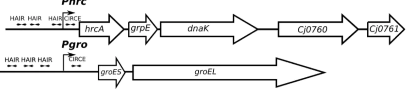
Previous studies identified the C. jejuni HspR regulon with a microarray approach, comparing transcript levels of wild type and a ΔhspR mutant strain (Andersen et al., 2005) (Holmes et al., 2010). The analysis highlighted thirty deregulated transcripts, seventeen of which appeared downregulated, and thirteen were upregulated. A further bioinformatics analysis was applied to identify putative HspR binding sites in proximity of deregulated promoters. Sequences with similarities to the HAIR-sequence motif from Streptomyces ssp were identified in four promoter regions, inferring the operons carrying these motifs as putative direct targets of HspR in C.jejuni. Figure 3 shows a schematic representation of these operons, consisting of the Pcbp, Pclp, Pgro, and Phrc promoters, which control transcription of the major
chaperone and hrcA and hspR regulatory genes. The scheme also displays the positions of the HAIR-like motif in the promoter regions. However, the functionality of HAIR-like sequences is not supported by experimental evidence, and a mechanism able to explain the HspR transcriptional repression effect is not identified (Andersen et al., 2005) (Holmes et al., 2010).
Figure 3: Schematic representation of the chaperone genes in C. jejuni. Open
arrows indicate coding genes. HAIR-like motifs are represented by convergent arrows. Bent arrowed indicate the start sites of transcription at the indicated promoters.
To start the characterization of CjHspR binding on these regions, and explore the role of the identified HAIR-like sequences in repressor binding, we set up DNase I footprinting assays with purified recombinant CjHspR on the Pcbp, Pclp, Pgro, and
Phrc promoter probes. Initiation of RNA transcription on these promoters was
identified by RNA-sequencing with a SuperGenome approach (Dugar et al., 2013) and verified in this study by primer extension analyses (data not shown).
3.1.1 CjHspR interacts with multiple sites of the heat-shock
promoters
To demonstrate the direct interaction of CjHspR to the potential target promoters, increasing concentrations of purified recombinant CjHspR were incubated with radiolabeled DNA probes spanning the Pcbp, Pclp, Pgro, and Phrc promoter regions and then digested with DNase I and fractionated on polyacrylamide denaturing gel. The result (fig. 4) is consistent with a DNase I footprinting assays obtained when the binding of a protein on the DNA probe occurs. Data reported in the figure clearly shows that upon increase of the amount of CjHspR in the reaction, area of DNase I protection are detected, due to the binding of the protein that precludes DNase I cut. In addition, binding of the protein give rise to the appearence of bands ofhypersensitivity to DNase I, likely an effect of structural DNA changes determined by the binding of the protein.
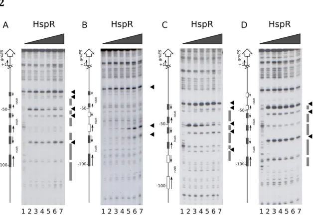
Figure 4: High resolution mapping of CjHspR binding sites on heat-shock
promoters. DNase I footprinting assays of CjHspR on Pcbp, Pclp, Pgro and Phrc promoter probes (panels A, B, C and D, respectively). Radiolabelled DNA probes were incubated with increasing concentrations of the HspR recombinant protein (0, 11, 22, 45, 90 and 180 nM CjHspR; lanes 1 to 6, respectively) at room temperature and subjected to limited DNase I digestion. In each panel, on the right the grey boxes highlight regions of DNase I protection and the black arrowheads indicate bands of hypersensitivity to DNase I digestion; while on the left, bent arrows indicate the transcriptional start site and vertical open arrows depict the open reading frames; numbers refer to the positions with respect to the transcriptional start sites.
The experiment carried out on the Pcbp promoter probe shows protection from DNase I digestion between the position -50 and the -20 above the transcriptional start site (Panel A, lanes 3-6). Also, two bands of hypersensitive DNase I sites appear in proximity of position -50 (lane 3). Upon addition of increasing concentration of
CjHspR in the reaction, a second area of protection and two additional
hypersensitive bands close to position -65 (lanes 4-6) were detected. A similar result was obtained on the Pclp promoter probe (panel B), where the experiment
displayed a protected region with flanking hypersensitive bands (lanes 3). Moreover, an area of protection emerges at high CjHspR concentration (lanes 4-6), and three hypersensitive bands is detected, two of which in the proximity of position +1. Besides, a protection region and hypersensitive bands are visible upstream to the -50 position. The DNase I footprinting assay on the Pgro (panel C), instead, shows two bands of DNase I hypersensitivity mapping to the position -50 and the -80 at lower repressor concentration (lane 3-6), and two protected regions between the -50 and -110 at higher CjHspR concentration (lanes 4-6). Finally, additional hypersensitive bands close to the position -40 (lanes 4-6) with an adjacent protection region (lanes 5-6) appear. On the Phrc promoter probe (panel D), hypersensitivity bands are identified close to the position -90 at low repressor concentration (lanes 3-6) with a protection region between the position -90 and the -55 (lanes 4-6). Increasing CjHspR concentration, two hypersensitivity bands appear between the position -100 and the -110 (lanes 4-6) with a protection region in proximity to the position -110. Moreover, an additional band tightly the -50 (lanes 3-6) and a protection region near the -30 is exhibited at higher protein concentration (lane 6). Of note, the protection pattern of CjHspR on the DNA probes show an interacting region already at lower protein concentration with additional binding areas detectable at higher protein concentration, suggesting the presence of high and low-affinity binding sites. In summary, low- and high-affinity binding sites are identified on the Pcbp and Pclp. Differently, a central high-affinity binding site with two flanking low-affinity sites are detected on the Pgro promoter. In contrast, the
Phrc reveal a central high-affinity site with a closely low-affinity binding-site and a
potential very low-affinity site.
3.1.2 Analysis of the HAIR-like motif sequence in C. jejuni
The HAIR motif proposed as a consensus sequence for the HspR protein of S.
coelicolor is constituted by IR spanning 21 bp (CTTGAGT-N7-ACTCAAG) (Grandvalet
et al., 1999). Inspection of the DNA regions interacting with CjHspR revealed the presence of five like sequences in addition to the previously reported HAIR-like sequences (Andersen et al., 2005) (Holmes et al., 2010) (fig. 3). Of these, two HAIRs map in the Pcbp and Pclp promoter regions and three HAIRs are located on
Pgro and Phrc promoters. The nucleotide sequences of the four promoters (Pcbp, Pclp, Pgro, and Phrc) and their key regulatory elements are summarized in figure 5.
Strikingly, while the HAIR-like sequences span over the Pcbp, Pclp, and Phrc core promoter elements (-10 and -35 boxes), no HAIR-like sequences overlap the Pgro promoter region, mapping upstream of the -35 region. This distinct binding architecture on the promoter regions suggests diversified mechanisms of repression exerted by CjHspR.
Figure 5: Features of the Pcbp, Pclp, Pgro and Phrc promoter sequences. For
each is indicated the promoter sequence, and numbers are referred to the transcriptional start site (+1). The -10 and 35 boxes are underlined bold on the coding DNA strand. The HAIR-like sequences are represented in boldface. The HAIR-like sequences identified in this study are grey highlight. The DNase I protection regions are highlight with boxes with different size, where the height is indication of HspR binding affinity for the specific region.
To gain insight on the role of the HAIR-like motif and identify key conserved nucleotides for CjHspR binding, HAIR sequences were aligned and submitted to WebLogo computer program with results shown in fig. 6. Alignment of all HAIR sequences highlighted nucleotides conservation within an imperfect IR of 21 bp. The most conserved nucleotides are the first three of the left emisite, CTT. Instead, the right emisite appears less conserved, and the higher conserved nucleotides are one
T and one A, which are, however, rather widespread as compared o the three nucleotides conserved in the left emisite (panel A). Restricting the analysis to the HAIR sequences found in high-affinity CjHspR binding sites, the dissimilarity of the two portions of the inverted repeat decreases (panel B), suggesting that the differences between the high- and low-affinity HAIR may reside in the right IR emisite conservation.
A B
Figure 6: : WebLogo representations of the HAIR sequences characterized through DNase I footprinting assays, A: all identified HAIR sequences, B: HAIR sequences of high affinity binding sites.
3.1.3 Elucidation of the repressor binding architecture and
the binding-site hierarchy
The above study on CjHspR-DNA interaction revealed an intriguing scenario based on multiple binding elements associated with each promoter and showing
a
variable propensity to interact with the repressor. The HAIR sequence analysis suggests that the diversity in protein interaction ability might reside in the different conservation of the two emisites. To investigate the relevance of the two portions of the HAIR motif, we generated mutants of one or both emisites, and assayed their interacting ability toCjHspR by DNase I footprinting experiments (fig. 7). Specifically, we selected the Pcbp
and Pgro promoters for the analysis, as representatives of two different binding architectures for CjHspR. Pcbp shows a promoter-proximal high-affinity binding site and an upstream low-affinity site. At the same time, Pgro carries a central high-affinity binding site flanked by two low-affinity sites, all upstream of the core promoter region. Base substitutions were applied to the high-affinity binding sites by changing each
base/position and maintaining the same base composition. A representation of the constructed mutants is depicted in figure 7 (1).
1
2
3
Figure 7: Features of the Pclp and Pgro wild type or mutants promoter sequences
(panel 1). For each are indicated promoter sequence, and numbers are referred to the transcriptional start site (+1). The HAIR-like sequences are represented in boldface, and the mutated sequences are grey highlight. DNase I footprinting assays of CjHspR on wild type Pcbp (panel A) or on mutants MHL (panel B), MHR (panel C) and MH (panel D) probes (pannel2), and on wild type Pgro (panel A) or on mutants MHL (panel B), MHR (panel C) and MH (panel D) probes (panel 3). Radiolabelled DNA probes were incubated with increasing concentrations of recombinant CjHspR protein (0, 22, 45, 90, 180, 360 and 720 nM CjHspR; lanes 1 to 7) at room temperature and subjected to partial DNase I digestion. On the right of each panel, grey boxes depict the regions of DNase I protection and black arrowheads indicate bands of hypersensitivity to DNase I digestion. On the left, schematic representation of the promoter region, where the bent arrows indicate the transcriptional start site, vertical open arrows depict the open reading frame; numbers refer to the positions with respect to the transcriptional start sites. Grey boxes represent the wild type HAIR sequences, while empty boxes represent the mutated sequences.
Comparing the DNase I footprinting assays results of CjHspR on Pcbp (2) wild type (panel A) to mutant probes (panels B, C, D), it emerged that mutation on one or both portions of the HAIR elicits loss of CjHspR protection. A partial loss of HspR affinity is visible in the mutant of the less conserved “right” emisite of the HAIR (probe MHR, panel B). Differently, the mutation of the more conserved “left” emisite (probe MHL, panel C) produces a more severe loss in protein affinity, which is reflected in a light protection region with three hypersensitive bands, and detectable only with higher HspR concentration. This result highlights a possible key role of the more conserved left emisite in the recruitment of the repressor on the Pcbp. Finally, mutations of both HAIRs emisites (probe MH, panel D) abolish almost totally the CjHspR binding to the site and allow the appearance of one residual hypersensitive band at extremely high HspR concentration. An analogous result was obtained on the Pgro promoter probe (3), where, however, the mutation on one of the two HAIR portions elicits a more gradual loss of CjHspR protection, if compared with the same mutation in Pcbp promoter. This allows appreciating the distinct effect generated by the specific mutations, harsher in the case of mutation on the more conserved “right” emisite (MHR, panel B) than in the “left” emisite (MHL). The consequences of mutations on one of the two emisites appear to be less strict on Pgro promoters then on Pcbp, presumably because of the presence of two low-affinity HAIR sites flanking the one mutated in the Pgro. Complete disruption of the high-affinity HAIR (MH, panel D) determined total loss of CjHspR protection pattern.
These results demonstrate that CjHspR binging to DNA requires a full HAIR motif and that mutations on the high-affinity HAIR also determine a severe reduction of CjHspR interaction with the low-affinity binding site. Nevertheless, these results are not sufficient to explain the role exerted by the additional HAIRs on the promoters. To grasp information on the function of multiple CjHspR binding sites on regulated promoters, we performed DNase I footprinting experiments on the Pgro promoter mutated in each HAIR motif (fig. 8).
1
2
Figure 8: Features of Pgro wild type or mutants promoter sequences (panel 1). For
each is indicated promoter sequence, numbers are referred to the transcriptional start site (+1). The HAIR-like sequences are represented in boldface, and the mutated sequences are grey highlight (1). And DNase I footprinting assays of
CjHspR on wild type Pgro or mutants MH (panel B), MM (panel C) and ML (panel
D) probes (panel 2). Radiolabelled DNA probes were incubated with different concentrations of recombinant CjHspR protein (0, 22, 45, 90, 180, 360 and 720 nM HspR; lanes 1 to 7) at room temperature and subjected to partial DNase I digestion. On the right of each panel, grey boxes depict the regions of DNase I protection and black arrowheads indicate bands of hypersensitivity to DNase I digestion. On the left, schematic representation of the promoter region, where the bent arrows indicate the transcriptional start site, vertical open arrows depict the open reading frame; numbers refer to the positions with respect to the transcriptional start sites. Grey boxes represent the wild type HAIR sequences, while white boxes represent the mutated sequences. The DNase I footprinting experiment results displays how disruption of the central high-affinity HAIR (MH, panel B) determined a drastic loss of the DNase I protection pattern by CjHspR. This evidence highlighted the essential role of the central high-affinity HAIR for the recruitment of the transcriptional repressor on the promoter. In contrast, DNase I footprinting assays on promoter probes carrying mutations in the two low-affinity HAIR motifs (MM, panel C and ML, panel D) exhibited a drastic loss of CjHspR binding
affinity exclusively for the mutated sequences, without altering the protection pattern on the remaining unaltered binding sites (C and D). This finding reveals that the flanking low-affinity HAIR motifs are not necessary for CjHspR interaction with high-affinity HAIR sequences. Moreover, the observation that a high-affinity HAIR site is required for the repressor recruitment on the promoter highlights the fundamental role that the high-affinity HAIR-CjHspR interaction has to enhance the repressor binding with close low-affinity site. Moreover, these results underline a binding hierarchy among the HAIRs located in a single promoter and depending not only on IR sequence conservation but also on the binding sites reciprocal position. This evidence suggests a potential role for the CjHspR-CjHspR intermolecular connections that might occur between repressors closely bound on the same promoter, and aimed to stabilize the repressor-promoter interaction.
3.2 Characterization of the CjHrcA binding on heat-shock promoters
In the same studies mentioned above, a microarray approach, was carried out to define the C. jejuni HrcA regulon. This whole transcriptomic approach was combined with an in silico search of putative CjHrcA binding sites looking for sequences with similarities tothe CIRCE-sequence motif from B. subtilis in the proximity of deregulated promoters. Specifically, the C. jejuni NCTC 11168 genome was analysed for the presence of CIRCE-like elements using fuzznuc program from EMBOSS (“the European Molecular Biology Open Software Suite”), leading to the identification of only one putative CjHrcA operator located in the Pgro promoter region (Holmes et al., 2010). It is worth mentioning that an additional CIRCE element situated in the Phrc promoter has been previously proposed (Thies, Karch, Hartung, & Giegerich, 1999). Figure 9 reports a schematic representation of the two potential target operons mentioned above. Starting from this information, we focussed our efforts on the study of CjHrcA-DNA binding ability on both the promoters, through in vitro DNA-binding assays.
Figure 9: hrc and gro operons’ schematic representation, the two potential CIRCE sequences are indicated on the promoters regions. Symbols are as in the legend to fig. 3.
3.2.1 CjHrcA specifically binds to Pgro and Phrc promoters
To investigate CjHrcA direct interaction with previously proposed CIRCE elements, we used the Pgro and the Phrc promoter regions as a probe for in vitro binding to a purifiedCjHrcA protein in DNase I footprinting assays. Figure 10 shows the CjHrcA ability to
bind and protect defined portions of the two probes. The protected regions overlap with the potential CIRCE elements on both promoters. More in detail, the DNase I footprinting experiment on Pgro (Figure 10; panel 1, A) shows three bands of hypersensitivity to DNase I digestion, which appear starting from lanes 2 and 3, and two DNase I protection regions noticeable from lane 4 and overlapping the transcriptional start site (+1 position). Figure 10; panel 1, B shows that upon the addition of CjHrcA to the labeled
Phrc probe, a clear region of protection appears, flanked on one side by a DNAse I
hypersensitive band. Also, in this case, the CjHrcA protected region encloses the transcriptional start site. We conclude that CjHrcA binds to the Pgro and Phrc promoters by contacting DNA regions overlapping the transcription start sites, and encompassing the previously proposed CIRCE elements.
33
1 A B
2
Pcbp
-80 -70 -60 -50 -40 -30 -20 -10 +1 +10 +20 +30AAAATTGTAAATAAAACCTTGAGTGATAAAAATTTATAAAACTTGATTGACTTAGGCTAAAGTTTATGTTATAATTTAATCCTCTATATAATCAAGTAAAAATTTTTAAGGAAATATAACAT TTTTAACATTTATTTTGGAACTCACTATTTTTAAATATTTTGAACTAACTGAATCCGATTTCAAATACAATATTAAATTAGGAGATATATTAGTTCATTTTTAAAAATTCCTTTATATTGTA
Pclp
-100 -90 -80 -70 -60 -50 -40 -30 -20 -10 +1 +10
TCTAAAATTAGTCATTTGTCCTATTATTTAAAAAATTTTACTCAATTAACTTGACATTATATGACTAAAGTTATATAATAGCAAGTCTATAAACTAAATAAAGGATAAAACATGGCAAATA AGATTTTAATCAGTAAACAGGATAATAAATTTTTTAAAATGAGTTAATTGAACTGTAATATACTGATTTCAATATATTATCGTTCAGATATTTGATTTATTTCCTATTTTGTACCGTTTAT
Pgro
-110 -100 -90 -80 -70 -60 -50 -40 -30 -20 -10 +1
TTTTATCCTTTAGTTTATTTTATAAAATAACTTTAGTCTATAAAACTAAACTTTTATAAATATTTTAATTTAAAGTATTGACAAAATTCATTTTTTATAGTATGATATTATCACTCTAAAT
AAAATAGGAAATCAAATAAAATATTTTATTGAAATCAGATATTTTGATTTGAAAATATTTATAAAATTAAATTTCATAACTGTTTTAAGTAAAAAATATCATACTATAATAGTGAGATTTA
Phrc
-110 -100 -90 -80 -70 -60 -50 -40 -30 -20 -10 +1GCTTATCCTTATAAAAAAGTAACTAATAAAACTTTAGTCATATTGACTAAATAAAATTAAGTATATTTTTTTATATTTATACTTGACAAAATAACACTCAATGTTGTATAATATATCTAG
CGAATAGGAATATTTTTTCATTGATTATTTTGAAATCAGTATAACTGATTTATTTTAATTCATATAAAAAAATATAAATATGAACTGTTTTATTGTGAGTTACAACATATTATATAGATC
Pgro
-40 -30 -20
∨
-10 +1∨
+10 +20∨
AAGTATTGACAAAATTCATTTTTTATAGTATGATATTATCACTCTAAATTAAAGAGTGCTAAAATCAATATTTTTAA TTCATAACTGTTTTAAGTAAAAAATATCATACTATAATAGTGAGATTTAATTTCTCACGATTTTAGTTATAAAAATT
Phrc
-40 -30 -20 -10
∨
+1 +10 +20TACTTGACAAAATAACACTCAATGTTGTATAATATATCTAGCAATCAAAAAATAGAGTGCTAGATATGAAGGAAAAT ATGAACTGTTTTATTGTGAGTTACAACATATTATATAGATCGTTAGTTTTTTATCTCACGATCTATACTTCCTTTTA
Figure 10: panel 1. High resolution mapping of CjHrcA binding sites on heat-shock
genes’ promoters. DNase I footprinting assays of CjHrcA on Pgro and Phrc promoters. Radiolabelled DNA probes were incubated with different concentrations of recombinant CjHrcA protein (0, 22, 45, 90, 180 and 360 nM CjHrcA; lanes 1 to 6, respectively) at room temperature, and followed by partial DNase I digestion. In each panel, on the right the grey boxes highlight regions of DNase I protection and the black arrowheads indicate bands of DNase I hypersensitivity; while on the left bent arrows indicate the transcriptional start site and vertical open arrows depict the open reading frames; numbers refer to the positions with respect to the transcriptional start sites. Panel 2.: Features of Pgro and Phrc promoter sequences. For each is indicated promoter sequence, and numbers are referred to the transcriptional start site (+1). The -10 and 35 boxes are underlined bold, and the CIRCE sequences are represented in boldface. DNase I protection regions are grey highlight and the hypersensitives DNase I cut are indicated with black tick.
3.3 Characterization of the CjHspR/CjHrcA binding on
the co-regulated promoters
Following DNase I footprinting assays described above, we have been able to identify extended CjHspR binding sites on Pgro and Phrc promoters located far upstream the core promoter region, in an atypical position for a transcriptional repressor. Moreover, on the same promoters, we demonstrated the presence of a CjHrcA binding site that is, instead, located within the core promoter overlapping the transcriptional start site. Notably, the distances between the CjHspR and CjHrcA binding sites on the Pgro and
Phrc promoters are 32 and 14 bp, suggesting the possibility of direct interaction and a
functional interplay between the two regulators, and potential cooperative interaction of the two repressors on these common targets.
Figure 11: hrc and gro operons’ schematic representation, on the promoters
3.3.1 CjHspR and CjHrcA mutual cooperative effect on DNA
binding to specific operators
To study possible interactions of CjHspR and CjHrcA, we assayed the DNA binding activities of both proteins by carrying out DNase I footprinting experiments on Pgro under competitive conditions (fig.12). Panel A shows the results of DNA-binding assays carried out using CjHspR in combination with CjHrcA (lanes 7 to 12) or with a control protein (GST, lanes 1 to 6). The addition of increasing amounts of CjHspR after binding of CjHrcA to the labelled probe resulted in enhanced footprinting patterns compared to those obtained by incubating CjHspR without CjHrcA. This effect is particularly evident by comparing the intensities of all the DNase I hypersensitive bands, which appear at lower CjHspR protein concentration in the presence of CjHrcA. Similar result is obtained when increased concentrations of CjHrcA are added to CjHspR bound to the same promoter probe (Figure 12, panel B). Also in this case, the CjHrcA footprinting pattern is enhanced by the incubation of the CjHrcA protein with a DNA probe binding to CjHspR (lanes 7 to 12), if compared to the control set (lanes 1 to 6). Consequently, we concluded that on Pgro co-regulated promoter and under the in vitro conditions used, the two regulatory proteins display a mutual cooperative mechanism of DNA binding.
Figure 12: DNase I footprinting assays of CjHspR on Pgro (0, 22, 45, 90, 180 and 360 nM, lanes 1 to 6 and 7 to 12; panel A) with GST (360 nM, lines 1 to 6) or CjHrcA (360 nM, lines 7 to 12) and CjHrcA (0, 22, 45, 90, 180 and 360 nM, lanes 1 to 6 and 7 to 12; panel B) with GST (180 nM, lines 1 to 6) or CjHspR (360 nM, lines 7 to 12). In each panel, on the right grey boxes highlight the regions of DNase I protection and the black arrowheads indicate bands of hypersensitivity to DNase I digestion of CjHspR or CjHrcA, panel A and B respectively. On the left, bent arrows indicate the transcriptional start site, vertical open arrows depict the open reading frame; numbers refer to the positions with respect to the transcriptional start sites. The HAIR and CIRCE sequences are indicated by dark and light grey boxes respectively.
3.4 Investigation of the CjHspR/CjHspR and
CjHspR/CjHrcA interactions
The DNA binding experiments described above allowed to discover a complex binding architecture. More in detail, the role played by the high-affinity HAIR in promoting the CjHspR binding on the flanking low-affinity site(s) may be explained by an interactionbetween functional units of CjHspR, each of which would make contact with one DNA binding site but also with the flanking CjHspR. Moreover, and as described previously, a CjHspR-CjHrcA interaction may be involved in the mutual cooperative effect of the two repressors. To test this hypothesis, we performed different protein-protein interaction assays.
3.4.1 CjHspR and CjHrcA interaction
To investigate a direct CjHrcA-CjHspR interaction, we performed an in vitro cross-linking assay with the two overexpressed and purified proteins (fig. 13).Figure 13: Cross-linking assay of
CjHspR+CjHrcA, CjHrcA+CjHrcA and CjHspR+CjHspR incubated with or without glutaraldehyde. R: CjHspR, A: CjHrcA and X: glutaraldehyde.
In detail, the CjHspR and CjHrcA proteins were incubated separately or mixed together with a crosslinker (glutaraldehyde) and fractionated on SDS-PAGE. The results obtained indicate that both proteins could form dimers in solution. The simples containing
CjHspR alone (Figure 13, lanes labeled RR and RR-X) exhibit a significant increase of
one band with an apparent molecular mass of ca. 30 kDa, only after glutaraldehyde treatment (compare lane RR with RR-X). The band observed following protein cross-link MW AR -X AR AA -X AA RR -X RR
might represent a dimeric form of the CjHspR protein, considering that the molecular weight of recombinant CjHspR is 15 kDa. An analogous observation is done when the crosslinker is added to the reaction containing CjHrcA alone. Even if in this case the purity of the protein preparation is lower, it is possible to observe a band with apparent molecular mass of ca. 62 kDa upon addition of glutaraldehyde (Figure 13, lane labelled AA-X), likely representing a dimer of CjHrcA (CjHrcA monomer molecular weight slightly exceed ca. 30 KDa). Interestingly, the incubation of glutaraldehyde with both proteins led to the appearance of a band of ca. 46 KDa, compatible with an HrcA-HspR complex (Figure 13, lane AR-X). To further confirm the direct interaction between CjHspR and
CjHrcA, we carried out a GST pulldown assay. To this aim, we expressed and purified the
recombinant GST-HspR and GST-HrcA fusion proteins, which were incubated with purified recombinant His-CjHspR. As a control, the same amount of purified His-CjHspR was incubated with a recombinant GST protein. The GST, GST-HrcA and GST-HspR proteins were recovered after incubation employing GSH-Sepharose slurry, and the protein composition of the three samples was analysed by SDS-PAGE.
Figure 14: GST pull down assay of purified His-CjHspR incubated with GST, GST-CjHrcA or GST-CjHspR bound to GSH-Sepharose slurry. 1. Samples analysed with SDS-page 2. Western blot analysis of samples stained with an anti-His antibody. R: CjHspR, A: CjHrcA and G: GST.
Using the GST-CjHspR fusion protein as bait, a protein band corresponding to the expected molecular mass of CjHspR was detected (Figure 14, panel 1, lane R+R). The
2
His-CjHspR R+R A+R G+R1
7563 48 35 25 20 17 11 GST-CjHrcA GST-CjHspR GST His-CjHspR MW GS H /G ST GS H /G ST -R GS H /G ST -A
CjHspR band was absent in the control sample in which the GST bait was used (Figure 14, panel 1, lane G+R). Intriguingly, also, when we used the GST-CjHrcA fusion protein as
bait, we detected a protein band likely corresponding to the CjHspR prey (Figure 14, panel 1, lane A+R). The same samples were also analysed by immunoblot assay stained with an anti-His tag antibody, able to detect only the His-tagged CjHspR (Figure 14, panel 2). These data strongly support the hypothesis of the propensity of CjHspR to form homodimers, but also of a direct interaction between the CjHrcA and CjHspR repressors. Finally, the dimerization ability of the two repressors was investigated using an in vivo system based on the Bacterial Adenylate Cyclase Two-Hybrid System (BACTH). This assay is based on the interaction-mediated reconstitution of the recombinant cyclase enzyme (CyaA) in E. coli (BTH101) strain, which is defective for adenylate-cyclase activity. It exploits the fact that the catalytic domain of CyaA from Bordetella
pertussis consists of two distinct subunits, T25 and T18. The two subunits are inactive
when physically separated, but if they are brought in close proximity to each other, the CyaA activity will be reconstituted. In the BACTH system, each one of the two enzyme fragments (T25 and T18) is fused to a polypeptide, CjHrcA, or CjHspR in our case. If the two analysed proteins can interact, dimerization of these hybrid proteins will take place, resulting in functional complementation between T25 and T18 subunits and in the reconstitution of the chimeric enzyme. Therefore, cAMP synthesis will occur and the cAMP will interact with the catabolite activator protein (CPR). The cAMP/CRP complex is a pleiotropic regulator of gene transcription in E. coli, which can turn on the expression of several resident genes, including lacZ that cods for the β-galactosidase (β-gal) enzymeone. Finally, the β-gal enzyme can be used as a reporter of the system, detecting the β-gal activity in a medium containing the X-gal compound, a chromogenic substrate of the enzyme. We assessed the capability of both repressors to form homo-dimers, as well as to form CjHrcA-CjHspR hetero-complexes. To this purpose, we set up BACTH assays, where were used the following two chimeric proteins: T25-CjHspR/T18-CjHspR, T25-CjHrcA/T18CjHrcA, T25-CjHrcA/T18-CjHspR or T25-CjHspR/T18-CjHrcA.