UNIVERSITA’ DEGLI STUDI DI ROMA “TOR VERGATA”
FACOLTA’ DI SCIENZE MATEMATICHE, FISICHE E
NATURALI
DOTTORATO DI RICERCA IN
BIOLOGIA CELLULARE E MOLECOLARE
XXII CICLO
THE FRAGILE X MENTAL RETARDATION PROTEIN
IN THE REGULATION OF NEURONAL
mRNA TRANSLATION AND STABILITY
Silvia De Rubeis
A.A. 2009/2010
Docente guida/Tutor. Prof. Claudia Bagni
Coordinatore: Prof. Gianni Cesareni
The Fragile X Syndrome is the most common cause of inherited mental retardation and is due to the absence of a single protein, the Fragile X Mental Retardation protein (FMRP). FMRP is an RNA-binding protein implicated in the regulation of mRNA transport, translation and stability in neurons. In the absence of FMRP, the expression of a large group of neuronal proteins is deregulated, resulting in impaired synaptic morphology and function.
Here, we investigated two critical steps of the posttranscriptional control mediated by FMRP, namely mRNA translation and stability. The modulation of protein synthesis involves the Cytoplasmic FMRP Interacting Protein 1 (CYFIP1), which represses the translation of the mRNAs associated with FMRP. Evidences reported here show that CYFIP1 may be also involved in the transport of the mRNAs and in the interplay between translation and cytoskeleton remodeling.
Besides the role in translation, FMRP regulates the decay of certain mRNA in neurons. In particular, FMRP stabilizes the mRNA encoding the PostSynaptic Density Protein 95 (PSD-95) in the hippocampus, but not in cortex. This study describes the molecular complexes associated with
PSD-95 mRNA that could account for the region-specific functions of FMRP.
In conclusions, this work provides further insights into the FMRP-dependent regulation of gene expression, therefore contributing to the understanding of Fragile X pathogenesis.
Preface
Neurons are one of the most fascinating examples of polarized cells. The highly specialized morphology of every neuron is essential to create and maintain the neural circuits which underlie the proper functioning of the brain.
Early during development, neurons acquire the polarity. From the cell body arise two types of neurites with specific functions: a single axon, which routes the neuron’s output, and one or multiple dendrites, which integrate inputs from other neurons (Arimura and Kaibuchi, 2007; da Silva and Dotti, 2002). In order to achieve their functional differentiation, the two processes acquire an array of morphological and biochemical specificities, including changes in the cytoskeleton, the membrane and the secretory pathway components (Conde and Caceres, 2009; Ye et al., 2007). Therefore, besides the cell body, which ensures the basic metabolic functions, the neurons possess subcellular compartments working as specialized domains.
The most emphasized specialization is reached at the synapses. In brain cortex, most of the excitatory synapses are formed by a bouton and a dendritic spine (Holtmaat and Svoboda, 2009). The bouton is a small axonal varicosity representing the presynaptic terminal, while the spine arises from the dendritic shaft and is the postsynaptic compartment (Holtmaat and Svoboda, 2009). Presynaptic compartments are characterized by the presence of an active zone with the synaptic vesicles containing the neurotransmitters (Ziv and Garner, 2004). The postsynaptic sites are defined by a local thickening beneath the membrane, the so-called Post Synaptic Density (PSD), which links the neurotransmitters receptors to signaling proteins and cytoskeleton (Kennedy et al., 2005).
The synapses are highly dynamic structures and undergo morphological changes in response to activity, underlying forms of experience-driven plasticity (Holtmaat and Svoboda, 2009). Such remodeling involves both the
14
architecture and the biochemistry and is thought to be crucial for sustaining and consolidating the processes at the base of complex phenomena, such as learning and memory.
Mental retardation
Mental retardation (MR) is a typical feature of a group of neurodevelopmental disorders defined by an early onset mental disability; the diagnostic criterion for MR is the IQ<70. In Western countries, MR affects 1.5-2% of the population and 0.3-0.5% are severely impaired (IQ<50) (Leonard and Wen, 2002; Ropers, 2008). While mild forms are likely due to complex interactions between genetic and environmental factors, severe MR is associated with circumstantial environmental events or specific genetic causes.
In fact, MR can occur as a consequence of premature birth, prenatal infections or perinatal asphyxia (Inlow and Restifo, 2004). Moreover, considering that MR is more frequent in developing countries, malnutrition, cultural deprivation and poor health care may also contribute (Leonard and Wen, 2002; Ropers, 2008).
More often, MR is due to genetic abnormalities, such as large chromosomal rearrangements (deletions, duplications or aneuploidies) or point mutations in individual genes. The chromosomal aberrations account for 15% of all cases (Leonard and Wen, 2002); for example, the Down’s syndrome (trisomy of chromosome 21) remains the most common cause of MR.
MR can occur as an isolated symptom, without other consistent features (non-syndromic MR) or can be syndromic. In this case patients present other physical characteristics in addition to the intellectual disability. Moreover, MR can be inherited as autosomal dominant, autosomal recessive and
X-15
chromosome-linked (XLMR). Up to 15% of the intellectual disabilities are attributable to XLMR and approximately 80 genes have been already discovered (Chiurazzi et al., 2008; Gécz et al., 2009). In order to identify novel candidate genes, the coding exons on the X chromosome from 208 families with XLMR have been recently subjected to a large-scale systematic resequencing (Tarpey et al., 2009).
Although MR is a heterogenous group of pathologies, one of the key hallmarks of MR is a neuroanatomical phenotype characterized by abnormal dendritic spines. As previously mentioned, the dendritic spines undergo a series of structural changes during development and in response to neuronal activity (Holtmaat and Svoboda, 2009). Pioneering studies showed that in mentally retarded patients, the dendritic spines exhibit an immature shape, suggesting synaptic impairment (Purpura, 1974).
The Fragile X Syndrome
The Fragile X Syndrome (FXS) is the most common cause of inherited mental retardation (1 in 4000 males and 1 in 6000 females) and the most frequent form of XLMR (Chiurazzi et al., 2008).
The patients show some physical features, such as large ears, elongated face and high arched palate that have been reported in 60% of prepubertal FXS boys. Other symptoms include connective tissue anomalies, which can lead to mitral valve prolapsed, scoliosis, flat feet and joint laxity. Recurrent otitis media and strabismus are also quite common. Macro-orchidism due to a hypothalamic dysfunction affects about 90% of FXS boys by age 14 years (Jacquemont et al., 2007).
The more severe feature is the complex neurological phenotype, with a broad spectrum of cognitive and behavioral deficits. The developmental delay is the most critical feature, with a mean IQ of 42 in boys and severe
16
mental retardation in about 25% of cases. Since the disorder is X-linked and the penetrance is variable, females are usually in a low-normal range, with an IQ ranging from 70 to 90 (Jacquemont et al., 2007).
Behavioral symptoms include anxiety (>70%), attention deficit hyperactivity disorder (ADHD, 80%) and autism (20-30%). Although in females those anomalies are less pronounced, shyness and social anxiety are frequent (Jacquemont et al., 2007).
Autism is the most severe form of an heterogenous group of neurodevelopmental pathologies referred as autism-spectrum disorders (ASDs) (Abrahams and Geschwind, 2008). ASDs are diagnostically defined by impairments in three domains, namely social interaction, language and range of interests. First, social interactions are featured by an impaired use of nonverbal communication (facial and body language) and poor spontaneous attempts in social contacts. Second, language is delayed or absent, frequently limited to echolalia (rote repetition of words spoken by others or of memorized scripts). Third, interests are restricted and/or repetitive, such as inflexibility to routines or rituals, motor stereotypes and compulsive actions (Abrahams and Geschwind, 2008).
FXS is the most frequent monogenic cause of ASDs, including autism, Asperger’s syndrome and pervasive developmental disorders not otherwise specified (Jacquemont et al., 2007). About 25% of FXS boys and 6% of girls meet criteria for ASDs, while 1-2% of patients affected by ASDs have FXS (Abrahams and Geschwind, 2008; Hatton et al., 2006). Shared neurobehavioral symptoms between ASDs and FXS are social anxiety, strong gaze avoidance, sensory hypersensitivity, tactile defensiveness, stereotypic movements, poor motor coordination, delayed speech development and echolalia (Belmonte and Bourgeron, 2006).
Epilepsy has been described in 13-18% of boys and 4% in girls and normally the seizures tend to resolve during childhood (Berry-Kravis, 2002).
17
Finally, the most prominent neuroanatomical feature is the dysgenesis of the dendritic spines, which appear longer and thinner than normal (Irwin et al., 2001). As mentioned, this is a key hallmark of mental retardation (Purpura, 1974).
The molecular basis of Fragile X and Tremor Ataxia Syndromes
In 1977, Sutherland described for the first time a fragile site on the q27.3 region of the X chromosome derived from cells of mentally retarded patients. Such mutation appeared to be inherited transmitted (Sutherland, 1977). Twenty years later, the molecular bases of the syndrome were discovered through the identification of the FMR1 gene (Fragile X Mental Retardation 1) (Verkerk et al., 1991). This gene is evolutionary conserved. Two autosomal paralogs, FXR1 and FXR2, and several orthologs in
Xenopus, Zebrafish, Drosophila, chicken, mouse and human have been
identified so far (Tucker et al., 2004; Verkerk et al., 1991; Zalfa and Bagni, 2004). The gene spans a region of over 40 kilobases (kb) and a 3.9 kb transcript composed by 17 exons. The mRNA presents a 5’untranslated region (5’UTR) of 0.2 kb, a coding region of 1.9 kb and a 3’UTR of 1.8 kb (Bardoni et al., 2001). Alternative splicing events on the primary transcript give rise to several protein isoforms (Denman and Sung, 2002).
In over 90% of FXS patients, the pathology is due to aberrations in the trinucleotide repeat (CGG) expansion in the 5’UTR of the FMR1 gene. This region is highly polymorphic in the normal population. Normally, the CGG expansion is within a range of 5-44 repeats, which are stably transmitted to the offspring (Fig. 1). However, for reasons still not understood yet the triplets can expand over 44 copies, giving rise to “grey-zone” alleles (45-54 repeats) or “premutation” alleles (55-200 copies) (Fig. 1). Both alleles are unstable and can evolve into a “full mutation” (>200 repeats) during the
18
transmission to the offspring (Jacquemont et al., 2007). It is clear that the expansion occurs during maternal transmission, since the spermatogenesis is unable to maintain the full mutations (Malter et al., 1997; O'Donnell and Warren, 2002).
While the grey-zone alleles require at least two generations before expanding to a full mutation (Fernandez-Carvajal et al., 2009), the premutation is highly unstable. In fact, the risk of transmitting an allele in the full mutation is a function of the repeat length (Hagerman and Hagerman, 2002) (Fig. 1).
Full mutation alleles are defined by the massive expansion of the triplet over 200 copies. In this condition, the CGG and the CpG islands upstream undergo hypermethylation, leading to transcriptional silencing of the gene. Therefore, no transcript and no FMRP protein are produced (O'Donnell and Warren, 2002).
The premutation alleles do not lead to FXS phenotype, but they can cause two distinct pathologies, namely the Premature Ovarian Failure (POF) and the Fragile X Tremor/Ataxia Syndrome (FXTAS) (Fig. 1).
POF is defined as menopause or hypoestrogenic amenorrhea occurring prior to age 40. Usually, POF affects about 1% of the general population; about 6% of women with POF are positive for premutation alleles (Hagerman and Hagerman, 2002).
FXTAS is a neurodegenerative disorder mainly featured by progressive cerebellar ataxia and intention tremor. The patients also show neuropsychiatric alterations (anxiety, hostility, depression) and cognitive dysfunctions, ranging from mild frontal executive and memory deficits to global dementia (Hagerman and Hagerman, 2002; Jacquemont et al., 2007). Although FXTAS mainly affects men, clinical cases of women with FXTAS have been reported (Hagerman et al., 2004).
The etiology of both pathologies is not clear. It is known that the premutation alleles produce an aberrant CGG-expanded mRNA, leading to
19
slightly reduced amount of FMRP. Since the mRNA has a reduced translational efficiency (Primerano et al., 2002), its levels are significantly increased (Tassone et al., 2000) (Fig. 1). Moreover, neuroanatomical studies revealed neuronal and astrocytic intranuclear inclusions in post mortem brains from FXTAS patients (Tassone et al., 2004). Interestingly, these inclusions contain the expanded CGG repeat-containing FMR1 mRNA, as
well as RNA-binding proteins. (Iwahashi et al., 2006; Tassone et al., 2004). These evidences raised the hypothesis that the premutation condition is a gain-of-function phenotype, due to RNA toxicity (Jacquemont et al., 2007; Swanson and Orr, 2007).
Fig. 1. Genotype-phenotype correlations for FMR1 alleles. Normal (5-44 CGG),
grey-zone (45-54), premutation (55-200) and full mutation (>200) alleles are represented. From the top to the bottom are represented: FMRP levels (in blue), FMR1 mRNA levels (in red), IQ (in
20
violet), the clinical features and the occurrence of expanding to a full mutation (in green). (Jacquemont et al., 2007)
21
The mouse model for the Fragile X Syndrome
Several model organisms of the FXS have been created. Since FMR1 gene is conserved along the evolution, the models are available for three organisms, namely mouse (Bakker, 1994; Mientjes et al., 2006), Drosophila (Zhang et al., 2001) and Zebrafish (Tucker et al., 2006).
The first model available was the FMR1 KO mouse created in 1994 by interrupting the exon 5 with a neomycin cassette (Bakker, 1994). Although this insertional mutation does not mimic the etiology of FXS in humans, it leads to the functional ablation of FMR1 gene. In fact, FMR1 mRNA is not intact and does not undergo translation (Bakker, 1994). This mouse model presents an array of anatomic, behavioral and neurological similarities to those observed in FXS patients.
First, no gross anatomical abnormalities are present in FMR1 KO brains, as observed in post mortem studies of human Fragile X brains (Bakker, 1994; Reyniers et al., 1999). However, FMR1 KO mice show abnormal dendritic spines with a typical immature phenotype, similar to those reported in FXS (Comery et al., 1997; Irwin et al., 2001). In addition, the mutant mice show macro-orchidism from day 15 after birth and at 6 months the size of the testis exceeds 30% compared with normal mice (Kooy, 2003).
FMR1 KO mice also present impairments in synaptic plasticity, namely changes in the strength and/or number of synaptic connections in response to the activity. Experimental protocols to measure those changes are the long-term potentiation (LTP), which corresponds to an increase in the synaptic strength, and long-term depression (LTP), which results in reduced synaptic strength. In FMR1 KO mice, the LTD dependent on the metabotropic glutamate receptors (mGluRs) is enhanced in both hippocampus and cerebellum (Hou et al., 2006; Huber et al., 2002; Pfeiffer and Huber, 2009). This abnormality is also accompanied by a widespread deficit in LTP. Several studies have shown the complete absence or the reduction of LTP in neocortex and hippocampus, respectively (Pfeiffer and Huber, 2009).
22
However, in both regions, LTP deficits can be rescued by increasing factors involved in LTP induction, such as acute application of the brain-derived neurotrophic factor (BDNF) (Pfeiffer and Huber, 2009). Another study suggests that, in FMR1 KO animals, there is an higher threshold for LTP induction (Meredith et al., 2007).
Finally, FMR1 KO mice are more prone to epileptic seizures, a symptom associated with FXS, and this susceptibility is age-dependent (Kooy, 2003).
The Fragile X Mental Retardation Protein
The Fragile X Mental Retardation Protein (FMRP) is an RNA binding protein (RBP). This class of molecules shuttle between the nucleus and cytoplasm and is involved in the regulation of posttranscriptional steps (splicing, nuclear export, stability, localization and translation) that can occur in a coordinated manner (see the “RNA-operon” theory by (Keene, 2007).
The severe neurological phenotype exhibited in FXS highlights the key role of FMRP in brain, where is highly expressed (Devys et al., 1993). At the subcellular level, FMRP is mainly localized in the cytoplasm, but is also present at low levels in the nucleus (Feng et al., 1997). In neurons, FMRP is present in the cell body, along the dendrites and at the base of the synaptic spines (Antar et al., 2004; Feng et al., 1997; Ferrari et al., 2007), as well as in axonal growth cones and mature axons (Antar et al., 2006; Centonze et al., 2008; Price et al., 2006).
From the soma to the synapse, FMRP is part of large messenger ribonucleoprotein particle (mRNP) containing a number of protein partners and specific mRNAs and noncoding RNAs (Fig. 2). These mRNPs are probably translationally silent as they travel along the dendrites. Like other mRNPs, FMRP-containing particles have a dynamic composition that undergo a series of rearrangements with its interacting proteins (Bagni and
23
Greenough, 2005). Once the FMRP-silent granule reaches the synapse, the translational repression would be released upon neuronal stimulation thereby contributing to local neuronal synaptic plasticity (Bramham and Wells, 2007; Costa-Mattioli et al., 2009) (Fig. 2).
Fig. 2. Speculative model for FMRP shuttling between the nucleus and the cytoplasm. (1)
FMRP enters the nucleus and form an mRNP possibly involved in the export from the nucleus to the cytoplasm. Once in the cytoplasm, the FMRP mRNP moves along the dendrites and regulates transport and translation. FMRP can also modulate mRNA stability (not shown). (2) FMRP could also be involved in the RNA-interference pathway that is associated with non coding RNAs. (Bagni and Greenough, 2005)
Biochemically, FMRP can be detected in large particles co-sedimenting with actively translating polyribosomes, in small particles co-sedimenting with silent ribosomal subunits, and with stalled (i.e., polysome associated but not translated) mRNP complexes. Cytologically, FMRP is a component of processing bodies (P bodies) and stress granules (Zalfa et al., 2006). It has been estimated that ~4% of the mRNAs in the mammalian brain are associated with FMRP (Ashley et al., 1993; Brown et al., 2001). Many FMRP target mRNAs encode important neuronal proteins; among the best
24
characterized are α-CaMKII mRNA (Dictenberg et al., 2008; Hou et al., 2006; Muddashetty et al., 2007; Napoli et al., 2008; Zalfa et al., 2003), Arc mRNA (Park et al., 2008; Zalfa et al., 2003), Map1b mRNA (Brown et al., 2001; Darnell et al., 2001; Dictenberg et al., 2008; Hou et al., 2006; Lu et al., 2004; Zalfa et al., 2003; Zhang et al., 2001), Sapap4 mRNA (Brown et al., 2001; Dictenberg et al., 2008) and Rac1 mRNA (Castets et al., 2005; Lee et al., 2003).
FMRP has been implicated in mRNA transport and translation, as well as in stability (Bassell and Warren, 2008; De Rubeis and Bagni, 2009). Both FMRP and associated mRNAs travel along dendrites, a dynamic process that is promoted by synaptic stimulation (Antar et al., 2004; Bassell and Warren, 2008; Ferrari et al., 2007). The transport of FMRP and associated RNAs can occur along microtubule tracks through the interactions with the motor protein kinesin (Antar et al., 2005; Davidovic et al., 2007; Dictenberg et al., 2008; Kanai et al., 2004). While some studies did not detect gross alterations in mRNA targeting in the absence of FMRP (Muddashetty et al., 2007; Steward et al., 1998; Zalfa et al., 2007), others showed that the dendritic localization of RGS5 mRNA was impaired (Miyashiro et al., 2003). More recently, some investigations demonstrated that FMRP is involved in activity-dependent dendritic transport of several target mRNAs, such as those encoding Map1b, α-CaMKII, Sapap4 (Dictenberg et al., 2008). From these data we can conclude that FMRP regulates mainly activity-dependent transport with the exception - so far - of RGS5 mRNA. Moreover, FMRP regulates local protein synthesis and mRNA stability, as extensively discussed in next chapters.
25
The control of protein synthesis
Protein synthesis
Protein synthesis is the final step of the gene expression and is finely tuned with other processes, such as splicing, mRNA export and stability (Keene, 2007). It is a sophisticated mechanism, which requires ribosomes, general translation factors and a plethora of modulators. All together, these components orchestrate the initiation, the elongation and finally the termination of the protein synthesis (Groppo and Richter, 2009).
In eukaryotes, the initiation starts with the formation of a ternary complex consisting of GTP, the methionyl tRNA specialized for the initiation (Met-tRNAi) and the initiation factor eIF2. Together with additional factors (eIF3,
eIF5, eIF1 and eEF1A), the ternary complex associates with the small 40S ribosomal subunit, forming the 43S ribosomal pre-initiation complex (PIC) (Sonenberg and Hinnebusch, 2009). This complex is then recruited to the mRNA by either cap-independent or cap-dependent mechanisms.
The cap-independent translation is less used and driven by RNA sequences called internal ribosome entry sites (IRESs) that are found in both viral RNAs and cellular mRNAs (Merrick, 2004). In the cap-dependent translation, the ribosome recognizes the 5’ cap structure (m7GpppX, where m is a methyl group and X is any nucleotide) through the trimeric complex eIF4F (Fig. 3). eIF4F is composed by the cap-binding protein (eIF4E), an ATP-dependent helicase which relaxes the secondary structure of the 5’UTR (eIF4A) and a scaffolding protein (eIF4G). eIF4G links the mRNA to the 43S PIC by interacting with eIF3 (Sonenberg and Hinnebusch, 2009). Moreover, the poly(A)-binding protein (PABP) associates with eIF4G, allowing the circularization of the mRNA (Tarun and Sachs, 1996). The PABP-eIF4G interaction would also promote the recognition of the 43
pre-26
initiation complex by stabilizing the eIF4F binding to the cap (Sonenberg and Hinnebusch, 2007).
Once bound, the 43 PIC scans the mRNA in 5’→3’ direction, until the initiation codon. Since the association of the 40S to the large ribosomal subunit 60S is prevented by the initiation factors, they are released. This event requires the hydrolysis of the eIF2-bound GTP, promoted by the GTPase eIF5B and the GTPase-activating protein eIF5 (Sonenberg and Hinnebusch, 2009) (Fig. 3).
After the two ribosomal subunits are joined together in the 80S, the elongation starts. During this step, the ribosome moves along the mRNA and the aminoacyl-tRNAs are recruited and delivered to the ribosomes. According to the reading frame, the aminoacids are added and linked via peptide bonds, giving rise to a nascent peptide. The elongation requires only two additional factors, eEF1A and eEF2. While eEF1A contributes to delivering the aminoacyl-tRNA to the ribosomes, eEF2 facilitates the translocation of the ribosome along the mRNA (Marshall et al., 2009; Richter and Klann, 2009).
Finally, the ribosome recognizes the stop codon and is released from the mRNA. The termination is mediated by the release factor eRF1, which binds the ribosome in place of a tRNA (Richter and Klann, 2009).
The regulation of the protein synthesis is essential to keep under control the basic expression of housekeeping proteins (Sonenberg and Hinnebusch, 2007). Alterations in the basic mechanisms of translation lead to pathological conditions. Rare pathologies are caused by mutations in the components of the translation machinery, such as ribosomal proteins, translational factors, tRNAs and aminoacyl-tRNA synthetases. Although general translation should be severely affected, these pathologies do not show overlapping phenotypes involving a broad range of tissues. This raises the idea that the components of the translation machinery could have
27
additional functions besides their canonical, housekeeping role (Scheper et al., 2007). In addition, several diseases are due to uncontrolled protein synthesis. This is the case for cancer and in heart pathologies, frequently associated with upregulated activity and/or expression of initiation factors (Sonenberg and Hinnebusch, 2007).
Moreover, the control of protein synthesis regulates a subset of mRNAs in certain tissues and/or developmental windows. For example, during early embryogenesis, the specification of the embryonic axis, the body pattern and the cellular differentiation rely on the translational control, since transcription is quiescent (Sonenberg and Hinnebusch, 2007). This has been largely demonstrated in Drosophila embryo and Xenopus oocytes, where maternal mRNAs are spatially restricted and stored in a translational silent state (Martin and Ephrussi, 2009).
In neurons, specific mRNAs are transported along the dendrites and locally translated at synapses (Bramham and Wells, 2007). This process underlies sophisticated phenomena, such as synaptic plasticity at the basis of learning and memory (Costa-Mattioli et al., 2009).
28
Fig. 3. Pathway of translation initiation in eukaryotes. The ternary complex
(eIF2•GTP•Met-tRNAi) associates with the small ribosomal subunit (40S). This binding is promoted by additional factors, such as eIF3, eIF1 (not shown) and eIF1A (1A) and generates the 43S pre-initiation complex. The cap-binding complex, consisting of eIF4E (4E), eIF4G and eIF4A (4A), binds the cap structure at the 5’ of the mRNA. eIF4G also interacts with the PABP, circularizing the mRNA. After the binding of eIF4G to eIF3 and the activation of the ATP-dependent helicase (eIF4A), the 43S pre-initiation complex binds the 5’UTR and scans the mRNA until the start codon (AUG). Once the eIF2-bound GTP is hydrolyzed by eIF5 (not shown) and eIF5B, the translational factors are released from the 40S and the joining of the two ribosomal subunits occurs (Klann and Dever, 2004)
29
Translational control in neurons
In the nervous system, the translational control is essential not only for basic gene expression, but also for the consolidation and storage of long-term memories. In fact, short-lasting forms of synaptic plasticity, such as the early phase of the long-term potentiation (E-LTP), rely on posttranslational modifications of pre-existing proteins. On the contrary, the long-lasting forms of plasticity, such as the late phase of the LTP (L-LTP), require de
novo protein synthesis (Klann and Dever, 2004).
As mentioned, protein synthesis does not occur only in neuronal soma, but also along the dendrites and at synapses. In fact, in these specialized compartments there are polyribosomes, translational factors and specific mRNAs (Bramham and Wells, 2007; Steward and Schuman, 2003). Cultured neurons or purified synapses (synaptoneurosomes) stimulated with BDNF activate local protein synthesis (Aakalu et al., 2001; Schratt et al., 2004; Takei et al., 2004). Moreover, BDNF-induced LTP in hippocampal brain slices can be blocked by translational inhibitors, even when the pre- and postsynapses are severed from the soma (Kang and Schuman, 1996). Another form of synaptic plasticity, the metabotropic glutamate receptors (mGluR)-dependent long-term depression (mGluR-LTD), also depends on local protein synthesis (Huber et al., 2000).
In neurons, as well as in other cells, several mechanisms of translational control occur at the initiation steps and mainly involve eIF2 and eIF4E. Once the eIF2-bound GTP is hydrolyzed, the GDP-eIF2 is recycled to GTP-eIF2 by eIF2B, a guanine nucleotide exchange factor (GEF). This process can be inhibited by four specific kinases during certain conditions (viral infections, low heme levels, endoplasmic stress reticulum and aminoacid limitation). Upon activation, the kinases phosphorylate one of the three subunits composing eIF2, namely the α subunit (eIF2α). The phosphorylated eIF2α
30
acts as a competitive inhibitor of eIF2B, impairing eIF2 recycling (Costa-Mattioli et al., 2009).
In the brain, the phosphorylation of eIF2 plays an important role in long-lasting synaptic plasticity. In fact, cultured neurons treated with BDNF have decreased levels of phosphorylated eIF2α (Takei et al., 2001). Moreover, L-LTP is associated with reduced eIF2α phosphorylation and can be blocked by inhibiting eIF2α dephosphorylation (Costa-Mattioli et al., 2007). Consistently, mice expressing a mutant eIF2α with impaired phosphorylation display decreased threshold for the induction of the L-LTP and enhanced memory (Costa-Mattioli et al., 2007). The four kinases responsible for eIF2α phosphorylation, especially that one responsive to aminoacid deprivation (GCN2), are expressed in the brain (Klann and Dever, 2004). According with the previous data, GCN2 mutant mice show a phenotype similar to the eIF2α mutant mice (Costa-Mattioli et al., 2005).
The direct phosphorylation of eIF4E also contributes to the regulation of protein synthesis initiation. In fact, eIF4E can be phosphorylated on a single site and this modification decreases the cap-binding affinity (Scheper et al., 2002). This event would release eIF4E from the cap, therefore promoting the ribosome scanning (Klann and Dever, 2004). In fact, both LTP and mGluR-LTD are associated with increased eIF4E phosphorylation (Banko et al., 2006; Kelleher et al., 2004). However, the mechanisms behind this effect are still controversial (Costa-Mattioli et al., 2009).
A finely tuned mechanism to modulate protein synthesis involves the eIF4E-binding proteins (4E-BPs). The 4E-BPs and eIF4G share a canonical eIF4E-binding site (YXXXXLΦ, where X is any aminoacid and Φ is a hydrophobic aminoacid) (Costa-Mattioli et al., 2009; Marcotrigiano et al., 1999). Therefore, they compete for binding to the surface of eIF4E and the 4E-BPs prevent eIF4E-eIF4G association (Costa-Mattioli et al., 2009; Marcotrigiano et al., 1999). This compromises the formation of eIF4F and
31
blocks translation. An array of physiological stimuli activates the extracellular signal-regulated kinase (ERK), phosphoinositide 3-kinase (PI3K) and mammalian target of rapamycin (mTOR) pathways, leading to the phosphorylation of the 4E-BPs (see below). This event disrupts the interaction with the eIF4E, relieving the translational inhibition (Richter and Klann, 2009) (Fig. 4).
Three canonical 4E-BPs have been identified in mammals. 4E-BP1 is mostly present in adipose tissues and in pancreas, 4E-BP3 in the liver and 4E-BP2 in the brain, which expresses little or no 4E-BP1 and 3 (Banko et al., 2005; Klann and Dever, 2004). Interestingly, 4E-BP2 KO mice show impaired hippocampal LTP (the E-LTP is converted in L-LTP) and memory deficits in several behavioral tests (Banko et al., 2007; Banko et al., 2005). Such phenotype highlights the importance of the 4E-BPs mechanism in regulating translation during synaptic plasticity.
The main kinase responsible for the phosphorylation of the 4E-BPs is the mammalian Target Of Rapamycin (mTOR), which takes part in two distinct complexes, mTORC1 and mTORC2.
While mTORC1 directly modulates initiation factors, mTORC2 is indirectly implicated the regulation of those factors (see below) (Ma and Blenis, 2009). mTORC1 comprises mTOR, Raptor (regulatory associated protein of mTOR) and LST8. Raptor recruits specific targets for the subsequent phosphorylation by mTOR. In fact, Raptor can bind the 4E-BPs, as well as the ribosomal protein kinase S6K, which in turn phosphorylates the ribosomal protein S6 and eIF4B. Unlike mTORC2, mTORC1 is sensitive to the antifungal rapamycin. This drug associates with the immunophilin FKBP12 and disrupts the mTOR-Raptor interaction (Ma and Blenis, 2009).
The dominant pathway modulating mTORC1 is the PI3K signaling (Fig. 4). In response to a plethora of stimuli, PI3K is activated and converts the membrane phospholipid phosphatidylinositol-4,5-bisphosphate (PIP2) in
32
and activated by the phosphoinositide-dependent kinase 1 (PDK1) and by mTORC2. Once activated, Akt inhibits the tuberous sclerosis complex (TSC), composed by TSC1 and TSC2. TSC2 is a GTPase activating protein for the small GTPase Rheb (Ras homologue enriched in brain) and reduces its activity. When TSC2 is inactive, Rheb is in the active, GTP-bound form, which is required for mTOR activation. Thus, the 4E-BPs are hyperphosphorylated, eIF4E is no longer sequestered and translation starts (Ma and Blenis, 2009; Richter and Klann, 2009) (Fig. 4).
In addition to the PI3K pathway, the extracellular signal-regulated kinase (ERK) can also activate mTOR. In fact, ERK phosphorylates and activates p90S6K (RSK), which can in turn activate PDK. Moreover, both ERK and RSK can phosphorylate and block TSC2 (Costa-Mattioli et al., 2009).
The modulation of mTOR pathway and downstream effectors is a critical event for translation-dependent synaptic plasticity. LTP triggers activation of mTOR, resulting in increased 4E-BP2 phosphorylation and eIF4F formation (Banko et al., 2005; Kelleher et al., 2004). Moreover, LTP induces S6K and subsequent S6 phosphorylation and these events require both mTOR and ERK signaling (Hoeffer et al., 2008; Tsokas et al., 2005; Tsokas et al., 2007). These changes occur locally along the dendrites (Tsokas et al., 2005). Similar events are induced also during mGluR-LTD (Antion et al., 2008; Banko et al., 2006; Hou et al., 2006; Ronesi and Huber, 2008). Finally, genetic alterations in the components of the mTOR pathway result in deficits in synaptic plasticity and behavioral anomalies. Besides the 4E-BP2 KO mice described above, interesting phenotypes have been observed in FKBP12 conditional KO mice and TSC2 heterozygous KO mice. As mentioned, FKBP12 mediates the rapamycin effects of mTORC1; consistently, the postnatal ablation of this gene results in increased mTORC1 (Hoeffer et al., 2008). The mice display enhanced contextual fear memory and perseverative/repetitive behaviors (Hoeffer et al., 2008). Moreover, TSC2 heterozygous KO mice show an array of memory deficits, but a brief
33
treatment with rapamycin in adult mice rescues both synaptic plasticity and behavior alterations (Ehninger et al., 2008a).
Fig. 4. Signaling pathways involved in translational regulation during L-LTP and
mGluR-dependent LTD. Stimulation of mGluRs and NMDA receptors triggers the ERK and the PI3K pathways. The sequential activation of PI3K, PDK1/2, Akt and mTOR results in the phosphorylation of S6K and 4E-BPs. The phosphorylation of 4E-BPs leads to the dissociation of the eIF4E. Therefore, eIF4F (eIF4E-eIF4A-eIF4G) is formed and translation starts. Of note, these signal transduction cascades are also activated by BDNF (not shown). (Klann and Dever, 2004)
34
The role of FMRP in translation
As mentioned above, FMRP is a modulator of mRNA translation. First, microarray analysis revealed that 251 mRNAs associated with FMRP have an abnormal polysomal distribution in cells derived from FXS patients, indicating a role of FMRP in the regulation of protein synthesis (Brown et al., 2001). Moreover, FMRP represses protein synthesis both in vitro (Laggerbauer et al., 2001; Li et al., 2001) and in vivo (Lu et al., 2004; Muddashetty et al., 2007; Napoli et al., 2008; Zalfa et al., 2003). Therefore, in mouse brain from FMR1 KO mice, a subset of target mRNAs, namely
α-CaMKII, Arc and Map1b, are distributed preferentially on the actively
translating fractions (polysomes) along sucrose gradients (see below) (Zalfa et al., 2003). Consequently, the levels of the proteins encoded by those mRNAs are significantly increased in the absence of FMRP (Zalfa et al., 2003). Noteworthy, these changes are also present in purified synaptoneurosomes, indicating that FMRP negatively regulates protein synthesis at synapses (Muddashetty et al., 2007; Zalfa et al., 2003).
A key co-player in mediating FMRP-dependent repression is the Brain
Cytoplasmic 1 (BC1) RNA, a small non coding RNA also expressed along
the dendrites (Rao and Steward, 1993). BC1 links FMRP to some target mRNAs, such as α-CaMKII, Arc and Map1b mRNAs (Zalfa et al., 2003). In fact, BC1 anneals with the mRNAs by base-pairing and in turn binds to FMRP (Zalfa et al., 2005; Zalfa et al., 2003).
In addition to the modulation of the basic levels of protein synthesis, FMRP is also responsible for the control of activity-dependent translation (Bassell and Warren, 2008). As extensively discussed, during BDNF-induced LTP or mGluRs-dependent LTD, synaptic protein synthesis is activated (Costa-Mattioli et al., 2009). Interestingly, the activation of group I mGluRs (mGluR1 and 5) with the agonist 3,5-dihydroxyphenylglycine (DHPG) increases FMRP at synapses by either recruiting the dendritic FMRP or activating local synthesis of FMR1 mRNA (Antar et al., 2004;
35
Weiler et al., 1997). DHPG treatment activates translation in WT, but not FMR1 KO synaptosomes (Muddashetty et al., 2007; Todd et al., 2003; Westmark and Malter, 2007). Furthermore, the target mRNAs are not longer recruited on polysomes in FMR1 KO synaptoneurosomes upon stimulation, indicating that the lack of FMRP impairs mGluR-induced translation (Muddashetty et al., 2007). In conclusion, FMRP controls both basal and activity-dependent synaptic protein synthesis (Muddashetty et al., 2007; Zalfa et al., 2003).
These observations, together with the deficits in mGluR-dependent LTD observed in FMR1 KO animals (Pfeiffer and Huber, 2009), contribute to create the so-called “mGluR theory” (Bear et al., 2004). This theory suggests that FMRP normally represses translation downstream of mGluRs and, in the absence of FMRP, uncontrolled protein synthesis results in excessive AMPA internalization and increased LTD (Bear et al., 2004). In support of this theory, some morphological, physiological and behavioral features of FXS can be rescued in the model organisms either by administration of a mGluR antagonist (MPEP) (McBride et al., 2005; Tucker et al., 2006; Yan et al., 2005) or genetic reduction of mGluR5 (Dolen et al., 2007).
The mechanisms behind FMRP-mediated translational control, namely whether FMRP intervenes during initiation or elongation, are still debated. Sedimentation along sucrose gradients, which measures complex size after ultracentrifugation, indicates the translational state of an mRNP (Zalfa et al., 2006). Usually, translational initiation complexes are smaller than a ribosome (80S) and are referred as light mRNPs; actively translating particles (polysomes) are bigger than the 80S. However, stalled initiation complexes, such as stress granules and P bodies, can aggregate in particles as heavy as polysomes (Zalfa et al., 2006). Some studies detected FMRP co-fractionating with polysomes (Ceman et al., 2003; Khandjian et al., 2004; Stefani et al., 2004), while some others showed co-sedimentation with
36
mRNPs (Ishizuka et al., 2002; Siomi et al., 2002; Siomi et al., 1996; Zalfa et al., 2003).
New insights into the posttranslational modifications of FMRP could reconcile these observations. FMRP can be either phosphorylated (Ceman et al., 2003) and methylated (Dolzhanskaya et al., 2006; Stetler et al., 2006). Ceman et al. (2003) reported that exogenous FMRP associates with apparently stalled polysomes, while mutants that cannot be phosphorylated are run-off from those fractions (Ceman et al., 2003). It is tempting to hypothesize that FMRP could shuttle between the two fractions depending on whether it is posttranslationally modified or not, although the mechanism still needs to be elucidated. Interestingly, the change in phosphorylation does not affect FMRP association to RNA. Moreover, two recent reports indicate that the endogenous phosphorylation of FMRP through the PP2A/S6K1 pathway would modulate the translational repression of FMRP mRNA targets in response to mGluRs signaling (Narayanan et al., 2007; Narayanan et al., 2008).
Furthermore, the distribution of FMRP on both mRNPs and polysomes could account for different functions of FMRP in either repressing or activating translation (Zalfa et al., 2006). In fact, Brown and colleagues found that out of 251 mRNAs displaying different polysomal distribution in cells from FXS patients, 136 were increased on polysomes and 115 decreased (Brown et al., 2001). Additionally, a recent report suggested that FMRP could promote the translation of a novel target mRNA, namely the mRNA encoding the superoxide dismutase 1 (SOD1) (Bechara et al., 2009).
The idea that FMRP is implicated in the repression of the initiation is also supported by the recent discovery that the Cytoplasmic FMRP Interacting Protein 1 (CYFIP1) is a novel neuronal 4E-BP (Napoli et al., 2008). CYFIP1 was early identified as a partner of FMRP in neurons (Schenck et al., 2003; Schenck et al., 2001) and as a crucial component of the actin cytoskeleton (Takenawa and Suetsugu, 2007). In fact, CYFIP1 is part of the complex with
37
the WASP-family verprolin-homologous (WAVE) family of proteins and could be the linker between upstream signaling and the activation of the complex (Eden et al., 2002; Kobayashi et al., 1998; Schenck et al., 2003).
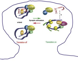
Our laboratory found that CYFIP1, as well as FMRP, co-fractionates with light mRNPs and that it is part of the cap-binding complex, together with eIF4E and PABP (Napoli et al., 2008). Interestingly, the association of CYFIP1 with eIF4E is competed by exogenous 4E-BP2. In addition, CYFIP1 binds directly eIF4E, although it does not have the canonical eIF4E-binding sequence (YXXXXLΦ, where X is any aminoacid and Φ is a hydrophobic aminoacid) (Costa-Mattioli et al., 2009; Marcotrigiano et al., 1999). Surprisingly, the “non canonical” sequence is predicted to form a peculiar “reverse L shaped” with two α helices turns; this structure overlaps with the region of the 4E-BPs fitting into the eIF4E pocket (Marcotrigiano et al., 1999; Napoli et al., 2008). The FMRP-CYFIP1-eIF4E complex is present also in synaptoneurosomes and contains BC1 RNA, as well as the FMRP targets α-CaMKII, Map1b, App and Arc. Noteworthy, stimulation with either BDNF or DHPG reduces the binding of CYFIP1 to eIF4E and the presence of Map1b mRNA and BC1 RNA in the complex (Napoli et al., 2008). According to all these observations, CYFIP1 downregulation in cultured neurons, as well as genetic depletion of CYFIP1 (CYFIP1 +/- mice), causes a significant increase in the protein levels of α-CaMKII, MAP1B and APP (Napoli et al., 2008). All these data support the model that at synapses FMRP tethers specific mRNAs on CYFIP1, which in turn sequesters eIF4E and represses translation initiation. Upon synaptic stimulation, FMRP-CYFIP1 dissociates from eIF4E and translation takes place (Fig. 5).
38
Fig. 5. Proposed model for mRNA translational repression and activation by
CYFIP1-FMRP complex. CYFIP1-CYFIP1-FMRP and CYFIP1-CYFIP1-FMRP-BC1 mRNPs are transported to the synapses in a translationally dormant state. After synaptic stimulation, CYFIP1-FMRP complex is released from eIF4E and local translation takes place. (Napoli et al., 2008)
CYFIP is not the only “specific” 4E-BP. In fact, the model proposed by Napoli and colleagues closely resembles the mechanism described for two regulatory complexes: vertebrate Maskin/Neuroguidin-CPEB and
Drosophila Cup-Bruno. In such cases, a protein (CYFIP1, Maskin or
Neuroguidin, Cup) sequesters the initiation factor eIF4E and simultaneously binds an RNA-binding protein (i.e., FMRP, CPEB, and Bruno, respectively); this configuration tethers the repression complex to a specific subset of mRNAs (Richter and Klann, 2009) (Fig. 6).
Besides the well-documented role in translational control, FMRP has been recently implicated in the regulation of mRNA stability (De Rubeis and Bagni, 2009).
39
Fig. 6. Translational control by 4E-BPs. Cap-dependent translation depends on
interactions between eIF4E, eIF4G, eIF3 and the small ribosomal subunit 40S. The eIF4E-eIF4G interaction can be disrupted by the “canonical” 4E-BPs (4E-BP1/2/3) or by “specific” 4E-BPs, such as Maskin, Neuroguidin (Ngd), Cup or CYFIP1. Because of specific RNA-binding proteins, such as CPEB, Bruno or FMRP, the inhibitory complex is driven on a subset of mRNAs. (Richter and Klann, 2009)
The control of mRNA stability
The regulation of protein stability and degradation
mRNA stability is a highly regulated posttranscriptional step tightly coordinated with mRNA translation. A specific RNA surveillance mechanism, the Nonsense-Mediated Decay (NMD), removes mRNAs harboring premature stop codons to prevent the accumulation of aberrant, possibly dominant-negative, proteins (Behm-Ansmant and Izaurralde, 2006). Once mRNAs overcome NMD control, they can be translated or sequestered such that they can undergo translation or degradation at a later time. In the latter case, several factors can modulate the decay rate of mRNAs and mediate the communication between translation and degradation.
40
Moreover, the interplay between translation and degradation may take place in cytoplasmic foci referred as P bodies, where mRNAs can be degraded or stored to later re-enter the translating pool of mRNAs (Parker and Sheth, 2007). These bodies are functionally related to other cytoplasmic aggregates, namely stress granules, which are composed by stalled translational pre-initiation complex (Kedersha et al., 2005). The nature of these bodies remains to be elucidated.
Normally, housekeeping genes produce invariantly stable transcripts while the turnover of some mRNAs undergoes a tight regulation to rearrange gene expression to certain cellular and/or developmental stimuli. Many mRNAs have a fast decay rate, resulting in a low steady-state level of protein under basal conditions. After stimulation, the mRNAs are rapidly induced by transcriptional activation and a modest increase in the amount leads to a significant variation in their expression (Khabar, 2007). In neurons, among the mRNAs regulated at the stability level are those encoding proteins related to neuronal growth (GAP-43, NGF, Tau), enzymes or enzyme inhibitors (acetylcholinesterase, neuroserpin), receptors (D2 dopamine receptor, m4 muscarinic receptor, β1-adrenergic receptor) and
transcription factors (c-Fos, N-Myc/c-Myc) (Bolognani and Perrone-Bizzozero, 2008).
The decay rate of mRNAs depends on cis-acting elements frequently located in their 3’UTRs as well as their associated trans-acting factors. A well characterized sequence involved in mRNA stability is a 50-150 nucleotide sequence rich in adenosine and uridine, the so-called AU-rich element (ARE). These sequences are located in the 3’ UTRs of the mRNAs that are regulated by the AU-rich RNA binding proteins (AUBPs). In some cases, these sequences are rich in different residues such as GU or C (Kim and Gorospe, 2008; Vlasova et al., 2008). The importance of AREs in regulating gene expression is highlighted by the fact that 5-8% of human genes encode ARE-containing transcripts (Bakheet et al., 2001). Although
41
AREs were originally defined as an AUUUA core associated with instability (Shaw and Kamen, 1986), it became clear over the years that ARE motifs can vary somewhat and regulate mRNA stability in both directions (Barreau et al., 2005). In fact, the interaction between ARE sequences and ARE-binding proteins can block or enhance the recruitment of the mRNA decay machinery and lead to a rapid modification of gene expression in response to environmental and developmental conditions.
Several RNA binding proteins that associate with these RNA elements are AU-binding factor 1 (AUF1) (Zhang et al., 1993), Tristetraprolin (TTP) (Carballo et al., 1998), Hu/ELAV (Dalmau et al., 1990), CUG triplet RNA-binding protein 1 (CUG-BP1) (Vlasova et al., 2008), and butyrate response factor-1 (BRF1) (Stoecklin et al., 2002). While the first two proteins are detected in all tissues, some of the Hu protein are neuro-specific (Hambardzumyan et al., 2009). In general AUF1, TTP, CUG-BP are destabilizing factors that decrease the half-life of mRNA while the Hu proteins are stabilizing factors that promote stability and translation. AUF-1 might promote stability and degradation depending on the mRNA and the cell type (Sela-Brown et al., 2000; Xu et al., 2001). In addition, microRNAs also affect mRNA stability. In a genome-wide microarray analysis it has been shown that some microRNAs downregulate many target mRNAs (Lim et al., 2005). Further studies have also identified the molecular mechanism and the protein complex(es) involved (Bagga et al., 2005; Behm-Ansmant et al., 2006; Wu et al., 2006).
In mammals, two major pathways for mRNA degradation have been described. The first step is the removal of the poly(A) tail, which opens both 5’ and 3’ ends for exonucleolytic attack (Fig. 7). In fact, the interaction of the poly(A) and the 5’ end of the mRNA, via the PABP-eIF4G complex, forms a closed-loop state of the mRNA that is not accessible to the exonucleases (Mazumder et al., 2003).
42
In the first pathway, after deadenylation, the decapping enzymes Dcp1 and Dcp2 eliminate the 5’ cap and the mRNA body is degraded by the 5’→ 3’ exonuclease Xrn (Wilusz and Wilusz, 2004). Alternatively, the decay occurs in 3’→ 5’ direction catalyzed by the exosome, a large exonucleolytic complex. The residual cap is degraded by the scavenger enzyme DcpS (Wilusz and Wilusz, 2004) (Fig. 7).
One of the most well described neuronal mRNAs regulated at the stability level is that encoding Growth-Associated Protein 43 (GAP-43), a developmentally-regulated protein involved in axon elongation in both developing and regenerating neurons (Korshunova and Mosevitsky, 2008). The expression of GAP-43 is posttranscriptionally regulated by HuD, a neuronal ARE-binding protein belonging to the family of ELAV/Hu proteins. HuD recognizes an U-rich element in the 3’UTR of GAP-43 mRNA and stabilizes it by interfering with the removal of the poly(A) tail (Beckel-Mitchener et al., 2002; Chung et al., 1997). Indeed, increased levels of HuD correspond to higher GAP-43 expression (Anderson et al., 2001; Bolognani et al., 2006; Pascale et al., 2004), leading to changes in neurite outgrowth (Anderson et al., 2001) and in synaptic plasticity (Bolognani et al., 2007b; Pascale et al., 2004).
43
Fig. 7. The pathways of the mRNA decay. The first step is the deadenylation, followed
by 5’→3’ or 3’→5’ degradation. The 5’→3’ decay starts with the removal of the cap by the Dcp1 and Dcp2 enzymes, followed by the degradation via the exonuclease Xrn1. The 3’→5’ decay is catalyzed by a large complex, the exosome, and the remaining cap is degraded by the scavenger protein DcpS. (Wilusz and Wilusz, 2004)
Neuronal regulators of mRNA stability
Hu/ELAV proteins. One of the few specific proteins that have been described as key regulators of mRNA turnover in brain is the Hu family of proteins. The Hu proteins were first described as autoantigens in patients affected by paraneoplastic encephalomyelitis (Dalmau et al., 1990). The human proteins were then recognized to be orthologous of the Drosophila Embryonic Lethal Abnormal Vision (ELAV) protein, a splicing regulator important for neural development (Simionato et al., 2007). In mammals, four ELAV/Hu proteins have been described: HuR (alias HuA), HuB, HuC and HuD (Brennan and Steitz, 2001). While HuR is ubiquitous and HuB is present in the brain and in germ cells, the expression of HuC and HuD is restricted to neurons. The neuronal ELAV/Hu proteins (nELAV) are essential for development of the nervous system. In fact, Hu proteins are reported as early markers of neuronal commitment and show a specific timing of expression in the developing brain (Okano and Darnell, 1997; Pascale et al., 2008). Studies in vivo or in primary neurons in which the nELAV expression has been perturbed by either overexpression or downregulation directly implicate these proteins in neuronal differentiation as well as in learning and memory (Akamatsu et al., 2005; Anderson et al., 2001; Bolognani et al., 2004; Bolognani et al., 2007a; Pascale et al., 2004; Quattrone et al., 2001). In neurons, the ELAV proteins are expressed in the cell body and along the dendrites (Bolognani et al., 2004; Tiruchinapalli et al., 2008a; Tiruchinapalli et al., 2008b); HuD has been also found in the neurites of in PC12 cells (Aranda-Abreu et al., 1999; Smith et al., 2004). Like many other RNA-binding proteins, the Hu proteins are detected both in
44
the nucleus and in the cytoplasm, suggesting different roles in different compartments. Although a role in splicing and nuclear polyadenylation is possible, the best characterized functions are the control of mRNA decay and the regulation of protein synthesis (Hinman and Lou, 2008). Focusing on the mRNA stability process, the Hu proteins can positively modulate the half-life of a subset of mRNAs critical for neuronal differentiation and maintenance. This group includes mRNAs encoding key transcription factors (c-Fos, c-Myc), molecules involved in neurite outgrowth and synapse functionality (the above-mentioned GAP-43, Tau, acetylcholinesterase, neuroserpin) and determinant of neural differentiation (Musashi-1) (Pascale et al., 2008).
AUF1 and KSRP. While the Hu proteins are mostly implicated in the stabilization of mRNAs, other neuronal decay-promoting factors have also been described. One of the first identified is the AU-binding factor 1 (AUF1), also known as hnRNP D (Zhang et al., 1993). AUF1, which shuttles between the nucleus and cytoplasm, consists of four isoforms generated by alternative splicing of a single transcript (Sarkar et al., 2003a; Wagner et al., 1998). The different isoforms are expressed in the brain and show specific activities in binding and modulating the decay of ARE-containing transcripts (Dobi et al., 2006; Raineri et al., 2004; Sarkar et al., 2003b). Although the mRNAs associated with AUF1 are well characterized in non-neuronal cells (Bhattacharya et al., 1999; Mazan-Mamczarz et al., 2009), few target mRNAs have been identified in the brain. One example is the mRNA encoding the α2 subunit of the nitric-oxide sensitive guanylyl cyclase in the
cerebellar granule cells; the messenger is bound by AUF1 but upon the activation of the N-methyl-D-aspartate (NMDA) glutamate receptors, AUF1 is downregulated and the α2 mRNA is stabilized (Jurado et al., 2006). In this case, there is an activity-dependent regulation mediated by AUF1. It has also been suggested that AUF1 is involved in integrating genetic and epigenetic signals during cortical development. It is specifically expressed in subsets of
45
proliferating neural precursors and differentiating postmitotic neurons of the developing cortex (Lee et al., 2008). Recently AUF-1 has been shown to promote the degradation of some target mRNAs but increase the stability and translation of other mRNAs ; this duality may be due to relative abundance of AUF1 (Mazan-Mamczarz et al., 2009).
Another destabilizing factor present in neurons is the human K homology splicing regulatory protein (KSRP), a protein originally described as a splicing regulator (Min et al., 1997). KSRP is homologous to the murine Zipcode Binding Protein 2 (ZBP2), involved in β-actin mRNA localization in neurons (Gu et al., 2002). The protein is expressed in neurons and in glia and it is distributed is both the nucleus and cytoplasm where it can interact with mRNAs and enhance their turnover (Chou et al., 2006; Gherzi et al., 2004; Snee et al., 2002). Despite the fact that KSRP is neuronal, no specific target mRNAs in the brain have been characterized.
The role of FMRP in stability control
In addition to its role in mRNA transport and translation, recent evidence shows that FMRP assumes an activity related to mRNA decay. Three studies have shown that mRNA abundance is affected when FMRP expression is abolished. Although such reports do not directly demonstrate a role of FMRP in mRNA stability, they provided a first cue that mRNA levels are altered in the absence of FMRP. First, in a high-throughput screen to identify FMRP target mRNAs, Brown et al. (2001) found that the levels of 144 genes were changed in lymphoblastoid cells from Fragile X patients (Brown et al., 2001).
In another analysis, Miyashiro et al. (2003) identified some of the same mRNAs as Brown et al. (2001) and described their expression pattern; 3 out of 11 mRNAs were reduced in the FMR1 KO versus wild type mouse
46
hippocampus (Miyashiro et al., 2003). For two of these mRNAs, those encoding the ribosomal component p40/LRP and the G-protein-coupled receptor kinase 4 (GRK4), the subcellular distribution was unaffected while for the dystroglycan-associated glycoprotein 1 (DAG1 mRNA), an altered dendritic localization as well as reduced abundance was observed (Miyashiro et al., 2003).
Moreover, FMRP loss may also lead to an impaired expression of GABAA receptors. The δ subunit mRNA, previously identified by Miyashiro
et al. (2003), has been found to be downregulated in FMR1 KO neurons in a genome-wide expression profiling study (Gantois et al., 2006) as well as in other studies addressing its localization (Dictenberg et al., 2008). Consistent with these results, El Idrissi et al. (2005) reported a decreased expression of the GABA β subunit in several brain areas from FMR1 KO mice (El Idrissi et al., 2005). Interestingly, a further indication of FMRP as neuronal mRNA stabilizing factor came from D’Hulst et al. (2006) who reported that the mRNAs encoding 8 out of the 18 known GABA subunits (α1, α3, α4, β1, β2,
γ1, γ2 as well as the above mentioned δ) were significantly reduced in the
cortex, but not in the cerebellum, from FMRP-lacking mice. In addition, the expression of all the three subunits conserved in Drosophila appears to be compromised as well (D'Hulst et al., 2006).
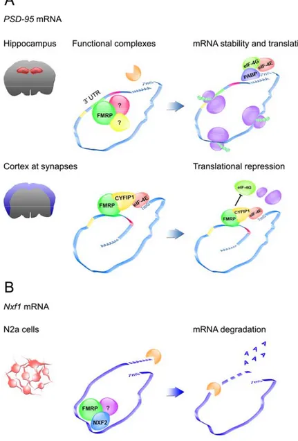
Finally, two recent reports implicate FMRP as a direct modulator of mRNA turnover (Fig. 7). First, FMRP has been shown to be involved in the regulation of PSD-95 mRNA stability in hippocampus (Zalfa et al., 2007). PSD-95 encodes a key scaffolding protein of the post-synaptic density (PSD), the signal transduction machinery at glutamatergic synapses (Kim and Sheng, 2004). Because PSD-95 loss compromises both the structure and the function of dendritic spines (Migaud et al., 1998; Vickers et al., 2006), alterations in PSD-95 expression could contribute to the cognitive impairment caused by the absence of FMRP. PSD-95 mRNA is part of the FMRP mRNP in vivo; the C-terminal domain of FMRP binds a G-rich
47
structure in its 3’UTR (shown not be structured as a G-quartet) (Zalfa et al., 2007). By inhibiting transcription in primary neurons with actinomycin D, it has been shown that FMRP protects PSD-95 mRNA from decay specifically in the hippocampus (Figure 7A). The stabilizing effect of FMRP can also be enhanced upon neuronal activity, such as the stimulation of metabotropic glutamate receptors (mGluRs) (Zalfa et al., 2007).
Furthermore, the region-specific effect of FMRP on PSD-95 mRNA stability is consistent with other reports indicating that PSD-95 synaptic translation is affected in the cortex of FMRP-lacking mice (Muddashetty et al., 2007; Todd et al., 2003) (Fig. 7A). Thus, it is possible that FMRP has multiple independent functions in the regulation of posttranscriptional control of gene expression (i.e. stability, mRNA translation) depending on the cellular context and developmental timing.
The FMRP complex regulating PSD-95 mRNA turnover has not yet been identified. The FMRP-binding site in the 3’UTR is close to three U-rich tracts; two of them contain AREs. These motifs could be crucial for PSD-95 mRNA half-life and the function of FMRP could prevent the action/binding of other destabilizing factors (Figure 7A). Moreover, FMRP is not a general regulator of mRNA decay. The stability assay revealed that only two out of 12 known FMRP targets (PSD-95, Map1b, α-CaMKII, Fxr1, G3bp, App,
RhoA, Ef-1α, Vdac1, HnRNPA2, Sapap4 and Mbp) have a compromised half-life in the absence of FMRP (Zalfa et al., 2007). The observation that
Myelin basic protein (Mbp) mRNA has also a decreased stability extends
this function to oligodendrocytes (Zalfa et al., 2007). Considering that previous work by Kooy and collaborators (D'Hulst et al., 2006; Gantois et al., 2006) has shown that in absence of FMRP a subset of mRNAs encoding the GABA receptors is downregulated, it is tempting to hypothesize that this reduced expression could occur at the level of mRNA stability.
48
FMRP has also been reported to contribute to Nxf1 mRNA stability (Zhang et al., 2007). Nxf1 mRNA encodes the large subunit of the mRNP export receptor involved in the transport of mature transcripts from the nucleus to the cytoplasm. NXF1 is the predominant component of the nuclear export factor family, which also includes NXF2, previously identified as a direct partner of FMRP in both testis and brain. The authors initially proposed that FMRP could act as an adaptor protein recruiting a specific subclass of mRNPs to NXF2 and to facilitate the nuclear export (Lai et al., 2006). More recently, using a mouse neuroblastoma cell line (N2a cells), they showed that upon NXF2 overexpression, the turnover of the messenger increased and consequently the mRNA levels were reduced. Because this effect was abolished by silencing FMRP in the neuroblastoma cell line, the authors proposed that FMRP could contribute to the degradation induced by NXF2 (Zhang et al., 2007) (Figure 7B).
49
Fig. 7. FMRP in the control of mRNA stability. Panel A shows the regulatory effect of
FMRP on PSD-95 mRNA in the hippocampus (upper panel, Zalfa et al., 2007) and in the cortex (lower panel, Muddashetty et al., 2007, Napoli et al., 2008). FMRP (in green) directly interacts with the 3’UTR of PSD-95 mRNA. The FMRP-binding site (G-rich region, in green)
50
is close to a U-rich stretch (in yellow) and two AU-rich tracts (in light blue and purple). In the hippocampus (upper panel), the FMRP complex, which could include other factors involved in the control of mRNA turnover (in yellow and purple), would block the entry of the exonucleases (in orange) and consequently mRNA degradation. Consequently, the stable mRNA can undergo translation. In the cortex (lower panel), the FMRP complex would tether
PSD-95 mRNA to a repression complex. One possibility is via CYFIP1. In this case, CYFIP1
would prevent eIF4E (in red) interacting with eIF4G (in light green), therefore inhibiting the initiation of translation. PSD-95 could also be possibly repressed via other mechanisms i.e. microRNAs (not shown here). Panel B depicts the role of FMRP in the regulation of Nxf1 mRNA half-life in a neuronal cell line (Zhang et al., 2007). FMRP (in green) directly binds NXF2 (in blue) and the complex associate with the messenger. It is still unknown which component of the complex mediate the recognition of the mRNA. The FMRP-NXF2 complex and possibly other proteins (purple) would facilitate the degradation of Nxf1 mRNA (De Rubeis and Bagni, 2009).
Aakalu, G., Smith, W. B., Nguyen, N., Jiang, C., and Schuman, E. M. (2001). Dynamic visualization of local protein synthesis in hippocampal neurons. Neuron 30, 489-502.
Abrahams, B. S., and Geschwind, D. H. (2008). Advances in autism genetics: on the threshold of a new neurobiology. Nat Rev Genet 9, 341-355.
Akamatsu, W., Fujihara, H., Mitsuhashi, T., Yano, M., Shibata, S., Hayakawa, Y., Okano, H. J., Sakakibara, S., Takano, H., Takano, T.,
et al. (2005). The RNA-binding protein HuD regulates neuronal cell
identity and maturation. Proc Natl Acad Sci U S A 102, 4625-4630. Anderson, K. D., Sengupta, J., Morin, M., Neve, R. L., Valenzuela, C. F.,
and Perrone-Bizzozero, N. I. (2001). Overexpression of HuD accelerates neurite outgrowth and increases GAP-43 mRNA expression in cortical neurons and retinoic acid-induced embryonic stem cells in vitro. Exp Neurol 168, 250-258.
Antar, L. N., Afroz, R., Dictenberg, J. B., Carroll, R. C., and Bassell, G. J. (2004). Metabotropic glutamate receptor activation regulates fragile x mental retardation protein and FMR1 mRNA localization differentially in dendrites and at synapses. J Neurosci 24, 2648-2655.
Antar, L. N., Dictenberg, J. B., Plociniak, M., Afroz, R., and Bassell, G. J. (2005). Localization of FMRP-associated mRNA granules and requirement of microtubules for activity-dependent trafficking in hippocampal neurons. Genes Brain Behav 4, 350-359.
Antar, L. N., Li, C., Zhang, H., Carroll, R. C., and Bassell, G. J. (2006). Local functions for FMRP in axon growth cone motility and activity-dependent regulation of filopodia and spine synapses. Mol Cell Neurosci 32, 37-48.
Antion, M. D., Hou, L., Wong, H., Hoeffer, C. A., and Klann, E. (2008). mGluR-dependent long-term depression is associated with increased phosphorylation of S6 and synthesis of elongation factor 1A but remains expressed in S6K-deficient mice. Mol Cell Biol 28, 2996-3007.
Aranda-Abreu, G. E., Behar, L., Chung, S., Furneaux, H., and Ginzburg, I. (1999). Embryonic lethal abnormal vision-like RNA-binding proteins regulate neurite outgrowth and tau expression in PC12 cells. J Neurosci 19, 6907-6917.
Arimura, N., and Kaibuchi, K. (2007). Neuronal polarity: from
extracellular signals to intracellular mechanisms. Nat Rev Neurosci
58
Ashley, C. T., Sutcliffe, J. S., Kunst, C. B., Leiner, H. A., Eichler, E. E., Nelson, D. L., and Warren, S. T. (1993). Human and murine FMR-1: alternative splicing and translational initiation downstream of the CGG-repeat. Nat Genet 4, 244-251.
Bagga, S., Bracht, J., Hunter, S., Massirer, K., Holtz, J., Eachus, R., and Pasquinelli, A. E. (2005). Regulation by let-7 and lin-4 miRNAs results in target mRNA degradation. Cell 122, 553-563.
Bagni, C., and Greenough, W. T. (2005). From mRNP trafficking to spine dysmorphogenesis: the roots of fragile X syndrome. Nat Rev Neurosci 6, 376-387.
Bakheet, T., Frevel, M., Williams, B. R., Greer, W., and Khabar, K. S. (2001). ARED: human AU-rich element-containing mRNA database reveals an unexpectedly diverse functional repertoire of encoded proteins. Nucleic Acids Res 29, 246-254.
Bakker, C. e. a. (1994). Fmr1 knockout mice: a model to study fragile X mental retardation. The Dutch-Belgian Fragile X Consortium. Cell
78, 23-33.
Banko, J. L., Hou, L., Poulin, F., Sonenberg, N., and Klann, E. (2006). Regulation of eukaryotic initiation factor 4E by converging signaling pathways during metabotropic glutamate receptor-dependent long-term depression. J Neurosci 26, 2167-2173. Banko, J. L., Merhav, M., Stern, E., Sonenberg, N., Rosenblum, K., and
Klann, E. (2007). Behavioral alterations in mice lacking the translation repressor 4E-BP2. Neurobiol Learn Mem 87, 248-256. Banko, J. L., Poulin, F., Hou, L., DeMaria, C. T., Sonenberg, N., and
Klann, E. (2005). The translation repressor 4E-BP2 is critical for eIF4F complex formation, synaptic plasticity, and memory in the hippocampus. J Neurosci 25, 9581-9590.
Bannai, H., Fukatsu, K., Mizutani, A., Natsume, T., Iemura, S., Ikegami, T., Inoue, T., and Mikoshiba, K. (2004). An RNA-interacting protein, SYNCRIP (heterogeneous nuclear ribonuclear protein Q1/NSAP1) is a component of mRNA granule transported with inositol 1,4,5-trisphosphate receptor type 1 mRNA in neuronal dendrites. J Biol Chem 279, 53427-53434.
Bardoni, B., Schenck, A., and Mandel, J. L. (2001). The Fragile X mental retardation protein. Brain Res Bull 56, 375-382.
Barreau, C., Paillard, L., and Osborne, H. B. (2005). AU-rich elements and associated factors: are there unifying principles? Nucleic Acids Res 33, 7138-7150.