A
A
l
l
m
m
a
a
M
M
a
a
t
t
e
e
r
r
S
S
t
t
u
u
d
d
i
i
o
o
r
r
u
u
m
m
–
–
U
U
n
n
i
i
v
v
e
e
r
r
s
s
i
i
t
t
à
à
d
d
i
i
B
B
o
o
l
l
o
o
g
g
n
n
a
a
DOTTORATO DI RICERCA
BIOCHIMICA
Ciclo XX
Settore/i scientifico disciplinari di afferenza: BIO/10
Biochemistry in Healthy and Neoplastic Human
Tissues: Metabolic Alteration Revealed by HR-MAS
Nuclear Magnetic Resonance Spectroscopy
VALERIA RIGHI
Coordinatore Dottorato
Relatori
Prof. Giorgio Lenaz
Dott. Vitaliano Tugnoli
Prof.ssa Adele Mucci
A
A
l
l
m
m
a
a
M
M
a
a
t
t
e
e
r
r
S
S
t
t
u
u
d
d
i
i
o
o
r
r
u
u
m
m
–
–
U
U
n
n
i
i
v
v
e
e
r
r
s
s
i
i
t
t
à
à
d
d
i
i
B
B
o
o
l
l
o
o
g
g
n
n
a
a
DOTTORATO DI RICERCA
BIOCHIMICA
Ciclo XX
Settore/i scientifico disciplinari di afferenza: BIO/10
Biochemistry in Healthy and Neoplastic Human
Tissues: Metabolic Alteration Revealed by HR-MAS
Nuclear Magnetic Resonance Spectroscopy
VALERIA RIGHI
Coordinatore Dottorato
Relatori
Prof. Giorgio Lenaz
Dott. Vitaliano Tugnoli
Prof.ssa Adele Mucci
Ai miei Genitori
Abbrevation 5
Summary 8
Riassunto 13
Chapter 1 Introduction to metabolomics 18
Chapter 2 Nuclear Magnetic Resonance 27
Chapter 3 Statistical Analyses 61
Chapter 4 Brain Tumors 71
Chapter 5 Gastro-intestinal Cancer 121
Chapter 6 Kidney Diseases 179
Concluding Remarks 193
5
ABBREVIATIONS
Ac Acetate
AAG Gastric Atrophy Autoimmune
Ala Alanine Asp Aspartate Arg Arginine Asn Asparagines At Glycerol-3-phosphate acyltransferases Bet Glycine-betaine 13 C Carbon 13
CHESS Chemical Shift Selective saturation standard Ck Choline kinase
CNS Central Nervous System Cho free Choline
ChoCC/tCho Choline Containing Compounds or total Choline Chol Cholesterol
COSY COrrelation SpectroscopY CPMG Carr-Purcell-Meiboom-Gill
Cr Creatine
Ct Cytidylyltransferase
CT Computed Tomografy
Cys Cysteine
D2O Deuterated water
1D one-Dimensional
2D two-Dimensional
E Ethanolamine
FA Fatty Acids
FID Free Induction Decay FT Fourier Trasformer GABA γ-AminoButyric Acid α α α α-, ββββ-Glc α-, β-Glucose Gln Glutamine Glu Glutamate
Glx Glutamine plus Glutamate
6 GPC Glycerophosphocholine GPE Glycerophosphoethanolamine GSH γ-Glutamylcysteinylglycine, glutathione 1 H Proton His Histidine
HPF High Power Field
HR-MAS High-Resolution Magic Angle Spinning HMBC Heteronuclear Multiple Bond Correlation HMQC Heteronuclear Multiple Quantum Coherence HSQC Heteronuclear Single Quantum Coherence HTau HypoTaurine
Ile Isoleucine
Lac Lactate
LED Longitudinal-Eddy current Delay
Leu Leucine
Lip Lipids
Lpl Lysophospholipase
Lys Lysine
MRI Magnetic Resonance Imaging MRS Magnetic Resonance Spectroscopy
Met Methionine
Man Mannitol
MM MacroMolecoles or bonded amino-acids Myo-Ino Myo-Inositol
NAA N-acetylAspartate
NMR Nuclear Magnetic Resonance β-OHBut β-hydroxybutyrate
Pap Phosphatidate phosphohydrolase PCA Principal Components Analysis PCr Phosphocreatine
PC Phosphocholine
PE Phosphoethanolamine PEG Polyethylene glycol
Ph Phospholipids
Phe Phenylalanine
7 Pld Phospholipase D
PLS-DA Partial Least Squares-Discriminant Analysis PNET Primitive NEuroectodermal Tumor PRESS Point Resolved Spectroscopy
Pro Proline
Ptc Phosphocholine cytidylyl-transferase RCC Renal Cell Carcinoma
Scy-Ino Scyllo-Inositol Suc Succinate Tau Taurine TG Triglycerides Thr Threonine TMA Tetramethylammonium
TOCSY TOtal Correlation SpectroscopY
Tyr Tyrosine
UDP Uridine diphosphate UMP Uridine monophosphate
Val Valine
VOI Volume of interest
8
SUMMARY
This thesis is focused on the metabolomic study of human cancer tissues by ex vivo High Resolution-Magic Angle Spinning (HR-MAS) nuclear magnetic resonance (NMR) spectroscopy. This new technique allows for the acquisition of spectra directly on intact tissues (biopsy or surgery), and it has become very important for integrated metabonomics studies. The objective is to identify metabolites that can be used as markers for the discrimination of the different types of cancer, for the grading, and for the assessment of the evolution of the tumour. Furthermore, an attempt to recognize metabolites, that although involved in the metabolism of tumoral tissues in low concentration, can be important modulators of neoplastic proliferation, was performed. In addition, NMR data was integrated with statistical techniques in order to obtain semi-quantitative information about the metabolite markers. In the case of gliomas, the NMR study was correlated with gene expression of neoplastic tissues.
Chapter 1 begins with a general description of a new “omics” study, the
metabolomics. The study of metabolism can contribute significantly to biomedical research and, ultimately, to clinical medical practice. This rapidly developing discipline involves the study of the metabolome: the total repertoire of small molecules present in cells, tissues, organs, and biological fluids. Metabolomic approaches are becoming increasingly popular in disease diagnosis and will play an important role on improving our understanding of cancer mechanism. Chapter 2 addresses in more detail the basis of NMR Spectroscopy, presenting the new HR-MAS NMR tool, that is gaining importance in the examination of tumour tissues, and in the assessment of tumour grade. Some advanced chemometric methodswere used in an attempt to enhance the interpretation and quantitative information of the HR-MAS NMR data
are and presented in chapter 3. Chemometric methods seem to have a high potential in the
study of human diseases, as it permits the extraction of new and relevant information from spectroscopic data, allowing a better interpretation of the results.
Chapter 4 reports results obtained from HR-MAS NMR analyses performed on
different brain tumours: medulloblastoma, meningioms and gliomas.
The medulloblastoma study is a case report of primitive neuroectodermal tumor (PNET) localised in the cerebellar region by Magnetic Resonance Imaging (MRI) in a 3-year-old child. In vivo single voxel 1H MRS shows high specificity in detecting the main metabolic
9
choline-containing compounds and of very low levels of creatine derivatives and N-acetylaspartate. Ex vivo HR-MAS NMR, performed at 9.4 Tesla on the neoplastic specimen collected during surgery, allows the unambiguous identification of several metabolites giving a more in-depth evaluation of the metabolic pattern of the lesion. The ex vivo HR-MAS NMR spectra show higher detail than that obtained in vivo. In addition, the spectroscopic data appear to correlate with some morphological features of the medulloblastoma. The present study shows that ex vivo HR-MAS 1H NMR is able to strongly improve the clinical possibility of in vivo MRS and can be used in conjunction with in vivo spectroscopy for clinical purposes.
Three histological subtypes of meningiomas (meningothelial, fibrous and oncocytic) were analysed both by in vivo and ex vivo MRS experiments. The ex vivo HR-MAS investigations are very helpful for the assignment of the in vivo resonances of human meningiomas and for the validation of the quantification procedure of in vivo MR spectra. By using one- and two dimensional experiments, several metabolites in different histological subtypes of meningiomas, were identified. The spectroscopic data confirmed the presence of the typical metabolites of these benign neoplasms and, at the same time, that meningomas with different morphological characteristics have different metabolic profiles, particularly regarding macromolecules and lipids.
The profile of total choline metabolites (tCho) and the expression of the Kennedy pathway genes in biopsies of human gliomas were also investigated using HR-MAS NMR, and microfluidic genomic cards. 1H HR-MAS spectra, allowed the resolution and relative quantification by LCModel of the resonances from choline (Cho), phosphorylcholine (PC) and glycerolphorylcholine (GPC), the three main components of the combined tCho peak observed in gliomas by in vivo 1H MRS spectroscopy. All glioma biopsies depicted an increase in tCho as calculated from the addition of Cho, PC and GPC HR-MAS resonances. However, the increase was constantly derived from augmented GPC in low grade NMR gliomas or increased PC content in the high grade gliomas, respectively. This circumstance allowed the unambiguous discrimination of high and low grade gliomas by 1H HR-MAS, which could not be achieved by calculating the tCho/Cr ratio commonly used by in vivo 1H MR spectroscopy. The expression of the genes involved in choline metabolism was investigated in the same biopsies. High grade gliomas depicted a significant upregulation of choline kinase and phospholipase C, as well as a downregulation of the cytidyltransferase gene, the balance of these being consistent with the accumulation of PC. In the low grade
10
became downregulated, supporting the accumulation of GPC. The present findings offer a convenient procedure to classify accurately glioma grade using 1H HR-MAS, providing in addition the genetic background for the alterations of choline metabolism observed in high and low gliomas grade.
Chapter 5 reports the study on human gastrointestinal tract (stomach and colon)
neoplasms. The human healthy gastric mucosa, and the characteristics of the biochemical profile of human gastric adenocarcinoma in comparison with that of healthy gastric mucosa were analyzed using ex vivo HR-MAS NMR. Healthy human mucosa is mainly characterized by the presence of small metabolites (more than 50 identified) and macromolecules. The adenocarcinoma spectra were dominated by the presence of signals due to triglycerides, that are usually very low in healthy gastric mucosa. The use of spin-echo experiments enable us to detect some metabolites in the unhealthy tissues and to determine their variation with respect to the healthy ones. Then, the ex vivo HR-MAS NMR analysis was applied to human gastric tissue, to obtain information on the molecular steps involved in the gastric carcinogenesis. It is possible to identify glycine, alanine, free choline and triglycerides as possible molecular markers related to the human gastric mucosa differentiation from pre-neoplastic to neoplastic conditions. A microscopic investigation was also carried out in order to identify and locate the lipids in the cellular and extra-cellular environments. Correlation of the morphological changes detected by transmission (TEM) and scanning (SEM) electron microscopy, with the metabolic profile of gastric mucosa in healthy, gastric atrophy autoimmune diseases (AAG),
Helicobacter pylori-related gastritis and adenocarcinoma subjects, were obtained. These
ultrastructural studies of AAG and gastric adenocarcinoma revealed lipid intra- and extra-cellularly accumulation associated with a severe prenecrotic hypoxia and mitochondrial degeneration.
A deep insight into the metabolic profile of human healthy and neoplastic colon tissues was gained using ex vivo HR-MAS NMR spectroscopy in combination with multivariate methods: Principal Component Analysis (PCA) and Partial Least Squares Discriminant Analysis (PLS-DA). The NMR spectra of healthy tissues highlight different metabolic profiles with respect to those of neoplastic and microscopically normal colon specimens (these last obtained at least 15 cm far from the adenocarcinoma). Furthermore, metabolic variations are detected not only for neoplastic tissues with different histological diagnosis, but also for those classified identical by histological analysis. These findings suggest that the same subclass of colon carcinoma is characterized, at a certain degree, by
11
crucial in order to find metabolic markers of the neoplastic state of colon tissues, and to correctly classify the samples. Significant different levels of choline containing compounds, taurine and myo-inositol, were observed. Particularly, in this case, the presence of lipids it is not a marker of the tumor or of the grade of malignity.
Chapter 6 deals with the metabolic profile of normal and tumoral renal human tissues
obtained by ex vivo HR-MAS NMR. The spectra of human normal cortex and medulla show the presence of differently distributed osmolytes as markers of physiological renal condition. The marked decrease or disappearance of these metabolites and the high lipid content (triglycerides and cholesteryl esters) is typical of clear cell renal carcinoma (RCC), while papillary RCC is characterized by the absence of lipids and very high amounts of taurine. This research is a contribution to the biochemical classification of renal neoplastic pathologies, especially for RCCs, which can be evaluated by in vivo MRS for clinical purposes. Moreover, these data help to gain a better knowledge of the molecular processes envolved in the onset of renal carcinogenesis.
12
RIASSUNTO
Lo scopo di questa tesi è stato di effettuare uno studio di metabolomica (scienza relativa al pool dei metaboliti a basso peso molecolare presenti in un tessuto o in un fluido biologico) eseguito su neoplasie umane, attraverso l’utilizzo della ex vivo High Resolution-Magic Angle Spinning (HR-MAS) Nuclear Magnetic Resonance (NMR) spectroscopy. Questa tecnica permette di ottenere spettri direttamente sul tessuto (ottenuto per biopsia o in seguito ad intervento chirurgico) senza nessun pretrattamento, ed è diventata molto importante per studi metabonomici, relativi all’integrazione del metaboloma con una robusta elaborazione statistica. Dopo aver identificato i pattern metabolici di vari tessuti e organi umani sani e quelli delle rispettive neoplasie di differente tipo e grado istologico, l’obbiettivo è stato quello di ricercare nel tessuto tumorale i metaboliti che potessero assumere il significato di markers delle neoplasie, in relazione anche al grado e all’evoluzione del tumore stesso, con evidenti implicazioni cliniche (diagnosi, trattamento e prognosi). Inoltre, si è cercato di individuare quei metaboliti, che anche se presenti in basse concentrazioni, appaiono tuttavia coinvolti nel metabolismo tumorale, e possono essere anch’essi importanti modulatori della proliferazione neoplastica. Come anticipato, per gli studi metabonomici, i risultati NMR sono stati integrati con analisi statistiche per ottenere informazioni semiquantitative sui marker metabolici. Nel caso dei gliomi lo studio di risonanza è stato inoltre correlato con l’espressione genica di enzimi coinvolti nel pattern delle “coline” a livello del tessuto neoplastico. La scelta delle “coline” si è resa indispensabile essendo ampiamente dimostrato che il ciclo di Kennedy è coinvolto in modo determinante nel processo tumorale.
Il capitolo 1 presenta una introduzione generale alla metablomica. Lo studio del metabolismo tumorale può contribuire significativamente alla ricerca biomedica e in definitiva alla pratica medico-clinica, rendendo disponibili informazioni molecolari relative all’insorgenza ed alla progressione della neoplasia. Il rapido sviluppo di questa disciplina riguarda lo studio del metaboloma, cioè l’insieme delle piccole molecole presenti in cellule, tessuti, organi, e fluidi biologici. L’approccio metabonomico è diventato sempre più popolare nella diagnosi di malattie e avrà un ruolo importante nel migliorare la comprensione nei meccanismi molecolari coinvolti nell’insorgenza e nello sviluppo delle neoplasie umane. Nel
capitolo 2, vengono riportate in dettaglio le basi della spettroscopia NMR. In particolare
13
rispettivi tessuti funzionale. Nel capitolo 3 sono descritti alcuni metodi chemometrici avanzati, utilizzati per migliorare l’interpretazione e le informazioni quantitative dei dati spettroscopici ottenuti con l’HR-MAS. La combinazione dei dati biochimico-spettroscopici con l’elaborazione statistica nello studio delle malattie umane, permette non solo l’estrazione di nuove e rilevanti informazioni dai dati ottenibili con la spettroscopia NMR, ma anche una migliore interpretazione degli stessi assegnando una significatività statistica ai marker biochimici ipotizzati.
Nel capitolo 4 sono presentati i risultati ottenuti attraverso l’analisi ex vivo HR-MAS NMR eseguita su differenti tipi di tumori cerebrali. Il primo tipo di neoplasia cerebrale riportata si riferisce al medulloblastoma, neoplasia a prevalente incidenza infantile. In particolare viene discusso il “case report” di un PNET (Neuro Ectodermal Primitive Tumour) localizzato nella regione cerebellare MRI (Magnetic Resonance Imaging) in un bambino di tre anni. La spettroscopia in vivo single voxel 1H MRS ha mostrato alta specificità nel rilevare le principali alterazioni metaboliche nella lesione cerebellare, come un alto contenuto dei composti contenenti colina e una ridotta quantità di creatina ed N-acetilaspartato. La ex vivo HR-MAS 1H NMR, eseguita a 9.4 Tesla sul campione neoplastico prelevato durante l’operazione, permette di identificare diversi metaboliti dando una approfondita e più ampia valutazione del pattern metabolico della lesione. Gli spettri HR-MAS mostrano che i dettagli spettrali sono maggiori rispetto a quelli che si ottengono dallo spettro in vivo. In più i dati spettroscopici sembrano essere correlati con alcune caratteristiche morfologiche del medulloblastoma. Lo studio mostra come l’ex vivo HR-MAS è capace di migliorare fortemente le possibilità cliniche della in vivo MRS e puo essere utilizzata in combinazione con la spettroscopia in vivo per scopi clinici. Tre sottotipi istologici di meningioma (meningoteliale, fibroso e oncocitico), neoplasia extracerebrale a carattere prevalentemente begnina, sono stati analizzati mediante spettroscopia in vivo ed ex vivo. L’analisi ex vivo HR-MAS NMR ha permesso una accurata descrizione del profilo metabolico ed facilitano l’interpretazione e l’assegnazione dei segnali degli spettri in vivo dei diversi meningiomi e anche la validazione del procedimento di quantificazione degli spettri in vivo. Utilizzando esperimenti mono- e bidimensionali, sono stati identificati diversi metaboliti nei differenti sottotipi istologici. I dati spettroscopici confermano la presenza di metaboliti tipici delle neoplasie benigne, e nello stesso tempo che i meningiomi con differenti caratteristiche morfologiche presentano un pattern metabolico diverso soprattutto per quanto riguarda componenti riferibili a macromolecole e lipidi.
14
Kennedy in biopsie umane di gliomi sono stati studiati utilizzando la spettroscopia HR-MAS NMR e sonde dedicate per lo studio di geni specifici. Lo spettro 1H HR-MAS permette la risoluzione e la relativa quantificazione attraverso un software particolare (LCmodel) delle risonanze della colina (Cho), fosforilcolina (PC) e glicerofoforilcolina (GPC), le tre componenti principali del singolo picco delle coline totali (tCho) osservato negli spettri in
vivo di gliomi. Tutte le biopsie di glioma mostrano una aumento delle tCho, calcolato come
somma delle risonanze rilevabili in HR-MAS di Cho, PC e GPC. Tuttavia il principale dato è relative all’aumento della GPC nei gliomi di basso grado e dall’aumento di PC nei gliomi di alto grado. Il comportameno di questi metaboliti permette una netta discriminazione tra gliomi di alto e basso grado con l’HR-MAS, situazione che non può essere ottenuta calcolando il rapporto tCho/Cr che comunemente viene considerato con la spettroscopia in vivo. Sulle stesse biopsie è stata studiata l’espressione genica del pattern enzimatico coinvolto nel metabolismo delle coline. I gliomi di alto grado mostrano una significativa up-regulation dell’enzima colina kinasi e fosfolipasi C, mentre mostra una down-regulation del gene che codifica per l’enzima citidil-trasferasi; l’andamento dell’espressione di questi enzimi giustifica l’accumulo della PC rilevato nella spettroscopia. Nei gliomi di basso grado, la fosfolipasi A e la lisofosfolipasi sono up-regulated, mentre la fosfolipasi D è down-regulated, spiegando in modo analogo l’accumulo di GPC. Questo studio presenta un’adatta procedura per classificare accuratamente il grado del glioma utilizzando l’HR-MAS, a condizione che parallelamente sia valutato il back-ground genetico alla base delle alterazioni del metabolismo delle coline osservato nei gliomi di basso e alto grado.
Lo studio di neoplasie umane dell’apparato gastrointestinale è presentato nel capitolo
5. Le caratteristiche dei profili metabolici della mucosa gastrica sana, della mucosa affetta da
gastrite per infezione da Helicobacter Pilory, della mucosa affetta da una condizione preneoplstica quale la gastrite atrofica autoimmune (AAG) e dell’adenocarcinoma gastrico sono analizzate utilizzando spettroscopia ex vivo HR-MAS NMR. Il profilo metabolico della mucosa gastrica sana e affetta da Helicobacter Pilory è risultato molto simile.Le due situazioni sono caratterizzate dalla presenza di più di 50 metaboliti. Al contrario gli spettri di adenocarcinoma sono dominati dalla presenza di risonanze dovute a lipidi, trigliceridi in particolare. Il contenuto di questi ultimi è basso o quasi assente nella mucosa sana. Inoltre il tessuto affetto da AAG ha mostrato un pattern molecolare intermedio fra la situazione funzionale e quella neoplastica. L’uso della sequenza spin-echo ha permesso di rilevare alcuni metaboliti distintivi del tessuto neoplastico e di evidenziare la loro variazione rispetto al sano.
15
possibili step molecolari coinvolti nel processo della carcinogenesi gastrica. È stato infatti possibile identificare alcuni metaboliti che appaiono particolarmente significativi quali glicina, alanina, colina libera e trigliceridi come possibili marker legati alla differenziazione della mucosa gastrica a partire da una condizione sana, attraverso la condizione preneoplastica fino ad arrivare a quella neoplastica. È stata effettuata anche una indagine microscopica per localizzare i lipidi (i trigliceridi rilevati con L’HR-MAS) nei tessuti gastrici sani, affetti da AAG e da adenocarcinoma. Lo studio ha avuto come obbiettivo di correlare i cambiamenti morfologici, rilevati con la microscopia elettronica a trasmissione (TEM) e a scansione (SEM), col profilo metabolico della mucosa gastrica sana, dell’AAG, della gastrite dovuta a H. pylori e dell’adenocarcinoma. Gli studi ultrastrutturali della AAG e dell’adenocarcinoma gastrico rivelano che l’accumulo di lipidi, sotto forma di “goccioline lipidiche” (lipids bodies) avviene sia negli spazi intra- che in quelli extra-cellulari ed è associato ad una forte ipossia prenecrotica e ad una degenerazione mitocondriale.
Nel capitolo viene riportata anche un’accurata analisi del profilo metabolico del tessuto sano e neoplastico del colon utilizzando la spettroscopia ex vivo HR-MAS NMR in combinazione con metodi statistici: Principal Component Analysis (PCA) e Partial Least Squares Discriminant Analysis (PLS-DA). Lo spettro NMR della mucosa sana presenta un profilo metabolico diverso rispetto al tessuto neoplastico e anche rispetto al tessuto prelevato a 15 cm di distanza dalla zona tumorale e definito macroscopicamente eistologicamente sano. Inoltre le variazioni metaboliche sono rilevate non solo tra carcinomi con diversa diagnosi istologica, ma anche per carcinomi che presentano la stessa diagnosi istologica. Questo suggerisce che la stessa sottoclasse tumorale è caratterizzata da un certa eterogeneità metabolica. L’analisi multivariata applicata ai dati NMR è stata determinante per identificare i marker metabolici del tumore del colon e per classificare correttamente il campione. Variazioni significative sono state osservate nelle quantità relative di metaboliti quali coline, taurina e mio-inositolo. I lipidi in questo tratto gastrointestinale non risultano essere un marker tumorale o del grado di malignità.
Il lavoro riportato nel capitolo 6 presenta la caratterizzazione del profilo metabolico di parenchima renale umano funzionale e neoplastico ottenuto mediante ex vivo HR-MAS NMR. Gli spettri di campioni chirurgici renali relativi alle zone corticali e midollari mostrano una diversa distribuzione dei metaboliti ad attività osmotica (osmoliti). La marcata diminuzione o assenza dei metaboliti e un elevato contenuto di lipidi (trigliceridi e esteri del colesterolo) è tipica dei carcinomi renali a cellule chiare, mentre i carcinomi papillari sono caratterizzati da
16
biochimico-molecolare ad una migliore comprensione delle patologie neoplastiche renali, soprattutto dei carcinomi a cellule chiare. Crea inoltre le premesse biochimiche fondamentali per future applicazioni cliniche della spettroscopia in vivo. Inoltre questi dati contribuiscono a migliorare la conoscenza del processo molecolare che è alla base della carcinogenesi renale.
1
INTRODUCTION TO
METABOLOMICS
18
GENERAL INTRODUCTION
The final decades of the twenteth and the beginning of the twenty-first centuries, have witnessed a revolution in biomedical research that has made it possible to move from the study of single genes, single mRNA transcripts, single proteins, or single metabolites to studies that encompass entire genomes, transcriptomes, proteomes, and metabolomes (Figure 1).
Figure 1. The biological organization of the ‘-omes’. The classical view of biological organization is to consider
the flow of information from the genome to the transcriptome, to the proteome and then the metabolome. However, each tier of organization depends on the other, so a perturbation in one network can affect another. Furthermore, the environment has a important impact not only on the expression and concentrations of transcripts, proteins and metabolites, but also on the genome by selecting for adaptive changes in subpopulations of cells within a tumour. Metabolomics can potentially investigate much more than normal metabolism and metabolic disorders, it can also be used to monitor changes in the genome or to measure the effects of downregulation or upregulation of a specific gene transcript.
Since the finalization of the sequencing of the human genome, the main goals of functional genomics have been to determine the role of the products of newly identified genes, as well as to determine those that might be therapeutically targeted. So far, functional genomic strategies have largely centred on gene-expression studies (transcriptomics) or protein profiles (proteomics) (1-3). These approaches have led to several successes in the field of cancer biology, such as the identification of new tumour subtypes, as well as transcriptional and protein biomarkers for certain types of cancer (4-8). Also, metabolomics, the study of metabolism at the global, has the potential to contribute significantly to biomedical research
Enviroment
Proteome Trascriptome
Metabolome Genome
19
and, ultimately, to clinical medical practice. This rapidly developing discipline involves the study of the metabolome, the total repertoire of small molecules present in cells, tissues, organs, and biological fluids. Metabolic activity can also be quantified, as various analytical tools have been developed to measure concentrations of low-molecular-weight metabolites. This is a particularly challenging task as low-molecular-weight metabolites represent a diverse range of chemicals. Perhaps the best description of this approach was offered by Steve Oliver of Manchester University, who used the term ‘metabolomics’ to describe “the complete set of metabolites/low-molecular-weight intermediates, which are context dependent, varying according to the physiology, developmental or pathological state of the cell, tissue, organ or organism” (9). This definition arose from the term that was originally used to describe a powerful tool for phenotyping yeast mutants (9).
Although ‘-omic’ technologies are complementary, analysis of the metabolome is an especially useful approach for identifying pathways that are perturbed in a given pathology, when compared with the transcriptome and proteome. Measuring metabolite concentrations is a more sensitive approach than following the rates of chemical reactions directly. Metabolic control analysis has demonstrated that although changes in enzyme concentrations and activities (‘the proteome’) have a small impact on metabolic fluxes (the rate of which metabolite passes through a given metabolic pathway), changes in flux have a significant impact on metabolite concentrations (10-12). This is because the control of the metabolic flux of a pathway is spread across all the enzymes present in the pathway, rather than being controlled by a rate determining step. Furthermore, there is not necessarily a good quantitative relation between mRNA concentrations and enzyme function, but as metabolites are downstream of both transcription and translation, they are potentially a better indicator of enzyme activity (13) (Figure 2). Metabolomic approaches also generate highly reproducible data sets. One disadvantage is that the number of metabolites that exist in a mammalian tissue is probably far smaller than the number of transcripts that are present in the mammalian transcriptome. Therefore, a given metabolite pattern can reflect several genomic changes. It is also not a trivial matter to connect the genome to the metabolome, complicating attempts of using metabolomics as a functional genomics tool in cancer research. Metabolomic approaches, however, will become increasingly popular in disease diagnosis and will have an important role in improving our understanding of the mechanisms of cancer.
20
Figure 2. The “Omics” sciences are characterized by complex data sets of related phenomena each of which
taken as a whole constitute a picture of an organism. Different techniques are used to analyze the different “omics” sciences.
The use of metabolic profiling to follow systemic changes in multicellular organisms has led Jeremy Nicholson to coin the word ‘metabonomics’. He defines metabonomics as “the quantitative measurement of the multivariate metabolic responses of multicellular systems to pathophysiological stimuli or genetic modification” (14). In addition to the terms ‘metabolomics’ and ‘metabonomics’, researchers have felt it necessary to distinguish the types of analytical techniques used in these approaches (15). ‘Metabolic profiling’ has been proposed as a means of measuring the total complement of individual metabolites in a given biological sample, whereas ‘metabolic fingerprinting’ refers to measuring a subclass of metabolites to create a ‘bar code’ of metabolism (16,17).
Metabolomics can be used to monitor tumour growth and regression, and can therefore increase our understanding of pathogenic mechanisms as well as improve monitoring of treatment regimens. It has already been used to analyse the function of Hypoxia Inducible Factor-1β in tumours and to monitor the progression of therapy-induced apoptosis in gliomas. Metabolomic analysis is therefore a promising approach to identify biomarkers that could be used in the non-invasive monitoring of anticancer therapies, particularly those that induce
21
apoptosis. This could also be used as a non-invasive tool for monitoring tumours progression in animal models, which cannot be achieved using histology, transcriptomics or proteomics. One drawback is that the number of metabolites that can be detected in vivo (directly on patients) is relatively small at present, making it difficult to determine exactly which metabolic pathways underlie a detected alteration. In vitro metabolomic studies, performed on the aqueous extracts of human tissues, have also demonstrated several differences between tumour types, in terms of various biochemical pathways (18,19) (Table I).
Table I. Example of metabolic biomarker of human tumors.
Metabolite Metabolic function Associated tumours/characteristics
Alanine
In conjunction with lactate, increases in tissues during hypoxia; made by transamination of pyruvate
to prevent further increases in lactate (20)
Brain tumours, including metastasis, gliomas, meningiomas and neuroepithelial tumours, clear
cell renal carcinomas.
ChoCC
Include free choline, phosphocholine, glycerophosphocholine and phosphatidilcholine; these are key constituents of phospholipid bilayer
cell membranes
Levels change during apoptosis and necrosis; the tumour types that these changes have been found
in include brain, sarcomas, prostate and hepatoma.
Glycine An amino acid and an essential precursor for de novo purine synthesis
Decreased following disruption of Hypoxia Inducible Factor (HIF-1) signalling pathway.
Lactate An end product of anaerobic glycolysis
Increases rapidly during hypoxia and ischaemia; poorly vascularised tumours have a low intracellular pH as a result of increased lactate production; increased rates of lactate production
have been associated with a range of tumours and, in particular, certain types of neoplasms (21)
Myo-Inositol Involved in osmoregulation and
volume regulation
Increased in colon adenocarcinoma, glioma, schwannomas,ovarian carcinoma, astrocytoma
and endometrial cells, decreased in breast tumours
Taurine
Important in osmoregulation and volume regulation; hypotaurine is also an antioxidant and might protect
cells from free-radical damage
Increased in squamous-cell carcinoma, prostate, cancer and liver metastasis
Fatty Acids Constituents of cell membranes Increased in glioma cells undergoing apoptosis and in dedifferentiated and pleomorphic
liposarcomas
Hence, metabolomics offers a particularly sensitive method to monitor changes in a biological system, through observed changes in the metabolic network. Different analytical techniques are utilized in metabolomic studies, including gas chromatography (GC), liquid chromatography (HPLC) and HPLC with electrochemical detection, mass spectrometry (MS)
22
and nuclear magnetic resonance (NMR) spectroscopy. In particular MS and NMR are used as tools to monitor changes in tumour metabolism (22,23) (The NMR in vitro was performed on the aqueous extracts obtained from the human tissues; the ex vivo was performed on the human specimens without any pretreatment; and in vivo was performed directly on healthy and functional tissues and organs of the human body). Such metabolomic approaches are being used to profile cell lines, tumours and systemic metabolism in human cancer tissue ex
vivo and in vivo, and will provide another functional genomic tool for cancer research (24).
Understanding disease processes through metabolic profiling is not a new concept and the NMR spectroscopy has been widely used as metabolic profiling tools since the early 1970s (25-27). NMR spectroscopy has also been used to differentiate between different cancer cell lines (18,19) and to monitor metabolic processes that occur in cancer cells. There are numerous reasons for employing NMR as a primary tool for structure based metabolomic investigations, many of which are the same as those that have attracted structural biologists to NMR. Modern NMR makes it possible to perform rigorous structural analysis of many metabolites in crude extracts, cell suspensions, intact tissues, or whole organisms. Structural determination of known metabolites using various one-dimensional (1D) and two-dimensional (2D) NMR methods is straightforward, whereas de novo structural analysis of unanticipated or even unknown metabolites is also feasible. In addition to popular high-sample-throughput applications, NMR is particularly powerful for metabolite structural determinations, including the atomic positions of isotopic labels (e.g., 13C, 15N, or 2H) in different isotopomers generated during stable isotope tracer studies (28-31). These latter applications provide detailed maps of biochemical pathways or networks (32,33). As a result, metabolic pathways can now be systematically mapped by NMR with unprecedented speed. Using NMR-based approaches, 20–40 metabolites can typically be detected in tissue extracts, and 100–200 can be detected in urine samples.
Today a new technique, called high-resolution magic angle spinning (HR-MAS) 1H NMR spectroscopy, can produce ex vivo high-resolution spectra from intact tissue (34-37). A biopsy or post-mortem sample of intact tissue is spun at an angle to the applied magnetic field. The spinning results in a significant improvement in the resolution of the spectrum obtained. This approach has several advantages over NMR spectroscopy of tissue extracts. The results from the ex vivo HR-MAS 1H NMR spectroscopy investigations of tumors have demonstrated the importance of being able to separate out co-resonant metabolites. It is also being used to confirm the metabolic events measured, in vivo, using MRS.
23 References
1. Chu S, DeRisi J, Eisen M, Mulholland J, Botstein D, Brown PO, Herskowitz I. The transcriptional program of sporulation in budding yeast. Science 1998; 282: 699-705.
2. Shalon D, Smith SJ, Brown PO. A DNA microarray system for analyzing complex DNA samples using two-color fluorescent probe hybridization. Genome Res. 1996; 6: 639-645.
3. Klose J, Nock C, Herrmann M, Stühler K, Marcus K, Blüggel M, Krause E, Schalkwyk LC, Rastan S, Brown SD, Büssow K, Himmelbauer H, Lehrach H. Genetic analysis of the mouse brain proteome. Nature Genet. 2002; 30: 385-393.
4. Golub TR, Slonim DK, Tamayo P, Huard C, Gaasenbeek M, Mesirov JP, Coller H, Loh ML, Downing JR, Caligiuri MA, Bloomfield CD, Lander ES. Molecular classification of cancer: class discovery and class prediction by gene expression monitoring. Science 1999: 286, 531-537.
5. Moch H, Schraml P, Bubendorf L, Mirlacher M, Kononen J, Gasser T, Mihatsch MJ, Kallioniemi OP, Sauter G. High-throughput tissue microarray analysis to evaluate genes uncovered by cDNA microarray screening in renal cell carcinoma. Am. J. Pathol. 1999; 154: 981-986.
6. Celis JE, Celis P, Ostergaard M, Basse B, Lauridsen JB, Ratz G, Rasmussen HH, Orntoft TF, Hein B, Wolf H, Celis A. Proteomics and immunohistochemistry define some of the steps involved in the squamous differentiation of the bladder transitional epithelium: a novel strategy for identifying metaplastic lesions. Cancer Res. 1999; 59: 3003-3009.
7. Seow TK, Ong SE, Liang RC, Ren EC, Chan L, Ou K, Chung MC. Two-dimensional electrophoresis map of the human hepatocellular carcinoma cell line, HCC-M, and identification of the separated proteins by mass spectrometry. Electrophoresis 2000; 21: 1787-1813.
8. Voss T, Ahorn H, Haberl P, Dohner H, Wilgenbus K. Correlation of clinical data with proteomics profiles in 24 patients with B-cell chronic lymphocytic leukemia. Int. J. Cancer 2001; 91: 180-186.
9. Oliver SG. Functional genomics: lessons from yeast. Phil. Trans. R. Soc. Lond. 2002; 357: 17-23. 10. Kell DB, Westerhoff HV. Towards a rational approach to the optimization of flux in microbial
biotransformations. Trends Biotehnol. 1986; 4: 137-142.
11. Fell DA. Understanding the Control of Metabolism Portland Press, London; 1996.
12. Mendes P, Kell DB, Westerhoff HV. Why and when channeling can decrease pool size at constant net flux in a simple dynamic channel. Biochim. Biophys. Acta 1996; 1289: 175-186.
13. ter Kuile BH, Westerhoff HV. Transcriptome meets metabolome: hierarchical and metabolic regulation of the glycolytic pathway. FEBS Letts. 2001; 500: 169-171.
14. Nicholson JK, Wilson I. Understanding ‘global’ systems biology: metabonomics and the continuum of metabolism. Nature Rev. Drug Discov. 2003; 2: 668-676.
24
15. Chung YL, Stubbs M, Griffiths JR. Metabolic Profiling, its Role in Biomarker Discovery and Gene Function Analysis Harrigan GC, Goodacre R. (eds). Kluwer Academic Publishing: Dordrecht 2003; 83-94.
16. Fiehn O. Combining genomics, metabolome analysis and biochemical modeling to understand metabolic networks. Comp. Funct. Genomics 2001; 2: 155-168.
17. Fiehn O. Metabolomics the link between genotypes and phenotypes. Plant Mol. Biol. 2002; 48: 155-171.
18. Florian CL, Preece NE, Bhakoo KK, Williams SR, Noble MD. Characteristic metabolic profiles revealed by 1H NMR spectroscopy for three types of human brain and nervous system tumours. NMR Biomed. 1995; 8: 253-264.
19. Florian CL, Preece NE, Bhakoo KK, Williams SR, Nobel MD. Cell type-specific fingerprinting of meningioma and meningeal cells by proton nuclear magnetic resonance spectroscopy. Cancer Res. 1995; 55: 420-427.
20. Ben-Yoseph O, Badar-Goffer RS, Morris PG, Bachelard HS. Glycerol 3-phosphate and lactate as indicators of the cerebral cytoplasmic redox state in severe and mild hypoxia respectively: a 13C- and 31P-n. m. r. study. Biochem J.1993; 291: 915-919.
21. Preul MC, Caramanos Z, Collins DL, Villemure JG, Leblanc R, Olivier A, Pokrupa R, Arnold DL. Accurate, noninvasive diagnosis of human brain tumors by using proton magnetic resonance spectroscopy. Nature Med. 1996; 2: 323-325.
22. Oliver SG, Winson MK, Kell DB, Baganz F. Systematic functional analysis of the yeast genome. Trends Biotechnol. 1998; 16: 373-378.
23. Nicholson JK, Lindon JC, Holmes E. ‘Metabonomics’: understanding the metabolic responses of living systems to pathophysiological stimuli via multivariate statistical analysis of biological NMR spectroscopic data. Xenobiotica 1999; 29: 1181-1189.
24. Griffin JL, Shockcor JP. Metabolic profiles of cancer cells. Nat. Rev. Cancer 2004; 4: 551-561. 25. Devaux PG, Horning MG, Horning EC. Benyzloxime derivatives of steroids; a new metabolic
profile procedure for human urinary steroids. Anal. Lett. 1971; 4: 151.
26. Horning EC, Horning MG. Human metabolic profiles obtained by GC and GC/MS. J. Chromatogr. Sci. 1971; 9: 129-140.
27. Fan TW. Metabolite profiling by one- and two-dimensional NMR analysis of complex mixtures. Prog. Nucl. Mag. Res. Spectrosc. 1996; 28: 161-219.
28. Fan X, Bai J, Shen P. Diagnosis of breast cancer using HPLC metabonomics fingerprints coupled with computational methods. Conf. Proc. IEEE Eng. Med. Biol. Soc. 2005; 6: 6081-6084.
29. de Graaf AA, Mahle M, Mllney M, Wiechert W, Stahmann PHS. Determination of full 13C isotopomer distributions for metabolic flux analysis using heteronuclear spin echo difference NMR spectroscopy. J. Biotechnol. 2000; 77: 25-35.
25
30. Lu D, Mulder H, Zhao P, Burgess SC, Jensen MV, Kamzolova S, Newgard CB, Sherry AD. 13C NMR isotopomer analysis reveals a connection between pyruvate cycling and glucosestimulated insulin secretion (GSIS). Proc. Natl. Acad. Sci. USA 2002; 99: 2708-2713.
31. Fan TW-M, Lane AN. Structure-based profiling of metabolites and isotopomers by NMR. Prog. NMR Spectrosc. 2007. In press.
32. Dauner M, Bailey JE, Sauer U. Metabolic flux analysis with a comprehensive isotopomer model in Bacillus subtilis. Biotechnol. Bioeng. 2001; 76:144-156.
33. Forbes NS, Meadows AL, Clark DS, Blanch HW. Estradiol stimulates the biosynthetic pathways of breast cancer cells: detection by metabolic flux analysis. Metab. Eng. 2006; 8: 639-652.
34. Cheng LL. Lean CL, Bogdanova A, Wright SC Jr, Ackerman JL, Brady TJ, Garrido L. Enhanced resolution of proton NMR spectra of malignant lymph nodes using magic angle spinning. Magn. Reson. Med. 1996; 36: 653-658.
35. Chen JH, Enloe BM, Fletcher CD, Cory DG, Singer S. Biochemical analysis using high-resolution magic angle spinning NMR spectroscopy distinguishes lipoma like well differentiated liposarcoma from normal fat. J. Am. Chem. Soc. 2001; 123: 9200-9201.
36. Millis K, Weybright P, Campbell N, Fletcher JA, Fletcher CD, Cory DG, Singer S. Classification of human liposarcoma and lipoma using ex vivo proton NMR spectroscopy. Magn. Reson. Med. 1999; 41: 257-267.
37. Tomlins A, Foxall PJ, Lynch MJ, Parkinson J, Everett JR, Nicholson JK. High resolution magic angle spinning 1H nuclear magnetic resonance analysis of intact prostatic hyperplastic and tumour
2
NUCLEAR MAGNETIC
RESONANCE
27
GENERAL INTRODUCTION
Nuclear Magnetic Resonance (NMR) Spectroscopy is a powerful tool for the structure determination of organic compounds, biomolecules and natural compounds, for the study of tissue extracts or intact tissues, and for the obtainment of detailed three-dimensional maps of the human body and organs. In the field of biomedicine, the two areas of NMR are represented by Magnetic Resonance Imaging (MRI) and by the multi-branched Magnetic Resonance Spectroscopy (MRS), where MRS is the acronym utilized in place of NMR. MRI uses the strong signal from water protons (1H) to provide a detailed anatomical maps and has proven to be an indispensable tool for both researchers and clinicians, but its specificity in defining the pathology is limited to only some diseases. More diagnostic information can be obtained by MRS because it can detect the much weaker signals from nuclei as proton, carbon and phosphorous of metabolites.
When MRS is applied to a small volume of interest (VOI) of an organ, exploiting the localization techniques employed in MRI, it is known as in vivo MRS. The in vivo MRS studies are focused on organs (muscles, liver, brain, breast and prostate) the study of which has been made accessible through the use of appropriate coils for the acquisition of the signals. In vivo MRS, owing to the low resolution experimentally obtainable, allows to detect and quantify the changes in concentration of a limited number of metabolites as lactate (Lac),
N-AcetylAspartate (NAA), Creatine (Cr) plus phosphocreatine (PCr) and choline containing
compounds (ChoCC) (1-4), and it can be used when the NMR molecular marker of the tissue states are well established by means of a detailed biochemical study. This important goal can be accomplished by the spectroscopic analysis on tissue extract, by in vitro MRS studies that can be performed both on water soluble and on lipid fractions (5-14), giving an overall profile of the metabolites present in the tissues (15). The biochemical detail attainable is far higher than that characteristic of in vivo MRS, and this renders in vitro studies extremely important for the correct interpretation of in vivo spectra (16-20). In these last years it has became possible to further improve the metabolite picture of a tissue, by using the ex vivo MRS directly on tissues specimens.
A new technique, HR-MAS NMR (High Resolution Magic Angle Spinning Nuclear Magnetic Resonance), working under magic-angle-spinning conditions (21-33), permits to obtain spectra on samples, which are otherwise troublesome for classical high-resolution,
28
much better than that of in vivo spectra. Ex vivo HR-MAS NMR represents another powerful tool, whose potentiality and relations with in vivo and in vitro MRS measurements have to explored (34-37).
BASIC PRINCIPLES
NMR is a spectroscopic technique (38-40) based on a quantum property of nuclei called spin I, described as an intrinsic angular momentum of quantum particles, that can only assume integer or half-integer values (0, 1/2, 1, 3/2,…). The quantum angular momentum of particle with spin I is given by:
L= 1/2
where ħ=1.054x10⁻³⁴Js is the Planck constant (h) divided by 2π. Nuclei also possess a magnetic moment µ proportional to their spin angular momentum L, through a constant characteristic for each nucleus called magnetogyric ratio γ (in units of rad s-1T-1), in the equation:
1/2
NMR spectroscopy can be in principle applied to all nuclei with spin I 0. The nuclei possessing I=1/2 are generally more easily studied than those having I>1/241 because they give sharper lines. The most important nuclei for organic chemists and biochemists are 1H, 13C, 15N,19F and 31P, which all have I=1/2. 1H, 19F, and 31P are the most receptive nuclei, being characterized by both high isotopic percentages and high γ. Conversely, 13C and 15N are less abundant (1.1% and 0.37%) and have low γ, hence are less facile nuclei. When a static magnetic field B0 (in units of T-Tesla) is applied, the energy levels of a nucleus with spin I become 2I+1. For instance, for a spin ½ the energy levels are two, the corresponding spin states are denoted as α and β (Figure 1) and are related to the parallel (low energy) and antiparallel (higher energy) orientation of the magnetic moment µ with respect to the B0 field.
29
Figure 1: Energy separation between nuclear spin states.
The energy difference between the spin energy levels is equal to hν0 (Planck’s law) where ν0 is referred to as Larmor frequency (of the order of MHz). Over a large number of spins at thermal equilibrium, the lower energy state is slightly more populated than the higher one, and a net magnetization M0 of the sample (longitudinal magnetization) is present parallel to the applied B0 field direction (z axis). The application of the radio-frequency field B1, close to the Larmor frequency ν0, perpendicular to B0 and turned on for a short time (pulse), causes the rotation of M0 around the B1 direction towards a plane perpendicular to B0 (the xy plane) and permits the creation of a transverse magnetization, the detection of which generates the NMR signal (Figure2).
Figure 2. Outline of the simplest NMR experiment in the rotating frame.
After the perturbation caused by B1 the magnetization relaxes to its equilibrium. The reconstruction of the longitudinal magnetization towards equilibrium is characterized by a time constant called longitudinal relaxation time T1, whereas the decay of the transverse magnetization is characterized by another time constant called transverse relaxation time T2.
m1= + 1/2 (α) x y z B0 B1 M0 x y z My detector ∆E=hν0 m2= -1/2 (β) Energy levels
B
030
The effect of T2 on the FID and the signals is shown in Figure 3. To reach the desired signal-to-noise ratio is often necessary to add more FIDs, waiting an appropriate delay (dependent on T1) between subsequent acquisitions to ensure that the magnetization has relaxed to its equilibrium value.
Figure 3. Illustration of the fact that the more rapidly the FID decays the broader is the line in the corresponding
spectrum. A series of FIDs are shown at the top of the figure and the corresponding spectra are below, all plotted on the same vertical scale. The integral of the peaks remains constant, so as they get broader the peak height decreases.
This signal, called FID (free induction decay), has to be processed with the Fourier transform (FT) algorithm to became interpretable, giving a spectrum consisting in a plot of signal intensity as a function of frequency (Figure 4).
Figure 4. From FID (a) to spectrum (b) through FT transformation. Fourier transformation is the mathematical
process which takes us from a function of time (the time domain), such as a FID, to a function of frequency, i. e. the spectrum.
The actions performed by the NMR spectrometer, pulses and delays of very precise lengths, can be programmed and constitute as a whole what is generally called a pulse sequence. Pulse sequences can be represented by simple diagrams in which pulses are denoted
31
by boxes of different lengths and are separated by time intervals which have to be accurately chosen in relation to specific NMR parameters. The simplest pulse is that commonly used to record 1H NMR spectra. (Figure 5)
Figure 5. Pulse sequence scheme for acquisition of 1H NMR spectrum. The index n indicates the repetition times
of the sequence and will be omitted later.
13
Carbon Nuclear Magnetic Resonance Spectroscopy
The element carbon consists of a stable isotopes 12C and 13C with 98.9% and 1.1% natural abundance, respectively. Only the 13C nucleus has a magnetic moment and has a spin I=1/2 while the 12C nucleus of the major isotope is non magnetic. The main difficult in 13C NMR is the low natural abundance of the carbon 13 nucleus (1.108%) and its low gyro-magnetic ratio γ. Low natural abundance and small gyro-gyro-magnetic ratio are the reason why 13C NMR is much less sensitive (1.59%) than 1H NMR (100%). The acquisition of 13C is used for determine the structure of compounds. The 13C chemical shift scale spans some 250 ppm. Because the 13C nucleus is isotopically rare, it is extremely unlikely that any two adjacent carbon atoms in a molecule will both be 13C. As a consequence 13C-13C coupling is not observed in 13C NMR spectra, i. e. there is no signal multiplicity or splitting in a 13C spectrum due 13C-13C coupling.
32
13C couples strongly to any protons which may be attached (1
JCH is typically about 125 Hz fro saturated carbon atoms in organic molecules). Spin coupling to one or more protons complicates the interpretation of the 13C spectra and thus it is normally removed using a variety of proton decoupling techniques (Figure 6). All decoupling procedures are based on the same principle, the broadband irradiation of all proton resonances with a high-power amplitude-modulated and monochromatic 1H frequency. The irradiation saturates the Boltzman energy levels of the proton spectrum, removing the macroscopic magnetization of all protons and making the scalar proton couplings disappear from the 13C spectrum. There are two main proton decoupling methods: broad band decoupling (BB) and composite pulse decoupling (WALTZ). They differ in the type of modulation of the proton frequencies. Whereas BB uses a continuous irradiation of proton frequencies with a train of rectangular pulses of identical duration and opposite phase (41) composite pulse decoupling uses blocks of 1H pulses of different pulse lengths and phases (42) WALTZ sequences are more efficient than BB, allowing decoupling 13C spectra using less decoupler power and diminishing also the potential dielectric heating of the sample.
NMR PARAMETERS
The structural identification of compounds is encoded into three principal spectral parameters: chemical shifts (δ, ppm), the spin-spin coupling constants (J, Hz) and the signal areas. The spectral shape is also dependent on the longitudinal and transverse relaxation times, T1 and T2, respectively.
Chemical shift
When a molecule is placed in a B0 field, the molecular electron distribution shields the
applied magnetic field with respect to the nuclei, and being this distribution generally anisotropic, the magnetic field experienced by nuclei at different sites is slightly different, as well as their Larmor frequencies (i.e. their signals in the NMR spectrum). This phenomenon is called chemical shift and, in principle, we expect to find, in the NMR spectrum of a certain isotope, a different signals for each chemically different nucleus. These differences are very low in the MHz scale and spectroscopists prefer to express them in terms of part per million (δ, ppm) with respect to a reference compound, which varies from nucleus to nucleus. For instance, for 1H, 13C and 29Si spectroscopy the reference, when organic solvents are employed,
33
is tetramethylsilane (TMS) whose 1H ,13C and 29Si NMR signals are placed at δ = 0 ppm in the corresponding chemical shift scales. In the case of water solutions the reference is sodium 2,2-dimethyl-2-silapentane-5-sulfonate (TSP). The chemical shift is very informative on the chemical environment of a given nucleus and permits the identification of the chemical groups present in a molecule. For organic compounds, 1H NMR signals are found in the range of 0-16 ppm (43), whereas 13C signals span approximately from 0 to 250 ppm [41, 43], 19F signals from +50 to -280 ppm (with respect to δ of trichlorofluoromethane set equal to 0), 31P signals from +250 to -180 ppm (with respect to δ of 85 % phosphoric acid set equal to 0) and 15N signals from +500 to -400 ppm (with respect to δ of nitromethane set equal to 0). The chemical shift range is different for the different nuclei, being dependent on the atomic number of the elements specie.The literature reports collections of chemical shifts for the most important metabolites (44-45) that can be used for assignment purposes.
Coupling constant
NMR signal of nuclei with spin I=½ posses a fine structure that is due to the interaction among nearby spins, mediated by bonding electrons and it is referred to as spinning coupling or scalar coupling constant, denoted J and given in Hz unit. The J coupling is an intramolecular interaction, independent of the applied magnetic field and is denoted as homonuclear and heteronuclear if the spins are of some or different isotopic type, respectively. The J coupling constants are shared between couples of spins. A group of spins coupled together is called spin system. The signal multiplicity of a nucleus in a spin system depends on the number of spins coupled with it and on the values of the coupling constants. The more observed proton-proton coupling constants J(H,H) are those over two and three bonds 2J(H,H) and 3J(H,H), respectively. The geminal coupling constants 2J(H,H) are
observed only in the case of diastereotopic methylene protons. For the 3J a further dependence
on the dihedral angle, expresses by a Karplus type relaxations, is found: the 3J value is larger
for an angle of 180°, smaller for an angle around 90° and reaches a relative maximum at 180°. The one-bonded 1J(C,H) are the largest carbon-proton couplings, hence the more easily
observed, whereas 3J(C,H) are usually higher than 2J(C,H) (41,43,46,47). The J coupling are
strongly dependent on the chemical structure in terms of geometry and constitution. The factors which influence more the values of coupling constants are γ of coupled nuclei, hybridization, bond length, bond angle, substituent electro-negativity. The phenomenon of
34
spin coupling is not restricted to carbon and proton, but involves other interesting nuclei such as 31P and 19F,the presence of which can further complicate proton and carbon spectra.
Integrated area
The area of NMR signals is directly proportional to the number of nuclei generating it, providing that an adequate delay between subsequent scans has to be used to allow for longitudinal relaxation. The estimate of the number of nuclei can be obtained from signal height only when comparing singlets of the same width at half height. Signal width is inversely proportional to transverse relaxation time, which, in turn, depends on molecular motion. Slowly tumbling molecules posses short T2 and give broad signals.
The peculiar advantage of NMR spectroscopy is based on its ability to identify a wide range of compounds present in complex mixtures, owing to the flexibility in the experimental implementation which, in turn, permits the choice among a great number of experiments in order to establish molecular connectivity and spatial relationships.
High Resolution NMR in solution
Liquid or powdered samples (e. g. those derived from perchloric acid -PCA- extraction and lyophilisation or from lyphophilic extraction) are usually dissolved in a suitable deuterated solvent, such as D2O or CDCl3. The usage of deuterated solvents permits to minimise the huge proton signals coming from the solvent itself, that can obscure the weaker ones from the solute. It also provides the deuterium lock signal utilised in high-resolution NMR for magnetic field stabilisation. When D2O is used signals coming from OH, NH and SH groups are lost due to proton-deuterium exchange. To avoid this effect a H2O/D2O 9/1 mixture is generally used, and suitable solvent suppression techniques employed.
HR-MAS (High Resolution Magic Angle Spinning)
High-resolution NMR spectra of biological samples have been usually obtained from homogeneous clear liquid extracts of tissues or cells. Recently, a new technique, HR-MAS, which stands for high-resolution magic-angle spinning, has been introduced. HR-MAS NMR can be considered a hybrid technique between solid-state NMR and classical solution state NMR. It uses, similarly to solid-state NMR, the magic angle spinning (MAS), (48) but it retains the classical solution NMR experiments. Similar to solution NMR, HR-MAS NMR
35
involves direct polarization transfer and not cross-polarization transfer (CP-MAS) (49) between 1H and heteronuclei (13C or 15N), thus distinguishing it from CP-MAS experiments on true solid state.
In liquids, molecules do not generally experience significant motional restriction and tumble at rates faster than the NMR observation frequency. Thus, significant spectral broadening effects due to the fact that the molecules are not motionally free are averaged, resulting in narrow spectral lines. In tissues, restriction of molecular motions and areas of different magnetic susceptibility result in spectral broadening that is not effectively averaged. In this type of sample, interactions such as dipolar couplings and chemical shift anisotropy produce spectral broadening with an angular dependence of (3cos2θ-1), where θ is the angle between the static magnetic field and the inter-nuclear vector responsible for the interaction. It is known that if a sample is spun mechanically, at a rate faster than the spectral broadening originating from these interactions, and at the magic angle of 54.7° (which meets the criterion of [3cos2 (54.7°)-1 = 0], the contribution from these interactions to the broadening of spin-one-half nuclei NMR bands can be significantly reduced (50,51).
Figure 7. Schematic representation of HR-MAS probe (left); HR-MAS rotor (right).
A rotor, which is filled with the tissue sample, is placed in a stator block. This rotor is spun at the magic angle at an appropriate frequency and the RF coil in the stator block allows for the application of high-frequency pulses to the sample and serves as a receiver coil thereafter. Nowadays, a magnetic field gradient coil, aligned along the magic angle axis, can also be included to allow NMR experiment in which gradients are used to select specific spin coherences or to allow the measurement of diffusion properties of the substances present.
36
HR-MAS uses dedicate probeheads capable of studying the samples in rapid rotation (3÷15 kHz) around an axis 54.7° tilted with respect to that of the static magnetic field (B0) (Figure 7). These probeheads drastically reduce contribution from dipolar couplings, chemical shift anisotropy, and susceptibly distortions, providing high-resolution spectra from semisolid samples, such as tissues. The quality of the obtained spectra is comparable to that obtained from aqueous extracts, and the acquisition techniques used are those employed in high-resolution NMR in solution. The advantage is that of carrying out measurements on intact tissues specimens (34,23), without any pretreatment, enabling the extractions step to be omitted, and aqueous and lipid fractions to be detected simultaneously, enhancing the possibility of identifying biochemical markers with diagnostic and prognostic value. After the first application of MAS NMR on human tissue specimens, some articles on ex vivo HR-MAS spectroscopic studies on different organ tissues have appeared (52-64).
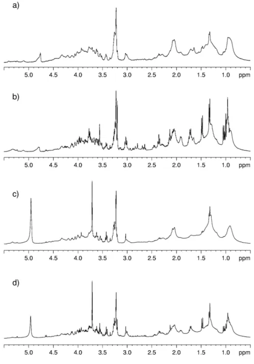
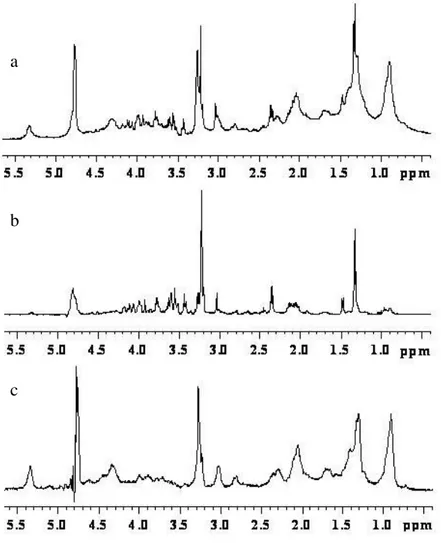
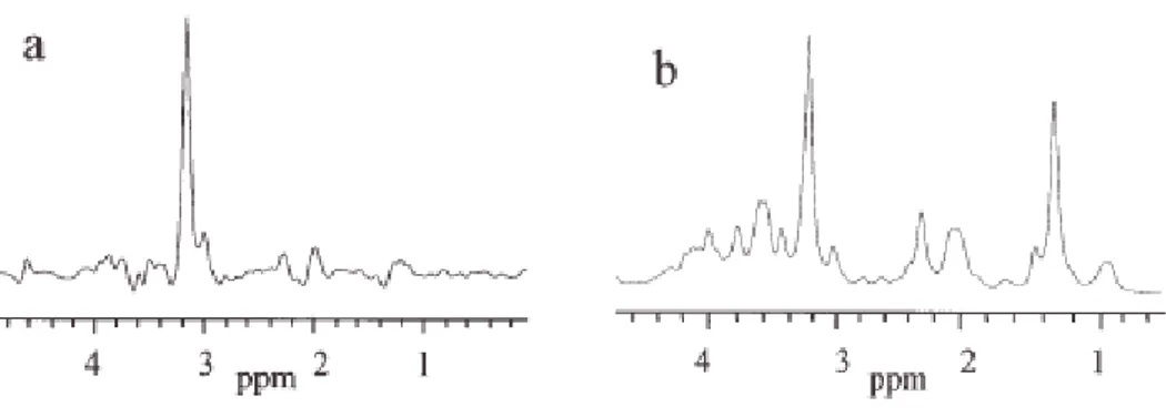
In order to evidence the quality of the spectra obtained with the HR-MAS probe with respect to those obtained using high resolution probe, I report in Figures 7 and 8 the spectra of kidney tissue samples. The sample for the high resolution probe was prepared by putting a piece of kidney directly in a 5mm glass tube. It is evident (see Figure 7 and 8) the better resolution obtained with HR-MAS.
Figure 7. 1D proton NMR spectra of kidney obtained with water-presaturation pulse sequence with composite
pulse to a) HR-MAS probe and b) High Resolution probe.
a)